User login
Is physician-assisted suicide compatible with the Hippocratic Oath?
Do we stand by our Hippocratic Oath when providing physician-assisted suicide?
Physician-assisted suicide (PAS) is a form of euthanasia in which a physician provides the patient with the pertinent information, and, in certain cases, a prescription for the necessary lethal drugs, so that the individual can willingly and successfully terminate his or her own life. The justification for PAS is the compassionate relief of intractable human suffering. Euthanasia and PAS are accepted practices in European countries such as the Netherlands, Belgium, and Luxembourg.

Such patients are mentally incapable of making the difficult decision to end their life and therefore should require a psychiatric evaluation to include counseling prior to making a decision to engage in PAS. For patients with mental illness, PAS is even more problematic than for terminally ill patients, because patients may lack the capacity to make rational and responsible decisions. Indeed, there are sizable loopholes where our medical system lacks safeguards and similarly lacks the requirements for a thorough, pre-PAS mental health examination, family notifications, and consultations, and for the minimally necessary legal pressure required to ensure patient cooperation.
Critical role of mental health workers
Although PAS has been legalized in those five U.S. states, its support and cases have stalled in recent years, indicating serious ethical concerns, mostly because of multileveled challenges of combating and delineating cultural stereotypes, quantifying mental capacity, gauging quality of life, and deciding where to situate psychiatrists in the PAS decision.
The psychiatrist’s role is being debated. In the United States, opponents take issue with current PAS-legal state’s legislation regarding psychiatric evaluations. For example, Oregon stipulates a psychiatrist referral only in cases where a physician other than a psychiatrist believes the patient’s judgment is impaired. It’s agreed that psychiatrists have the best skill set to assess a patient’s perceptions. Other PAS-legal states require a psychiatrist or psychologist assessment before making the decision. Unfortunately, though, physicians have rarely referred these patients to psychiatrists before offering PAS as an option.
PAS opponents target standards by which concepts like “quality of life” or “contributing member of society” are judged – specifically, that “unbearable suffering” and its ramifications are ill defined – people whose lives are deemed “not worth living” (including the terminally ill) would be susceptible to “sympathetic death” via PAS that might result from PAS legalization. Opponents also argue that recognizing a suicide “right” contradicts that a significant number of suicide attempters have mental illness and need help. They say that legalizing PAS would enable mentally incapacitated people to commit the irreversible act based on their distorted perceptions without providing them the expected assistance from their profession.1
The use of euthanasia or PAS gradually is trending from physically terminally ill patients toward psychiatrically complex patients. There are cases in which euthanasia or PAS was requested by psychiatric patients who had chronic psychiatric, medical, and psychosocial histories rather than purely physical ailment histories. In one study, Scott Y. H. Kim, MD, PhD, and his associates reviewed cases in the Netherlands in which either euthanasia or PAS for psychiatric disorders was deployed. The granted PAS requests appeared to involve physician judgment without psychiatric input.
The study reviewed 66 cases: 55% of patients had chronic severe conditions with extensive histories of attempted suicides and psychiatric hospitalizations, demonstrating that the granted euthanasia and PAS requests had involved extensive evaluations. However, 11% of cases had no independent psychiatric input, and 24% involved disagreement among the physicians.2 PAS proponents and opponents support the involvement and expertise of psychiatrists in all of this.
Psychiatrists have long contended that suicide attempts are often a “cry for help,” not an earnest act to end one’s life. Legalizing PAS tells suicidal individuals that society does not care whether they live or die – a truly un-Hippocratic stance. The stereotypes tossed around in the PAS debate, which could mean life or death, need to be unpacked with specific criteria attached, rather than preconceptions.
Autonomy of patients and implications of PAS
In the PAS debate, there are serious life concerns exhibited by terminally ill/psychiatrically ill patients over losing their autonomy and becoming a burden to their families and caregivers. Stereotypes must be deconstructed, yes, but such patients generally have been considered to be rendered, by the sum effect of their illnesses, mentally incapable of making the decision to end their lives. Addressing this order of patient life concerns generally has been the realm of social workers, psychologists, and, in the most desirable cases, psychiatrists. Therefore, even in keeping with established practice, some form of competent psychiatric evaluation should be required before considering PAS as a viable option.
The call for this requirement is backed up by a study, which reported that 47% of patients who had considered or committed suicide previously were diagnosed with psychiatric disorders, and 15% had undiagnosed psychiatric disorders.3 Also, inventories of thwarted suicide attempters reveal that most lack the conviction to end their lives, and this obverse phenomenon is backed by a study that revealed that 75% of 96 suicidal patients were found to be ambivalent in their intent to end their lives. Suicide attempters simply may be trying to communicate with people in their lives in order to test their love and care.4 This indicates that those who attempt suicide are predominantly psychiatrically ill and prone to distorted perceptions, impaired judgment, frustration, escapism, and manipulative guilt.
Suicide is abhorrent to most human beings’ sensibilities, because humanity has an innate will to live.5 It is wrong to offer patients life-ending options when they might rejuvenate or ease into death naturally. With the debilitated or elderly, PAS could violate their human rights. If the attitude that they are a burden to society persists, then a certain segment of society might be tempted to avoid their intergenerational obligations to elders, particularly those elders presenting with concurrent mental illnesses. PAS would additionally fuel that problem. This would represent a profound injustice and gross violation of human dignity, while also serving as a denial of basic human rights, particularly the right to life and care.6
Conclusion
The complex physical, psychological, and social challenges associated with PAS and the difficulty in enforcing its laws necessitate more adept alternatives. Instead of conditionally legalizing suicide, we should ease patient suffering with compassion and calibrated treatment.6,7
Terminally ill patients often suffer from depression, affecting their rationality. Adequate counseling, medical care, and psychological care should be the response to terminally ill patients considering suicide. If society accepts that ending a life is a reasonable response to human suffering that could otherwise be treated or reversed, then those with the most serious psychiatric conditions are destined to become more vulnerable. Therefore, physicians and psychiatrists should instead work harder to help patients recover, rather than participating in their quest for death.
Simplistic legal and regulatory oversight is insufficient, because the questions evoked by PAS are complicated by life, death, and ethics. Further research is needed to look into the mechanisms and morality of how psychiatrists and other physicians will make judgments and recommendations that are vital elements in developing regulatory oversight on PAS. Finally, future studies should look at which practices have been successful and which might be deemed unethical.
References
1. Clin Med (Lond). 2010 Aug;109(4):323-5.
2. JAMA Psychiatry. 2016;73(4):362-8.
3. “The Final Months: A Study of the Lives of 134 Persons Who Committed Suicide,” New York: Oxford University Press, 1981.
4. “Why We Shouldn’t Legalize Assisting Suicide, Part I.” Department of Medical Ethics, National Right to Life Committee.
5. “Working with Suicidal Individuals: A Guide to Providing Understanding, Assessment and Support,” Philadelphia: Jessica Kingsley Publishers, 2010.
6. “Always Care, Never Kill: How Physician-Assisted Suicide Endangers the Weak, Corrupts Medicine, Compromises the Family, and Violates Human Dignity and Equality,” Washington: The Heritage Foundation, 2015.
7. Ann Intern Med. 2012 Jan 3;156(1 Pt 2):73-104.
Dr. Ahmed is a 2nd-year resident in the psychiatry & behavioral sciences department at the Nassau University Medical Center, New York. Over the last 3 years, he has published and presented papers at national and international forums. His interests include public social psychiatry, health care policy, health disparities, mental health stigma, and undiagnosed and overdiagnosed psychiatric illnesses in children. Dr. Ahmed is a member of the American Psychiatric Association, the American Society of Clinical Psychopharmacology, and the American Association for Social Psychiatry.
Do we stand by our Hippocratic Oath when providing physician-assisted suicide?
Physician-assisted suicide (PAS) is a form of euthanasia in which a physician provides the patient with the pertinent information, and, in certain cases, a prescription for the necessary lethal drugs, so that the individual can willingly and successfully terminate his or her own life. The justification for PAS is the compassionate relief of intractable human suffering. Euthanasia and PAS are accepted practices in European countries such as the Netherlands, Belgium, and Luxembourg.
Such patients are mentally incapable of making the difficult decision to end their life and therefore should require a psychiatric evaluation to include counseling prior to making a decision to engage in PAS. For patients with mental illness, PAS is even more problematic than for terminally ill patients, because patients may lack the capacity to make rational and responsible decisions. Indeed, there are sizable loopholes where our medical system lacks safeguards and similarly lacks the requirements for a thorough, pre-PAS mental health examination, family notifications, and consultations, and for the minimally necessary legal pressure required to ensure patient cooperation.
Critical role of mental health workers
Although PAS has been legalized in those five U.S. states, its support and cases have stalled in recent years, indicating serious ethical concerns, mostly because of multileveled challenges of combating and delineating cultural stereotypes, quantifying mental capacity, gauging quality of life, and deciding where to situate psychiatrists in the PAS decision.
The psychiatrist’s role is being debated. In the United States, opponents take issue with current PAS-legal state’s legislation regarding psychiatric evaluations. For example, Oregon stipulates a psychiatrist referral only in cases where a physician other than a psychiatrist believes the patient’s judgment is impaired. It’s agreed that psychiatrists have the best skill set to assess a patient’s perceptions. Other PAS-legal states require a psychiatrist or psychologist assessment before making the decision. Unfortunately, though, physicians have rarely referred these patients to psychiatrists before offering PAS as an option.
PAS opponents target standards by which concepts like “quality of life” or “contributing member of society” are judged – specifically, that “unbearable suffering” and its ramifications are ill defined – people whose lives are deemed “not worth living” (including the terminally ill) would be susceptible to “sympathetic death” via PAS that might result from PAS legalization. Opponents also argue that recognizing a suicide “right” contradicts that a significant number of suicide attempters have mental illness and need help. They say that legalizing PAS would enable mentally incapacitated people to commit the irreversible act based on their distorted perceptions without providing them the expected assistance from their profession.1
The use of euthanasia or PAS gradually is trending from physically terminally ill patients toward psychiatrically complex patients. There are cases in which euthanasia or PAS was requested by psychiatric patients who had chronic psychiatric, medical, and psychosocial histories rather than purely physical ailment histories. In one study, Scott Y. H. Kim, MD, PhD, and his associates reviewed cases in the Netherlands in which either euthanasia or PAS for psychiatric disorders was deployed. The granted PAS requests appeared to involve physician judgment without psychiatric input.
The study reviewed 66 cases: 55% of patients had chronic severe conditions with extensive histories of attempted suicides and psychiatric hospitalizations, demonstrating that the granted euthanasia and PAS requests had involved extensive evaluations. However, 11% of cases had no independent psychiatric input, and 24% involved disagreement among the physicians.2 PAS proponents and opponents support the involvement and expertise of psychiatrists in all of this.
Psychiatrists have long contended that suicide attempts are often a “cry for help,” not an earnest act to end one’s life. Legalizing PAS tells suicidal individuals that society does not care whether they live or die – a truly un-Hippocratic stance. The stereotypes tossed around in the PAS debate, which could mean life or death, need to be unpacked with specific criteria attached, rather than preconceptions.
Autonomy of patients and implications of PAS
In the PAS debate, there are serious life concerns exhibited by terminally ill/psychiatrically ill patients over losing their autonomy and becoming a burden to their families and caregivers. Stereotypes must be deconstructed, yes, but such patients generally have been considered to be rendered, by the sum effect of their illnesses, mentally incapable of making the decision to end their lives. Addressing this order of patient life concerns generally has been the realm of social workers, psychologists, and, in the most desirable cases, psychiatrists. Therefore, even in keeping with established practice, some form of competent psychiatric evaluation should be required before considering PAS as a viable option.
The call for this requirement is backed up by a study, which reported that 47% of patients who had considered or committed suicide previously were diagnosed with psychiatric disorders, and 15% had undiagnosed psychiatric disorders.3 Also, inventories of thwarted suicide attempters reveal that most lack the conviction to end their lives, and this obverse phenomenon is backed by a study that revealed that 75% of 96 suicidal patients were found to be ambivalent in their intent to end their lives. Suicide attempters simply may be trying to communicate with people in their lives in order to test their love and care.4 This indicates that those who attempt suicide are predominantly psychiatrically ill and prone to distorted perceptions, impaired judgment, frustration, escapism, and manipulative guilt.
Suicide is abhorrent to most human beings’ sensibilities, because humanity has an innate will to live.5 It is wrong to offer patients life-ending options when they might rejuvenate or ease into death naturally. With the debilitated or elderly, PAS could violate their human rights. If the attitude that they are a burden to society persists, then a certain segment of society might be tempted to avoid their intergenerational obligations to elders, particularly those elders presenting with concurrent mental illnesses. PAS would additionally fuel that problem. This would represent a profound injustice and gross violation of human dignity, while also serving as a denial of basic human rights, particularly the right to life and care.6
Conclusion
The complex physical, psychological, and social challenges associated with PAS and the difficulty in enforcing its laws necessitate more adept alternatives. Instead of conditionally legalizing suicide, we should ease patient suffering with compassion and calibrated treatment.6,7
Terminally ill patients often suffer from depression, affecting their rationality. Adequate counseling, medical care, and psychological care should be the response to terminally ill patients considering suicide. If society accepts that ending a life is a reasonable response to human suffering that could otherwise be treated or reversed, then those with the most serious psychiatric conditions are destined to become more vulnerable. Therefore, physicians and psychiatrists should instead work harder to help patients recover, rather than participating in their quest for death.
Simplistic legal and regulatory oversight is insufficient, because the questions evoked by PAS are complicated by life, death, and ethics. Further research is needed to look into the mechanisms and morality of how psychiatrists and other physicians will make judgments and recommendations that are vital elements in developing regulatory oversight on PAS. Finally, future studies should look at which practices have been successful and which might be deemed unethical.
References
1. Clin Med (Lond). 2010 Aug;109(4):323-5.
2. JAMA Psychiatry. 2016;73(4):362-8.
3. “The Final Months: A Study of the Lives of 134 Persons Who Committed Suicide,” New York: Oxford University Press, 1981.
4. “Why We Shouldn’t Legalize Assisting Suicide, Part I.” Department of Medical Ethics, National Right to Life Committee.
5. “Working with Suicidal Individuals: A Guide to Providing Understanding, Assessment and Support,” Philadelphia: Jessica Kingsley Publishers, 2010.
6. “Always Care, Never Kill: How Physician-Assisted Suicide Endangers the Weak, Corrupts Medicine, Compromises the Family, and Violates Human Dignity and Equality,” Washington: The Heritage Foundation, 2015.
7. Ann Intern Med. 2012 Jan 3;156(1 Pt 2):73-104.
Dr. Ahmed is a 2nd-year resident in the psychiatry & behavioral sciences department at the Nassau University Medical Center, New York. Over the last 3 years, he has published and presented papers at national and international forums. His interests include public social psychiatry, health care policy, health disparities, mental health stigma, and undiagnosed and overdiagnosed psychiatric illnesses in children. Dr. Ahmed is a member of the American Psychiatric Association, the American Society of Clinical Psychopharmacology, and the American Association for Social Psychiatry.
Lower Extremity Occlusive Disease and Alternative Graft Options
Vascular surgeons spend much of their clinical time taking care of patients with lower extremity occlusive disease, so the “New Devices and Techniques for Treating Lower Extremity Occlusive Disease; Prosthetic Grafts and Heparin Bonding” session on Thursday afternoon will be particularly useful for the practicing surgeon, according to Dr. Russell Samson who will co-moderate the session along with Dr. Ali F. AbuRahma of West Virginia University.
Two recent advances with respect to such alternative graft options, which will be addressed during the session, are heparin-bonded ePTFE and spiral laminar flow grafts.
“Whether these two modalities improve patency rates compared to standard ePTFE grafts remains controversial,” Dr. Samson said.
Heparin-bonded ePTFE will be the topic of a debate presented by Dr. Richard Neville of George Washington University Hospital, and Dr. Jonathan Beard of Sheffield Vascular Institute, in the United Kingdom. These two experts will conduct a debate. Dr. Neville will discuss long-term patency data suggesting that heparin bonding is valuable in PTFE Fempop Bypass Grafts, and Dr. Beard will argue that it does not improve results.
Additionally, Dr. Yann Gouëffic of the University of Nantes, France, will address the cost-efficiency of heparin bonding.
“In an age where surgeons are becoming more cost conscious, new graft configurations, which are often more costly, need to offer significant improvement in long-term outcomes,” Dr. Samson said.
As for spiral laminar flow grafts, Dr. Hosam F. El Sayed of Ohio State University will provide insight into a specific format of PTFE that creates a spiral laminar flow that more accurately represents flow within the arterial system, Dr. Samson noted.
Therefore, Dr. Enrico Ascher of Lutheran Medical Center, who is highly regarded for his experience in performing distal tibial bypass, will provide insight into how to deal with “pipe-like” calcified arteries that some surgeons may consider inoperable, Dr. Samson added.
Further, since many surgeons may prefer an endovascular procedure as a first-line alternative to bypass, this session will also include a talk by Dr. Hiroyoshi Yokoi of Fukuoka Sanno Hospital in Japan on advanced guidewire techniques to open these distal vessels, and another by noted inventor, Dr. Timothy A.M. Chuter of the University of California, San Francisco, who will discuss advantages and early clinical experience with a novel balloon that does not straighten when it inflates.
Another talk during the session, which will be given by Dr. Joseph L. Mills of Baylor College of Medicine, will focus on the impact of foot infections on leg bypass outcomes in CLI patients and what can be done to offset that impact.
“Dr. Mills will remind us that no matter what treatment we choose, the final result can be spoiled by an infection, so managing foot infections in patients requiring surgical bypass becomes a preeminent consideration,” Dr. Samson said.
“The practicing vascular surgeon deals with challenging infrainguinal atherosclerotic conditions on a daily basis. Therefore, information gained from attending this program should enhance their ability to offer long-lasting therapies,” he added.
Session 59
“New Devices and Techniques for Treating Lower Extremity Occlusive Disease; Prosthetic Grafts and Heparin Bonding”
Thursday, 1:00 p.m. – 1:54 p.m.
Grand Ballroom West, 3rd Floor
Highlight on Innovative Devices for Embolectomy, Clot Retrieval, and Embolization
With the development of ever-more-sophisticated endovascular intervention systems, limb ischemia need no longer be an automatic sentence of lifelong pain or amputation.
But the burgeoning supply of these specialized devices brings its own challenge to the area of endovascular intervention. Which device is best for which patient? And which have strong data to back up their claims of effectiveness? Thursday’s session, “New Devices for Embolectomy, Clot Removal, and Embolization,” will offer some guidance.
“The presentations represent significant further advances in methods and technology that can treat patients with severe, limb-threatening, and life-altering vascular diseases,” said Dr. McNamara. “Also, comparisons are made between competing new technologies. This will be very helpful in deciding which method and/or technology to use. And it will, I think, be very helpful in deciding whether to use interventional or open surgical treatments.“These presentations will not only guide the vascular specialist, but help to substantiate that the interventional techniques continue to be more and more effective. This information could, and should, be shared with primary care physicians.”
This point can’t be underestimated, Dr. McNamara noted. Primary care physicians are on the front line of venous disorders, and should know that there are good treatment options for many patients – treatments than can save limbs and vastly improve pain and function.
“An increasing percentage of patients should be referred for consultation and possible treatments for arterial and venous disease. This will result in decreased morbidity and improved quality of life for an increasing number of patients.”
Surgeons should expect to see more and more such patients as the U.S. population continues to age – and effective treatment not only benefits each individual, but society as a whole.
“The disease significantly decreases the quality of life, and reduces the independence of our aging population. This significantly increases the costs, and increases the burden to society and families of the growing percentage of our population in their ‘Golden Years.’”
The session opens with a lecture by Dr. Ali Amin, a vascular surgeon from West Reading, Pa. He will discuss the role of mechanical thrombectomy and thrombolysis in acute limb ischemia, and when open surgery rather than endovascular intervention is indicated
Dr. Jos C. van den Berg of the University of Bern, Switzerland, will then tackle how to utilize suction techniques and devices to deal with the embolic complications of peripheral vascular interventions.
Rotarex and Aspirex, two mechanical debulking devices created by Straub Med, will be the topic of a presentation by Dr. Michael K.W. Lichtenberg chief of the Angiology Clinic and Venous Center at Klinikum Arnsberg, Germany.
There will be three presentations on thrombectomy with the Indigo System by Penumbra. Indigo provides mechanical clot engagement and vacuum extraction and can be used in both arteries and veins.
Dr. James F. Benenati, of Florida International University Herbert Wertheim College of Medicine, and Dr. Richard R. Saxon of San Diego Imaging, will discuss the PRISM Registry data. The registry recently reported that mechanical aspiration with the Indigo device is a safe and effective option for the treatment of peripheral or visceral arterial occlusions either as a frontline therapy or in patients who failed thrombolysis.
Dr. Frank R. Arko of Carolinas Healthcare System, will share his thoughts on why the Indigo system is an excellent choice for clot removal, and how it decreases the need for lytic therapy.
Dr. George L. Adams, MD, Director of Cardiovascular and Peripheral Vascular Research will make the case for why the Indigo system could be a “game-changer” in the treatment of acute limb ischemia and visceral artery thomboses and emboli.
Dr. Andrej Schmidt of the University Clinic of Leipzig, Germany, will discuss the advantages and limitations of several other Penumbra products, including the Ruby Coil for peripheral embolization, and the Penumbra Occlusion Device (POD). He will also touch on the Lantern microcatheter, Penumbra’s newest product, launched in April. The low-profile, high-flow microcatheter can pass through the distal vasculature, but can also be used for high-flow contrast injections.
Finally, Dr. Aaron Fischman of Mount Sinai Medical Center, will speak on the challenge of endovascular rescue procedures for acute visceral ischemia from thromboembolism.
Session 62
“New Devices for Embolectomy, Clot Removal, and Embolization”
4:32 p.m. – 5:30 p.m.
Grand Ballroom West, 3rd Floor
Bioactive lipid shows promise in atopic dermatitis
VIENNA – A novel oral agent known as DS107 showed promise as a safe and effective treatment for moderate-to-severe atopic dermatitis in a phase IIa proof-of-concept study, Diamant Thaçi, MD, reported at the annual congress of the European Academy of Dermatology and Venereology.
“Clear efficacy signals in the reduction of clinical symptoms of atopic dermatitis were detected within 2 weeks of treatment, with the maximum improvement in the endpoints observed between weeks 4 and 8,” said Dr. Thaçi, professor of dermatology and head of the Comprehensive Center for Inflammation Medicine at University Hospital Schleswig-Holstein, in Lübeck, Germany.
Dr. Thaçi presented the results of an 8-week, double-blind, randomized, placebo-controlled, multicenter phase IIa study that included 102 patients with moderate-to-severe AD. Participants averaged a baseline Investigator Global Assessment (IGA) score of 3.5 on the 0-5 scale. They were randomized to 2 g of oral DS107 once daily or an equal quantity of mineral oil as a placebo control.
Based upon the encouraging findings, a 300-patient phase IIb study will get underway soon. It will examine the effects of 1 g of DS107 per day as well as 2 g in the hope that the lower dose will cut down on the high rate of minor GI side effects seen at 2 g/day while preserving the efficacy of the higher dose.
The primary efficacy endpoint in the phase IIa study was at least a 2-point drop from baseline in IGA plus an end-of-treatment IGA of 0 or 1, meaning clear or almost clear. In the intent-to-treat analysis, this was achieved in 21.6% of the group assigned to DS107, compared with 11.8% of controls. In the 71 participants who actually completed 8 weeks of treatment – 35 in the DS107 arm, 36 controls – the composite efficacy endpoint was achieved in 31.4% of those on active treatment and 16.7% on mineral oil.
Significant separation between the active treatment and control arms in terms of itch visual analog scores was seen by week 4. This was a particularly encouraging finding, since patients report pruritis to be the most troublesome symptom of AD, Dr. Thaçi noted.
Significantly greater improvement in quality of life as measured by the Patient-Oriented Eczema Measure (POEM) was also seen by week 4 in the DS107 group as compared with controls.
No severe adverse events occurred in the study. However, more than one-quarter of subjects interrupted or discontinued participation because of mild nausea, loose stools, and/or abdominal pain. These issues were equally common in the DS107 and mineral oil groups, and Dr. Thaçi and his coinvestigators suspect that for many patients it was simply a matter of too much oil in the stomach. The GI symptoms resolved quickly without intervention after a brief halt of therapy, but some patients never returned to participation.
“This problem can be solved in the future with a different dosing design or even a different method of delivering the DGLA,” the dermatologist added.
Asked about the significant placebo response seen in the study, Dr. Thaçi shrugged it off as “quite understandable.”
“Placebo is not always a placebo. There is the feeling of fullness in the stomach, there is some emollient effect, the continuous contact with the physician. You see this in all the clinical trials in atopic dermatitis: in the beginning, the first 2-3 weeks, you have some influence of placebo,” he said.
The placebo effect was greatly diminished in patients with more severe AD. In the subset with a baseline IGA of 4 or 5, none of the control subjects achieved the primary efficacy endpoint, while more than 20% on DS107 did, Dr. Thaçi noted.
The mechanism of benefit of DGLA is not fully understood as yet. Animal studies point to an antibacterial effect, and DGLA also reduces levels of inflammatory cytokines. Eosinophilia was reduced to a much greater extent in the DS107 group than controls.
Dr. Thaçi reported serving as a consultant to DS Pharma, the privately held biopharmaceutical company that is developing oral DS107, as well as to numerous other pharmaceutical companies.
[email protected]
AT THE EADV CONGRESS
Key clinical point:
Major finding: Once-daily oral DS107, which contains a bioactive lipid, achieved significant improvement in moderate-to-severe atopic dermatitis in 31.4% of patients, compared with 16.7% on placebo.
Data source: This phase IIa, double-blind, randomized, placebo-controlled, multicenter 8-week trial included 102 adults with moderate-to-severe atopic dermatitis.
Disclosures: The study presenter reported serving as a consultant to DS Pharma, the privately held biopharmaceutical company that is developing oral DS107, as well as to numerous other pharmaceutical companies.
Study of Bioengineered Vessels for Dialysis Vascular Access Promising
The idea of using bioengineered human acellular vessels for dialysis vascular access used to be a pie-in-the-sky notion, but research presented today will demonstrate just how close they are to becoming a realistic option.
Dr. Jeffrey H. Lawson will present results from two Phase II single-arm trials of bioengineered human acellular vessels for dialysis access in 60 patients with end-stage renal disease conducted in six hospitals: three in the United States and three in Poland. His presentation is entitled “Tissue Engineered Blood Vessels for Arterial Bypass and Dialysis Access: Midterm Results n Patients.” To date, the early clinical experience with these vessels “suggests that they are safe to implant as a dialysis access vessel and lower leg bypass graft,” said Dr. Lawson, chief medical officer at Humacyte and professor of surgery and pathology in the department of surgery at Duke University, Durham, N.C. “They enjoy enduring patency, tolerate needed interventions, appear durable and have a low risk of infection.”
The researchers observed that during 82 patient-years of follow-up, only one vessel became infected. The vessels had no dilatation and rarely had postcannulation bleeding. At 6 months, 63% of patients had primary patency, 73% had primary assisted patency, and 97% had secondary patency, with most loss of primary patency because of thrombosis. At 1 year, 28% had primary patency, 38% had primary assisted patency, and 89% had secondary patency. “Following implantation into patients, the human acellular vessel (HAV) appears to completely repopulate with the host’s (recipient’s) own vascular tissue,” Dr. Lawson said. “The vessel is filled with cells that look like vascular smooth muscle cells and the implanted vessel is lined with endothelial cells from the host, suggesting that the manufactured acellular vessel becomes repopulated with the hosts own cells making it part of their own body. I will also be discussing some unpublished work that we have done for lower leg arterial bypass work in 20 patients to date.”
Dr. Lawson acknowledged that the Phase II experience at a few clinical sites is a limitation of the current analysis. “These findings need to be confirmed and validated in a large Phase III clinical trial, which is now underway,” he said.
Session 60
“New Developments in Arterial Grafts; Stents and Stent-Grafts; Concepts and Techniques to Improve Their Use and Results”
Thursday, 1:54 p.m.– 3:24 p.m.
Grand Ballroom East, 3rd Floor
ASVAL Can Save Refluxing Saphenous Veins
Ambulatory selective varicose vein ablation under local anesthesia (ASVAL) can abolish reflux while preserving the great saphenous vein, and benefits can last at least a decade, said Dr. Silvain Chastanet, who helped create the technique and will discuss it Thursday morning.
Historically, a refluxing great saphenous vein was treated by removing it. The ASVAL method preserves the vein by using microphlebotomy to instead remove the varicose reservoir, said Dr. Chastanet of the Riviera Vein Institute in Monaco. “Because each patient is different, this strategy offers tailored, à la carte treatment, as opposed to systematic ablation,” he said. “Doing precise and thorough microphlebectomies is not as simple as one might think, but we will share tips and tricks for performing saphenous sparing venous surgery in an easy and elegant way.”
The ASVAL technique is based on the premise that reflux usually ascends from the tributaries toward the saphenous trunk, rather than following the opposite path, said Dr. Chastanet. Although the ascending pattern is not universal, increasing evidence suggests that it is most common. For example, a study of more than 700 consecutive patients with chronic venous disease showed that patients of CEAP category C4-C6 had significantly greater involvement of the saphenofemoral junction and saphenopopliteal trunk than patients with less advanced venous disease. The authors concluded that reflux usually ascends up from the tributaries and accessory saphenous veins (J Vasc Surg. 2010;51(1):96-103).
Thus, when evaluating a patient with saphenous reflux, surgeons should always ask whether they can spare the great saphenous vein, Dr. Chastanet said. Evidence indicates that if the patient is classified as C2 (varicose veins) and the great saphenous vein is dilated less than 10 mm, ASVAL may be a valid option, especially if reflux is localized above the knee, with the varices localized to the thigh.
Techniques such as ASVAL are a major advance, but questions persist about surgically treating varicose disease, Dr. Chastanet noted. For example, although ascending disease appears to be most common, it can convert to descending disease or patients can have a refluxing saphenous trunk without tributaries. “We still do not know which patients are likely to have descending or ascending disease, and why,” Dr. Chastanet said. Likewise, experts continue to debate whether phlebectomy should be performed together with saphenous ablation. “Considering that some varicosities will disappear after endothermal ablation of the saphenous vein, some surgeons consider concomitant phlebectomy to be overtreatment,” Dr. Chastenet noted. “Conversely, if we consider that the disease begins at the tributary level according to the ascending theory, it seems obvious to focus treatment on the varicose tributaries, which are the main concern for the majority of patients.”
Bringing techniques such as ASVAL into community practice involves practical challenges, too. Microphlebectomy is “clearly” the best cosmetic option, but is not taught during surgical or phlebology training, Dr. Chastanet said. “There is a need to develop new tools for treating the varicose tributaries in an easier and faster way.” Such advances could help transition procedures such as ASVAL to outpatient settings, which is not now the norm in most countries, he noted. “Using local anesthesia, mini-invasive surgical procedures with immediate ambulation will enable us to reach this goal.”
Session 63
“Venous Clinical Examination and Hemodynamics”
Thursday 8:13 a.m. – 8:18 a.m.
Trianon Ballroom, 3rd Floor
Sunscreen and Sperm: Can Chemical UV Filters Alter Sperm Function?

In an article published online on September 1 in Endocrinology, Rehfeld et al discussed their results after testing 29 UV filters. They found that 13 of 29 filters tested had in vitro effects on Ca2+: 4-methylbenzylidene camphor, 3-benzylidene camphor, menthyl anthranilate, isoamyl p-methoxycinnamate, ethylhexyl salicylate, benzylidene camphor sulfonic acid, homosalate, ethylhexyl methoxycinnamate, octcrylene, butyl methoxydibenzoylmethane, and diethylamino hydroxybenzoyl hexyl benzoate.
This study was prompted by a prior study by Schiffer et al (EMBO Rep. 2014;15:758-765) on multiple endocrine disrupting chemicals of which 33 of 96 tested chemicals induced Ca2+ signals in human sperm cells in vitro. Of these previously tested chemicals, some of the chemical sunscreen filters were the most potent, leading to the current study.
Rehfeld et al sought to determine how the UV filters affected calcium signaling, which is a pathway that is essential for sperm cells to be able to swim healthily. These calcium-signaling pathways usually are triggered by progesterone, but the authors showed that 13 of 29 UV filters (45%) also commenced calcium signaling. This effect began at low doses of the chemicals, below the levels of some UV filters found in people after whole-body application of sunscreens.
What’s the issue?
Are these chemical UV filters mimicking progesterone in vivo and could it be interfering with sperm motility? A suboptimal progesterone-induced Ca2+ influx has been associated with reduced male fertility and CatSper (cation channel of sperm) is essential for male fertility (Hum Reprod. 1995;10:120-124).
The UV filters tested are widely available in Europe and the United States. Although this study was in vitro, the in vivo effects will need to be explored. It has been reported by Chivsvert et al (Anal Chim Acta. 2012;752:11-29) that some UV filters can be transcutaneously absorbed into bodily tissues, which could be potentially important for men trying to conceive or for reproductively challenged couples.
What do you discuss with your patients regarding sunscreen safety?

Genetically corrected skin grafts explored in dystrophic EB
Genetically corrected autologous skin grafts produced wound healing in a phase I trial involving four men with recessive dystrophic epidermolysis bullosa in what the investigators described as the first human trial of cutaneous gene therapy for this indication, according to a report published in JAMA.
The wound healing varied according to graft site and across the four patients, and generally declined over the course of 1 year of follow-up. Given that this study focused on safety outcomes and that the treatment’s safety profile was deemed “acceptable,” the Food and Drug Administration has permitted a phase IIA trial (NCT01263379) that is currently enrolling adolescents with the disease and will focus on clinical outcomes. Longer-term follow-up of these four patients will continue, and “controlled trials are needed with a broader range of patients to better understand the potential long-term efficacy of genetically corrected autologous epidermal grafts,” said Zurab Siprashvili, Ph.D., of Stanford (Calif.) University and associates.
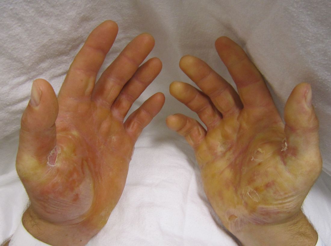
The investigators assessed whether grafting of type VII collagen gene-corrected autologous keratinocytes onto existing wounds would promote their healing. They first harvested and cultured keratinocytes from biopsies of the patients’ unwounded, unscarred skin and transduced these samples with COL7A1-containing retrovirus, producing eight gene-corrected grafts for each patient. All the biopsy sites healed completely, without complications.
The wound beds were prepared for engraftment by cauterization to minimize any retained epidermal stem cells. Then grafts were applied using dissolvable sutures to six wound sites on each patient; the sites had been selected for their accessibility, their potential to enhance the patients’ quality of life, and their ability to ease the period of immobilization after grafting.
All 24 grafts were well tolerated, and no serious adverse events occurred during 1 year of follow-up, which represented approximately 10 epidermal turnover cycles. The most common adverse events – pruritus (three patients) and drainage (two patients) at the graft site – were mild. None of the patients showed systemic autoimmune symptoms or increased blistering outside of the grafted areas, and no clinical signs of malignancy developed.
The gene-corrected graft sites showed type VII collagen localization to anchoring fibrils at the dermal-epidermal junction, in contrast to control sites that showed no such localization or fibril formation. “Gene-corrected graft sites showed fully differentiated epidermis with spinous and granular layers, which were positive for epidermal markers keratin 14, keratin 1, and loricirin resembling normal skin,” Dr. Siprashvili and his associates wrote.
At 1-month follow-up, 20 of the 24 grafts showed 75% or greater wound healing, compared with baseline, and the other 4 grafts showed 50%-74% wound healing. At 3 months, 21 of the 24 graft sites showed 75% or greater wound healing while the remaining 3 grafts showed 50%-74% wound healing. At 6 months, 16 of 24 graft sites showed 75% or greater wound healing and 5 graft sites showed 50%-74% wound healing, but 3 graft sites showed extensive blisters or erosions and were considered graft failures. At 12 months, only 12 of the 24 graft sites showed 75% or greater wound healing.
This general decline in efficacy might be related to the relatively small number of stem cells available for transplantation in these patients, who had only small areas of unscarred skin for harvesting. Or it may be that uncorrected cells in the wound beds began competing with corrected cells within the graft and eventually overcame them, the researchers said.
This study was supported by the National Institutes of Health, the Epidermolysis Bullosa Medical Research Foundation, the Epidermolysis Bullosa Research Partnership, and the Palo Alto VA Medical Center. Dr. Siprashvili and some associates reported having U.S. patents pending; two associates also reported ties to Scioderm and Fibrocell.
Genetically corrected autologous skin grafts produced wound healing in a phase I trial involving four men with recessive dystrophic epidermolysis bullosa in what the investigators described as the first human trial of cutaneous gene therapy for this indication, according to a report published in JAMA.
The wound healing varied according to graft site and across the four patients, and generally declined over the course of 1 year of follow-up. Given that this study focused on safety outcomes and that the treatment’s safety profile was deemed “acceptable,” the Food and Drug Administration has permitted a phase IIA trial (NCT01263379) that is currently enrolling adolescents with the disease and will focus on clinical outcomes. Longer-term follow-up of these four patients will continue, and “controlled trials are needed with a broader range of patients to better understand the potential long-term efficacy of genetically corrected autologous epidermal grafts,” said Zurab Siprashvili, Ph.D., of Stanford (Calif.) University and associates.
The investigators assessed whether grafting of type VII collagen gene-corrected autologous keratinocytes onto existing wounds would promote their healing. They first harvested and cultured keratinocytes from biopsies of the patients’ unwounded, unscarred skin and transduced these samples with COL7A1-containing retrovirus, producing eight gene-corrected grafts for each patient. All the biopsy sites healed completely, without complications.
The wound beds were prepared for engraftment by cauterization to minimize any retained epidermal stem cells. Then grafts were applied using dissolvable sutures to six wound sites on each patient; the sites had been selected for their accessibility, their potential to enhance the patients’ quality of life, and their ability to ease the period of immobilization after grafting.
All 24 grafts were well tolerated, and no serious adverse events occurred during 1 year of follow-up, which represented approximately 10 epidermal turnover cycles. The most common adverse events – pruritus (three patients) and drainage (two patients) at the graft site – were mild. None of the patients showed systemic autoimmune symptoms or increased blistering outside of the grafted areas, and no clinical signs of malignancy developed.
The gene-corrected graft sites showed type VII collagen localization to anchoring fibrils at the dermal-epidermal junction, in contrast to control sites that showed no such localization or fibril formation. “Gene-corrected graft sites showed fully differentiated epidermis with spinous and granular layers, which were positive for epidermal markers keratin 14, keratin 1, and loricirin resembling normal skin,” Dr. Siprashvili and his associates wrote.
At 1-month follow-up, 20 of the 24 grafts showed 75% or greater wound healing, compared with baseline, and the other 4 grafts showed 50%-74% wound healing. At 3 months, 21 of the 24 graft sites showed 75% or greater wound healing while the remaining 3 grafts showed 50%-74% wound healing. At 6 months, 16 of 24 graft sites showed 75% or greater wound healing and 5 graft sites showed 50%-74% wound healing, but 3 graft sites showed extensive blisters or erosions and were considered graft failures. At 12 months, only 12 of the 24 graft sites showed 75% or greater wound healing.
This general decline in efficacy might be related to the relatively small number of stem cells available for transplantation in these patients, who had only small areas of unscarred skin for harvesting. Or it may be that uncorrected cells in the wound beds began competing with corrected cells within the graft and eventually overcame them, the researchers said.
This study was supported by the National Institutes of Health, the Epidermolysis Bullosa Medical Research Foundation, the Epidermolysis Bullosa Research Partnership, and the Palo Alto VA Medical Center. Dr. Siprashvili and some associates reported having U.S. patents pending; two associates also reported ties to Scioderm and Fibrocell.
FROM JAMA
Key clinical point: Genetically corrected autologous skin grafts produced wound healing in a phase I study of four patients with recessive dystrophic epidermolysis bullosa.
Major finding: All 24 grafts were well tolerated, and no serious adverse events occurred during 1 year of follow-up; none of the patients showed systemic autoimmune symptoms or increased blistering outside of the grafted areas, and no clinical signs of malignancy developed.
Data source: A single-center open-label phase I study involving four men followed for 1 year.
Disclosures: This study was supported by the National Institutes of Health, the Epidermolysis Bullosa Medical Research Foundation, the Epidermolysis Bullosa Research Partnership, and the Palo Alto VA Medical Center. Dr. Siprashvili and some associates reported having U.S. patents pending; two associates also reported ties to Scioderm and Fibrocell.
Top 3 things I learned at the NAMS 2016 annual meeting
- Visit NAMS
- Visit NAMS
- Visit NAMS

Mastoid stimulation significantly reduces episodic migraine frequency
Episodic migraines were safely and effectively treated using percutaneous mastoid electrical stimulators in a randomized, double-blind, sham-controlled, multicenter trial.
Yang Juan of the department of neurology at the Second People’s Hospital of Chengdu (China), and associates reported that a group of 39 migraine patients who received percutaneous mastoid electrical stimulation (PMES) experienced significantly fewer migraine days, compared with a control group of 37 patients who received sham electrical treatment. After 3 months, migraine days were reduced by 71.3% from baseline, compared with a 14.4% reduction in the control group. About a third of PMES patients experienced no migraines in the third month, whereas no patients in the control group had more than a 75% reduction in migraine incidence.
There were no adverse reactions reported either in the PMES group or in the control group during the treatment period.
“The best treatment mode including current intensity and duration is unclear. Whether extension of the time interval (e.g., once or twice a week) of PMES treatment may also have preventive effect remains to be determined. In addition, as the patients recruited in this study were not highly disabled, the preventive effect of PMES treatment in patients with more frequent migraine episodes and in patients with chronic migraine needs further study,” the investigators wrote.
Read the full study in Cephalalgia (2016 Nov 7. doi: 10.1177/0333102416678623).