User login
Not so peripheral anymore: VIVA late-breakers address PAD/CLI trials
After years of relative neglect, compared with the attention given to aortic disease, studies and devices designed to expand and improve treatment of peripheral arterial disease are moving to the forefront.
At this year’s VIVA Vascular meeting, the majority of late-breaking clinical trials have a focus on peripheral arterial disease (PAD) and critical limb ischemia (CLI), emphasizing the tremendous growth of research in this area. The following are some of the latest trials for which results will be presented:
On Monday morning, Michael P. Murphy, MD, will present results from the MOBILE clinical trial, which examines the use of the MarrowStim™ PAD Kit for the treatment of CLI in subjects with severe PAD. The treatment involves removing bone marrow from the hips, which is then processed in the MarrowStim device to separate stem cells for delivery by injection to multiple sites in the affected leg.
A report on the 6-month results from the two-phase DISRUPT PAD study to assess the safety and performance of the Shockwave Lithoplasty® System in treating calcified peripheral vascular lesions will then be presented by Thomas Zeller, MD, principal investigator for the study. “Lithoplasty is a novel technology utilizing integrated lithotripsy emitters that generate mechanical pulse waves to disrupt both superficial and deep calcium normalizing vessel wall compliance prior to low-pressure balloon dilatation,” according to a company report.
In addition, results from the IN.PACT SFA randomized trial will be presented by Prakash Krishnan, MD, and data from the VIRTUS US Iliofemoral Stenting IDE Trial will be addressed by Stephen Black, MD, and Michael R. Jaff, DO, will present insights on the use of drug-coated balloon treatment for patients with intermittent claudication based on results from the IN.PACT Global full clinical cohort.
On Tuesday morning, Stefan Müller-Hülsbeck, MD, will present the 2-year results plus subgroup analysis of the Majestic Trial, which examined the efficacy benefit of using the Eluvia drug-eluting stent for treating lesions in the femoropopliteal arteries.
This will be followed by Antonio Micari, MD, PhD, who will present the 2-year results of from the SFA-Long Study, which examines the use of paclitaxel coated balloons for long femoropopliteal artery disease.
On Wednesday morning, the 3-Year Results of the Evaluation of the ESPRIT Bioresorbable Vascular Scaffold (ESPRIT BVS) in The Treatment of Patients with Occlusive Vascular Disease of the Superficial Femoral (SFA) or Common or External Iliac Arteries (ESPRIT I Trial) will be presented by Michael R. Jaff, DO.
This will be followed by a presentation of the 12-month results from the DANCE Trial Atherectomy Cohort by Chris D. Owens, MD, and John Laird, MD, discussing the 9-month results of the Prospective, Multicenter BOLSTER Trial, which examined the use of an expanded polytetrafluoroethylene balloon-expandable covered stent for obstructive lesions in the iliac artery.
In further research addressing the peripheral vascular system, Dr. Laird will also address the 24-month results from the TIGRIS randomized trial, assessing the use of a novel nitinol stent for long lesions in the SFA and the proximal popliteal artery.
Afterwards, Richard Saxon, MD, will discuss the PRISM Indigo Aspiration System as a potential frontline tool for vacuum-assisted aspiration thrombectomy in the periphery.
After years of relative neglect, compared with the attention given to aortic disease, studies and devices designed to expand and improve treatment of peripheral arterial disease are moving to the forefront.
At this year’s VIVA Vascular meeting, the majority of late-breaking clinical trials have a focus on peripheral arterial disease (PAD) and critical limb ischemia (CLI), emphasizing the tremendous growth of research in this area. The following are some of the latest trials for which results will be presented:
On Monday morning, Michael P. Murphy, MD, will present results from the MOBILE clinical trial, which examines the use of the MarrowStim™ PAD Kit for the treatment of CLI in subjects with severe PAD. The treatment involves removing bone marrow from the hips, which is then processed in the MarrowStim device to separate stem cells for delivery by injection to multiple sites in the affected leg.
A report on the 6-month results from the two-phase DISRUPT PAD study to assess the safety and performance of the Shockwave Lithoplasty® System in treating calcified peripheral vascular lesions will then be presented by Thomas Zeller, MD, principal investigator for the study. “Lithoplasty is a novel technology utilizing integrated lithotripsy emitters that generate mechanical pulse waves to disrupt both superficial and deep calcium normalizing vessel wall compliance prior to low-pressure balloon dilatation,” according to a company report.
In addition, results from the IN.PACT SFA randomized trial will be presented by Prakash Krishnan, MD, and data from the VIRTUS US Iliofemoral Stenting IDE Trial will be addressed by Stephen Black, MD, and Michael R. Jaff, DO, will present insights on the use of drug-coated balloon treatment for patients with intermittent claudication based on results from the IN.PACT Global full clinical cohort.
On Tuesday morning, Stefan Müller-Hülsbeck, MD, will present the 2-year results plus subgroup analysis of the Majestic Trial, which examined the efficacy benefit of using the Eluvia drug-eluting stent for treating lesions in the femoropopliteal arteries.
This will be followed by Antonio Micari, MD, PhD, who will present the 2-year results of from the SFA-Long Study, which examines the use of paclitaxel coated balloons for long femoropopliteal artery disease.
On Wednesday morning, the 3-Year Results of the Evaluation of the ESPRIT Bioresorbable Vascular Scaffold (ESPRIT BVS) in The Treatment of Patients with Occlusive Vascular Disease of the Superficial Femoral (SFA) or Common or External Iliac Arteries (ESPRIT I Trial) will be presented by Michael R. Jaff, DO.
This will be followed by a presentation of the 12-month results from the DANCE Trial Atherectomy Cohort by Chris D. Owens, MD, and John Laird, MD, discussing the 9-month results of the Prospective, Multicenter BOLSTER Trial, which examined the use of an expanded polytetrafluoroethylene balloon-expandable covered stent for obstructive lesions in the iliac artery.
In further research addressing the peripheral vascular system, Dr. Laird will also address the 24-month results from the TIGRIS randomized trial, assessing the use of a novel nitinol stent for long lesions in the SFA and the proximal popliteal artery.
Afterwards, Richard Saxon, MD, will discuss the PRISM Indigo Aspiration System as a potential frontline tool for vacuum-assisted aspiration thrombectomy in the periphery.
After years of relative neglect, compared with the attention given to aortic disease, studies and devices designed to expand and improve treatment of peripheral arterial disease are moving to the forefront.
At this year’s VIVA Vascular meeting, the majority of late-breaking clinical trials have a focus on peripheral arterial disease (PAD) and critical limb ischemia (CLI), emphasizing the tremendous growth of research in this area. The following are some of the latest trials for which results will be presented:
On Monday morning, Michael P. Murphy, MD, will present results from the MOBILE clinical trial, which examines the use of the MarrowStim™ PAD Kit for the treatment of CLI in subjects with severe PAD. The treatment involves removing bone marrow from the hips, which is then processed in the MarrowStim device to separate stem cells for delivery by injection to multiple sites in the affected leg.
A report on the 6-month results from the two-phase DISRUPT PAD study to assess the safety and performance of the Shockwave Lithoplasty® System in treating calcified peripheral vascular lesions will then be presented by Thomas Zeller, MD, principal investigator for the study. “Lithoplasty is a novel technology utilizing integrated lithotripsy emitters that generate mechanical pulse waves to disrupt both superficial and deep calcium normalizing vessel wall compliance prior to low-pressure balloon dilatation,” according to a company report.
In addition, results from the IN.PACT SFA randomized trial will be presented by Prakash Krishnan, MD, and data from the VIRTUS US Iliofemoral Stenting IDE Trial will be addressed by Stephen Black, MD, and Michael R. Jaff, DO, will present insights on the use of drug-coated balloon treatment for patients with intermittent claudication based on results from the IN.PACT Global full clinical cohort.
On Tuesday morning, Stefan Müller-Hülsbeck, MD, will present the 2-year results plus subgroup analysis of the Majestic Trial, which examined the efficacy benefit of using the Eluvia drug-eluting stent for treating lesions in the femoropopliteal arteries.
This will be followed by Antonio Micari, MD, PhD, who will present the 2-year results of from the SFA-Long Study, which examines the use of paclitaxel coated balloons for long femoropopliteal artery disease.
On Wednesday morning, the 3-Year Results of the Evaluation of the ESPRIT Bioresorbable Vascular Scaffold (ESPRIT BVS) in The Treatment of Patients with Occlusive Vascular Disease of the Superficial Femoral (SFA) or Common or External Iliac Arteries (ESPRIT I Trial) will be presented by Michael R. Jaff, DO.
This will be followed by a presentation of the 12-month results from the DANCE Trial Atherectomy Cohort by Chris D. Owens, MD, and John Laird, MD, discussing the 9-month results of the Prospective, Multicenter BOLSTER Trial, which examined the use of an expanded polytetrafluoroethylene balloon-expandable covered stent for obstructive lesions in the iliac artery.
In further research addressing the peripheral vascular system, Dr. Laird will also address the 24-month results from the TIGRIS randomized trial, assessing the use of a novel nitinol stent for long lesions in the SFA and the proximal popliteal artery.
Afterwards, Richard Saxon, MD, will discuss the PRISM Indigo Aspiration System as a potential frontline tool for vacuum-assisted aspiration thrombectomy in the periphery.

VIDEO: Medical apps will disrupt health care in a good way
BOSTON – Smartphones coupled with the rapidly growing number of medical applications are the latest disruptive technologies reshaping the way physicians conduct business and interact with patients, according to Paul Alan Wetter, MD, founder and chairman of the Society of Laparoendoscopic Surgeons.
“The message really is to the physicians out there: We need to be prepared and understand as much about this as we can,” said Dr. Wetter, clinical professor emeritus at the University of Miami. “We don’t want to be, 5 years from now ... wondering what’s going, what is this change?”
In a video interview, Dr. Wetter explained how mobile devices and apps could potentially improve technology-based tools that doctors already use, such as electronic health records, by allowing patients to carry accurate and up-to-date medical information with them.
Dr. Wetter spoke at the annual Minimally Invasive Surgery Week, held by the Society of Laparoendoscopic Surgeons. He did not report any relevant financial disclosures.
The video associated with this article is no longer available on this site. Please view all of our videos on the MDedge YouTube channel
BOSTON – Smartphones coupled with the rapidly growing number of medical applications are the latest disruptive technologies reshaping the way physicians conduct business and interact with patients, according to Paul Alan Wetter, MD, founder and chairman of the Society of Laparoendoscopic Surgeons.
“The message really is to the physicians out there: We need to be prepared and understand as much about this as we can,” said Dr. Wetter, clinical professor emeritus at the University of Miami. “We don’t want to be, 5 years from now ... wondering what’s going, what is this change?”
In a video interview, Dr. Wetter explained how mobile devices and apps could potentially improve technology-based tools that doctors already use, such as electronic health records, by allowing patients to carry accurate and up-to-date medical information with them.
Dr. Wetter spoke at the annual Minimally Invasive Surgery Week, held by the Society of Laparoendoscopic Surgeons. He did not report any relevant financial disclosures.
The video associated with this article is no longer available on this site. Please view all of our videos on the MDedge YouTube channel
BOSTON – Smartphones coupled with the rapidly growing number of medical applications are the latest disruptive technologies reshaping the way physicians conduct business and interact with patients, according to Paul Alan Wetter, MD, founder and chairman of the Society of Laparoendoscopic Surgeons.
“The message really is to the physicians out there: We need to be prepared and understand as much about this as we can,” said Dr. Wetter, clinical professor emeritus at the University of Miami. “We don’t want to be, 5 years from now ... wondering what’s going, what is this change?”
In a video interview, Dr. Wetter explained how mobile devices and apps could potentially improve technology-based tools that doctors already use, such as electronic health records, by allowing patients to carry accurate and up-to-date medical information with them.
Dr. Wetter spoke at the annual Minimally Invasive Surgery Week, held by the Society of Laparoendoscopic Surgeons. He did not report any relevant financial disclosures.
The video associated with this article is no longer available on this site. Please view all of our videos on the MDedge YouTube channel
AT MINIMALLY INVASIVE SURGERY WEEK

FDA panel gives nod to removing boxed warning on varenicline
FROM AN FDA ADVISORY PANEL MEETING
The majority of attendees at a joint meeting of two Food and Drug Administration advisory committees voted Sept. 14 to eliminate the black box warning about neuropsychiatric risks for the smoking cessation drug varenicline.
Panelists weighed the need for greater adoption of effective smoking cessation interventions against the possibility that varenicline (Chantix) might increase the risk of such serious adverse events (AEs) as aggression and suicidal behavior. In assessing findings of a clinical trial designed to sort out neuropsychiatric risk, they also had to wade through differing interpretations of the clinical trial data presented by the drug’s sponsor and the FDA.
Members of the Psychopharmacologic Drugs Advisory and the Drug Safety and Risk Management committees heard Chantix’s sponsor, Pfizer, present results of the postmarketing EAGLES trial. The global multisite trial compared varenicline with bupropion, also approved for smoking cessation, as well as with nicotine replacement therapy and placebo.

Of the 19 voting advisory committee members, 10 voted in favor of eliminating the boxed warning, while 4 members voted for a modification of the existing boxed warning, and 5 voted to retain the warning. The vote represents a reversal for the committees. In 2014, the joint panels recommended retaining the black box warning on the drug. The FDA usually follows the recommendations of its advisory panels, but the recommendations are not binding.
The boxed warning, which advises prescribers of the potential for “serious neuropsychiatric events,” arose from individual postmarketing reports of agitation, hostility, depressed mood, and suicidal ideation and behavior. Though postmarketing observational studies had not shown a consistent signal for increased risk of neuropsychiatric events, a plausible psychopharmacologic mechanism existed for the reports, and a clinical trial examining neuropsychiatric safety was designed to settle the question.
The primary objectives of EAGLES were to assess the risk of “clinically significant” AEs for individuals using varenicline, bupropion, nicotine replacement, or placebo, and to determine whether a past history of psychiatric disorders increased the risk of neuropsychiatric AEs when either prescription medication was used for smoking cessation.
A total of 8,000 patients were randomized 1:1:1:1 to receive varenicline, bupropion, nicotine replacement via patch, or placebo. The double blind, triple dummy design meant that all patients took pills and applied a patch daily. Each study arm was evenly divided so that half of the participants had a prior psychiatric diagnosis of a mood, anxiety, psychotic, or personality disorder. Overall, 70.7% of the psychiatric cohort had a mood disorder, and just under half were receiving a psychotropic medication at baseline.
The other half of patients in each study arm had no current or past psychiatric diagnoses. Approximately 80% of participants completed the trial overall.
The FDA and Pfizer worked together to develop a composite neuropsychiatric safety endpoint that broadly included the postmarketing AEs that had been reported for varenicline. The composite was designed to increase sensitivity, but only “moderate” or “severe” events were included in the analysis to increase specificity by minimizing inclusion of symptoms that might be attributable to nicotine withdrawal.
Results were similar across treatment arms for the primary composite endpoint, with higher incidence of events for the psychiatric cohort regardless of treatment, according to Pfizer’s analysis of the data. Since the study was not designed with a specific hypothesis, the results were not expressed in terms of statistical significance, but rather in risk differences with accompanying confidence intervals (CIs). The confidence interval crossed zero for all study drugs (including nicotine), except when varenicline was compared with placebo in the nonpsychiatric cohort (risk difference, 1.28; 95% CI, –2.40 to –0.15).
Overall, Pfizer found that about 2% of smokers without mental illness experienced neuropsychiatric AEs, compared with 5%-7% in the psychiatric cohort, regardless of treatment arm.
The FDA also found a small decrease in AEs for varenicline when compared with placebo in those without mental illness, and a higher incidence of neuropsychiatric events for those patients with mental illness who took varenicline and bupropion than those on placebo. In the FDA’s analysis, 6.5% of those on varenicline and 6.7% of those on bupropion met the primary composite AE endpoint. However, the FDA found that severe neuropsychiatric events and scoring on the Columbia Suicide Severity Rating Scale (C-SSRS), though lower for participants without mental illness, were similar across treatment arms in both cohorts.
Reporting problems also complicated the panel’s task. As the FDA conceived it, said Celia Winchell, MD, each AE was to be accompanied by a narrative that captured not just test scores and coded clinician assessments, but also the verbatim patient report and any other relevant data, such as the police report of a traffic accident, or family members’ appraisals of the event. Instead, she said, many of the reports appeared to have been automatically generated and were lacking in detail. Compared with other studies she’s reviewed, “The quality of the narratives that were submitted were unusually uninformative,” said Dr. Winchell, clinical team leader in the FDA’s Division of Anesthesia, Analgesia, and Addiction Products (DAAAP).
Clinical trials were run at 139 sites in 16 countries, treating a total of 8,058 patients. Despite Pfizer’s extensive efforts to standardize adverse event categorization and reporting, the FDA and some panelists remained concerned that language difficulty and cultural variation in interpreting neuropsychiatric symptoms may have resulted in miscategorization or underreporting of such events. In addition, Pfizer’s own surveillance revealed data reporting problems at two overseas sites that resulted in those sites’ data being eliminated from analysis.
All of the trial data, together with a review of several observational studies, had to be weighed against the individual and public health risks of smoking. Individuals with serious mental illness are significantly more likely to smoke heavily and to relapse after quitting compared with the general population. Yet, those individuals are less likely to be prescribed medication to help stop smoking, Anne Eden Evins, MD, director of the Center for Addiction Medicine at Massachusetts General Hospital, Boston, said in her testimony on Pfizer’s behalf.
“The implication of EAGLES is that we can offer treatment to all smokers, including those with stable mental illness,” she said. “It is imperative we find ways to increase use of the most effective smoking cessation treatment for our patients who try time and again to quit smoking.”
Many committee members agreed, providing anecdotal evidence that the current boxed warning deters both prescribers and patients from considering varenicline for smoking cessation in patients with mental illness.
For David Pickar, MD, who voted to remove the warning, the decision was easy. “I have no ambivalence, whatsoever,” he said. “The risk-benefit ratio to me is as clear as anything I have seen. I have never, ever, ever seen a schizophrenia patient on this drug,” said Dr. Pickar, a psychiatrist affiliated with the Uniformed Services University of the Health Sciences, Bethesda, Md.
However, many panelists were disturbed by the variability seen between sites during the clinical trials and by the lack of depth in Pfizer’s adverse events reporting. Said Rajesh Narendran, MD, professor of radiology and psychiatry at the University of Pittsburgh: “I don’t think the trial was conducted with the same cleanliness and elegance” of a trial for a new drug application. “I wasn’t fully reassured, and I think that removing the black box does send the wrong message that this is now a safe drug,” he said.
Although the joint committees’ chair, Ruth Murphey Parker, MD, voted for modification of the boxed warning, she agreed. “I also felt that removal of the black box would be a signal for safety,” she said.
Others who voted for modification felt that prescribers should talk to their patients about risks and benefits. Said Christianne Roumie, MD, professor of internal medicine and pediatrics at Vanderbilt University, Nashville, Tenn., and a staff physician at Veterans Affairs Tennessee Valley Healthcare System, also in Nashville: “As a clinician, I always have the discussion. This should be a conversation and not ... carte blanche.”
On Twitter @karioakes
FROM AN FDA ADVISORY PANEL MEETING
The majority of attendees at a joint meeting of two Food and Drug Administration advisory committees voted Sept. 14 to eliminate the black box warning about neuropsychiatric risks for the smoking cessation drug varenicline.
Panelists weighed the need for greater adoption of effective smoking cessation interventions against the possibility that varenicline (Chantix) might increase the risk of such serious adverse events (AEs) as aggression and suicidal behavior. In assessing findings of a clinical trial designed to sort out neuropsychiatric risk, they also had to wade through differing interpretations of the clinical trial data presented by the drug’s sponsor and the FDA.
Members of the Psychopharmacologic Drugs Advisory and the Drug Safety and Risk Management committees heard Chantix’s sponsor, Pfizer, present results of the postmarketing EAGLES trial. The global multisite trial compared varenicline with bupropion, also approved for smoking cessation, as well as with nicotine replacement therapy and placebo.
Of the 19 voting advisory committee members, 10 voted in favor of eliminating the boxed warning, while 4 members voted for a modification of the existing boxed warning, and 5 voted to retain the warning. The vote represents a reversal for the committees. In 2014, the joint panels recommended retaining the black box warning on the drug. The FDA usually follows the recommendations of its advisory panels, but the recommendations are not binding.
The boxed warning, which advises prescribers of the potential for “serious neuropsychiatric events,” arose from individual postmarketing reports of agitation, hostility, depressed mood, and suicidal ideation and behavior. Though postmarketing observational studies had not shown a consistent signal for increased risk of neuropsychiatric events, a plausible psychopharmacologic mechanism existed for the reports, and a clinical trial examining neuropsychiatric safety was designed to settle the question.
The primary objectives of EAGLES were to assess the risk of “clinically significant” AEs for individuals using varenicline, bupropion, nicotine replacement, or placebo, and to determine whether a past history of psychiatric disorders increased the risk of neuropsychiatric AEs when either prescription medication was used for smoking cessation.
A total of 8,000 patients were randomized 1:1:1:1 to receive varenicline, bupropion, nicotine replacement via patch, or placebo. The double blind, triple dummy design meant that all patients took pills and applied a patch daily. Each study arm was evenly divided so that half of the participants had a prior psychiatric diagnosis of a mood, anxiety, psychotic, or personality disorder. Overall, 70.7% of the psychiatric cohort had a mood disorder, and just under half were receiving a psychotropic medication at baseline.
The other half of patients in each study arm had no current or past psychiatric diagnoses. Approximately 80% of participants completed the trial overall.
The FDA and Pfizer worked together to develop a composite neuropsychiatric safety endpoint that broadly included the postmarketing AEs that had been reported for varenicline. The composite was designed to increase sensitivity, but only “moderate” or “severe” events were included in the analysis to increase specificity by minimizing inclusion of symptoms that might be attributable to nicotine withdrawal.
Results were similar across treatment arms for the primary composite endpoint, with higher incidence of events for the psychiatric cohort regardless of treatment, according to Pfizer’s analysis of the data. Since the study was not designed with a specific hypothesis, the results were not expressed in terms of statistical significance, but rather in risk differences with accompanying confidence intervals (CIs). The confidence interval crossed zero for all study drugs (including nicotine), except when varenicline was compared with placebo in the nonpsychiatric cohort (risk difference, 1.28; 95% CI, –2.40 to –0.15).
Overall, Pfizer found that about 2% of smokers without mental illness experienced neuropsychiatric AEs, compared with 5%-7% in the psychiatric cohort, regardless of treatment arm.
The FDA also found a small decrease in AEs for varenicline when compared with placebo in those without mental illness, and a higher incidence of neuropsychiatric events for those patients with mental illness who took varenicline and bupropion than those on placebo. In the FDA’s analysis, 6.5% of those on varenicline and 6.7% of those on bupropion met the primary composite AE endpoint. However, the FDA found that severe neuropsychiatric events and scoring on the Columbia Suicide Severity Rating Scale (C-SSRS), though lower for participants without mental illness, were similar across treatment arms in both cohorts.
Reporting problems also complicated the panel’s task. As the FDA conceived it, said Celia Winchell, MD, each AE was to be accompanied by a narrative that captured not just test scores and coded clinician assessments, but also the verbatim patient report and any other relevant data, such as the police report of a traffic accident, or family members’ appraisals of the event. Instead, she said, many of the reports appeared to have been automatically generated and were lacking in detail. Compared with other studies she’s reviewed, “The quality of the narratives that were submitted were unusually uninformative,” said Dr. Winchell, clinical team leader in the FDA’s Division of Anesthesia, Analgesia, and Addiction Products (DAAAP).
Clinical trials were run at 139 sites in 16 countries, treating a total of 8,058 patients. Despite Pfizer’s extensive efforts to standardize adverse event categorization and reporting, the FDA and some panelists remained concerned that language difficulty and cultural variation in interpreting neuropsychiatric symptoms may have resulted in miscategorization or underreporting of such events. In addition, Pfizer’s own surveillance revealed data reporting problems at two overseas sites that resulted in those sites’ data being eliminated from analysis.
All of the trial data, together with a review of several observational studies, had to be weighed against the individual and public health risks of smoking. Individuals with serious mental illness are significantly more likely to smoke heavily and to relapse after quitting compared with the general population. Yet, those individuals are less likely to be prescribed medication to help stop smoking, Anne Eden Evins, MD, director of the Center for Addiction Medicine at Massachusetts General Hospital, Boston, said in her testimony on Pfizer’s behalf.
“The implication of EAGLES is that we can offer treatment to all smokers, including those with stable mental illness,” she said. “It is imperative we find ways to increase use of the most effective smoking cessation treatment for our patients who try time and again to quit smoking.”
Many committee members agreed, providing anecdotal evidence that the current boxed warning deters both prescribers and patients from considering varenicline for smoking cessation in patients with mental illness.
For David Pickar, MD, who voted to remove the warning, the decision was easy. “I have no ambivalence, whatsoever,” he said. “The risk-benefit ratio to me is as clear as anything I have seen. I have never, ever, ever seen a schizophrenia patient on this drug,” said Dr. Pickar, a psychiatrist affiliated with the Uniformed Services University of the Health Sciences, Bethesda, Md.
However, many panelists were disturbed by the variability seen between sites during the clinical trials and by the lack of depth in Pfizer’s adverse events reporting. Said Rajesh Narendran, MD, professor of radiology and psychiatry at the University of Pittsburgh: “I don’t think the trial was conducted with the same cleanliness and elegance” of a trial for a new drug application. “I wasn’t fully reassured, and I think that removing the black box does send the wrong message that this is now a safe drug,” he said.
Although the joint committees’ chair, Ruth Murphey Parker, MD, voted for modification of the boxed warning, she agreed. “I also felt that removal of the black box would be a signal for safety,” she said.
Others who voted for modification felt that prescribers should talk to their patients about risks and benefits. Said Christianne Roumie, MD, professor of internal medicine and pediatrics at Vanderbilt University, Nashville, Tenn., and a staff physician at Veterans Affairs Tennessee Valley Healthcare System, also in Nashville: “As a clinician, I always have the discussion. This should be a conversation and not ... carte blanche.”
On Twitter @karioakes
FROM AN FDA ADVISORY PANEL MEETING
The majority of attendees at a joint meeting of two Food and Drug Administration advisory committees voted Sept. 14 to eliminate the black box warning about neuropsychiatric risks for the smoking cessation drug varenicline.
Panelists weighed the need for greater adoption of effective smoking cessation interventions against the possibility that varenicline (Chantix) might increase the risk of such serious adverse events (AEs) as aggression and suicidal behavior. In assessing findings of a clinical trial designed to sort out neuropsychiatric risk, they also had to wade through differing interpretations of the clinical trial data presented by the drug’s sponsor and the FDA.
Members of the Psychopharmacologic Drugs Advisory and the Drug Safety and Risk Management committees heard Chantix’s sponsor, Pfizer, present results of the postmarketing EAGLES trial. The global multisite trial compared varenicline with bupropion, also approved for smoking cessation, as well as with nicotine replacement therapy and placebo.
Of the 19 voting advisory committee members, 10 voted in favor of eliminating the boxed warning, while 4 members voted for a modification of the existing boxed warning, and 5 voted to retain the warning. The vote represents a reversal for the committees. In 2014, the joint panels recommended retaining the black box warning on the drug. The FDA usually follows the recommendations of its advisory panels, but the recommendations are not binding.
The boxed warning, which advises prescribers of the potential for “serious neuropsychiatric events,” arose from individual postmarketing reports of agitation, hostility, depressed mood, and suicidal ideation and behavior. Though postmarketing observational studies had not shown a consistent signal for increased risk of neuropsychiatric events, a plausible psychopharmacologic mechanism existed for the reports, and a clinical trial examining neuropsychiatric safety was designed to settle the question.
The primary objectives of EAGLES were to assess the risk of “clinically significant” AEs for individuals using varenicline, bupropion, nicotine replacement, or placebo, and to determine whether a past history of psychiatric disorders increased the risk of neuropsychiatric AEs when either prescription medication was used for smoking cessation.
A total of 8,000 patients were randomized 1:1:1:1 to receive varenicline, bupropion, nicotine replacement via patch, or placebo. The double blind, triple dummy design meant that all patients took pills and applied a patch daily. Each study arm was evenly divided so that half of the participants had a prior psychiatric diagnosis of a mood, anxiety, psychotic, or personality disorder. Overall, 70.7% of the psychiatric cohort had a mood disorder, and just under half were receiving a psychotropic medication at baseline.
The other half of patients in each study arm had no current or past psychiatric diagnoses. Approximately 80% of participants completed the trial overall.
The FDA and Pfizer worked together to develop a composite neuropsychiatric safety endpoint that broadly included the postmarketing AEs that had been reported for varenicline. The composite was designed to increase sensitivity, but only “moderate” or “severe” events were included in the analysis to increase specificity by minimizing inclusion of symptoms that might be attributable to nicotine withdrawal.
Results were similar across treatment arms for the primary composite endpoint, with higher incidence of events for the psychiatric cohort regardless of treatment, according to Pfizer’s analysis of the data. Since the study was not designed with a specific hypothesis, the results were not expressed in terms of statistical significance, but rather in risk differences with accompanying confidence intervals (CIs). The confidence interval crossed zero for all study drugs (including nicotine), except when varenicline was compared with placebo in the nonpsychiatric cohort (risk difference, 1.28; 95% CI, –2.40 to –0.15).
Overall, Pfizer found that about 2% of smokers without mental illness experienced neuropsychiatric AEs, compared with 5%-7% in the psychiatric cohort, regardless of treatment arm.
The FDA also found a small decrease in AEs for varenicline when compared with placebo in those without mental illness, and a higher incidence of neuropsychiatric events for those patients with mental illness who took varenicline and bupropion than those on placebo. In the FDA’s analysis, 6.5% of those on varenicline and 6.7% of those on bupropion met the primary composite AE endpoint. However, the FDA found that severe neuropsychiatric events and scoring on the Columbia Suicide Severity Rating Scale (C-SSRS), though lower for participants without mental illness, were similar across treatment arms in both cohorts.
Reporting problems also complicated the panel’s task. As the FDA conceived it, said Celia Winchell, MD, each AE was to be accompanied by a narrative that captured not just test scores and coded clinician assessments, but also the verbatim patient report and any other relevant data, such as the police report of a traffic accident, or family members’ appraisals of the event. Instead, she said, many of the reports appeared to have been automatically generated and were lacking in detail. Compared with other studies she’s reviewed, “The quality of the narratives that were submitted were unusually uninformative,” said Dr. Winchell, clinical team leader in the FDA’s Division of Anesthesia, Analgesia, and Addiction Products (DAAAP).
Clinical trials were run at 139 sites in 16 countries, treating a total of 8,058 patients. Despite Pfizer’s extensive efforts to standardize adverse event categorization and reporting, the FDA and some panelists remained concerned that language difficulty and cultural variation in interpreting neuropsychiatric symptoms may have resulted in miscategorization or underreporting of such events. In addition, Pfizer’s own surveillance revealed data reporting problems at two overseas sites that resulted in those sites’ data being eliminated from analysis.
All of the trial data, together with a review of several observational studies, had to be weighed against the individual and public health risks of smoking. Individuals with serious mental illness are significantly more likely to smoke heavily and to relapse after quitting compared with the general population. Yet, those individuals are less likely to be prescribed medication to help stop smoking, Anne Eden Evins, MD, director of the Center for Addiction Medicine at Massachusetts General Hospital, Boston, said in her testimony on Pfizer’s behalf.
“The implication of EAGLES is that we can offer treatment to all smokers, including those with stable mental illness,” she said. “It is imperative we find ways to increase use of the most effective smoking cessation treatment for our patients who try time and again to quit smoking.”
Many committee members agreed, providing anecdotal evidence that the current boxed warning deters both prescribers and patients from considering varenicline for smoking cessation in patients with mental illness.
For David Pickar, MD, who voted to remove the warning, the decision was easy. “I have no ambivalence, whatsoever,” he said. “The risk-benefit ratio to me is as clear as anything I have seen. I have never, ever, ever seen a schizophrenia patient on this drug,” said Dr. Pickar, a psychiatrist affiliated with the Uniformed Services University of the Health Sciences, Bethesda, Md.
However, many panelists were disturbed by the variability seen between sites during the clinical trials and by the lack of depth in Pfizer’s adverse events reporting. Said Rajesh Narendran, MD, professor of radiology and psychiatry at the University of Pittsburgh: “I don’t think the trial was conducted with the same cleanliness and elegance” of a trial for a new drug application. “I wasn’t fully reassured, and I think that removing the black box does send the wrong message that this is now a safe drug,” he said.
Although the joint committees’ chair, Ruth Murphey Parker, MD, voted for modification of the boxed warning, she agreed. “I also felt that removal of the black box would be a signal for safety,” she said.
Others who voted for modification felt that prescribers should talk to their patients about risks and benefits. Said Christianne Roumie, MD, professor of internal medicine and pediatrics at Vanderbilt University, Nashville, Tenn., and a staff physician at Veterans Affairs Tennessee Valley Healthcare System, also in Nashville: “As a clinician, I always have the discussion. This should be a conversation and not ... carte blanche.”
On Twitter @karioakes

Noninvasive ventilation prevents rehospitalization in COPD patients
LONDON – Patients with chronic obstructive pulmonary disease (COPD) and persistent hypercapnia were half as likely to be readmitted to hospital 1 year after an acute hypercapnic exacerbation if they had received home mechanical ventilation (HMV) in addition to home oxygen therapy (HOT) than if they had not.
The median admission-free survival time in the HOT-HMV U.K. trial was 4.3 months when HMV was used in addition to HOT, versus 1.4 months for HOT alone (unadjusted hazard ratio = 0.54, P = .007).

“I think what’s really important is that we now have a treatment that we know that if we direct toward [patients with persistent hypercapnia after acute hypercapnic exacerbation] that we effect a significant change in their outcomes,” said study investigator Patrick Murphy, MBBS, PhD, a consultant physician and honorary senior lecturer at the Lane Fox Respiratory Unit at Guy’s and St Thomas’ NHS Foundation Trust (London).
Speaking at the annual congress of the European Respiratory Society, he added: “We need to titrate the home ventilation to control nocturnal hypoventilation, and although I’ve not presented the data as time is short, there is no deleterious effect on quality of life.”
Nicholas Hart, MBBS, PhD, co–study investigator and clinical and academic director of Lane Fox Respiratory Unit, said in a statement issued by Philips Respironics that the results “have the ability to change the way that COPD patients are treated worldwide.”
“We’re looking forward to continuing the trial over the next 5 years to monitor survival rates, which we hope will rise, and readmission rates, which will hopefully fall,” he added.
The HOT-HMV UK Trial was conducted in 15 centers and involved patients with severe COPD who had persistent hypercapnia 2-4 weeks after experiencing an acute, life-threatening hypercapnic exacerbation requiring hospitalization. Persistent hypercapnia was defined as a pH of 7.3 or more and a PaCO2 of 7 kPa or higher. Patients had to have a 20-year or more pack year history of smoking, a forced expiratory volume in 1 second (FEV1) of 50% or less, and FEV1 to forced vital capacity (FVC) ratio of below 60%.
Dr. Murphy observed that the trial design assumed that the rate of hospital readmission at 1 year could be reduced from 55% to 25% with the use of noninvasive ventilation (NIV). The hypothesis was that HMV plus long-term HOT would increase admission-free survival compared with HOT alone.
More than 2,000 patients were initially screened for inclusion in the trial, with 116 randomized. Of the excluded patients, 1,609 did not meet inclusion criteria, 296 declined to participate, and 8 patients were not included for other reasons.
The average age of patients participating in the study was 67 years. The patients had a median body mass index of 21.6 kg/m2 and most (61%) were female. Prior long-term oxygen therapy had been used by most (80%), and 61% had three or more COPD-related hospital admissions in the last year.
Putting the primary endpoint data into perspective, Dr. Murphy said that six patients with persistent hypercapnia after treatment for an acute exacerbation needed to be treated with HMV to prevent one readmission in the following 12-month period.
Improved nocturnal hypercapnia and sleep-disordered breathing led to improved daytime hypercapnia, he observed. The change in daytime hypercapnia after 6 weeks and 3 months showed a clear statistical benefit for the combined HMV/HOT approach over HOT alone, although this lost statistical significance after 6 and 12 months’ follow-up. “That’s in part explained by the fact that the patient numbers were reduced, but also by the fact that, as part of the trial protocol, once [HOT only] patients had reached the primary outcome we allowed them to move onto HMV.”
The study was supported by Guy’s and St. Thomas’ Charity, Philips Respironics, ResMed, and the ResMed Foundation. Dr. Murphy has received hospitality for conferences and lecturing from Philips Respironics, lecturing from Fisher & Paykel Healthcare, and hospitality for conferences from ResMed.
LONDON – Patients with chronic obstructive pulmonary disease (COPD) and persistent hypercapnia were half as likely to be readmitted to hospital 1 year after an acute hypercapnic exacerbation if they had received home mechanical ventilation (HMV) in addition to home oxygen therapy (HOT) than if they had not.
The median admission-free survival time in the HOT-HMV U.K. trial was 4.3 months when HMV was used in addition to HOT, versus 1.4 months for HOT alone (unadjusted hazard ratio = 0.54, P = .007).
“I think what’s really important is that we now have a treatment that we know that if we direct toward [patients with persistent hypercapnia after acute hypercapnic exacerbation] that we effect a significant change in their outcomes,” said study investigator Patrick Murphy, MBBS, PhD, a consultant physician and honorary senior lecturer at the Lane Fox Respiratory Unit at Guy’s and St Thomas’ NHS Foundation Trust (London).
Speaking at the annual congress of the European Respiratory Society, he added: “We need to titrate the home ventilation to control nocturnal hypoventilation, and although I’ve not presented the data as time is short, there is no deleterious effect on quality of life.”
Nicholas Hart, MBBS, PhD, co–study investigator and clinical and academic director of Lane Fox Respiratory Unit, said in a statement issued by Philips Respironics that the results “have the ability to change the way that COPD patients are treated worldwide.”
“We’re looking forward to continuing the trial over the next 5 years to monitor survival rates, which we hope will rise, and readmission rates, which will hopefully fall,” he added.
The HOT-HMV UK Trial was conducted in 15 centers and involved patients with severe COPD who had persistent hypercapnia 2-4 weeks after experiencing an acute, life-threatening hypercapnic exacerbation requiring hospitalization. Persistent hypercapnia was defined as a pH of 7.3 or more and a PaCO2 of 7 kPa or higher. Patients had to have a 20-year or more pack year history of smoking, a forced expiratory volume in 1 second (FEV1) of 50% or less, and FEV1 to forced vital capacity (FVC) ratio of below 60%.
Dr. Murphy observed that the trial design assumed that the rate of hospital readmission at 1 year could be reduced from 55% to 25% with the use of noninvasive ventilation (NIV). The hypothesis was that HMV plus long-term HOT would increase admission-free survival compared with HOT alone.
More than 2,000 patients were initially screened for inclusion in the trial, with 116 randomized. Of the excluded patients, 1,609 did not meet inclusion criteria, 296 declined to participate, and 8 patients were not included for other reasons.
The average age of patients participating in the study was 67 years. The patients had a median body mass index of 21.6 kg/m2 and most (61%) were female. Prior long-term oxygen therapy had been used by most (80%), and 61% had three or more COPD-related hospital admissions in the last year.
Putting the primary endpoint data into perspective, Dr. Murphy said that six patients with persistent hypercapnia after treatment for an acute exacerbation needed to be treated with HMV to prevent one readmission in the following 12-month period.
Improved nocturnal hypercapnia and sleep-disordered breathing led to improved daytime hypercapnia, he observed. The change in daytime hypercapnia after 6 weeks and 3 months showed a clear statistical benefit for the combined HMV/HOT approach over HOT alone, although this lost statistical significance after 6 and 12 months’ follow-up. “That’s in part explained by the fact that the patient numbers were reduced, but also by the fact that, as part of the trial protocol, once [HOT only] patients had reached the primary outcome we allowed them to move onto HMV.”
The study was supported by Guy’s and St. Thomas’ Charity, Philips Respironics, ResMed, and the ResMed Foundation. Dr. Murphy has received hospitality for conferences and lecturing from Philips Respironics, lecturing from Fisher & Paykel Healthcare, and hospitality for conferences from ResMed.
LONDON – Patients with chronic obstructive pulmonary disease (COPD) and persistent hypercapnia were half as likely to be readmitted to hospital 1 year after an acute hypercapnic exacerbation if they had received home mechanical ventilation (HMV) in addition to home oxygen therapy (HOT) than if they had not.
The median admission-free survival time in the HOT-HMV U.K. trial was 4.3 months when HMV was used in addition to HOT, versus 1.4 months for HOT alone (unadjusted hazard ratio = 0.54, P = .007).
“I think what’s really important is that we now have a treatment that we know that if we direct toward [patients with persistent hypercapnia after acute hypercapnic exacerbation] that we effect a significant change in their outcomes,” said study investigator Patrick Murphy, MBBS, PhD, a consultant physician and honorary senior lecturer at the Lane Fox Respiratory Unit at Guy’s and St Thomas’ NHS Foundation Trust (London).
Speaking at the annual congress of the European Respiratory Society, he added: “We need to titrate the home ventilation to control nocturnal hypoventilation, and although I’ve not presented the data as time is short, there is no deleterious effect on quality of life.”
Nicholas Hart, MBBS, PhD, co–study investigator and clinical and academic director of Lane Fox Respiratory Unit, said in a statement issued by Philips Respironics that the results “have the ability to change the way that COPD patients are treated worldwide.”
“We’re looking forward to continuing the trial over the next 5 years to monitor survival rates, which we hope will rise, and readmission rates, which will hopefully fall,” he added.
The HOT-HMV UK Trial was conducted in 15 centers and involved patients with severe COPD who had persistent hypercapnia 2-4 weeks after experiencing an acute, life-threatening hypercapnic exacerbation requiring hospitalization. Persistent hypercapnia was defined as a pH of 7.3 or more and a PaCO2 of 7 kPa or higher. Patients had to have a 20-year or more pack year history of smoking, a forced expiratory volume in 1 second (FEV1) of 50% or less, and FEV1 to forced vital capacity (FVC) ratio of below 60%.
Dr. Murphy observed that the trial design assumed that the rate of hospital readmission at 1 year could be reduced from 55% to 25% with the use of noninvasive ventilation (NIV). The hypothesis was that HMV plus long-term HOT would increase admission-free survival compared with HOT alone.
More than 2,000 patients were initially screened for inclusion in the trial, with 116 randomized. Of the excluded patients, 1,609 did not meet inclusion criteria, 296 declined to participate, and 8 patients were not included for other reasons.
The average age of patients participating in the study was 67 years. The patients had a median body mass index of 21.6 kg/m2 and most (61%) were female. Prior long-term oxygen therapy had been used by most (80%), and 61% had three or more COPD-related hospital admissions in the last year.
Putting the primary endpoint data into perspective, Dr. Murphy said that six patients with persistent hypercapnia after treatment for an acute exacerbation needed to be treated with HMV to prevent one readmission in the following 12-month period.
Improved nocturnal hypercapnia and sleep-disordered breathing led to improved daytime hypercapnia, he observed. The change in daytime hypercapnia after 6 weeks and 3 months showed a clear statistical benefit for the combined HMV/HOT approach over HOT alone, although this lost statistical significance after 6 and 12 months’ follow-up. “That’s in part explained by the fact that the patient numbers were reduced, but also by the fact that, as part of the trial protocol, once [HOT only] patients had reached the primary outcome we allowed them to move onto HMV.”
The study was supported by Guy’s and St. Thomas’ Charity, Philips Respironics, ResMed, and the ResMed Foundation. Dr. Murphy has received hospitality for conferences and lecturing from Philips Respironics, lecturing from Fisher & Paykel Healthcare, and hospitality for conferences from ResMed.
AT THE ERS CONGRESS 2016
Key clinical point: Using home mechanical ventilation (HMV) plus home oxygen therapy (HOT) significantly improves the length of time patients stay out of the hospital.
Major finding: The median admission-free survival time was 4.3 months for HMV plus HOT versus 1.4 months for HOT alone (hazard ratio = 0.54, P = .007).
Data source: Multicenter, randomized, open-label, controlled trial of HMV plus HOT in 116 patients with chronic obstructive pulmonary disease after an acute hypercapnic exacerbation.
Disclosures: The study was supported by Guy’s and St. Thomas’ Charity, Philips Respironics, ResMed, and the ResMed Foundation. Dr. Murphy has received hospitality for conferences and lecturing from Philips Respironics, lecturing from Fisher & Paykel Healthcare, and hospitality for conferences from ResMed.

Trials confirm benefits of triple COPD therapy
LONDON – Phase III evidence confirms the multiple benefits of using a triple, fixed-dose combination (FDC) therapy over standard options in patients with severe chronic obstructive pulmonary disease (COPD), according to a presentation on two trials at the annual congress of the European Respiratory Society.
In the TRINITY trial, the combination of the inhaled corticosteroid (ICS) beclometasone diproprionate (BDP), the long-acting beta-agonist (LABA) formoterol fumarate (FF), and the long-acting muscarinic antagonist (LAMA) glycopyrronium bromide (GB) delivered via a single pressurized metered-dose inhaler (pMDI), was more effective at reducing exacerbations than was tiotropium bromide (Spiriva, Boehringer Ingelheim) monotherapy.
Results of the TRILOGY trial, which were simultaneously published in The Lancet (doi: 10.1016/S0140-6736(16)31354-X) at the time of their presentation at the ERS meeting, showed that the novel single-inhaler, triple fixed-dose combination could induce greater improvements in lung function when compared to a double fixed-dose combination of BDP and FF (Foster, Chiesi Farmaceutici SpA).
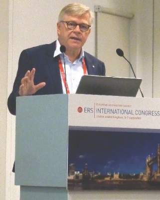
“LAMA monotherapy or ICS/LABA are standard options for treating patients with advanced COPD,” Jørgen Vestbo, MD, president of ERS and professor of respiratory medicine at the University of Manchester (England), said in an interview.
Dr. Vestbo, who was an investigator in both the TRINITY and TRILOGY trials, added that the Global Initiative for Chronic Obstructive Pulmonary Disease (GOLD) guidelines also mention that these drugs can be combined in patients who continue to experience COPD exacerbations. “But the evidence behind that is fairly weak,” he observed.
Although many patients are already being treated with triple therapy, this is via two inhalers, and “there have not been that many really good, long-term outcome studies” that have proven this approach to be the best way to manage those at risk for continued exacerbations of COPD, he said.
Drug companies are now starting to combine these three drugs into one inhaler, however, and this means that registration studies need to be done to get the products licensed, and so “there is an interest in coming up with the evidence,” Dr. Vestbo said.
“What is good about these two studies is that they are both 1-year studies and they are of sufficient size to give quite good estimates … These are studies that we should have done 5 years ago,” he said. Although the ideal is to have patients on as little therapy as possible, the results of TRINITY and TRILOGY now provide much needed evidence that it will work better than either LAMA or ICS/LABA.
The piece of evidence that is still missing is what the benefit, if any, is over a LAMA/LABA combination, a fact noted during discussion following the presentations of these data at the ERS meeting and in an editorial by Peter Calverley, MD, of the University of Liverpool (England) that accompanied the published TRILOGY findings (Lancet. 2016;388:937-8). There also is a question over whether twice daily is really better than once daily dosing, or vice versa.
“Until these next studies become available, we can be comforted by the knowledge that three therapies can be combined in a single inhaler which offers more effective therapy than at least one of the recommended treatment regimens for patients with severe COPD,” Dr. Calverley observed in reference to TRILOGY only.
Dr. Vestbo noted that at the time the TRILOGY and TRINITY studies were designed, there wasn’t the evidence from other studies such as the FLAME study (N Engl J Med. 2016. doi: 10.1056/NEJMoa1516385), showing the benefit of the LABA/LAMA combination over ICS/LABA. The TRILOGY and TRINITY studies “give that degree of evidence that was needed,” he said.
“I am not sure that the guidelines [for treating severe COPD] will change much, but at least they can say with better certainty that you can use the triple,” he added.
TRINITY – can triple better LAMA monotherapy?
The TRINITY study looked at whether patients with GOLD 3-4 COPD would be better off treated with LAMA monotherapy (tiotropium 18 mcg, one puff per day), the triple fixed-dose combination of BDP (100 mcg), FF (6 mcg), and GB (12.5 mcg) given via the novel pressurized metered-dose inhaler (two puffs twice daily), or a “free” triple combination of BDP (100 mcg) and FF (6 mcg) given via one pressurized metered-dose inhaler (two puffs daily) plus the same dose of the once-daily LAMA.
In all, 2,690 patients were randomized to these three treatments arms. The mean age of patients was 63 years and the majority (74%-77%) were men, with an average FEV1 predicted of 36% and one COPD exacerbation in the past year. Just under half of the study population were current smokers. Most (75%) had received prior treatment with an ICS/LABA combination, with about 11% receiving LAMA, and the rest either ICS/LAMA (3%), or LABA/LAMA (12%)
The annualized exacerbation rate (the primary endpoint) was 0.457 in the 1,077 patients who were treated with the triple fixed-dose combination versus 0.571 in the 1,074 patients who received tiotropium alone. The rate ratio was 0.8 indicating a 20% reduction in exacerbations was achieved (P = .003).
The annualized exacerbation rate in the 532 patients given the “free” triple combination (BDP/FF plus tiotropium) was 0.452, with a rate ratio of 0.790 (P =.010) versus those who received the LAMA as monotherapy.
There was no significant difference between the two triple combination strategies.
Presenting these data, Dr. Vestbo noted that the benefit was seen in preventing both severe and moderate COPD exacerbations. Significantly improved lung function, as measured by the change in FEV1 from baseline to week 52, was also observed to a greater degree with the triple therapy approaches than with the LAMA monotherapy.
“All three treatments were well tolerated and there were no particular safety concerns in this study,” he said.
TRILOGY – are three drugs better than two?
In contrast to the TRINITY study, the TRILOGY study looked at whether patients with severe COPD would be better off taking an ICS/LABA or the new triple fixed-dose combination pressurized metered-dose inhaler.
Just over 1,200 patients were recruited into the study, which had two co–primary endpoints: change from baseline to week 26 in predose morning FEV1 and 2-hour postdose FEV1, and the change in transition dyspnea index focal score at week 26.
Results showed that the triple fixed-dose combination improved predose FEV1 by 0.081 L and 2-hour postdose FEV1 by 0.117 L compared with the ICS/LABA combination (P less than .001 for both comparisons). Mean transition dyspnea index scores were 1.71 and 1.50, with a nonsignificant difference of 0.21.
“To be honest, I don’t think we had expected that [the triple combination] would mean much for patients, but we were hoping there would be a significant increase in lung function and a reduction of symptoms,” Dr. Vestbo said about the TRILOGY study. “What we saw was there was [symptomatic improvement] but it was not quite as impressive as we thought, but we reduced exacerbations.”
There was a significant, 23% reduction in the annualized exacerbation rate via the triple combination versus the ICS/LABA combination (0.41 vs 0.53, adjusted rate ratio 0.77, P = .005).
The triple approach was well tolerated, with no increase in adverse events versus the dual combination. The results support the idea that instead of giving patients an ICS/LABA at the start, better disease control can be achieved with a triple fixed-dose combination, Dr. Vestbo suggested.
Writing in The Lancet, Dr. Calverley noted: “The inability to meet one part of a co–primary endpoint clouds the interpretation of the other findings in the study, a familiar problem in COPD trials.” He added that there was a significant difference in the overall St George’s Respiratory Questionnaire scores favoring the triple over double therapy.
Chiesi Farmaceutici SpA funded the studies. Dr. Vestbo was an investigator for both TRINITY and TRILOGY and has received honoraria for advising and presenting from AstraZeneca, Boehringer Ingelheim, Chiesi, GlaxoSmithKline, and Novartis. Dr. Calverley has consulted for Boehringer Ingelheim, GlaxoSmithKline, and AstraZeneca.
LONDON – Phase III evidence confirms the multiple benefits of using a triple, fixed-dose combination (FDC) therapy over standard options in patients with severe chronic obstructive pulmonary disease (COPD), according to a presentation on two trials at the annual congress of the European Respiratory Society.
In the TRINITY trial, the combination of the inhaled corticosteroid (ICS) beclometasone diproprionate (BDP), the long-acting beta-agonist (LABA) formoterol fumarate (FF), and the long-acting muscarinic antagonist (LAMA) glycopyrronium bromide (GB) delivered via a single pressurized metered-dose inhaler (pMDI), was more effective at reducing exacerbations than was tiotropium bromide (Spiriva, Boehringer Ingelheim) monotherapy.
Results of the TRILOGY trial, which were simultaneously published in The Lancet (doi: 10.1016/S0140-6736(16)31354-X) at the time of their presentation at the ERS meeting, showed that the novel single-inhaler, triple fixed-dose combination could induce greater improvements in lung function when compared to a double fixed-dose combination of BDP and FF (Foster, Chiesi Farmaceutici SpA).
“LAMA monotherapy or ICS/LABA are standard options for treating patients with advanced COPD,” Jørgen Vestbo, MD, president of ERS and professor of respiratory medicine at the University of Manchester (England), said in an interview.
Dr. Vestbo, who was an investigator in both the TRINITY and TRILOGY trials, added that the Global Initiative for Chronic Obstructive Pulmonary Disease (GOLD) guidelines also mention that these drugs can be combined in patients who continue to experience COPD exacerbations. “But the evidence behind that is fairly weak,” he observed.
Although many patients are already being treated with triple therapy, this is via two inhalers, and “there have not been that many really good, long-term outcome studies” that have proven this approach to be the best way to manage those at risk for continued exacerbations of COPD, he said.
Drug companies are now starting to combine these three drugs into one inhaler, however, and this means that registration studies need to be done to get the products licensed, and so “there is an interest in coming up with the evidence,” Dr. Vestbo said.
“What is good about these two studies is that they are both 1-year studies and they are of sufficient size to give quite good estimates … These are studies that we should have done 5 years ago,” he said. Although the ideal is to have patients on as little therapy as possible, the results of TRINITY and TRILOGY now provide much needed evidence that it will work better than either LAMA or ICS/LABA.
The piece of evidence that is still missing is what the benefit, if any, is over a LAMA/LABA combination, a fact noted during discussion following the presentations of these data at the ERS meeting and in an editorial by Peter Calverley, MD, of the University of Liverpool (England) that accompanied the published TRILOGY findings (Lancet. 2016;388:937-8). There also is a question over whether twice daily is really better than once daily dosing, or vice versa.
“Until these next studies become available, we can be comforted by the knowledge that three therapies can be combined in a single inhaler which offers more effective therapy than at least one of the recommended treatment regimens for patients with severe COPD,” Dr. Calverley observed in reference to TRILOGY only.
Dr. Vestbo noted that at the time the TRILOGY and TRINITY studies were designed, there wasn’t the evidence from other studies such as the FLAME study (N Engl J Med. 2016. doi: 10.1056/NEJMoa1516385), showing the benefit of the LABA/LAMA combination over ICS/LABA. The TRILOGY and TRINITY studies “give that degree of evidence that was needed,” he said.
“I am not sure that the guidelines [for treating severe COPD] will change much, but at least they can say with better certainty that you can use the triple,” he added.
TRINITY – can triple better LAMA monotherapy?
The TRINITY study looked at whether patients with GOLD 3-4 COPD would be better off treated with LAMA monotherapy (tiotropium 18 mcg, one puff per day), the triple fixed-dose combination of BDP (100 mcg), FF (6 mcg), and GB (12.5 mcg) given via the novel pressurized metered-dose inhaler (two puffs twice daily), or a “free” triple combination of BDP (100 mcg) and FF (6 mcg) given via one pressurized metered-dose inhaler (two puffs daily) plus the same dose of the once-daily LAMA.
In all, 2,690 patients were randomized to these three treatments arms. The mean age of patients was 63 years and the majority (74%-77%) were men, with an average FEV1 predicted of 36% and one COPD exacerbation in the past year. Just under half of the study population were current smokers. Most (75%) had received prior treatment with an ICS/LABA combination, with about 11% receiving LAMA, and the rest either ICS/LAMA (3%), or LABA/LAMA (12%)
The annualized exacerbation rate (the primary endpoint) was 0.457 in the 1,077 patients who were treated with the triple fixed-dose combination versus 0.571 in the 1,074 patients who received tiotropium alone. The rate ratio was 0.8 indicating a 20% reduction in exacerbations was achieved (P = .003).
The annualized exacerbation rate in the 532 patients given the “free” triple combination (BDP/FF plus tiotropium) was 0.452, with a rate ratio of 0.790 (P =.010) versus those who received the LAMA as monotherapy.
There was no significant difference between the two triple combination strategies.
Presenting these data, Dr. Vestbo noted that the benefit was seen in preventing both severe and moderate COPD exacerbations. Significantly improved lung function, as measured by the change in FEV1 from baseline to week 52, was also observed to a greater degree with the triple therapy approaches than with the LAMA monotherapy.
“All three treatments were well tolerated and there were no particular safety concerns in this study,” he said.
TRILOGY – are three drugs better than two?
In contrast to the TRINITY study, the TRILOGY study looked at whether patients with severe COPD would be better off taking an ICS/LABA or the new triple fixed-dose combination pressurized metered-dose inhaler.
Just over 1,200 patients were recruited into the study, which had two co–primary endpoints: change from baseline to week 26 in predose morning FEV1 and 2-hour postdose FEV1, and the change in transition dyspnea index focal score at week 26.
Results showed that the triple fixed-dose combination improved predose FEV1 by 0.081 L and 2-hour postdose FEV1 by 0.117 L compared with the ICS/LABA combination (P less than .001 for both comparisons). Mean transition dyspnea index scores were 1.71 and 1.50, with a nonsignificant difference of 0.21.
“To be honest, I don’t think we had expected that [the triple combination] would mean much for patients, but we were hoping there would be a significant increase in lung function and a reduction of symptoms,” Dr. Vestbo said about the TRILOGY study. “What we saw was there was [symptomatic improvement] but it was not quite as impressive as we thought, but we reduced exacerbations.”
There was a significant, 23% reduction in the annualized exacerbation rate via the triple combination versus the ICS/LABA combination (0.41 vs 0.53, adjusted rate ratio 0.77, P = .005).
The triple approach was well tolerated, with no increase in adverse events versus the dual combination. The results support the idea that instead of giving patients an ICS/LABA at the start, better disease control can be achieved with a triple fixed-dose combination, Dr. Vestbo suggested.
Writing in The Lancet, Dr. Calverley noted: “The inability to meet one part of a co–primary endpoint clouds the interpretation of the other findings in the study, a familiar problem in COPD trials.” He added that there was a significant difference in the overall St George’s Respiratory Questionnaire scores favoring the triple over double therapy.
Chiesi Farmaceutici SpA funded the studies. Dr. Vestbo was an investigator for both TRINITY and TRILOGY and has received honoraria for advising and presenting from AstraZeneca, Boehringer Ingelheim, Chiesi, GlaxoSmithKline, and Novartis. Dr. Calverley has consulted for Boehringer Ingelheim, GlaxoSmithKline, and AstraZeneca.
LONDON – Phase III evidence confirms the multiple benefits of using a triple, fixed-dose combination (FDC) therapy over standard options in patients with severe chronic obstructive pulmonary disease (COPD), according to a presentation on two trials at the annual congress of the European Respiratory Society.
In the TRINITY trial, the combination of the inhaled corticosteroid (ICS) beclometasone diproprionate (BDP), the long-acting beta-agonist (LABA) formoterol fumarate (FF), and the long-acting muscarinic antagonist (LAMA) glycopyrronium bromide (GB) delivered via a single pressurized metered-dose inhaler (pMDI), was more effective at reducing exacerbations than was tiotropium bromide (Spiriva, Boehringer Ingelheim) monotherapy.
Results of the TRILOGY trial, which were simultaneously published in The Lancet (doi: 10.1016/S0140-6736(16)31354-X) at the time of their presentation at the ERS meeting, showed that the novel single-inhaler, triple fixed-dose combination could induce greater improvements in lung function when compared to a double fixed-dose combination of BDP and FF (Foster, Chiesi Farmaceutici SpA).
“LAMA monotherapy or ICS/LABA are standard options for treating patients with advanced COPD,” Jørgen Vestbo, MD, president of ERS and professor of respiratory medicine at the University of Manchester (England), said in an interview.
Dr. Vestbo, who was an investigator in both the TRINITY and TRILOGY trials, added that the Global Initiative for Chronic Obstructive Pulmonary Disease (GOLD) guidelines also mention that these drugs can be combined in patients who continue to experience COPD exacerbations. “But the evidence behind that is fairly weak,” he observed.
Although many patients are already being treated with triple therapy, this is via two inhalers, and “there have not been that many really good, long-term outcome studies” that have proven this approach to be the best way to manage those at risk for continued exacerbations of COPD, he said.
Drug companies are now starting to combine these three drugs into one inhaler, however, and this means that registration studies need to be done to get the products licensed, and so “there is an interest in coming up with the evidence,” Dr. Vestbo said.
“What is good about these two studies is that they are both 1-year studies and they are of sufficient size to give quite good estimates … These are studies that we should have done 5 years ago,” he said. Although the ideal is to have patients on as little therapy as possible, the results of TRINITY and TRILOGY now provide much needed evidence that it will work better than either LAMA or ICS/LABA.
The piece of evidence that is still missing is what the benefit, if any, is over a LAMA/LABA combination, a fact noted during discussion following the presentations of these data at the ERS meeting and in an editorial by Peter Calverley, MD, of the University of Liverpool (England) that accompanied the published TRILOGY findings (Lancet. 2016;388:937-8). There also is a question over whether twice daily is really better than once daily dosing, or vice versa.
“Until these next studies become available, we can be comforted by the knowledge that three therapies can be combined in a single inhaler which offers more effective therapy than at least one of the recommended treatment regimens for patients with severe COPD,” Dr. Calverley observed in reference to TRILOGY only.
Dr. Vestbo noted that at the time the TRILOGY and TRINITY studies were designed, there wasn’t the evidence from other studies such as the FLAME study (N Engl J Med. 2016. doi: 10.1056/NEJMoa1516385), showing the benefit of the LABA/LAMA combination over ICS/LABA. The TRILOGY and TRINITY studies “give that degree of evidence that was needed,” he said.
“I am not sure that the guidelines [for treating severe COPD] will change much, but at least they can say with better certainty that you can use the triple,” he added.
TRINITY – can triple better LAMA monotherapy?
The TRINITY study looked at whether patients with GOLD 3-4 COPD would be better off treated with LAMA monotherapy (tiotropium 18 mcg, one puff per day), the triple fixed-dose combination of BDP (100 mcg), FF (6 mcg), and GB (12.5 mcg) given via the novel pressurized metered-dose inhaler (two puffs twice daily), or a “free” triple combination of BDP (100 mcg) and FF (6 mcg) given via one pressurized metered-dose inhaler (two puffs daily) plus the same dose of the once-daily LAMA.
In all, 2,690 patients were randomized to these three treatments arms. The mean age of patients was 63 years and the majority (74%-77%) were men, with an average FEV1 predicted of 36% and one COPD exacerbation in the past year. Just under half of the study population were current smokers. Most (75%) had received prior treatment with an ICS/LABA combination, with about 11% receiving LAMA, and the rest either ICS/LAMA (3%), or LABA/LAMA (12%)
The annualized exacerbation rate (the primary endpoint) was 0.457 in the 1,077 patients who were treated with the triple fixed-dose combination versus 0.571 in the 1,074 patients who received tiotropium alone. The rate ratio was 0.8 indicating a 20% reduction in exacerbations was achieved (P = .003).
The annualized exacerbation rate in the 532 patients given the “free” triple combination (BDP/FF plus tiotropium) was 0.452, with a rate ratio of 0.790 (P =.010) versus those who received the LAMA as monotherapy.
There was no significant difference between the two triple combination strategies.
Presenting these data, Dr. Vestbo noted that the benefit was seen in preventing both severe and moderate COPD exacerbations. Significantly improved lung function, as measured by the change in FEV1 from baseline to week 52, was also observed to a greater degree with the triple therapy approaches than with the LAMA monotherapy.
“All three treatments were well tolerated and there were no particular safety concerns in this study,” he said.
TRILOGY – are three drugs better than two?
In contrast to the TRINITY study, the TRILOGY study looked at whether patients with severe COPD would be better off taking an ICS/LABA or the new triple fixed-dose combination pressurized metered-dose inhaler.
Just over 1,200 patients were recruited into the study, which had two co–primary endpoints: change from baseline to week 26 in predose morning FEV1 and 2-hour postdose FEV1, and the change in transition dyspnea index focal score at week 26.
Results showed that the triple fixed-dose combination improved predose FEV1 by 0.081 L and 2-hour postdose FEV1 by 0.117 L compared with the ICS/LABA combination (P less than .001 for both comparisons). Mean transition dyspnea index scores were 1.71 and 1.50, with a nonsignificant difference of 0.21.
“To be honest, I don’t think we had expected that [the triple combination] would mean much for patients, but we were hoping there would be a significant increase in lung function and a reduction of symptoms,” Dr. Vestbo said about the TRILOGY study. “What we saw was there was [symptomatic improvement] but it was not quite as impressive as we thought, but we reduced exacerbations.”
There was a significant, 23% reduction in the annualized exacerbation rate via the triple combination versus the ICS/LABA combination (0.41 vs 0.53, adjusted rate ratio 0.77, P = .005).
The triple approach was well tolerated, with no increase in adverse events versus the dual combination. The results support the idea that instead of giving patients an ICS/LABA at the start, better disease control can be achieved with a triple fixed-dose combination, Dr. Vestbo suggested.
Writing in The Lancet, Dr. Calverley noted: “The inability to meet one part of a co–primary endpoint clouds the interpretation of the other findings in the study, a familiar problem in COPD trials.” He added that there was a significant difference in the overall St George’s Respiratory Questionnaire scores favoring the triple over double therapy.
Chiesi Farmaceutici SpA funded the studies. Dr. Vestbo was an investigator for both TRINITY and TRILOGY and has received honoraria for advising and presenting from AstraZeneca, Boehringer Ingelheim, Chiesi, GlaxoSmithKline, and Novartis. Dr. Calverley has consulted for Boehringer Ingelheim, GlaxoSmithKline, and AstraZeneca.
AT THE ERS CONGRESS 2016
Key clinical point: A triple, fixed-dose combination therapy delivered by a single inhaler could be a new treatment option for severe COPD.
Major finding: Exacerbations were reduced by 20% with the triple combination versus current standards of care for COPD.
Data source: TRINITY and TRILOGY: Two 1-year, multicenter, randomized, double-blind, active controlled, parallel group, phase III studies of more than 4,000 patients with severe COPD.
Disclosures: Chiesi Farmaceutici SpA funded the studies. Dr. Vestbo was an investigator for both TRINITY and TRILOGY and has received honoraria for advising and presenting from AstraZeneca, Boehringer Ingelheim, Chiesi, GlaxoSmithKline, and Novartis. Dr. Calverley has consulted for Boehringer Ingelheim, GlaxoSmithKline, and AstraZeneca.
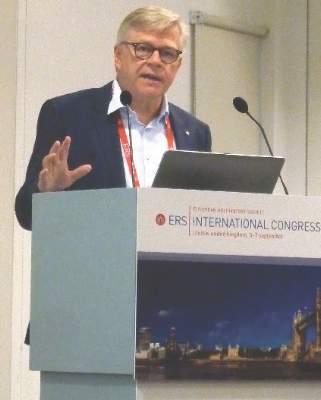
Sequential protocol benefits patients with field cancerization
BOSTON – Field cancerization poses unique treatment challenges, but useful approaches to managing patients with such extreme dermatoheliosis are emerging, according to Anokhi Jambusaria-Pahlajani, MD.
In patients with extensive sun damage to the head and neck, she said she performs initial curettage of any hyperkeratotic actinic keratoses, followed by topical 0.5% 5-fluorouracil applied twice daily on postcurettage days 1 through 5. On day 6, she performs aminolevulinic acid photodynamic therapy (PDT) with 1 hour of incubation.
“Basically, on day zero they come into my office, and I look for anything that’s hyperkeratotic, and I numb it up and curette it. The goal of that is to get rid of the scale so that when I do the topical 5-fluorouracil or I do the photodynamic therapy, the medicine is actually getting to where we want it to go,” Dr. Jambusaria-Pahlajani, a dermatologist in Round Rock, Tex., said at the American Academy of Dermatology summer meeting.
She discussed one such patient – a lung transplant recipient – in whom this approach worked particularly well. At 6 months’ follow-up, the patient had had a significant response, and during a follow-up of at least 2 years, he did not develop a single actinic keratosis.
Early results with this combination approach were so encouraging that she and her colleagues published a case series of four solid organ transplant recipients. “All four patients tolerated the approach well, demonstrated excellent response to treatment with complete or near complete resolution of actinic keratosis and squamous cell carcinoma in situ lesions,” she said. At 1-6 months following treatment, “basically patients were clear,” she added (Dermatol Surg. 2016 Jan;42:S66-72).
Dr. Jambusaria-Pahlajani also said she has seen “great results” in most of the 30-40 other patients she has treated using this approach. “I’ve followed up patients for years, and they really haven’t had a problem like what they had prior to getting this treatment,” she said.
She noted that, anecdotally, many patients who had been treated with multiple courses of topical 5-fluorouracil monotherapy or photodynamic therapy actually preferred the combination approach. They felt that they were out of commission for a shorter period of time, and most of the patients “tended to be kind of red and inflamed for about 7-10 days total from start to finish, which was a lot better than with, for example, 5-fluorouracil,” she said.
One patient – another lung transplant recipient – initially responded to combination therapy, but returned in 3 months with numerous recurrent lesions. Based on findings from a 2010 study, a cyclic approach to PDT was tried in this patient. In that study of 12 solid organ transplant recipients, cyclic PDT reduced the incidence of squamous cell carcinoma (Dermatol Surg. 2010 May;36[5]:652-8).
The study subjects had a high burden of squamous cell carcinoma in situ. The cyclic PDT they received included blue light PDT with 1 hour of incubation every 4-8 weeks for 2 years. The median number of new invasive and in situ squamous cell carcinomas declined from about 20 to 4 in the first year (a 79% reduction vs. 1 month prior to first treatment) and to 1 at 2 years (a 95% reduction).
“This patient happened to be a transplant patient as well, but I think you could also use [this approach] in nontransplant patients,” Dr. Jambusaria-Pahlajani said. His response was “pretty significant,” and after 18 months of treatment he was doing well.
“The main thing that you have to remember, though, is that these patients have to be willing to come in every 4-8 weeks to get the PDT,” which, she added, “is not for every patient.” It’s also not for every provider. Aside from staff or resource access limitations, these treatments can be very time consuming and costly, she noted. “Treating these patients can be a challenge, but it can be rewarding, as well,” she commented.
She reported having no relevant financial disclosures.
BOSTON – Field cancerization poses unique treatment challenges, but useful approaches to managing patients with such extreme dermatoheliosis are emerging, according to Anokhi Jambusaria-Pahlajani, MD.
In patients with extensive sun damage to the head and neck, she said she performs initial curettage of any hyperkeratotic actinic keratoses, followed by topical 0.5% 5-fluorouracil applied twice daily on postcurettage days 1 through 5. On day 6, she performs aminolevulinic acid photodynamic therapy (PDT) with 1 hour of incubation.
“Basically, on day zero they come into my office, and I look for anything that’s hyperkeratotic, and I numb it up and curette it. The goal of that is to get rid of the scale so that when I do the topical 5-fluorouracil or I do the photodynamic therapy, the medicine is actually getting to where we want it to go,” Dr. Jambusaria-Pahlajani, a dermatologist in Round Rock, Tex., said at the American Academy of Dermatology summer meeting.
She discussed one such patient – a lung transplant recipient – in whom this approach worked particularly well. At 6 months’ follow-up, the patient had had a significant response, and during a follow-up of at least 2 years, he did not develop a single actinic keratosis.
Early results with this combination approach were so encouraging that she and her colleagues published a case series of four solid organ transplant recipients. “All four patients tolerated the approach well, demonstrated excellent response to treatment with complete or near complete resolution of actinic keratosis and squamous cell carcinoma in situ lesions,” she said. At 1-6 months following treatment, “basically patients were clear,” she added (Dermatol Surg. 2016 Jan;42:S66-72).
Dr. Jambusaria-Pahlajani also said she has seen “great results” in most of the 30-40 other patients she has treated using this approach. “I’ve followed up patients for years, and they really haven’t had a problem like what they had prior to getting this treatment,” she said.
She noted that, anecdotally, many patients who had been treated with multiple courses of topical 5-fluorouracil monotherapy or photodynamic therapy actually preferred the combination approach. They felt that they were out of commission for a shorter period of time, and most of the patients “tended to be kind of red and inflamed for about 7-10 days total from start to finish, which was a lot better than with, for example, 5-fluorouracil,” she said.
One patient – another lung transplant recipient – initially responded to combination therapy, but returned in 3 months with numerous recurrent lesions. Based on findings from a 2010 study, a cyclic approach to PDT was tried in this patient. In that study of 12 solid organ transplant recipients, cyclic PDT reduced the incidence of squamous cell carcinoma (Dermatol Surg. 2010 May;36[5]:652-8).
The study subjects had a high burden of squamous cell carcinoma in situ. The cyclic PDT they received included blue light PDT with 1 hour of incubation every 4-8 weeks for 2 years. The median number of new invasive and in situ squamous cell carcinomas declined from about 20 to 4 in the first year (a 79% reduction vs. 1 month prior to first treatment) and to 1 at 2 years (a 95% reduction).
“This patient happened to be a transplant patient as well, but I think you could also use [this approach] in nontransplant patients,” Dr. Jambusaria-Pahlajani said. His response was “pretty significant,” and after 18 months of treatment he was doing well.
“The main thing that you have to remember, though, is that these patients have to be willing to come in every 4-8 weeks to get the PDT,” which, she added, “is not for every patient.” It’s also not for every provider. Aside from staff or resource access limitations, these treatments can be very time consuming and costly, she noted. “Treating these patients can be a challenge, but it can be rewarding, as well,” she commented.
She reported having no relevant financial disclosures.
BOSTON – Field cancerization poses unique treatment challenges, but useful approaches to managing patients with such extreme dermatoheliosis are emerging, according to Anokhi Jambusaria-Pahlajani, MD.
In patients with extensive sun damage to the head and neck, she said she performs initial curettage of any hyperkeratotic actinic keratoses, followed by topical 0.5% 5-fluorouracil applied twice daily on postcurettage days 1 through 5. On day 6, she performs aminolevulinic acid photodynamic therapy (PDT) with 1 hour of incubation.
“Basically, on day zero they come into my office, and I look for anything that’s hyperkeratotic, and I numb it up and curette it. The goal of that is to get rid of the scale so that when I do the topical 5-fluorouracil or I do the photodynamic therapy, the medicine is actually getting to where we want it to go,” Dr. Jambusaria-Pahlajani, a dermatologist in Round Rock, Tex., said at the American Academy of Dermatology summer meeting.
She discussed one such patient – a lung transplant recipient – in whom this approach worked particularly well. At 6 months’ follow-up, the patient had had a significant response, and during a follow-up of at least 2 years, he did not develop a single actinic keratosis.
Early results with this combination approach were so encouraging that she and her colleagues published a case series of four solid organ transplant recipients. “All four patients tolerated the approach well, demonstrated excellent response to treatment with complete or near complete resolution of actinic keratosis and squamous cell carcinoma in situ lesions,” she said. At 1-6 months following treatment, “basically patients were clear,” she added (Dermatol Surg. 2016 Jan;42:S66-72).
Dr. Jambusaria-Pahlajani also said she has seen “great results” in most of the 30-40 other patients she has treated using this approach. “I’ve followed up patients for years, and they really haven’t had a problem like what they had prior to getting this treatment,” she said.
She noted that, anecdotally, many patients who had been treated with multiple courses of topical 5-fluorouracil monotherapy or photodynamic therapy actually preferred the combination approach. They felt that they were out of commission for a shorter period of time, and most of the patients “tended to be kind of red and inflamed for about 7-10 days total from start to finish, which was a lot better than with, for example, 5-fluorouracil,” she said.
One patient – another lung transplant recipient – initially responded to combination therapy, but returned in 3 months with numerous recurrent lesions. Based on findings from a 2010 study, a cyclic approach to PDT was tried in this patient. In that study of 12 solid organ transplant recipients, cyclic PDT reduced the incidence of squamous cell carcinoma (Dermatol Surg. 2010 May;36[5]:652-8).
The study subjects had a high burden of squamous cell carcinoma in situ. The cyclic PDT they received included blue light PDT with 1 hour of incubation every 4-8 weeks for 2 years. The median number of new invasive and in situ squamous cell carcinomas declined from about 20 to 4 in the first year (a 79% reduction vs. 1 month prior to first treatment) and to 1 at 2 years (a 95% reduction).
“This patient happened to be a transplant patient as well, but I think you could also use [this approach] in nontransplant patients,” Dr. Jambusaria-Pahlajani said. His response was “pretty significant,” and after 18 months of treatment he was doing well.
“The main thing that you have to remember, though, is that these patients have to be willing to come in every 4-8 weeks to get the PDT,” which, she added, “is not for every patient.” It’s also not for every provider. Aside from staff or resource access limitations, these treatments can be very time consuming and costly, she noted. “Treating these patients can be a challenge, but it can be rewarding, as well,” she commented.
She reported having no relevant financial disclosures.
EXPERT ANALYSIS FROM AAD SUMMER ACADEMY 2016
Durable Efficacy of Cladribine Tablets After Conversion to Clinically Definite MS

LONDON—Significant treatment effect versus placebo of cladribine tablets given to patients with clinically isolated syndrome during the initial treatment period continues to be observed in patients who convert to clinically definite multiple sclerosis (MS) and switch to treatment with a different disease modifying drug (ie, subcutaneous interferon beta-1a), according to data presented at the 32nd Congress of the European Committee for Treatment and Research in Multiple Sclerosis (ECTRIMS).
Giancarlo Comi, MD, Professor of Neurology and Chairman of the Department of Neurology at Vita-Salute San Raffaele University in Milan, Italy, and colleagues reported that patients with clinically isolated syndrome who had been treated with cladribine tablets and who had converted to MS during the Oral Cladribine in Early Multiple Sclerosis (ORACLE-MS) initial treatment period had lower annualized relapse rates during the open-label maintenance period, relative to those patients who had received placebo during the ORACLE-MS initial treatment period.
The CLARITY (CLAdRIbine Tablets treating multiple sclerosis orallY) study in patients with active MS showed that annualized relapse rates and sustained disability worsening were reduced in patients treated with cladribine tablets annually for two years in short-duration courses, compared with placebo. The efficacy observed in CLARITY was maintained without further active treatment during the CLARITY extension study. In the ORACLE-MS study in patients with a first demyelinating event, cladribine tablets (3.5 mg/kg and 5.25 mg/kg) significantly reduced the risk of conversion to clinically definite MS, compared with placebo. If clinically definite MS occurred in the double-blinded, initial treatment period, patients were treated with subcutaneous interferon beta-1a in an open-label maintenance period.
The present study was designed to assess the annualized relapse rate during the ORACLE-MS open-label maintenance period in patients randomized to cladribine (3.5 mg/kg and 5.25 mg/kg) or placebo in the initial treatment period.
Similar to previous trials, participation in the ORACLE-MS open-label maintenance period was dependent upon the clinical course of the patient’s disease in the initial treatment period. Patients in ORACLE-MS who converted to clinically definite MS (according to Poser criteria) during the initial treatment period entered the open-label maintenance period and were treated with subcutaneous interferon beta-1a (titrated over four weeks up to the dose of 44 μg) administered three times per week.
A total of 109 patients in ORACLE-MS converted to clinically definite MS in the initial treatment period and received at least one dose of interferon beta-1a. The median time on interferon beta-1a was 56.0 weeks. Estimated annualized relapse rates in the open-label maintenance period were 0.14 for patients (n = 25) originally treated with cladribine 3.5 mg/kg, 0.24 for patients (n = 24) originally treated with cladribine 5.25 mg/kg, and 0.42 for patients (n = 60) who originally received placebo in the initial treatment period.
According to the researchers, durable efficacy of cladribine tablets in ORACLE-MS into the open-label maintenance period is consistent with results of the CLARITY and CLARITY extension studies.
This study was sponsored by EMD Serono Inc.
—Glenn S. Williams
Suggested Reading
Cook S, Vermersch P, Comi G, et al. Safety and tolerability of cladribine tablets in multiple sclerosis: the CLARITY (CLAdRIbine Tablets treating multiple sclerosis orallY) study. Mult Scler. 2011;17(5):578-593.
Leist TP, Comi G, Cree BA, et al. Effect of oral cladribine on time to conversion to clinically definite multiple sclerosis in patients with a first demyelinating event (ORACLE MS): a phase 3 randomised trial. Lancet Neurol. 2014;13(3):257-267.
LONDON—Significant treatment effect versus placebo of cladribine tablets given to patients with clinically isolated syndrome during the initial treatment period continues to be observed in patients who convert to clinically definite multiple sclerosis (MS) and switch to treatment with a different disease modifying drug (ie, subcutaneous interferon beta-1a), according to data presented at the 32nd Congress of the European Committee for Treatment and Research in Multiple Sclerosis (ECTRIMS).
Giancarlo Comi, MD, Professor of Neurology and Chairman of the Department of Neurology at Vita-Salute San Raffaele University in Milan, Italy, and colleagues reported that patients with clinically isolated syndrome who had been treated with cladribine tablets and who had converted to MS during the Oral Cladribine in Early Multiple Sclerosis (ORACLE-MS) initial treatment period had lower annualized relapse rates during the open-label maintenance period, relative to those patients who had received placebo during the ORACLE-MS initial treatment period.
The CLARITY (CLAdRIbine Tablets treating multiple sclerosis orallY) study in patients with active MS showed that annualized relapse rates and sustained disability worsening were reduced in patients treated with cladribine tablets annually for two years in short-duration courses, compared with placebo. The efficacy observed in CLARITY was maintained without further active treatment during the CLARITY extension study. In the ORACLE-MS study in patients with a first demyelinating event, cladribine tablets (3.5 mg/kg and 5.25 mg/kg) significantly reduced the risk of conversion to clinically definite MS, compared with placebo. If clinically definite MS occurred in the double-blinded, initial treatment period, patients were treated with subcutaneous interferon beta-1a in an open-label maintenance period.
The present study was designed to assess the annualized relapse rate during the ORACLE-MS open-label maintenance period in patients randomized to cladribine (3.5 mg/kg and 5.25 mg/kg) or placebo in the initial treatment period.
Similar to previous trials, participation in the ORACLE-MS open-label maintenance period was dependent upon the clinical course of the patient’s disease in the initial treatment period. Patients in ORACLE-MS who converted to clinically definite MS (according to Poser criteria) during the initial treatment period entered the open-label maintenance period and were treated with subcutaneous interferon beta-1a (titrated over four weeks up to the dose of 44 μg) administered three times per week.
A total of 109 patients in ORACLE-MS converted to clinically definite MS in the initial treatment period and received at least one dose of interferon beta-1a. The median time on interferon beta-1a was 56.0 weeks. Estimated annualized relapse rates in the open-label maintenance period were 0.14 for patients (n = 25) originally treated with cladribine 3.5 mg/kg, 0.24 for patients (n = 24) originally treated with cladribine 5.25 mg/kg, and 0.42 for patients (n = 60) who originally received placebo in the initial treatment period.
According to the researchers, durable efficacy of cladribine tablets in ORACLE-MS into the open-label maintenance period is consistent with results of the CLARITY and CLARITY extension studies.
This study was sponsored by EMD Serono Inc.
—Glenn S. Williams
LONDON—Significant treatment effect versus placebo of cladribine tablets given to patients with clinically isolated syndrome during the initial treatment period continues to be observed in patients who convert to clinically definite multiple sclerosis (MS) and switch to treatment with a different disease modifying drug (ie, subcutaneous interferon beta-1a), according to data presented at the 32nd Congress of the European Committee for Treatment and Research in Multiple Sclerosis (ECTRIMS).
Giancarlo Comi, MD, Professor of Neurology and Chairman of the Department of Neurology at Vita-Salute San Raffaele University in Milan, Italy, and colleagues reported that patients with clinically isolated syndrome who had been treated with cladribine tablets and who had converted to MS during the Oral Cladribine in Early Multiple Sclerosis (ORACLE-MS) initial treatment period had lower annualized relapse rates during the open-label maintenance period, relative to those patients who had received placebo during the ORACLE-MS initial treatment period.
The CLARITY (CLAdRIbine Tablets treating multiple sclerosis orallY) study in patients with active MS showed that annualized relapse rates and sustained disability worsening were reduced in patients treated with cladribine tablets annually for two years in short-duration courses, compared with placebo. The efficacy observed in CLARITY was maintained without further active treatment during the CLARITY extension study. In the ORACLE-MS study in patients with a first demyelinating event, cladribine tablets (3.5 mg/kg and 5.25 mg/kg) significantly reduced the risk of conversion to clinically definite MS, compared with placebo. If clinically definite MS occurred in the double-blinded, initial treatment period, patients were treated with subcutaneous interferon beta-1a in an open-label maintenance period.
The present study was designed to assess the annualized relapse rate during the ORACLE-MS open-label maintenance period in patients randomized to cladribine (3.5 mg/kg and 5.25 mg/kg) or placebo in the initial treatment period.
Similar to previous trials, participation in the ORACLE-MS open-label maintenance period was dependent upon the clinical course of the patient’s disease in the initial treatment period. Patients in ORACLE-MS who converted to clinically definite MS (according to Poser criteria) during the initial treatment period entered the open-label maintenance period and were treated with subcutaneous interferon beta-1a (titrated over four weeks up to the dose of 44 μg) administered three times per week.
A total of 109 patients in ORACLE-MS converted to clinically definite MS in the initial treatment period and received at least one dose of interferon beta-1a. The median time on interferon beta-1a was 56.0 weeks. Estimated annualized relapse rates in the open-label maintenance period were 0.14 for patients (n = 25) originally treated with cladribine 3.5 mg/kg, 0.24 for patients (n = 24) originally treated with cladribine 5.25 mg/kg, and 0.42 for patients (n = 60) who originally received placebo in the initial treatment period.
According to the researchers, durable efficacy of cladribine tablets in ORACLE-MS into the open-label maintenance period is consistent with results of the CLARITY and CLARITY extension studies.
This study was sponsored by EMD Serono Inc.
—Glenn S. Williams
Suggested Reading
Cook S, Vermersch P, Comi G, et al. Safety and tolerability of cladribine tablets in multiple sclerosis: the CLARITY (CLAdRIbine Tablets treating multiple sclerosis orallY) study. Mult Scler. 2011;17(5):578-593.
Leist TP, Comi G, Cree BA, et al. Effect of oral cladribine on time to conversion to clinically definite multiple sclerosis in patients with a first demyelinating event (ORACLE MS): a phase 3 randomised trial. Lancet Neurol. 2014;13(3):257-267.
Suggested Reading
Cook S, Vermersch P, Comi G, et al. Safety and tolerability of cladribine tablets in multiple sclerosis: the CLARITY (CLAdRIbine Tablets treating multiple sclerosis orallY) study. Mult Scler. 2011;17(5):578-593.
Leist TP, Comi G, Cree BA, et al. Effect of oral cladribine on time to conversion to clinically definite multiple sclerosis in patients with a first demyelinating event (ORACLE MS): a phase 3 randomised trial. Lancet Neurol. 2014;13(3):257-267.
More Ammunition in the Battle Against Opioid Overdoses
In 2014, more people died of drug overdoses than in any other year, and the majority of the deaths involved an opioid. In fact, “more Americans die from drug overdoses than car crashes,” said Sylvia Burwell, HHS secretary, in announcing new actions the department is taking to combat the opioid epidemic.
Related: Call for App to Help Opioid Rehab
The actions build on the HHS Opioid Initiative, launched March 2015, and the National Pain Strategy, the government’s first coordinated plan to reduce the burden of chronic pain in the U.S. The programs focus on 3 priorities: improving opioid prescribing practices, expanding access to medication-assisted treatment, and increasing use of naloxone to reverse overdoses.
Among the changes: SAMHSA finalized a rule to allow practitioners who can prescribe buprenorphine for up to 100 patients for a year or more now to treat up to 275 patients. Practitioners can obtain the waiver for the increase if they have additional credentialing in addiction medicine or addiction psychiatry from a specialty medical board and/or professional society, or practice in a qualified setting.
Related: Lowering Veterans’ Opioid Use and Reducing Overdose Risk
Another important change was to the IHS Prescription Drug Monitoring Program (PDMP) policy. Although many IHS clinicians already use PDMP databases, opioid prescribers and pharmacists will now be required to check state PDMP databases before prescribing or dispensing any opioid for > 7 days. The goal is to help improve pain management care, identify patients who may have a misuse problem, and prevent diversion of drugs. The new policy is effective immediately for > 1,200 clinicians working in IHS federally operated facilities. The IHS has also announced that it will train hundreds of law enforcement officers of the Bureau of Indian Affairs on how to use naloxone and provide them with the drug.
The VA is releasing a new policy as well that requires health care providers who prescribe controlled substances to check State PDMPs before prescribing, at least once a year and/or when clinically indicated for renewal or continuation of therapy.
Related: Veterans’ Health and Opioid Safety–Contexts, Risks, and Outreach Implications
President Obama’s budget requests $1.1 billion in new mandatory and discretionary investments over fiscal years 2017 and 2018, to expand access to treatment and prevent opioid misuse and abuse.
In 2014, more people died of drug overdoses than in any other year, and the majority of the deaths involved an opioid. In fact, “more Americans die from drug overdoses than car crashes,” said Sylvia Burwell, HHS secretary, in announcing new actions the department is taking to combat the opioid epidemic.
Related: Call for App to Help Opioid Rehab
The actions build on the HHS Opioid Initiative, launched March 2015, and the National Pain Strategy, the government’s first coordinated plan to reduce the burden of chronic pain in the U.S. The programs focus on 3 priorities: improving opioid prescribing practices, expanding access to medication-assisted treatment, and increasing use of naloxone to reverse overdoses.
Among the changes: SAMHSA finalized a rule to allow practitioners who can prescribe buprenorphine for up to 100 patients for a year or more now to treat up to 275 patients. Practitioners can obtain the waiver for the increase if they have additional credentialing in addiction medicine or addiction psychiatry from a specialty medical board and/or professional society, or practice in a qualified setting.
Related: Lowering Veterans’ Opioid Use and Reducing Overdose Risk
Another important change was to the IHS Prescription Drug Monitoring Program (PDMP) policy. Although many IHS clinicians already use PDMP databases, opioid prescribers and pharmacists will now be required to check state PDMP databases before prescribing or dispensing any opioid for > 7 days. The goal is to help improve pain management care, identify patients who may have a misuse problem, and prevent diversion of drugs. The new policy is effective immediately for > 1,200 clinicians working in IHS federally operated facilities. The IHS has also announced that it will train hundreds of law enforcement officers of the Bureau of Indian Affairs on how to use naloxone and provide them with the drug.
The VA is releasing a new policy as well that requires health care providers who prescribe controlled substances to check State PDMPs before prescribing, at least once a year and/or when clinically indicated for renewal or continuation of therapy.
Related: Veterans’ Health and Opioid Safety–Contexts, Risks, and Outreach Implications
President Obama’s budget requests $1.1 billion in new mandatory and discretionary investments over fiscal years 2017 and 2018, to expand access to treatment and prevent opioid misuse and abuse.
In 2014, more people died of drug overdoses than in any other year, and the majority of the deaths involved an opioid. In fact, “more Americans die from drug overdoses than car crashes,” said Sylvia Burwell, HHS secretary, in announcing new actions the department is taking to combat the opioid epidemic.
Related: Call for App to Help Opioid Rehab
The actions build on the HHS Opioid Initiative, launched March 2015, and the National Pain Strategy, the government’s first coordinated plan to reduce the burden of chronic pain in the U.S. The programs focus on 3 priorities: improving opioid prescribing practices, expanding access to medication-assisted treatment, and increasing use of naloxone to reverse overdoses.
Among the changes: SAMHSA finalized a rule to allow practitioners who can prescribe buprenorphine for up to 100 patients for a year or more now to treat up to 275 patients. Practitioners can obtain the waiver for the increase if they have additional credentialing in addiction medicine or addiction psychiatry from a specialty medical board and/or professional society, or practice in a qualified setting.
Related: Lowering Veterans’ Opioid Use and Reducing Overdose Risk
Another important change was to the IHS Prescription Drug Monitoring Program (PDMP) policy. Although many IHS clinicians already use PDMP databases, opioid prescribers and pharmacists will now be required to check state PDMP databases before prescribing or dispensing any opioid for > 7 days. The goal is to help improve pain management care, identify patients who may have a misuse problem, and prevent diversion of drugs. The new policy is effective immediately for > 1,200 clinicians working in IHS federally operated facilities. The IHS has also announced that it will train hundreds of law enforcement officers of the Bureau of Indian Affairs on how to use naloxone and provide them with the drug.
The VA is releasing a new policy as well that requires health care providers who prescribe controlled substances to check State PDMPs before prescribing, at least once a year and/or when clinically indicated for renewal or continuation of therapy.
Related: Veterans’ Health and Opioid Safety–Contexts, Risks, and Outreach Implications
President Obama’s budget requests $1.1 billion in new mandatory and discretionary investments over fiscal years 2017 and 2018, to expand access to treatment and prevent opioid misuse and abuse.
Metric measures influence of research

Photo by Rhoda Baer
Researchers have developed a metric that uses citation rates to determine the influence of a scientific article.
The team says the metric, known as the Relative Citation Ratio (RCR), measures a scientific publication’s influence in a way that is article-level and field-independent.
George Santangelo, PhD, of the National Institutes of Health in Bethesda, Maryland, and his colleagues described this metric in PLOS Biology.
The researchers noted that citation is the primary mechanism for scientists to recognize the importance of each other’s work, but citation practices vary widely between fields.
RCR incorporates a novel method for field-normalization: the co-citation network. This network is formed from the reference lists of articles that cite the article in question.
For example, if Article X is cited by Article A, Article B, and Article C, the co-citation network of Article X would contain all the articles from the reference lists of Articles A, B, and C. Comparing the citation rate of Article X to the citation rate in the co-citation network allows each article to create its own individualized field.
In addition to using the co-citation network, RCR is also benchmarked to a peer comparison group so that it’s easy to determine the relative impact of an article.
The researchers said this benchmarking step is particularly important as it allows “apples-to-apples” comparisons for groups of papers; eg, comparing research output between similar types of institutions or between developing nations.
To test RCR, Dr Santangelo and his colleagues analyzed 88,835 articles published between 2003 and 2010.
The team said the National Institutes of Health awardees listed as authors of those articles “occupy relatively stable positions of influence across all disciplines.” Furthermore, the values generated by RCR correlated with the opinions of subject matter experts.
Still, the researchers acknowledged that RCR should not be used as a substitute for expert opinion.
“No number can fully represent the impact of an individual work or investigator,” Dr Santangelo said. “Neither RCR nor any other metric can quantitate the underlying value of a study nor measure the importance of making progress in solving a particular problem.”
Dr Santangelo said that, although expert opinion will remain the gold standard, RCR can assist in “the dissemination of a dynamic way to measure the influence of articles on their respective fields.”
A beta version of “iCite,” a web tool for calculating the RCR of articles listed in PubMed, is available at https://icite.od.nih.gov. 
Photo by Rhoda Baer
Researchers have developed a metric that uses citation rates to determine the influence of a scientific article.
The team says the metric, known as the Relative Citation Ratio (RCR), measures a scientific publication’s influence in a way that is article-level and field-independent.
George Santangelo, PhD, of the National Institutes of Health in Bethesda, Maryland, and his colleagues described this metric in PLOS Biology.
The researchers noted that citation is the primary mechanism for scientists to recognize the importance of each other’s work, but citation practices vary widely between fields.
RCR incorporates a novel method for field-normalization: the co-citation network. This network is formed from the reference lists of articles that cite the article in question.
For example, if Article X is cited by Article A, Article B, and Article C, the co-citation network of Article X would contain all the articles from the reference lists of Articles A, B, and C. Comparing the citation rate of Article X to the citation rate in the co-citation network allows each article to create its own individualized field.
In addition to using the co-citation network, RCR is also benchmarked to a peer comparison group so that it’s easy to determine the relative impact of an article.
The researchers said this benchmarking step is particularly important as it allows “apples-to-apples” comparisons for groups of papers; eg, comparing research output between similar types of institutions or between developing nations.
To test RCR, Dr Santangelo and his colleagues analyzed 88,835 articles published between 2003 and 2010.
The team said the National Institutes of Health awardees listed as authors of those articles “occupy relatively stable positions of influence across all disciplines.” Furthermore, the values generated by RCR correlated with the opinions of subject matter experts.
Still, the researchers acknowledged that RCR should not be used as a substitute for expert opinion.
“No number can fully represent the impact of an individual work or investigator,” Dr Santangelo said. “Neither RCR nor any other metric can quantitate the underlying value of a study nor measure the importance of making progress in solving a particular problem.”
Dr Santangelo said that, although expert opinion will remain the gold standard, RCR can assist in “the dissemination of a dynamic way to measure the influence of articles on their respective fields.”
A beta version of “iCite,” a web tool for calculating the RCR of articles listed in PubMed, is available at https://icite.od.nih.gov.
Photo by Rhoda Baer
Researchers have developed a metric that uses citation rates to determine the influence of a scientific article.
The team says the metric, known as the Relative Citation Ratio (RCR), measures a scientific publication’s influence in a way that is article-level and field-independent.
George Santangelo, PhD, of the National Institutes of Health in Bethesda, Maryland, and his colleagues described this metric in PLOS Biology.
The researchers noted that citation is the primary mechanism for scientists to recognize the importance of each other’s work, but citation practices vary widely between fields.
RCR incorporates a novel method for field-normalization: the co-citation network. This network is formed from the reference lists of articles that cite the article in question.
For example, if Article X is cited by Article A, Article B, and Article C, the co-citation network of Article X would contain all the articles from the reference lists of Articles A, B, and C. Comparing the citation rate of Article X to the citation rate in the co-citation network allows each article to create its own individualized field.
In addition to using the co-citation network, RCR is also benchmarked to a peer comparison group so that it’s easy to determine the relative impact of an article.
The researchers said this benchmarking step is particularly important as it allows “apples-to-apples” comparisons for groups of papers; eg, comparing research output between similar types of institutions or between developing nations.
To test RCR, Dr Santangelo and his colleagues analyzed 88,835 articles published between 2003 and 2010.
The team said the National Institutes of Health awardees listed as authors of those articles “occupy relatively stable positions of influence across all disciplines.” Furthermore, the values generated by RCR correlated with the opinions of subject matter experts.
Still, the researchers acknowledged that RCR should not be used as a substitute for expert opinion.
“No number can fully represent the impact of an individual work or investigator,” Dr Santangelo said. “Neither RCR nor any other metric can quantitate the underlying value of a study nor measure the importance of making progress in solving a particular problem.”
Dr Santangelo said that, although expert opinion will remain the gold standard, RCR can assist in “the dissemination of a dynamic way to measure the influence of articles on their respective fields.”
A beta version of “iCite,” a web tool for calculating the RCR of articles listed in PubMed, is available at https://icite.od.nih.gov.
Certain nuclear facilities may increase risk of leukemia

Photo by Rafaël Delaedt
Living near a certain type of nuclear facility may increase a child’s risk of developing acute leukemia, a new study suggests.
The research showed a 2- to 3-fold increased risk of acute leukemia among children living near 1 nuclear facility in Belgium—Mol-Dessel.
However, children who lived near the 3 other Belgian nuclear facilities studied—Doel, Fleurus, and Tihange—had no such increased risk.
Researchers said this study does not confirm a causal relationship between the Mol-Dessel facility and the increased risk of leukemia observed.
However, they did say the facility seems a plausible cause of the increased risk because the data showed significant associations between acute leukemia incidence and 3 surrogate measures of exposure—distance from the facility, prevailing winds, and simulated discharges of the radionuclide Ar-41.
An Van Nieuwenhuyse, MD, PhD, of the Scientific Institute of Public Health in Brussels, Belgium, and colleagues conducted this research and reported the results in the European Journal of Cancer Prevention.
Facilities
The study included 4 nuclear facilities in Belgium—Doel, Mol-Dessel, Fleurus, and Tihange. (The researchers also evaluated a fifth facility, Chooz, but they said it was not possible to draw conclusions regarding this site because the region around it is sparsely populated, which limits the number of cancer cases.)
Doel and Tihange are electricity-generating nuclear power plants that started up in 1975. The nuclear facility at Fleurus has produced radionuclides for medicine and industry since 1971.
The Mol-Dessel facility started up in 1956, and a combination of nuclear activities have taken place there, including scientific and technological research, applied research and metrology, operational waste management, the Belgian Underground Research Laboratory, and the production of fuel assemblies for pressurized-water reactors based on uranium oxide and mixed oxides.
Analysis
The researchers set out to determine whether there was an excess incidence of acute leukemia among children ages 0 to 14 who lived in the vicinity of the aforementioned nuclear facilities. The team analyzed data spanning the period from 2002 to 2008.
For all 4 facilities, the researchers used 2 different measures of surrogate exposure to radionuclide gaseous discharges—residential proximity to the nuclear site and prevailing wind directions.
For the Mol-Dessel site, the researchers also used estimated discharges of the radionuclide Ar-41 as a surrogate measure of exposure.
Results
The data did not show an increased incidence of acute leukemia among children living near the Doel, Tihange, or Fleurus facilities. However, children living within 15 km of the Mol-Dessel site had a significantly increased risk of acute leukemia.
In fact, the researchers said they observed statistically significant associations as a function of distance, prevailing winds, and simulated Ar-41 discharges, which suggests a potential link between acute leukemia incidence and the Mol-Dessel site.
The rate ratios for the Mol-Dessel facility, which were adjusted for age, sex, and socioeconomic status, were:
- 2.70 (95% CI: 1.15–6.33) for children living 0 to 5 km from the site (5 cases of acute leukemia)
- 1.82 (95% CI: 1.02–3.25) for children living 0 to 10 km from the site (11 cases of acute leukemia)
- 1.96 (95% CI: 1.19–3.22) for children living 0 to 15 km from the site (15 cases of acute leukemia)
- 1.09 (95% CI: 0.71–1.61) for children living 0 to 20 km from the site (21 cases of acute leukemia).
The 3 communities lying in the dominant wind direction of the Mol-Dessel nuclear site had rate ratios of 6.81 (95% CI: 2.28–20.32), 4.39 (95% CI: 1.46–13.17), and 3.74 (95% CI: 0.98–14.27).
The researchers calculated P values using Stone’s test, Bithell’s linear risk score test (LRS), and Bithell’s linear risk score test with corresponding ranks as a function of the different measures of surrogate exposure (LRSr).
All 3 tests showed a significant association between acute leukemia incidence and proximity to the Mol-Dessel site (P<0.01 for all). However, only LRS and LRSr showed a significant association for wind direction (P=0.01 and 0.03, respectively) and simulated radioactive discharges by Ar-41 (P<0.01 for both).
The researchers noted that, although these results suggest an association between acute childhood leukemia and the Mol-Dessel site, this study had limitations, and more research is needed.
Photo by Rafaël Delaedt
Living near a certain type of nuclear facility may increase a child’s risk of developing acute leukemia, a new study suggests.
The research showed a 2- to 3-fold increased risk of acute leukemia among children living near 1 nuclear facility in Belgium—Mol-Dessel.
However, children who lived near the 3 other Belgian nuclear facilities studied—Doel, Fleurus, and Tihange—had no such increased risk.
Researchers said this study does not confirm a causal relationship between the Mol-Dessel facility and the increased risk of leukemia observed.
However, they did say the facility seems a plausible cause of the increased risk because the data showed significant associations between acute leukemia incidence and 3 surrogate measures of exposure—distance from the facility, prevailing winds, and simulated discharges of the radionuclide Ar-41.
An Van Nieuwenhuyse, MD, PhD, of the Scientific Institute of Public Health in Brussels, Belgium, and colleagues conducted this research and reported the results in the European Journal of Cancer Prevention.
Facilities
The study included 4 nuclear facilities in Belgium—Doel, Mol-Dessel, Fleurus, and Tihange. (The researchers also evaluated a fifth facility, Chooz, but they said it was not possible to draw conclusions regarding this site because the region around it is sparsely populated, which limits the number of cancer cases.)
Doel and Tihange are electricity-generating nuclear power plants that started up in 1975. The nuclear facility at Fleurus has produced radionuclides for medicine and industry since 1971.
The Mol-Dessel facility started up in 1956, and a combination of nuclear activities have taken place there, including scientific and technological research, applied research and metrology, operational waste management, the Belgian Underground Research Laboratory, and the production of fuel assemblies for pressurized-water reactors based on uranium oxide and mixed oxides.
Analysis
The researchers set out to determine whether there was an excess incidence of acute leukemia among children ages 0 to 14 who lived in the vicinity of the aforementioned nuclear facilities. The team analyzed data spanning the period from 2002 to 2008.
For all 4 facilities, the researchers used 2 different measures of surrogate exposure to radionuclide gaseous discharges—residential proximity to the nuclear site and prevailing wind directions.
For the Mol-Dessel site, the researchers also used estimated discharges of the radionuclide Ar-41 as a surrogate measure of exposure.
Results
The data did not show an increased incidence of acute leukemia among children living near the Doel, Tihange, or Fleurus facilities. However, children living within 15 km of the Mol-Dessel site had a significantly increased risk of acute leukemia.
In fact, the researchers said they observed statistically significant associations as a function of distance, prevailing winds, and simulated Ar-41 discharges, which suggests a potential link between acute leukemia incidence and the Mol-Dessel site.
The rate ratios for the Mol-Dessel facility, which were adjusted for age, sex, and socioeconomic status, were:
- 2.70 (95% CI: 1.15–6.33) for children living 0 to 5 km from the site (5 cases of acute leukemia)
- 1.82 (95% CI: 1.02–3.25) for children living 0 to 10 km from the site (11 cases of acute leukemia)
- 1.96 (95% CI: 1.19–3.22) for children living 0 to 15 km from the site (15 cases of acute leukemia)
- 1.09 (95% CI: 0.71–1.61) for children living 0 to 20 km from the site (21 cases of acute leukemia).
The 3 communities lying in the dominant wind direction of the Mol-Dessel nuclear site had rate ratios of 6.81 (95% CI: 2.28–20.32), 4.39 (95% CI: 1.46–13.17), and 3.74 (95% CI: 0.98–14.27).
The researchers calculated P values using Stone’s test, Bithell’s linear risk score test (LRS), and Bithell’s linear risk score test with corresponding ranks as a function of the different measures of surrogate exposure (LRSr).
All 3 tests showed a significant association between acute leukemia incidence and proximity to the Mol-Dessel site (P<0.01 for all). However, only LRS and LRSr showed a significant association for wind direction (P=0.01 and 0.03, respectively) and simulated radioactive discharges by Ar-41 (P<0.01 for both).
The researchers noted that, although these results suggest an association between acute childhood leukemia and the Mol-Dessel site, this study had limitations, and more research is needed.
Photo by Rafaël Delaedt
Living near a certain type of nuclear facility may increase a child’s risk of developing acute leukemia, a new study suggests.
The research showed a 2- to 3-fold increased risk of acute leukemia among children living near 1 nuclear facility in Belgium—Mol-Dessel.
However, children who lived near the 3 other Belgian nuclear facilities studied—Doel, Fleurus, and Tihange—had no such increased risk.
Researchers said this study does not confirm a causal relationship between the Mol-Dessel facility and the increased risk of leukemia observed.
However, they did say the facility seems a plausible cause of the increased risk because the data showed significant associations between acute leukemia incidence and 3 surrogate measures of exposure—distance from the facility, prevailing winds, and simulated discharges of the radionuclide Ar-41.
An Van Nieuwenhuyse, MD, PhD, of the Scientific Institute of Public Health in Brussels, Belgium, and colleagues conducted this research and reported the results in the European Journal of Cancer Prevention.
Facilities
The study included 4 nuclear facilities in Belgium—Doel, Mol-Dessel, Fleurus, and Tihange. (The researchers also evaluated a fifth facility, Chooz, but they said it was not possible to draw conclusions regarding this site because the region around it is sparsely populated, which limits the number of cancer cases.)
Doel and Tihange are electricity-generating nuclear power plants that started up in 1975. The nuclear facility at Fleurus has produced radionuclides for medicine and industry since 1971.
The Mol-Dessel facility started up in 1956, and a combination of nuclear activities have taken place there, including scientific and technological research, applied research and metrology, operational waste management, the Belgian Underground Research Laboratory, and the production of fuel assemblies for pressurized-water reactors based on uranium oxide and mixed oxides.
Analysis
The researchers set out to determine whether there was an excess incidence of acute leukemia among children ages 0 to 14 who lived in the vicinity of the aforementioned nuclear facilities. The team analyzed data spanning the period from 2002 to 2008.
For all 4 facilities, the researchers used 2 different measures of surrogate exposure to radionuclide gaseous discharges—residential proximity to the nuclear site and prevailing wind directions.
For the Mol-Dessel site, the researchers also used estimated discharges of the radionuclide Ar-41 as a surrogate measure of exposure.
Results
The data did not show an increased incidence of acute leukemia among children living near the Doel, Tihange, or Fleurus facilities. However, children living within 15 km of the Mol-Dessel site had a significantly increased risk of acute leukemia.
In fact, the researchers said they observed statistically significant associations as a function of distance, prevailing winds, and simulated Ar-41 discharges, which suggests a potential link between acute leukemia incidence and the Mol-Dessel site.
The rate ratios for the Mol-Dessel facility, which were adjusted for age, sex, and socioeconomic status, were:
- 2.70 (95% CI: 1.15–6.33) for children living 0 to 5 km from the site (5 cases of acute leukemia)
- 1.82 (95% CI: 1.02–3.25) for children living 0 to 10 km from the site (11 cases of acute leukemia)
- 1.96 (95% CI: 1.19–3.22) for children living 0 to 15 km from the site (15 cases of acute leukemia)
- 1.09 (95% CI: 0.71–1.61) for children living 0 to 20 km from the site (21 cases of acute leukemia).
The 3 communities lying in the dominant wind direction of the Mol-Dessel nuclear site had rate ratios of 6.81 (95% CI: 2.28–20.32), 4.39 (95% CI: 1.46–13.17), and 3.74 (95% CI: 0.98–14.27).
The researchers calculated P values using Stone’s test, Bithell’s linear risk score test (LRS), and Bithell’s linear risk score test with corresponding ranks as a function of the different measures of surrogate exposure (LRSr).
All 3 tests showed a significant association between acute leukemia incidence and proximity to the Mol-Dessel site (P<0.01 for all). However, only LRS and LRSr showed a significant association for wind direction (P=0.01 and 0.03, respectively) and simulated radioactive discharges by Ar-41 (P<0.01 for both).
The researchers noted that, although these results suggest an association between acute childhood leukemia and the Mol-Dessel site, this study had limitations, and more research is needed.