User login
Low doses may revive targeted therapy for chronic cough
LONDON – AF-219, a promising targeted therapy for chronic cough derailed by taste disturbances, has been revived by new studies suggesting that there is a therapeutic window that preserves benefits but reduces the risk of the adverse effect, according to new data presented at the annual congress of the European Respiratory Society.
The median duration of chronic cough of the patients on which the new data is based was 13 years. For patients with this type of durable cough history, there is a major unmet need for effective agents, reported Dr. Jacky Smith, MB, ChB, PhD, and professor of respiratory medicine at the University of Manchester (England).
The P2X3 antagonist AF-219 “is showing real promise as an antitussive agent when used at low doses,” Dr. Smith said.
P2X3 receptors are expressed by afferent neurons on the vagus nerves and appear to be a strong trigger of cough when stimulated, according to previous work by Dr. Smith and others. AF-219 is an oral antagonist of P2X3 and produced a 75% reduction in cough frequency when administered in a dose of 600 mg twice daily in a previously reported double-blind, placebo-controlled pilot study (Abdulqawi R et al. Lancet. 2015;385:1198-205). “However, there was a small wrinkle. All of the patients had taste disturbances. At this dose, it was primarily loss of taste,” Dr. Smith explained. As P2X3 is also found on neurons mediating taste, the adverse event was consistent with the mechanism of AF-219.
A series of studies have since been conducted to show that much lower doses than the twice-daily 600 mg dose employed in the original trial provide an antitussive effect but impose a much reduced risk of affecting taste.
In the latest dose-ranging study, 30 patients, who on average were aged 60 years, were randomized in a crossover design to receive placebo or active therapy in sequential doses over 4 days each of 7.5 mg, 15 mg, 30 mg, or 50 mg twice daily. At the end of the initial 16-day study period and a washout of 14 to 21 days, the patients who were initially randomized to placebo were evaluated on the sequential doses of active therapy, and those previously treated with active therapy took placebo.
On placebo, there was no change in cough frequency. On active therapy, there were incremental reductions in cough at 7.5 and 15 mg, but the differences relative to placebo did not reach statistical significance. Significant reductions in cough frequency relative to placebo were reached on both the 30 mg (P = 0.001) and the 50 mg dose (P = 0.002). The reductions on these two doses, however, were not significantly different from each other, suggesting that 30 mg may be an adequate dose to achieve clinically relevant antitussive benefits.
Taste disturbances, which were reported in 6.7% of patients taking both the 7.5 mg and 15 mg dose, increased to 46.7% in those taking the 30 mg dose and then to 53.3% of those taking the 50 mg dose. Lack of taste was only reported by 6.7% of those taking the 50 mg dose and none of those taking lower doses. Other adverse events, such as nasal dryness and rhinitis, were infrequent (less than 10%) and not dose related.
“Significantly lower doses than we originally tested appear to provide near maximum antitussive effects but with a much reduced risk of changes in taste,” Dr. Smith reported.
She added that in this dose-ranging study, there was a correlation between increasing dose and increasing cough-specific measures of quality of life.
“These data support a separation of the dose response relationships for antitussive effects and taste disturbance,” Dr. Smith reported. “Studies of longer duration are needed to test sustained efficacy and tolerability.”
AT THE EUROPEAN RESPIRATORY SOCIETY INTERNATIONAL CONGRESS 2016
Key clinical point: An effective therapy for chronic cough derailed for taste disturbances may be resurrected with low doses.
Major finding: The acceptable dose for the targeted P2X3 antagonist AF-219 appears to be 30 mg – a fraction of the dose evaluated in phase II trials.
Data source: A randomized, double-blind, placebo-controlled, crossover, dose-ranging study of 30 patients with a median cough duration of 13 years.
Disclosures: Dr. Smith reports that she has no relevant financial relationships.

Smoking thickens LV wall, worsens function
Current smoking, as well as higher levels of cumulative cigarette exposure from past smoking, were both associated with higher left ventricular mass, a higher LV mass-to-volume ratio, and worse diastolic function in an elderly community-based population with no overt indications of coronary artery disease or heart failure, according to a report published online Sept. 13 in Circulation: Cardiovascular Imaging.
“These findings suggest that smoking is associated with subtle alterations in LV structure and function, which might help explain the higher risk of heart failure [HF] reported for smokers, independent of coronary artery disease [CAD],” said Wilson Nadruz Jr., MD, of the cardiovascular division, Brigham and Women’s Hospital, Boston, and his associates.

They analyzed links between smoking and echocardiographic features using data from the Atherosclerosis Risk in Communities (ARIC) study, an ongoing prospective observational study involving community-dwelling adults who were aged 45-64 years at baseline in 1987-1989. For their study, Dr. Nadruz and his colleagues assessed echocardiographic images taken for 4,580 ARIC participants at follow-up roughly 25 years later. None of these adults had any indication of CAD or HF; 287 (6.3%) were current smokers, 2,316 (50.5%) were former smokers, and 1,977 (43.2%) never smoked.
Compared with never smokers, current smokers showed a greater LV mass index (80.4 vs. 76.7), a greater LV mass-to-volume ratio (1.93 vs. 1.83), and a higher prevalence of LV hypertrophy (15% vs. 9%), as well as a higher prevalence of concentric LV hypertrophy and worse LV diastolic function. The same association was found between never smokers and former smokers who had higher levels of cumulative cigarette exposure, the investigators said (Circ Cardiovasc Imag. 2016 Sep 13. doi: 10.1161/circimaging.116.004950).

This association between smoking and altered LV structure and function remained robust after the data were adjusted to account for numerous cardiac risk factors such as older age, higher BMI, diabetes, hypertension, greater alcohol consumption, and higher heart rate. It also didn’t vary by patient sex, race, or income level. In contrast, there was no association between smoking and right ventricular structure or function.
“These data suggest that smoking can independently lead to thickening of the heart and worsening of heart function, which may lead to a higher risk for heart failure, even in people who don’t have heart attacks,” Dr. Nadruz said in a statement.
Looking at the results in a more positive light, senior author Scott D. Solomon, MD, professor of medicine at Harvard University, Boston, said “The good news is that former smokers had similar heart structure and function, compared with never smokers,” suggesting that “the potential effects of tobacco on the myocardium might be reversible after smoking cessation.”
Current smoking, as well as higher levels of cumulative cigarette exposure from past smoking, were both associated with higher left ventricular mass, a higher LV mass-to-volume ratio, and worse diastolic function in an elderly community-based population with no overt indications of coronary artery disease or heart failure, according to a report published online Sept. 13 in Circulation: Cardiovascular Imaging.
“These findings suggest that smoking is associated with subtle alterations in LV structure and function, which might help explain the higher risk of heart failure [HF] reported for smokers, independent of coronary artery disease [CAD],” said Wilson Nadruz Jr., MD, of the cardiovascular division, Brigham and Women’s Hospital, Boston, and his associates.
They analyzed links between smoking and echocardiographic features using data from the Atherosclerosis Risk in Communities (ARIC) study, an ongoing prospective observational study involving community-dwelling adults who were aged 45-64 years at baseline in 1987-1989. For their study, Dr. Nadruz and his colleagues assessed echocardiographic images taken for 4,580 ARIC participants at follow-up roughly 25 years later. None of these adults had any indication of CAD or HF; 287 (6.3%) were current smokers, 2,316 (50.5%) were former smokers, and 1,977 (43.2%) never smoked.
Compared with never smokers, current smokers showed a greater LV mass index (80.4 vs. 76.7), a greater LV mass-to-volume ratio (1.93 vs. 1.83), and a higher prevalence of LV hypertrophy (15% vs. 9%), as well as a higher prevalence of concentric LV hypertrophy and worse LV diastolic function. The same association was found between never smokers and former smokers who had higher levels of cumulative cigarette exposure, the investigators said (Circ Cardiovasc Imag. 2016 Sep 13. doi: 10.1161/circimaging.116.004950).
This association between smoking and altered LV structure and function remained robust after the data were adjusted to account for numerous cardiac risk factors such as older age, higher BMI, diabetes, hypertension, greater alcohol consumption, and higher heart rate. It also didn’t vary by patient sex, race, or income level. In contrast, there was no association between smoking and right ventricular structure or function.
“These data suggest that smoking can independently lead to thickening of the heart and worsening of heart function, which may lead to a higher risk for heart failure, even in people who don’t have heart attacks,” Dr. Nadruz said in a statement.
Looking at the results in a more positive light, senior author Scott D. Solomon, MD, professor of medicine at Harvard University, Boston, said “The good news is that former smokers had similar heart structure and function, compared with never smokers,” suggesting that “the potential effects of tobacco on the myocardium might be reversible after smoking cessation.”
FROM CIRCULATION: CARDIOVASCULAR IMAGING
Key clinical point: Current smoking was associated with higher left ventricular mass, a higher LV mass-to-volume ratio, and worse diastolic function.
Major finding: Compared with never smokers, current smokers showed a greater LV mass index (80.4 vs. 76.7), a greater LV mass-to-volume ratio (1.93 vs. 1.83), and a higher prevalence of LV hypertrophy (15% vs. 9%).
Data source: A secondary analysis of data for 4,580 elderly participants in ARIC, a large community-based cohort.
Disclosures: This study was supported by Brigham and Women’s Hospital, Boston. Dr. Nadruz and his associates reported having no relevant financial disclosures.

HIV research update: Late August 2016
A large volume of HIV and AIDS research enters the medical literature every month. It can be difficult to monitor everything, so here’s a quick look at some notable news items and journal articles published over the past few weeks.
Emotion dysregulation in people with HIV/AIDS may interact with HIV symptom severity to negatively impact certain aspects of quality of life, according to a study in AIDS Care.

Concurrent multitype HPV infection is common in HIV-seropositive women and frequency rises as CD4 count declines, a recent study revealed, but multitype infection does not increase precancer risk.
A study published in HIV Clinical Trials found an association between increased homocysteine plasma level and shortened prothrombin time in HIV-infected patients.
Lower levels of gamma-linolenic acid are associated with lower CD4 counts in HIV patients and an increased risk of death or hospitalization, a recent study revealed. The authors said the results suggest a potential for using n6-fatty acids to improve outcomes from antiretroviral therapy.
A recent study found that the early spread of HIV virus from blood plasma to genital tract, and the complex viral interplay between these compartments, suggests that viral eradication efforts will require monitoring viral subpopulations in anatomic sites and viral trafficking during the course of infection.
A study of aviremic HIV-2–infected individuals in Guinea-Bissau revealed that increased frequencies of CD4+ T cells with an activated/exhausted phenotype correlate with exacerbated immunodeficiency.
Treatment interventions to curb the hepatitis C virus epidemic among HIV-infected men who have sex with men could be effective if high-risk behavior does not increase as it has during the last decade, according to a study in Hepatology.
A German study found that short treatment with 8 weeks of sofosbuvir and ledipasvir seems highly effective and safe in selected hepatitis C virus mono- and HIV-HCV–coinfected patients in a real-world setting.
A study of antiretroviral treatment in Swaziland found the attrition rate to be 10.3% in 16,423 participants that initiated ART in 2012.
Mycobacterium tuberculosis in HIV-infected patients had lower expression of the DosR regulon genes, a critical metabolic and immunomodulatory switch induced by nitric oxide, carbon monoxide, and hypoxia, according to a study in the Journal of Infectious Diseases.
The influence of the Affordable Care Act on insurance coverage for HIV patients and other factors affecting HIV care likely varies by jurisdiction, reported a study in AIDS Care.
A Danish study found that HIV-exposed uninfected children had an increased risk of overall hospital admission mainly due to observation/nonspecific diagnoses, not to infectious disease.
A recent study found that mobile technology is a promising approach to intervention delivery for both younger and older HIV-positive black men who have sex with men.
Interventions or programmatic decisions regarding preconception counseling methods for young women living with HIV are necessary and potentially transferable between populations, new research suggests.
A meta-analysis in HIV Medicine found that the life expectancy of HIV-positive people after starting combination antiretroviral therapy (cART) improved over time. The authors said monitoring life expectancy into the future is important to assess how changes to cART guidelines will affect patient long-term outcomes.
Involving people living with HIV/AIDS as patient instructors (PHA-PIs) reduced HIV-related stigma among medical students and increased comfort in providing HIV-related care, according to a study published in AIDS Care.
A recent study found a benefit of antidepressant treatment in people with HIV, depression, and alcohol use, and found no evidence that either alcohol use or illicit drug use moderates the antidepressant treatment response.
It is possible to successfully integrate a patient retention protocol into the existing infrastructure of an HIV clinic and reengage a majority of out-of-care patients into medical care, according to investigators in North Carolina.
On Twitter @richpizzi
A large volume of HIV and AIDS research enters the medical literature every month. It can be difficult to monitor everything, so here’s a quick look at some notable news items and journal articles published over the past few weeks.
Emotion dysregulation in people with HIV/AIDS may interact with HIV symptom severity to negatively impact certain aspects of quality of life, according to a study in AIDS Care.
Concurrent multitype HPV infection is common in HIV-seropositive women and frequency rises as CD4 count declines, a recent study revealed, but multitype infection does not increase precancer risk.
A study published in HIV Clinical Trials found an association between increased homocysteine plasma level and shortened prothrombin time in HIV-infected patients.
Lower levels of gamma-linolenic acid are associated with lower CD4 counts in HIV patients and an increased risk of death or hospitalization, a recent study revealed. The authors said the results suggest a potential for using n6-fatty acids to improve outcomes from antiretroviral therapy.
A recent study found that the early spread of HIV virus from blood plasma to genital tract, and the complex viral interplay between these compartments, suggests that viral eradication efforts will require monitoring viral subpopulations in anatomic sites and viral trafficking during the course of infection.
A study of aviremic HIV-2–infected individuals in Guinea-Bissau revealed that increased frequencies of CD4+ T cells with an activated/exhausted phenotype correlate with exacerbated immunodeficiency.
Treatment interventions to curb the hepatitis C virus epidemic among HIV-infected men who have sex with men could be effective if high-risk behavior does not increase as it has during the last decade, according to a study in Hepatology.
A German study found that short treatment with 8 weeks of sofosbuvir and ledipasvir seems highly effective and safe in selected hepatitis C virus mono- and HIV-HCV–coinfected patients in a real-world setting.
A study of antiretroviral treatment in Swaziland found the attrition rate to be 10.3% in 16,423 participants that initiated ART in 2012.
Mycobacterium tuberculosis in HIV-infected patients had lower expression of the DosR regulon genes, a critical metabolic and immunomodulatory switch induced by nitric oxide, carbon monoxide, and hypoxia, according to a study in the Journal of Infectious Diseases.
The influence of the Affordable Care Act on insurance coverage for HIV patients and other factors affecting HIV care likely varies by jurisdiction, reported a study in AIDS Care.
A Danish study found that HIV-exposed uninfected children had an increased risk of overall hospital admission mainly due to observation/nonspecific diagnoses, not to infectious disease.
A recent study found that mobile technology is a promising approach to intervention delivery for both younger and older HIV-positive black men who have sex with men.
Interventions or programmatic decisions regarding preconception counseling methods for young women living with HIV are necessary and potentially transferable between populations, new research suggests.
A meta-analysis in HIV Medicine found that the life expectancy of HIV-positive people after starting combination antiretroviral therapy (cART) improved over time. The authors said monitoring life expectancy into the future is important to assess how changes to cART guidelines will affect patient long-term outcomes.
Involving people living with HIV/AIDS as patient instructors (PHA-PIs) reduced HIV-related stigma among medical students and increased comfort in providing HIV-related care, according to a study published in AIDS Care.
A recent study found a benefit of antidepressant treatment in people with HIV, depression, and alcohol use, and found no evidence that either alcohol use or illicit drug use moderates the antidepressant treatment response.
It is possible to successfully integrate a patient retention protocol into the existing infrastructure of an HIV clinic and reengage a majority of out-of-care patients into medical care, according to investigators in North Carolina.
On Twitter @richpizzi

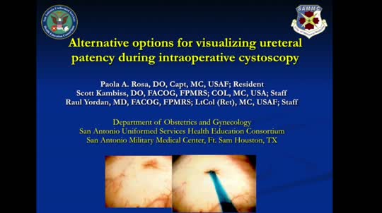
Alternative options for visualizing ureteral patency during intraoperative cystoscopy
For more videos from the Society of Gynecologic Surgeons, click here
Visit the Society of Gynecologic Surgeons online: sgsonline.org
Related Articles:
- Use of suprapubic Carter-Thomason needle to assist in cystoscopic excision of an intravesical foreign object
- Uterine artery ligation: Advanced techniques and considerations for the difficult laparoscopic hysterectomy
- Cervical injection of methylene blue for identification of sentinel lymph nodes in cervical cancer
- Misplaced hysteroscopic sterilization micro-insert in the peritoneal cavity: A corpus alienum
- Laparoscopic cystectomy for large, bilateral ovarian dermoids
This video brought to you by ![]()

Meta-analysis suggests earlier twin delivery to prevent stillbirth
Delivery for uncomplicated dichorionic twin pregnancies should be considered at 37 weeks’ gestation, a week earlier than is generally recommended in the United States, according to a systematic review and meta-analysis of studies that reported rates of stillbirth and neonatal mortality at various gestational ages.
The researchers assessed the competing risks in twin pregnancies of stillbirth from expectant management versus neonatal death from early delivery, looking for an optimal gestational age at which these risks were balanced.

In dichorionic pregnancies continuing beyond 34 weeks, these perinatal risks were balanced at 37 weeks. Beyond that, “the risks of stillbirth significantly outweighed the risk of neonatal death from delivery,” with a 1-week delay in delivery (to 38 weeks’ gestation) leading to an additional 8.8 perinatal deaths per 1,000 pregnancies due to an increase in stillbirth, Fiona Cheong-See, MD, of the Queen Mary University of London and her colleagues reported (BMJ 2016;354:i4353. doi: 10.1136/bmj.i4353).
The review included 32 studies published in the past 10 years (observational cohort studies and cohorts nested in randomized studies) that reported rates of stillbirth and/or neonatal outcomes, including neonatal mortality, in monochorionic and/or dichorionic twins. Neonatal death was defined as death up to 28 days after delivery.
The study authors shared unpublished aggregate and individual patient data with the meta-analysis researchers, which enabled an analysis at weekly intervals. The data included in the review covered 35,171 women with twin gestations (29,685 dichorionic and 5,486 monochorionic pregnancies).
Pregnancies with unclear chorionicity, monoamnionity, and twin-to-twin transfusion syndrome were excluded from the analysis.
In monochorionic pregnancies continuing beyond 34 weeks (2,149 pregnancies), there was a trend after 36 weeks toward stillbirth risk being higher than the risk of neonatal death, but the risk difference was not significant.
More data are needed, the researchers said, but “based on our findings, there is no clear evidence to recommend early preterm delivery routinely before 36 weeks in monochorionic pregnancies.”
A committee opinion published by the American College of Obstetricians and Gynecologists and the Society for Maternal-Fetal Medicine (ACOG-SMFM) on medically indicated late-preterm and early-term deliveries states that decisions regarding the timing of delivery should be individualized and should take into account relative maternal and newborn risks, practice environment, and patient preferences (Obstet Gynecol. 2013;121:908-10).
Still, the ACOG-SMFM document offers delivery recommendations: 38 0/7-38 6/7 weeks of gestation for dichorionic-diamniotic pregnancies, and 34 0/7-36 6/7 weeks of gestation for monochorionic-diamniotic pregnancies.
The opinion was published in 2013 and reflects recommendations made 2 years earlier by the National Institute of Child Health and Human Development and SMFM after a workshop on indicated preterm birth (Obstet Gynecol 2011;118:323-33). ACOG and SMFM reaffirmed their document in 2015.

Brigid McCue, MD, chief of ob.gyn. at Beth Israel Deaconess Hospital–Plymouth (Mass.) and a member of ACOG’s Committee on Obstetric Practice, said the meta-analysis is of “high quality,” with “helpful and valid” findings for dichorionic twins.
The risk of stillbirth was 1.2/1,000 pregnancies at 34 weeks’ gestation, while the risk of neonatal death from delivery was 6.7/1,000 pregnancies. The risk of stillbirth remained significantly lower than the risk of neonatal death from delivery at 35 weeks, and was lower at 36 weeks as well. At 37 weeks, the two categories of perinatal death risk were basically balanced, with the risk of stillbirth at 3.4/1,000 pregnancies.
Beyond that, at 38 weeks’ gestation, the risk of stillbirth (10.6/1,000) significantly outweighed the risk of neonatal death from delivery (1.5/1,000) for a pooled risk difference of 8.8.
“The finding that stillbirth risk is higher when you allow someone to go from 37 to 38 weeks – I think this is true,” Dr. McCue said in an interview.
Further research needs to account, however, for the risks of neonatal morbidity at 37 and 38 weeks. “The next question in coming up with a point of inflection is to ask, What are other contributors to the balance of timing of the delivery?” Dr. McCue said. “There’s a bigger picture that needs more analysis.”
“We don’t induce singletons prior to 39 weeks because they can have more respiratory distress, more hypoglycemia, more temperature instability, and less success breastfeeding, for example,” she added. “These complications pale in comparison to a stillbirth, but the numbers of stillbirth are so low and the numbers for [these other morbidities] are much higher.”
The authors of the meta-analysis noted that their findings were limited by the common policy of planned delivery beyond 37 and 38 weeks’ gestation. “This reduced the sample size near term, particularly in monochorionic pregnancies, and could have led to underestimation of risk of stillbirth in the last weeks of pregnancy,” they wrote.
The rates of assisted ventilation, respiratory distress syndrome, admission to the neonatal intensive care unit, and septicemia showed a consistent reduction with increasing gestational age in babies of both monochorionic and dichorionic pregnancies, the researchers noted.
The researchers reported that they received no funding support from any organization and had no relevant financial disclosures.
Delivery for uncomplicated dichorionic twin pregnancies should be considered at 37 weeks’ gestation, a week earlier than is generally recommended in the United States, according to a systematic review and meta-analysis of studies that reported rates of stillbirth and neonatal mortality at various gestational ages.
The researchers assessed the competing risks in twin pregnancies of stillbirth from expectant management versus neonatal death from early delivery, looking for an optimal gestational age at which these risks were balanced.
In dichorionic pregnancies continuing beyond 34 weeks, these perinatal risks were balanced at 37 weeks. Beyond that, “the risks of stillbirth significantly outweighed the risk of neonatal death from delivery,” with a 1-week delay in delivery (to 38 weeks’ gestation) leading to an additional 8.8 perinatal deaths per 1,000 pregnancies due to an increase in stillbirth, Fiona Cheong-See, MD, of the Queen Mary University of London and her colleagues reported (BMJ 2016;354:i4353. doi: 10.1136/bmj.i4353).
The review included 32 studies published in the past 10 years (observational cohort studies and cohorts nested in randomized studies) that reported rates of stillbirth and/or neonatal outcomes, including neonatal mortality, in monochorionic and/or dichorionic twins. Neonatal death was defined as death up to 28 days after delivery.
The study authors shared unpublished aggregate and individual patient data with the meta-analysis researchers, which enabled an analysis at weekly intervals. The data included in the review covered 35,171 women with twin gestations (29,685 dichorionic and 5,486 monochorionic pregnancies).
Pregnancies with unclear chorionicity, monoamnionity, and twin-to-twin transfusion syndrome were excluded from the analysis.
In monochorionic pregnancies continuing beyond 34 weeks (2,149 pregnancies), there was a trend after 36 weeks toward stillbirth risk being higher than the risk of neonatal death, but the risk difference was not significant.
More data are needed, the researchers said, but “based on our findings, there is no clear evidence to recommend early preterm delivery routinely before 36 weeks in monochorionic pregnancies.”
A committee opinion published by the American College of Obstetricians and Gynecologists and the Society for Maternal-Fetal Medicine (ACOG-SMFM) on medically indicated late-preterm and early-term deliveries states that decisions regarding the timing of delivery should be individualized and should take into account relative maternal and newborn risks, practice environment, and patient preferences (Obstet Gynecol. 2013;121:908-10).
Still, the ACOG-SMFM document offers delivery recommendations: 38 0/7-38 6/7 weeks of gestation for dichorionic-diamniotic pregnancies, and 34 0/7-36 6/7 weeks of gestation for monochorionic-diamniotic pregnancies.
The opinion was published in 2013 and reflects recommendations made 2 years earlier by the National Institute of Child Health and Human Development and SMFM after a workshop on indicated preterm birth (Obstet Gynecol 2011;118:323-33). ACOG and SMFM reaffirmed their document in 2015.
Brigid McCue, MD, chief of ob.gyn. at Beth Israel Deaconess Hospital–Plymouth (Mass.) and a member of ACOG’s Committee on Obstetric Practice, said the meta-analysis is of “high quality,” with “helpful and valid” findings for dichorionic twins.
The risk of stillbirth was 1.2/1,000 pregnancies at 34 weeks’ gestation, while the risk of neonatal death from delivery was 6.7/1,000 pregnancies. The risk of stillbirth remained significantly lower than the risk of neonatal death from delivery at 35 weeks, and was lower at 36 weeks as well. At 37 weeks, the two categories of perinatal death risk were basically balanced, with the risk of stillbirth at 3.4/1,000 pregnancies.
Beyond that, at 38 weeks’ gestation, the risk of stillbirth (10.6/1,000) significantly outweighed the risk of neonatal death from delivery (1.5/1,000) for a pooled risk difference of 8.8.
“The finding that stillbirth risk is higher when you allow someone to go from 37 to 38 weeks – I think this is true,” Dr. McCue said in an interview.
Further research needs to account, however, for the risks of neonatal morbidity at 37 and 38 weeks. “The next question in coming up with a point of inflection is to ask, What are other contributors to the balance of timing of the delivery?” Dr. McCue said. “There’s a bigger picture that needs more analysis.”
“We don’t induce singletons prior to 39 weeks because they can have more respiratory distress, more hypoglycemia, more temperature instability, and less success breastfeeding, for example,” she added. “These complications pale in comparison to a stillbirth, but the numbers of stillbirth are so low and the numbers for [these other morbidities] are much higher.”
The authors of the meta-analysis noted that their findings were limited by the common policy of planned delivery beyond 37 and 38 weeks’ gestation. “This reduced the sample size near term, particularly in monochorionic pregnancies, and could have led to underestimation of risk of stillbirth in the last weeks of pregnancy,” they wrote.
The rates of assisted ventilation, respiratory distress syndrome, admission to the neonatal intensive care unit, and septicemia showed a consistent reduction with increasing gestational age in babies of both monochorionic and dichorionic pregnancies, the researchers noted.
The researchers reported that they received no funding support from any organization and had no relevant financial disclosures.
FROM BMJ
Key clinical point: Delivery of uncomplicated dichorionic twin pregnancies at 37 weeks’ gestation could minimize perinatal deaths.
Major finding: A delay in delivery to 38 weeks’ gestation led to an additional 8.8 perinatal deaths per 1,000 pregnancies because of an increase in stillbirth.
Data source: A systematic review and meta-analysis of 32 studies.
Disclosures: The researchers reported that they received no funding support from any organization and had no relevant financial disclosures.

Four-step screen IDs silent heart attack in type 2 diabetes
MUNICH – A four-component imaging/biomarker screen was highly accurate for identifying silent myocardial infarction among asymptomatic patients with type 2 diabetes.
The screen is far more accurate than the current standards of invasive imaging or only looking for pathologic Q waves, Peter Swoboda, MD, said at the annual meeting of the European Association for the Study of Diabetes.

“By combining these four factors we came up with a tool that has a diagnostic area under the curve [AUC] of 0.85,” said Dr. Swoboda of Leeds (England) University. “This is far better than the 0.58 AUC that we have with Q waves only – a sensitivity of just 25%.”
The study was published online in June in the Journal of Clinical Endocrinology and Metabolism (JCEM 2016. doi: 10.1210/jc.2016-1318).
The screen employs noninvasive imaging and biomarkers to tap multiple clinical hallmarks of silent MI. The components are:
• electrocardiogram.
• echocardiography.
• biomarker assessment.
• cardiac magnetic resonance imaging, focusing on left ventricular ejection fraction and late gadolinium enhancement.
The study cohort comprised 100 patients with type 2 diabetes without known heart disease and with no new cardiac symptoms. They underwent cardiac MRI, a 12-lead electrocardiogram, echocardiography, and serum biomarker assessment. Late gadolinium enhancement identified evidence of silent MI in 17 patients (17%).
There were few differences in the clinical characteristics of those who had experienced MI and those who had not, Dr. Swoboda noted. There were no differences at all in diabetes-related measures, including disease duration or hemoglobin A1c levels. Blood pressures were similar. Patients with MI were significantly older (65 vs. 60 years).
In cardiac-specific measures, left ventricular ejection fraction was similar, as was left ventricular mass, end diastolic volume, and left atrial volume. There were however, other very important differences, Dr. Swoboda noted.
Imaging included a measure called “feature tracking analysis,” which measured the peak global longitudinal strain, systolic strain rate, and early and late diastolic strain rates during contraction. This analysis noted a significant difference in global longitudinal strain between the MI and non-MI groups.
Ventricular filling velocities as measured by the E/A ratio on ECG were also significantly different between the MI and non-MI groups (0.75 vs. 0.89, respectively). ECG also found pathologic Q waves in significantly more MI patients (24% vs. 7%).
Finally, the serum biomarker panel showed a very strong increase in B-type natriuretic peptide (NT-proBNP) among MI patients, compared with non-MI patients (105 vs. 52 ng/L). There were no significant differences in the other biomarkers, including C-reactive protein and high-sensitive cardiac troponin.
Dr. Swoboda and his team then compiled these findings into a composite measure, assigning them optimum cutoff measures:
• Age older than 62 years.
• E/A ratio 0.72 or lower.
• Global longitudinal strain of at least 18.4%.
• NT-proBNP more than 29 ng/L.
The system resulted in a diagnostic accuracy AUC of 0.85 – significantly better than any of the AUCs generated by the individual components. All patients who scored 0 or 1 were free of MI. Among the 28 with a score of 2, only three had experienced a silent MI. Among the 21 with a score of 3, seven had experienced a silent MI and 14 had not. Among the 16 with a score of 4, seven had experienced a silent MI and nine had not.
While Dr. Swoboda called the screening method “simple” during discussion, a colleague in the audience disagreed with that.
“A simple test is something like a blood test only, not an MRI. Not imaging. That is expensive and takes time,” said Naveed Sattar, MD, of the University of Glasgow, Scotland. “However, I do think your data add more to the evidence that BNP can be a really valuable marker of cardiovascular risk in patients with diabetes.”
Dr. Sattar recently examined the value of cardiac serum biomarkers in predicting cardiovascular disease and mortality in nearly 100,000 people without a history of heart disease. In these subjects, he wrote, “NT-proBNP assessment strongly predicted first-onset heart failure and augmented coronary heart disease and stroke prediction, suggesting that NT-proBNP concentration assessment could be used to integrate heart failure into cardiovascular disease primary prevention.”
The paper appeared online in Lancet Diabetes in September (Lancet Diab. 2016. doi: 10.1016/S2213-8587[16]30196-6).
Dr. Swoboda agreed that data continue to support the increased use of NT-proBNP as a marker of heart disease.
“I think that in the future, diabetes medicine is moving toward individualized patient care, based on individualized risk factors. The future of assessing asymptomatic cardiac patients might be a combination of BNP and MRI.”
Dr. Swoboda had no financial disclosures. Some of Dr. Sattar’s coauthors reported relationships with pharmaceutical companies.
MUNICH – A four-component imaging/biomarker screen was highly accurate for identifying silent myocardial infarction among asymptomatic patients with type 2 diabetes.
The screen is far more accurate than the current standards of invasive imaging or only looking for pathologic Q waves, Peter Swoboda, MD, said at the annual meeting of the European Association for the Study of Diabetes.
“By combining these four factors we came up with a tool that has a diagnostic area under the curve [AUC] of 0.85,” said Dr. Swoboda of Leeds (England) University. “This is far better than the 0.58 AUC that we have with Q waves only – a sensitivity of just 25%.”
The study was published online in June in the Journal of Clinical Endocrinology and Metabolism (JCEM 2016. doi: 10.1210/jc.2016-1318).
The screen employs noninvasive imaging and biomarkers to tap multiple clinical hallmarks of silent MI. The components are:
• electrocardiogram.
• echocardiography.
• biomarker assessment.
• cardiac magnetic resonance imaging, focusing on left ventricular ejection fraction and late gadolinium enhancement.
The study cohort comprised 100 patients with type 2 diabetes without known heart disease and with no new cardiac symptoms. They underwent cardiac MRI, a 12-lead electrocardiogram, echocardiography, and serum biomarker assessment. Late gadolinium enhancement identified evidence of silent MI in 17 patients (17%).
There were few differences in the clinical characteristics of those who had experienced MI and those who had not, Dr. Swoboda noted. There were no differences at all in diabetes-related measures, including disease duration or hemoglobin A1c levels. Blood pressures were similar. Patients with MI were significantly older (65 vs. 60 years).
In cardiac-specific measures, left ventricular ejection fraction was similar, as was left ventricular mass, end diastolic volume, and left atrial volume. There were however, other very important differences, Dr. Swoboda noted.
Imaging included a measure called “feature tracking analysis,” which measured the peak global longitudinal strain, systolic strain rate, and early and late diastolic strain rates during contraction. This analysis noted a significant difference in global longitudinal strain between the MI and non-MI groups.
Ventricular filling velocities as measured by the E/A ratio on ECG were also significantly different between the MI and non-MI groups (0.75 vs. 0.89, respectively). ECG also found pathologic Q waves in significantly more MI patients (24% vs. 7%).
Finally, the serum biomarker panel showed a very strong increase in B-type natriuretic peptide (NT-proBNP) among MI patients, compared with non-MI patients (105 vs. 52 ng/L). There were no significant differences in the other biomarkers, including C-reactive protein and high-sensitive cardiac troponin.
Dr. Swoboda and his team then compiled these findings into a composite measure, assigning them optimum cutoff measures:
• Age older than 62 years.
• E/A ratio 0.72 or lower.
• Global longitudinal strain of at least 18.4%.
• NT-proBNP more than 29 ng/L.
The system resulted in a diagnostic accuracy AUC of 0.85 – significantly better than any of the AUCs generated by the individual components. All patients who scored 0 or 1 were free of MI. Among the 28 with a score of 2, only three had experienced a silent MI. Among the 21 with a score of 3, seven had experienced a silent MI and 14 had not. Among the 16 with a score of 4, seven had experienced a silent MI and nine had not.
While Dr. Swoboda called the screening method “simple” during discussion, a colleague in the audience disagreed with that.
“A simple test is something like a blood test only, not an MRI. Not imaging. That is expensive and takes time,” said Naveed Sattar, MD, of the University of Glasgow, Scotland. “However, I do think your data add more to the evidence that BNP can be a really valuable marker of cardiovascular risk in patients with diabetes.”
Dr. Sattar recently examined the value of cardiac serum biomarkers in predicting cardiovascular disease and mortality in nearly 100,000 people without a history of heart disease. In these subjects, he wrote, “NT-proBNP assessment strongly predicted first-onset heart failure and augmented coronary heart disease and stroke prediction, suggesting that NT-proBNP concentration assessment could be used to integrate heart failure into cardiovascular disease primary prevention.”
The paper appeared online in Lancet Diabetes in September (Lancet Diab. 2016. doi: 10.1016/S2213-8587[16]30196-6).
Dr. Swoboda agreed that data continue to support the increased use of NT-proBNP as a marker of heart disease.
“I think that in the future, diabetes medicine is moving toward individualized patient care, based on individualized risk factors. The future of assessing asymptomatic cardiac patients might be a combination of BNP and MRI.”
Dr. Swoboda had no financial disclosures. Some of Dr. Sattar’s coauthors reported relationships with pharmaceutical companies.
AT EASD 2016
Key clinical point: A four-component screen accurately identified silent myocardial infarction in asymptomatic patients with type 2 diabetes
Major finding: The tool had an 82% sensitivity and 72% specificity for silent MI.
Data source: It was created in a cohort of 100 patients with type 2 diabetes and no history of heart disease.
Disclosures: Dr. Swoboda had no financial disclosures.

Study: Health spending related to opioid treatment rose more than 1,300%
The nation’s ongoing opioid problem comes with staggering physical and emotional costs to patients and families. But the dollar cost to the health system has been harder to peg. Now a new report shows a more than 1,300 percent rise in spending by health insurers in a four-year period on patients with a diagnosis of opioid dependence or abuse.
From 2011 to 2015, insurers’ payments to hospitals, laboratories, treatment centers and other medical providers for these patients grew from $32 million to $446 million – a 1,375 percent increase.
While that’s a small portion of the overall spending on medical care in the United States, the rapid rise is cause for concern, says Robin Gelburd, president of Fair Health, a nonprofit databank that provides cost information to the health industry and consumers.
“That really shows the stress on the health system and the impact on the individuals,” said Gelburd.
The Fair Health study found a sharp difference in how much insurers spend on individual patients with such a diagnosis.
On average, insurers spend $3,435 a year on an individual patient, but for those with an opioid dependence or abuse diagnosis, that amount jumps to $19,333. Those numbers reflect what insurers actually paid. The report also includes data on what providers charged, amounts that are lowered by their contracts with insurers.
The study, set to be released Tuesday, builds on one Fair Health released in early August that found a 3,000 percent increase in the volume of insurance claims related to opioid dependence diagnoses between 2007 and 2014.
The latest study – part of a series – offers amounts associated with claims billed by providers and paid by insurers for the types of medical services used.
Both studies use de-identified claims data from insurers representing more than 150 million insured Americans who either have insurance through work or buy coverage on their own.
There have been other efforts by several researchers to quantify the cost of the opioid problem on the overall economy, estimated in the tens of billions of dollars.
The new report adds to the available data “that it’s not just the human cost associated with the opioid crisis that is enormous, but also that the economic costs are staggering,” said Dr. Andrew Kolodny, senior scientist at Brandeis University. He did not work on the study.
The surge in spending on patients with opioid diagnoses is likely a combination of factors, the report notes. As media attention focuses on drug dependency, more people may be seeking treatment. At the same time, prescription and illegal use of narcotics may also be increasing.
The study found that emergency room visits and laboratory tests accounted for much of the spending.
Based on claims volume, the fastest-growing set of services in terms of utilization were for alcohol or drug therapy. Lab tests, including checks for barbiturate or opioid use, were not far behind.
Researchers did not use 2015 data for lab test costs, noting that a change in billing codes was made that increased the number of categories – and, in some cases, appear to generate higher payments by insurers. It is too early to estimate the long-term effects of the change, Gelburd said.
The report gives some examples of the changes, however. For example, one billing code for a test on opiate use commonly brought in a $31 payment from insurers prior to the change. The two billing codes that replaced it now are commonly paid at $78 and $156.
The new billing codes may reflect new technology in testing, said Gelburd. She said some observers speculate that the rapid increase in lab spending might reflect that, with more patients in therapy, the tests are being used to ensure they are taking their proper medications and not abusing narcotics.
But the spending might also reflect a growing use of very expensive urine and blood tests when less expensive ones would be sufficient, said Kolodny.
“I worry about profiteering,” said Kolodny. “We do need tests, but not the expensive ones. A lot of clinics are making extra money off these lab tests.”
The overall increase in spending across all types of medical services “is a societal issue,” said Gelburd, who says policymakers need to ensure that changes are made to address the problem.
“Are medical school curricula adjusting to recognize the growing need for these services? Are insurers increasing the number of providers in their networks to ensure sufficient access? Are consumers being educated? It’s an issue that has to be dealt with in all quadrants.”
Kaiser Health News is a national health policy news service that is part of the nonpartisan Henry J. Kaiser Family Foundation.
First-generation DES looking good at 10 years
ROME – There’s good news for the millions of patients living with a first-generation metallic drug-eluting stent for coronary revascularization implanted in years past: The devices perform reassuringly well a full decade after implantation, Lorenz Räber, MD, reported at the annual congress of the European Society of Cardiology.
That’s the key message of the SIRTAX VERY LATE study, the only randomized trial of first-generation drug-eluting stents (DES) that didn’t turn out the lights at a maximum of 5 years of follow-up. In fact, at the 10-year mark, SIRTAX VERY LATE shows that regardless of whether the first-generation DES was paclitaxel- or sirolimus-eluting, the risk of major adverse cardiac events due to device failure was substantially lower in the second half-decade than in the first 5 years after deployment, according to Dr. Räber of Bern (Switzerland) University Hospital.

More specifically, the cumulative risk of ischemia-driven target lesion revascularization was 14.6% at 5 years and 17.7% at 10 years, while the 5- and 10-year cumulative risks of definite stent thrombosis were 4.5% and 5.6%, respectively.
The annual risk of ischemia-driven target lesion revascularization dropped by 64%, from 1.8%/year during years 1-5 to 0.7% during years 6-10. Similarly, the annual risk of definite stent thrombosis fell from 0.67%/year to 0.23%/year after year 5, a 69% relative risk reduction. And importantly, these attenuations in risk occurred independent of age.
“The lower risk of late clinical events suggests stabilization of delayed arterial healing over time after first-generation DES implantation, with reduced chronic inflammation and neoatherosclerosis,” he said.
This is reassuring in light of the stormy history of the first-generation DES. Three years after the devices came on the U.S. market, the so-called ESC firestorm erupted. At the 2006 ESC congress, investigators presented meta-analyses suggesting the devices carried a possible late increased thrombotic risk beyond the then-recommended 3-6 months of prescribed dual-antiplatelet therapy. The use of these devices declined sharply in response, even though a Food and Drug Administration advisory panel charged with looking at the totality of evidence concluded that concerns about thrombosis didn’t outweigh the benefits of the first-generation DES over bare-metal stents.
The reductions in very late stent thrombosis and ischemia-driven target lesion revascularization beyond 5 years seen in the SIRTAX VERY LATE trial occurred despite the fact that only 15% of patients were on dual-antiplatelet therapy throughout the first 5 years and 11% were on dual-antiplatelet therapy afterwards, Dr. Räber noted.
“Our findings may have implications for secondary prevention after PCI with a first-generation DES, including the need for long-term antiplatelet therapy,” the cardiologist added.
The previously reported 5-year results of SIRTAX (the Sirolimus-Eluting Versus Paclitaxel-Eluting Stents for Coronary Revascularization trial) showed a steady increase over time in late lumen loss and an ongoing risk of very late stent thrombosis (Circulation. 2011 Jun 21;123(24):2819-28). Much the same was seen at the 5-year mark in the other major trials of first-generation DES, including RAVEL, SIRIUS, and TAXUS.
However, all those studies ended at 5 years, leaving unanswered the key question of what happens later. Cardiologists have wondered if the first-generation DES they put in their patients years ago were associated with a continued steady climb in the risk of device-related adverse events, or if the risk plateaued or even dropped off. The SIRTAX VERY LATE study was conducted in order to provide answers.
SIRTAX included 1,012 Swiss patients randomized to coronary revascularization using a first-generation sirolimus- or paclitaxel-eluting stent in 2003-2004. Roughly half had stable coronary artery disease and half presented with acute coronary syndromes. The 10-year follow-up conducted in the SIRTAX VERY LATE study captured 895 (88%) of the original 1,012 subjects.
The cumulative incidence of major cardiac adverse events – a composite of cardiac death, MI, and ischemia-driven target lesion revascularization – was 20.8% at 5 years and 33.8% at 10 years. The rate was similar between years 1-5 and 6-10.
The cumulative all-cause mortality rate was 10.4% at 5 years and 24.2% at 10 years. The rate was 2.0%/year during years 1-5 and accelerated significantly to 3.1%/year in years 6-10. However, this increase appears to be largely due to the background impact of advancing age rather than to any effect of having a first-generation DES. The 5- and 10-year all-cause mortality rates in the age- and sex-matched general Swiss population are similar to those seen in SIRTAX VERY LATE, at 9.6% and 22.1%, respectively, the cardiologist observed.
The cumulative incidence of MI in the study population was 7% at 5 years and 9.7% at 10 years. Between years 1-5 the rate was 0.9%/year, dropping to 0.6%/year during years 6-10.
One of the useful potential purposes for the new SIRTAX VERY LATE follow-up data beyond 5 years is that the results could serve as a benchmark in evaluating the long-term safety and efficacy of the much newer drug-eluting fully bioresorbable vascular scaffolds, since the potential benefits of these new devices may not appear until relatively late, after the devices themselves have disappeared. SIRTAX VERY LATE sets the bar for stent-related adverse events 5 years or more after device implantation at an annual risk of less than 0.3%/year for stent thrombosis and less than 1%/year for ischemia-driven target lesion revascularization.
Session co-chair Hector Bueno, MD, drew attention to the fact that no significant differences in clinical outcomes were seen at either 5 or 10 years between the sirolimus- and paclitaxel-eluting stent recipients. That’s noteworthy because more than a decade ago when the primary endpoint of SIRTAX was reported, much was made of the finding that the 9-month rate of major adverse cardiac events was significantly lower in the sirolimus-eluting stent group (N Engl J Med. 2005 Aug 18;353[7]:653-62). Over time, any outcome differences between the two devices were erased, observed Dr. Bueno of Complutense University of Madrid.
The SIRTAX VERY LATE study was funded by grants from Bern University Hospital. Dr. Räber reported having no relevant financial interests.
Simultaneously with his presentation in Rome at ESC 2016, the study results were published online (Eur Heart J. 2016 Aug 30. doi: 10.1093/eurheartj/ehw343).
ROME – There’s good news for the millions of patients living with a first-generation metallic drug-eluting stent for coronary revascularization implanted in years past: The devices perform reassuringly well a full decade after implantation, Lorenz Räber, MD, reported at the annual congress of the European Society of Cardiology.
That’s the key message of the SIRTAX VERY LATE study, the only randomized trial of first-generation drug-eluting stents (DES) that didn’t turn out the lights at a maximum of 5 years of follow-up. In fact, at the 10-year mark, SIRTAX VERY LATE shows that regardless of whether the first-generation DES was paclitaxel- or sirolimus-eluting, the risk of major adverse cardiac events due to device failure was substantially lower in the second half-decade than in the first 5 years after deployment, according to Dr. Räber of Bern (Switzerland) University Hospital.
More specifically, the cumulative risk of ischemia-driven target lesion revascularization was 14.6% at 5 years and 17.7% at 10 years, while the 5- and 10-year cumulative risks of definite stent thrombosis were 4.5% and 5.6%, respectively.
The annual risk of ischemia-driven target lesion revascularization dropped by 64%, from 1.8%/year during years 1-5 to 0.7% during years 6-10. Similarly, the annual risk of definite stent thrombosis fell from 0.67%/year to 0.23%/year after year 5, a 69% relative risk reduction. And importantly, these attenuations in risk occurred independent of age.
“The lower risk of late clinical events suggests stabilization of delayed arterial healing over time after first-generation DES implantation, with reduced chronic inflammation and neoatherosclerosis,” he said.
This is reassuring in light of the stormy history of the first-generation DES. Three years after the devices came on the U.S. market, the so-called ESC firestorm erupted. At the 2006 ESC congress, investigators presented meta-analyses suggesting the devices carried a possible late increased thrombotic risk beyond the then-recommended 3-6 months of prescribed dual-antiplatelet therapy. The use of these devices declined sharply in response, even though a Food and Drug Administration advisory panel charged with looking at the totality of evidence concluded that concerns about thrombosis didn’t outweigh the benefits of the first-generation DES over bare-metal stents.
The reductions in very late stent thrombosis and ischemia-driven target lesion revascularization beyond 5 years seen in the SIRTAX VERY LATE trial occurred despite the fact that only 15% of patients were on dual-antiplatelet therapy throughout the first 5 years and 11% were on dual-antiplatelet therapy afterwards, Dr. Räber noted.
“Our findings may have implications for secondary prevention after PCI with a first-generation DES, including the need for long-term antiplatelet therapy,” the cardiologist added.
The previously reported 5-year results of SIRTAX (the Sirolimus-Eluting Versus Paclitaxel-Eluting Stents for Coronary Revascularization trial) showed a steady increase over time in late lumen loss and an ongoing risk of very late stent thrombosis (Circulation. 2011 Jun 21;123(24):2819-28). Much the same was seen at the 5-year mark in the other major trials of first-generation DES, including RAVEL, SIRIUS, and TAXUS.
However, all those studies ended at 5 years, leaving unanswered the key question of what happens later. Cardiologists have wondered if the first-generation DES they put in their patients years ago were associated with a continued steady climb in the risk of device-related adverse events, or if the risk plateaued or even dropped off. The SIRTAX VERY LATE study was conducted in order to provide answers.
SIRTAX included 1,012 Swiss patients randomized to coronary revascularization using a first-generation sirolimus- or paclitaxel-eluting stent in 2003-2004. Roughly half had stable coronary artery disease and half presented with acute coronary syndromes. The 10-year follow-up conducted in the SIRTAX VERY LATE study captured 895 (88%) of the original 1,012 subjects.
The cumulative incidence of major cardiac adverse events – a composite of cardiac death, MI, and ischemia-driven target lesion revascularization – was 20.8% at 5 years and 33.8% at 10 years. The rate was similar between years 1-5 and 6-10.
The cumulative all-cause mortality rate was 10.4% at 5 years and 24.2% at 10 years. The rate was 2.0%/year during years 1-5 and accelerated significantly to 3.1%/year in years 6-10. However, this increase appears to be largely due to the background impact of advancing age rather than to any effect of having a first-generation DES. The 5- and 10-year all-cause mortality rates in the age- and sex-matched general Swiss population are similar to those seen in SIRTAX VERY LATE, at 9.6% and 22.1%, respectively, the cardiologist observed.
The cumulative incidence of MI in the study population was 7% at 5 years and 9.7% at 10 years. Between years 1-5 the rate was 0.9%/year, dropping to 0.6%/year during years 6-10.
One of the useful potential purposes for the new SIRTAX VERY LATE follow-up data beyond 5 years is that the results could serve as a benchmark in evaluating the long-term safety and efficacy of the much newer drug-eluting fully bioresorbable vascular scaffolds, since the potential benefits of these new devices may not appear until relatively late, after the devices themselves have disappeared. SIRTAX VERY LATE sets the bar for stent-related adverse events 5 years or more after device implantation at an annual risk of less than 0.3%/year for stent thrombosis and less than 1%/year for ischemia-driven target lesion revascularization.
Session co-chair Hector Bueno, MD, drew attention to the fact that no significant differences in clinical outcomes were seen at either 5 or 10 years between the sirolimus- and paclitaxel-eluting stent recipients. That’s noteworthy because more than a decade ago when the primary endpoint of SIRTAX was reported, much was made of the finding that the 9-month rate of major adverse cardiac events was significantly lower in the sirolimus-eluting stent group (N Engl J Med. 2005 Aug 18;353[7]:653-62). Over time, any outcome differences between the two devices were erased, observed Dr. Bueno of Complutense University of Madrid.
The SIRTAX VERY LATE study was funded by grants from Bern University Hospital. Dr. Räber reported having no relevant financial interests.
Simultaneously with his presentation in Rome at ESC 2016, the study results were published online (Eur Heart J. 2016 Aug 30. doi: 10.1093/eurheartj/ehw343).
AT THE ESC CONGRESS 2016
Key clinical point: The annual risks of ischemia-driven target lesion revascularization and stent thrombosis significantly decreased starting 5 years after implantation of a first-generation sirolimus- or paclitaxel-eluting stent.
Major finding: The annual risk of ischemia-driven target lesion revascularization was 1.8%/year between 1 and 5 years after implantation of a first-generation drug-eluting stent, but only 0.7%/year during years 6-10.
Data source: This was a unique extended 10-year follow-up of 88% of the original 1,012 participants in a randomized, assessor-blinded Swiss trial of coronary revascularization using a first-generation sirolimus- or paclitaxel-eluting stent.
Disclosures: The SIRTAX VERY LATE study was funded by grants from Bern University Hospital. The presenter reported having no relevant financial interests.

Lowering LDL-C yields usual benefits in rheumatoid arthritis
Lowering LDL cholesterol level using statins appears to yield the same cardiovascular benefits in rheumatoid arthritis patients as in the general population, a study showed.
Cardiovascular (CV) disease is a common comorbidity in rheumatoid arthritis (RA) patients, who carry an estimated 50% increased risk of CV events or early mortality, compared with the general population. Yet epidemiologic studies have reported that RA patients generally have lower lipid levels than members of the general population, and studies assessing statin therapy in this patient population have yielded mixed results, said JaeJin An, PhD, of the department of pharmacy practice and administration, Western University of Health Sciences, Pomona, Calif., and her associates.

This may be because RA alters lipid metabolism in a complex manner, due to its associated systemic inflammation, the effects of RA drug therapy, and several genetic factors that characterize RA, they noted.
To assess the benefit of statin therapy for primary prevention in RA, the investigators performed a retrospective cohort study using the electronic medical records of adults enrolled in a single large California health care system. They focused on RA patients taking at least one disease-modifying antirheumatic drug who also had hyperlipidemia. In one cohort, 1,522 RA patients were matched for age and sex with 6,511 control subjects who were also in the health care system, and in a second cohort 1,746 RA patients were matched with 2,554 patients who had osteoarthritis (OA).
These study participants were followed for a median of 3-4 years for the development of MI, angina, stroke, transient ischemic attack, intermittent claudication, heart failure, or CV death. Lipid-lowering medications they were taking included atorvastatin, simvastatin, fluvastatin, lovastatin, rosuvastatin, and pravastatin. The study was supported by Bristol-Myers Squibb, maker of pravastatin.
Similar proportions of patients in the three study groups lowered their LDL-C level to clinically recommended levels based on their baseline CV risk: 79% of the RA group, 79% of the general control group, and 80% of the OA control group.
There were no differences in the link between lowering LDL-C level and improved CV outcomes among the three study groups. A reduction in LDL-C was associated with a 29% reduction in the risk of CV events in the RA group plus the general control group, and it was associated with a 50% reduction in the risk of CV events in the RA group plus the OA control group, the investigators reported (J Rheumatol. 2016 Sep 1. doi: 10.3899/jrheum.160110). In a further analysis of the data that adjusted for multiple CV risk factors, lowering LDL-C levels was associated with a similar degree of reduction in CV events in RA patients in the first cohort (hazard ratio, 0.68) and the second cohort (HR, 0.67).
“Our analyses further verified that having RA itself increased CV risk by 76% [compared with general control patients], even after adjusting for traditional risk factors such as age, sex, smoking, hypertension, and diabetes. These findings are consistent with previous findings, which suggest about a 50% higher risk of CV events and mortality among patients with RA relative to the general population,” Dr. An and her associates wrote.
Their results also are consistent with those of the recently reported TRACE-RA (Trial of Atorvastatin for the Primary Prevention of Cardiovascular Events in Rheumatoid Arthritis) study, which found that lipid-lowering therapy with atorvastatin was associated with a 34% reduction in the primary composite CV endpoint in RA patients.
Bristol-Myers Squibb supported the study, and several coauthors are employees of the company.
Lowering LDL cholesterol level using statins appears to yield the same cardiovascular benefits in rheumatoid arthritis patients as in the general population, a study showed.
Cardiovascular (CV) disease is a common comorbidity in rheumatoid arthritis (RA) patients, who carry an estimated 50% increased risk of CV events or early mortality, compared with the general population. Yet epidemiologic studies have reported that RA patients generally have lower lipid levels than members of the general population, and studies assessing statin therapy in this patient population have yielded mixed results, said JaeJin An, PhD, of the department of pharmacy practice and administration, Western University of Health Sciences, Pomona, Calif., and her associates.
This may be because RA alters lipid metabolism in a complex manner, due to its associated systemic inflammation, the effects of RA drug therapy, and several genetic factors that characterize RA, they noted.
To assess the benefit of statin therapy for primary prevention in RA, the investigators performed a retrospective cohort study using the electronic medical records of adults enrolled in a single large California health care system. They focused on RA patients taking at least one disease-modifying antirheumatic drug who also had hyperlipidemia. In one cohort, 1,522 RA patients were matched for age and sex with 6,511 control subjects who were also in the health care system, and in a second cohort 1,746 RA patients were matched with 2,554 patients who had osteoarthritis (OA).
These study participants were followed for a median of 3-4 years for the development of MI, angina, stroke, transient ischemic attack, intermittent claudication, heart failure, or CV death. Lipid-lowering medications they were taking included atorvastatin, simvastatin, fluvastatin, lovastatin, rosuvastatin, and pravastatin. The study was supported by Bristol-Myers Squibb, maker of pravastatin.
Similar proportions of patients in the three study groups lowered their LDL-C level to clinically recommended levels based on their baseline CV risk: 79% of the RA group, 79% of the general control group, and 80% of the OA control group.
There were no differences in the link between lowering LDL-C level and improved CV outcomes among the three study groups. A reduction in LDL-C was associated with a 29% reduction in the risk of CV events in the RA group plus the general control group, and it was associated with a 50% reduction in the risk of CV events in the RA group plus the OA control group, the investigators reported (J Rheumatol. 2016 Sep 1. doi: 10.3899/jrheum.160110). In a further analysis of the data that adjusted for multiple CV risk factors, lowering LDL-C levels was associated with a similar degree of reduction in CV events in RA patients in the first cohort (hazard ratio, 0.68) and the second cohort (HR, 0.67).
“Our analyses further verified that having RA itself increased CV risk by 76% [compared with general control patients], even after adjusting for traditional risk factors such as age, sex, smoking, hypertension, and diabetes. These findings are consistent with previous findings, which suggest about a 50% higher risk of CV events and mortality among patients with RA relative to the general population,” Dr. An and her associates wrote.
Their results also are consistent with those of the recently reported TRACE-RA (Trial of Atorvastatin for the Primary Prevention of Cardiovascular Events in Rheumatoid Arthritis) study, which found that lipid-lowering therapy with atorvastatin was associated with a 34% reduction in the primary composite CV endpoint in RA patients.
Bristol-Myers Squibb supported the study, and several coauthors are employees of the company.
FROM JOURNAL OF RHEUMATOLOGY
Key clinical point: Lowering LDL cholesterol yields the same cardiovascular benefits in rheumatoid arthritis patients as in the general population.
Major finding: A reduction in LDL-C was associated with a 29% reduction in the risk of cardiovascular events in the RA group plus the general control group, and with a 50% reduction in the risk of CV events in the RA group plus the osteoarthritis control group.
Data source: An industry-sponsored retrospective cohort study involving 3,268 RA patients, 6,511 general controls, and 2,554 controls with osteoarthritis.
Disclosures: Bristol-Myers Squibb supported the study, and several coauthors are employees of the company.

Low caregiver self-care linked with depression, anxiety
There is increased anxiety and depression among family caregivers who do not take care of themselves, according to a study to be presented at the 2016 ASCO Palliative Care in Oncology Symposium.
Nearly a quarter of 294 caregivers of Medicare patients with advanced cancer reported high depression scores (23%) and 34% reported borderline or high anxiety scores. Worse caregiver anxiety, depression, and mental health–related quality of life scores were significantly associated with lower scores in every self-care measure (P less than .05 for all). Lower self-care behavior scores were associated with longer durations, higher hours, and more days/week of caregiving and with fair or poor patient health.
The cross-sectional survey was conducted in community settings of eight cancer centers in Alabama, Florida, and Tennessee. The family caregivers of Medicare beneficiaries diagnosed with pancreatic, lung, brain, ovarian, head & neck, hematologic, or stage IV cancer completed measures of self-care behaviors, including health responsibility, physical activity, nutrition, spiritual growth, interpersonal relations, stress management, and sleep. Caregivers averaged 66 years and were mostly female (72.8%), white (91.2%), Protestant (76.2%), retired (54.4%), and patients’ spouse/partner (60.2%). Approximately half were rural dwellers (46.9%) and had incomes less than $50,000 (53.8%). The majority provided support 6-7 days per week (71%) for greater than 1 year (68%).
“This research serves as an important call to action for the oncology community to implement support networks and services that care for the caregiver,” ASCO representative Andrew Epstein, MD, of Memorial Sloan Kettering Cancer Center, New York, said in a written statement ahead of the symposium.
“We hope our research rallies the oncology palliative care communities to develop assessment tools and services that support caregivers,” lead author James Nicholas Dionne-Odom, PhD, RN, of the University of Alabama at Birmingham, said in the statement.
On Twitter @jessnicolecraig
FROM THE 2016 ASCO PALLIATIVE CARE IN ONCOLOGY SYMPOSIUM
Key clinical point: There is increased anxiety and depression among family caregivers who do not take care of themselves.
Major finding: Worse caregiver anxiety, depression, and mental health–related quality of life scores were significantly associated with lower scores in every self-care measure (P less than .05 for all).
Data source: A multistate and cross-sectional survey of 294 family caregivers.
Disclosures: The University of Alabama at Birmingham funded the study. One investigator reported receiving financial compensation and honoraria from Medscape, Carevive Systems, and PackHealth.