User login
Use of topical hemostatic agents in gynecologic surgery
Sutures, hemoclips, and electrocautery are the primary means of achieving hemostasis during gynecologic surgery. When these are inadequate or infeasible, topical hemostatic agents can be employed. Use of these agents has increased by 10%-21% since 2000, yet studies evaluating their use in gynecologic surgery are limited (J. Surg. Res. 2014;186:458-66).
Oxidized regenerated cellulose
Oxidized regenerated cellulose (Surgicel) is made from dissolved oxidized cellulose woven into a dry gauze sheet (J. Urol. 2006;176:2367-74). It is applied directly to tissue, creating a scaffold for platelet aggregation and decreasing tissue pH, further activating the clotting cascade (Surg. Infect. (Larchmt.) 2003;4:255-62). It is absorbed in 14 days, but can persist for 1 year.

Oxidized regenerated cellulose (ORC) is easily passed through laparoscopic trocars. One study found ORC efficacious in controlling tubal hemorrhage during laparoscopic sterilization (Int. J. Gynaecol. Obstet. 2003;82:221-2). It has also been shown to have bactericidal activity (Surg. Infect. (Larchmt.) 2003; 4:255-62) and prevent development of peritoneal adhesions (Acta. Chir. Scand. 1978;144:375-8).
Microfibrillar collagen
Microfibrillar collagen (Avitene) is made from bovine collagen in a powder or sponge sheet, and acts as a scaffold for platelet aggregation. It is applied directly to tissue and is absorbed in 3 months. One study found microfibrillar collagen (MC) use during cold knife conization resulted in nonsignificant reduction in operative time and similar hemostatic results compared to Sturmdorf suture (Obstet. Gynecol. 1978;51:118-22). MC also has been used to treat bleeding following uterine perforation and during laparoscopic hysterectomy.

Gelatins
Gelatins (Gelfoam, Surgifoam) are made of porcine collagen in a powder or foam (J. Urol. 2006;176:2367-74). It is applied directly to tissue, acting as a sponge to absorb blood. Pressure for several minutes is necessary for optimal hemostasis. Some surgeons moisten gelatins with topical thrombin prior to use, though no trials exist evaluating the efficacy of this maneuver.
Gelatin is absorbed in 4-6 weeks (J. Urol. 2006;176:2367-74) and can be passed through laparoscopic trocars. No studies have evaluated gelatins in gynecologic surgery so its applications are extrapolated from vascular and urologic surgery (J. Urol. 2006;176:2367-74).
Microporous polysaccharide spheres
Microporous polysaccharide spheres (Arista) form a polysaccharide powder made from potato starch. It absorbs water, concentrating platelets and other proteins to accelerate clot formation. It is applied to a dry surgical field and followed with gentle pressure. MPS is absorbed in 48 hours. No studies specifically evaluate the use of MPS in gynecologic surgery.
Topical thrombins
Thrombin (Thrombin-JMI, Evithrom, Recothrom) is derived from bovine, human, or recombinant sources. It converts fibrinogen to fibrin and activates factor XIII, platelets, and smooth muscle constriction (Biologics 2008;2:593-9). Thrombin is a spray or syringe, and is often used with gelatin foam (Thrombi-Gel) or matrix (FloSeal) (Biologics 2008;2:593-9). FloSeal use has been reported during ovarian cystectomy (J. Minim. Invasive. Gynecol. 2009;16:153-6), hysterotomy repair (J. Obstet. Gynaecol. 2012;32:34-5). During myomectomy, it was associated with decreased blood loss, transfusions, and shorter length of stay (Fertil. Steril. 2009;92:356-60).
Fibrin sealants
Fibrin sealants (Tisseel, TachoSil) are made of thrombin and concentrated fibrinogen from human plasma. They must be mixed prior to application and act by forming a fibrin clot. Tisseel can reduce hemorrhage after loop electrosurgical excision procedure (Gynecol. Obstet. Invest. 2012;74:1-5) and decreases operative time, time to hemostasis, and blood loss during laparoscopic myomectomy (Surg. Endosc. 2012;26:2046-53). Case reports describe the use of fibrin sealants in the management of obstetrical hemorrhage and hysterotomy repair.
Cost and complications
Hemostatic agents vary significantly in cost, but no comparative cost analyses exist. One study found that commercial insurance was associated with topical hemostatic agent use during gynecologic surgery (J. Surg. Res. 2014;186:458-66).
Use of ORC has been associated with postoperative abscess and imitation of abscess without true infection, and MC and gelatins can also increase infection risk. The dry hemostatic agents have been associated with thromboembolism. The complications of thrombins and fibrins are related to immune responses or transmission of pathogens. Recombinant thrombin is believed to be the safest option (J. Am. Coll. Surg. 2007;205:256-65). Floseal has been reported to cause diffuse pelvic inflammation and postoperative small bowel obstruction. Because of possible complications, it is important to use only the needed amount of product, and to dictate use in the operative note.
Despite widespread use of topical hemostatic agents in gynecologic surgery, studies are limited and these agents should be recommended only as adjuncts to conventional methods of achieving hemostasis.
Topical hemostatic agents are recommended for surgical fields that are less amenable to electrocautery, including denuded areas on peritoneal surfaces, and around important heat-sensitive structures such as nerves. The dry matrix agents (ORC, MC, gelatin, and MPS) are most useful in slowly bleeding areas or in patients with a bleeding diathesis. Thrombin and fibrin can be useful in situations when more significant bleeding is encountered. Complications arising from topical hemostatic agents are few.
Given current limited studies, the choice of product continues to depend on patient characteristics and surgeon preference.
Dr. Wysham is currently a fellow in the department of gynecologic oncology at the University of North Carolina at Chapel Hill. Dr. Roque is a fellow in the gynecologic oncology program at UNC-Chapel Hill. Dr. Soper is a professor of gynecologic oncology at UNC-Chapel Hill.
Sutures, hemoclips, and electrocautery are the primary means of achieving hemostasis during gynecologic surgery. When these are inadequate or infeasible, topical hemostatic agents can be employed. Use of these agents has increased by 10%-21% since 2000, yet studies evaluating their use in gynecologic surgery are limited (J. Surg. Res. 2014;186:458-66).
Oxidized regenerated cellulose
Oxidized regenerated cellulose (Surgicel) is made from dissolved oxidized cellulose woven into a dry gauze sheet (J. Urol. 2006;176:2367-74). It is applied directly to tissue, creating a scaffold for platelet aggregation and decreasing tissue pH, further activating the clotting cascade (Surg. Infect. (Larchmt.) 2003;4:255-62). It is absorbed in 14 days, but can persist for 1 year.
Oxidized regenerated cellulose (ORC) is easily passed through laparoscopic trocars. One study found ORC efficacious in controlling tubal hemorrhage during laparoscopic sterilization (Int. J. Gynaecol. Obstet. 2003;82:221-2). It has also been shown to have bactericidal activity (Surg. Infect. (Larchmt.) 2003; 4:255-62) and prevent development of peritoneal adhesions (Acta. Chir. Scand. 1978;144:375-8).
Microfibrillar collagen
Microfibrillar collagen (Avitene) is made from bovine collagen in a powder or sponge sheet, and acts as a scaffold for platelet aggregation. It is applied directly to tissue and is absorbed in 3 months. One study found microfibrillar collagen (MC) use during cold knife conization resulted in nonsignificant reduction in operative time and similar hemostatic results compared to Sturmdorf suture (Obstet. Gynecol. 1978;51:118-22). MC also has been used to treat bleeding following uterine perforation and during laparoscopic hysterectomy.
Gelatins
Gelatins (Gelfoam, Surgifoam) are made of porcine collagen in a powder or foam (J. Urol. 2006;176:2367-74). It is applied directly to tissue, acting as a sponge to absorb blood. Pressure for several minutes is necessary for optimal hemostasis. Some surgeons moisten gelatins with topical thrombin prior to use, though no trials exist evaluating the efficacy of this maneuver.
Gelatin is absorbed in 4-6 weeks (J. Urol. 2006;176:2367-74) and can be passed through laparoscopic trocars. No studies have evaluated gelatins in gynecologic surgery so its applications are extrapolated from vascular and urologic surgery (J. Urol. 2006;176:2367-74).
Microporous polysaccharide spheres
Microporous polysaccharide spheres (Arista) form a polysaccharide powder made from potato starch. It absorbs water, concentrating platelets and other proteins to accelerate clot formation. It is applied to a dry surgical field and followed with gentle pressure. MPS is absorbed in 48 hours. No studies specifically evaluate the use of MPS in gynecologic surgery.
Topical thrombins
Thrombin (Thrombin-JMI, Evithrom, Recothrom) is derived from bovine, human, or recombinant sources. It converts fibrinogen to fibrin and activates factor XIII, platelets, and smooth muscle constriction (Biologics 2008;2:593-9). Thrombin is a spray or syringe, and is often used with gelatin foam (Thrombi-Gel) or matrix (FloSeal) (Biologics 2008;2:593-9). FloSeal use has been reported during ovarian cystectomy (J. Minim. Invasive. Gynecol. 2009;16:153-6), hysterotomy repair (J. Obstet. Gynaecol. 2012;32:34-5). During myomectomy, it was associated with decreased blood loss, transfusions, and shorter length of stay (Fertil. Steril. 2009;92:356-60).
Fibrin sealants
Fibrin sealants (Tisseel, TachoSil) are made of thrombin and concentrated fibrinogen from human plasma. They must be mixed prior to application and act by forming a fibrin clot. Tisseel can reduce hemorrhage after loop electrosurgical excision procedure (Gynecol. Obstet. Invest. 2012;74:1-5) and decreases operative time, time to hemostasis, and blood loss during laparoscopic myomectomy (Surg. Endosc. 2012;26:2046-53). Case reports describe the use of fibrin sealants in the management of obstetrical hemorrhage and hysterotomy repair.
Cost and complications
Hemostatic agents vary significantly in cost, but no comparative cost analyses exist. One study found that commercial insurance was associated with topical hemostatic agent use during gynecologic surgery (J. Surg. Res. 2014;186:458-66).
Use of ORC has been associated with postoperative abscess and imitation of abscess without true infection, and MC and gelatins can also increase infection risk. The dry hemostatic agents have been associated with thromboembolism. The complications of thrombins and fibrins are related to immune responses or transmission of pathogens. Recombinant thrombin is believed to be the safest option (J. Am. Coll. Surg. 2007;205:256-65). Floseal has been reported to cause diffuse pelvic inflammation and postoperative small bowel obstruction. Because of possible complications, it is important to use only the needed amount of product, and to dictate use in the operative note.
Despite widespread use of topical hemostatic agents in gynecologic surgery, studies are limited and these agents should be recommended only as adjuncts to conventional methods of achieving hemostasis.
Topical hemostatic agents are recommended for surgical fields that are less amenable to electrocautery, including denuded areas on peritoneal surfaces, and around important heat-sensitive structures such as nerves. The dry matrix agents (ORC, MC, gelatin, and MPS) are most useful in slowly bleeding areas or in patients with a bleeding diathesis. Thrombin and fibrin can be useful in situations when more significant bleeding is encountered. Complications arising from topical hemostatic agents are few.
Given current limited studies, the choice of product continues to depend on patient characteristics and surgeon preference.
Dr. Wysham is currently a fellow in the department of gynecologic oncology at the University of North Carolina at Chapel Hill. Dr. Roque is a fellow in the gynecologic oncology program at UNC-Chapel Hill. Dr. Soper is a professor of gynecologic oncology at UNC-Chapel Hill.
Hospitalist Vivek Murthy, 37, Confirmed as U.S. Surgeon General
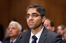
He has aged a year since President Obama nominated him for U.S. Surgeon General in November 2013, but on Monday Boston hospitalist Vivek Murthy, MD, was confirmed as the highest physician in America.
According to multiple sources, Dr. Murthy’s outspoken support for stricter gun laws and belief that guns are a public health issue delayed his confirmation due to opposition from the National Rifle Association (NRA), which in a letter to Senate leadership in February said Dr. Murthy’s confirmation would be a “prescription for disaster for America’s gun owners.”
Despite this, Senate Democrats approved his four-year appointment in a 51-43 vote that cut along party lines. In his confirmation hearing in February, Dr. Murthy said he does not “intend to use the surgeon general’s office as a bully pulpit for gun control.”
Dr. Murthy, 37, earned his medical and business degrees from Yale and for the last decade has worked as both an internist and a hospitalist at Brigham and Women’s Hospital in Boston. He is the youngest Surgeon General ever, and the first of Indian-American descent.
"On behalf of America's 44,000 hospitalists, I congratulate Dr. Murthy, a fellow hospitalist and one of our SHM members, on his historic appointment to U.S. Surgeon General," says Society of Hospital Medicine President Burke Kealey, MD, SFHM. "Being America’s doctor requires many of the same traits required of hospitalists: leadership, sharp clinical skills, and the ability to engage with patients. And, like hospitalists in thousands of hospitals across the country, I am confident Dr. Murthy will become an agent of change to improve delivery of care in our country."
In 2008, Dr. Murthy founded Doctors for Obama, a non-profit, grassroots organization of 16,000 physicians and medical students dedicated to transforming the healthcare system. After the election, he changed the name of the organization to Doctors for America. He also started the software company TrialNetworks in 2007 to aid in drug development, and, in 1995, he started an HIV and AIDS education non-profit in India called VISIONS Worldwide.
In a statement from the White House Monday, President Obama applauded the Senate for Dr. Murthy’s confirmation, saying: “Vivek’s confirmation makes us better positioned to save lives around the world and protect the American people here at home.”
Dr. Murthy replaces acting Surgeon General Boris Lushniak, who took over when Regina Benjamin resigned in July 2013. The surgeon general is the U.S.’ top spokesperson on all matters of public health and oversees the 6,700 members of the U.S. Public Health Service Commissioned Corps.
Kelly April Tyrrell is a freelance writer in Madison, Wis.
Information for this report was published online at cnn.com and usatoday.com.
Researchers show CTL019 cells proliferate and persist
SAN FRANCISCO—Two goals for cell therapy with chimeric antigen receptor (CAR) T cells are significant levels of in vivo proliferation and persistence after the cells are infused.
Researchers at the University of Pennsylvania, working with CTL019 cells, are beginning to see both of these phenomena in children with relapsed, refractory acute lymphoblastic leukemia (ALL).
Stephan Grupp, MD, PhD, described these results at the 2014 ASH Annual Meeting (abstract 380).*
CTL019 is a second-generation chimeric protein engineered using a single-chain variable fragment of an antibody that targets CD19 on B cells. It is combined with the intracellular signaling domains 4-1BB and CD3 zeta and expanded ex vivo with anti-CD3/anti-CD28.
“We take T cells from the patient—this is an individualized or personalized product,” Dr Grupp explained. “We transfect the T cells with a virus, and, in our case, we are using a lentiviral vector. This permanently modifies the T cells.”
“And this allows the expression of the CAR protein in the T cells, which then drives the interaction between the T cell and the cancer cell, hopefully killing the cancer cell but also, and I think this is extraordinarily important, allowing for T-cell activation and significant proliferation.”
More than 130 patients have been treated with CTL019, including patients with CLL whose results were reported at the 2014 ASCO Annual Meeting.
Updated results
At ASH, Dr Grupp provided an update on the 39 children with relapsed, refractory ALL treated with CTL019. He and his colleagues previously reported results in children and adults with ALL in NEJM.
Thirty-six patients (92%) achieved complete remission within a month after infusion. Ten patients relapsed, of whom 5 were CD19+ and 5 were CD19-.
Dr Grupp explained that CD19+ relapses represent waning T cells, and CD19- relapses represent true antigen escape. The latter patients still have CTL019 cells.
Patients were followed for a median of 6 months, ranging from 6 weeks to 31 months. And 15 patients have been followed for more than 1 year.
The patient followed for 31 months “represents the first patient treated who remains in remission with no further therapy,” Dr Grupp said.
Three patients went on to have a stem cell transplant, and 2 had other treatments. One patient had a donor-lymphocyte infusion, and 1 patient who developed myelodysplastic syndrome received treatment for that condition.
“And I think this is a key point,” Dr Grupp noted. “[I]t was a possibility to consider not continuing with a second, third, or, in one case, a fourth transplant.”
Another important point is disease burden, he said. Patients with more than 50% bone marrow blasts at the time of their T-cell infusion had a similar response rate (82%) to those patients who had a lower disease burden of 5% blasts or more (88%). Relapse occurred in all levels of disease burden in a small number of patients.
To date, there has been no graft-vs-host disease.
In terms of efficacy, there appeared to be no significant difference if the patient had received a transplant before CAR therapy or not. Eighty-nine percent of patients who had received an allogeneic transplant responded, compared to 100% who had not had a transplant.
Persistence and proliferation
“Q-PCR detection of CAR cells shows enormous proliferation,” Dr Grupp said. “We have an extraordinary amount of expansion of these cells that’s nearly universal.”
Specifically, the researchers saw 100,000- to 110,000-fold expansions of CAR-positive cells.
Two-thirds of patients have circulating CAR cells 6 months out from their CTL019 infusion. And a group of patients have kept their CAR cells for longer than 12 months. In the group that loses their cells more quickly, CD19+ recurrence is overrepresented, Dr Grupp noted.
Event-free survival is 70% at 6 months, and 76% of patients had a 6-month duration of response.
Toxicity
Cytokine release syndrome (CRS) is a “significant toxicity,” Dr Grupp said, but investigators are beginning to understand some correlates that impact treatment.
Patients with extraordinary levels of the cytokine interleukin-6 (IL-6)—those who require blood pressure or respiratory support—have significantly more severe CRS than those with lower IL-6 levels (P<0.001). Responding patients have high IL-6 levels as well, but patients with severe CRS have very high levels.
The effector cytokine IFNγ, which may be required for the T-cell response, is also elevated in patients with severe CRS compared to those without CRS (P<0.001).
“The thing that I think we’ve really learned from these patients is the impact of disease burden,” Dr Grupp said.
Patients with high disease burden—those with more than 50% bone marrow blasts—have a high likelihood of developing severe CRS. Patients with less burden—fewer than 50% blasts—have a low likelihood.
Dr Grupp pointed out that only 2 patients with more than 50% blasts did not have severe CRS, and they did not respond to therapy.
“This is highly significant and quite predictive for our patients,” he said, adding that CRS is quite controllable via IL-6 blockade with tocilizumab.
B-cell aplasia is “inevitable” as long as these patients have their CAR T cells, Dr Grupp noted. Patients require IVIg replacement therapy for the entire period.
Macrophage activation syndrome, the flip side of CRS, is also a concern, and neurotoxicity, consisting of confusion and aphasia, occurred in a small number of patients and required no therapy.
Given these results, the investigators believe that CTL019 cells may be able to provide long-term response without subsequent therapy.
CTL019 recently received breakthrough therapy designation from the US Food and Drug Administration.
CTL019 was invented at The University of Pennsylvania but has been licensed to Novartis. Several researchers involved in this study reported research funding and/or consultancy payments from Novartis, and 2 researchers are employed by the company. ![]()
*Data in the presentation differ from the abstract.
Iron chelation improves survival in lower-risk MDS
Photo courtesy of ASH
SAN FRANCISCO—Iron chelation therapy significantly improves survival for patients with lower-risk myelodysplastic syndrome (MDS) and delays the progression to acute myeloid leukemia (AML), a new study suggests.
“There is a signal for survival with an impressive difference with chelation therapy,” said study investigator Roger Lyons, MD, of Cancer Care Centers of South Texas in San Antonio.
“If this is real, then everyone with lower-risk MDS will go on chelation therapy upfront.”
Dr Lyons presented the results of this research at the 2014 ASH Annual Meeting (abstract 1350).
He and his colleagues initiated a US registry to collect prospective data on clinical outcomes of patients with lower-risk MDS who received chelation or non-chelation therapy.
The registry enrolled 599 adult patients, with a median age of 76 years, from 107 US centers. The patients had transfusional iron overload with serum ferritin ≥ 1000 µg/L and/or ≥ 20 packed red blood cell units and/or ≥ 6 units every 12 weeks.
Patients were divided into 2 groups: those who had never been chelated and those who had used iron chelation. The researchers also looked at a subgroup of patients: those who received chelation therapy for 6 months or more.
The team evaluated patients every 5 months for 5 years or until death, assessing patient characteristics, survival, disease status, comorbidities, cause of death, and MDS therapy.
At enrollment, the 271 chelated patients had a greater median number of lifetime units transfused compared to the 328 non-chelated patients. Additionally, fewer patients receiving chelation therapy had cardiac, vascular, or endocrine concomitant conditions.
Of the chelated patients, 187 (69%) were chelated with deferasirox, 40 (14.8%) with deferasirox and deferoxamine, and 32 (11.8%) with deferoxamine. For 12 patients (4.5%), the researchers did not know which chelator was used.
The cumulative duration of chelation was 18.9 months in patients who had ever used iron chelation and 27 months in patients with at least 6 months of iron chelation.
Patient outcomes
“From the date of diagnosis, the overall survival for patients receiving chelation therapy was significantly longer than for patients receiving non-chelation therapy, including those with cardiovascular or endocrine concomitant conditions,” Dr Lyons noted.
However, there was a potential clinical bias in patient selection, since patients with longer predicted survival may have been chosen for chelation therapy.
At 5 years of follow-up, the mortality rate was 72.9% for non-chelated patients and 59.4% for patients who received chelation therapy (P=0.0005).
Among patients chelated for 6 months or more, the mortality rate was 56.9% (P=0.0002, compared to non-chelated patients). The most common causes of death were MDS/AML and cardiac conditions.
The time from MDS diagnosis to AML progression was significantly longer for chelated patients than for non-chelated patients—72.1 months and 46.4 months, respectively (P<0.0001).
Among patients chelated for 6 months or more, the time to AML transformation was 78.8 months (P<0.0001, compared to non-chelated patients).
Twice as many patients developed AML in the non-chelation group (n=34, 10.4%) than in the chelation group (n=17, 6.3%). However, this difference was not statistically significant.
Taken together, these results suggest chelation can benefit patients with lower-risk MDS, according to Dr Lyons and his colleagues.
“If you think a lower-risk MDS patient will live 1 or 2 years or is a candidate for transplant, get the patient’s iron levels down by chelation, if possible,” Dr Lyons advised.
Three researchers involved in this study are employed by Novartis, and 1 reported research funding from the company, which manufactures deferasirox (Exjade). ![]()
TBI increases risk of cognitive decline in young kids

Credit: Petr Kratochvil
Young children who undergo total body irradiation (TBI) in preparation for hematopoietic stem cell transplant (HSCT) are at a higher risk for a decline in IQ, according to research published in the Journal of Clinical Oncology.
The study showed that most young patients don’t experience lasting effects on their IQ following HSCT.
However, patients who underwent HSCT at 3 years of age or younger and received TBI had a greater risk of intellectual decline after transplant.
“For the great majority of patients, these findings provide reassurance that transplantation will not have a significant negative impact on cognitive development,” said study author Sean Phipps, PhD, of St Jude Children’s Research Hospital in Memphis, Tennessee.
“We have also identified a high-risk group of younger patients who may benefit from more intensive interventions, including developmental stimulation and other rehabilitative therapies designed to prevent a decline in intellectual functioning and aid in recovery.”
Dr Phipps and his colleagues tracked the IQ scores of 170 St Jude patients before HSCT and for 5 years after the procedure. The patients were 4 months to 23 years of age when their transplants occurred.
Before HSCT, the average IQ scores of all patients were in the normal range. One year after transplant, average IQ scores of patients aged 5 and younger had declined sharply.
But the scores of most patients rebounded in subsequent years. Five years after the procedure, IQ scores for most patients, even the youngest survivors, had largely recovered and fell within the range of normal intelligence.
Patients in the high-risk group were the lone exception. The IQ scores of patients who were both aged 3 or younger when their transplants occurred and who received TBI failed to recover from the first-year decline.
Five years after HSCT, these survivors had average IQ scores in the low-normal range of intelligence. Their scores were more than 16 points lower than the scores of patients who were just as young when their transplants occurred but did not receive TBI.
Furthermore, of the 72 patients in this study whose transplants included TBI, there was a long-term impact on intellectual functioning only for patients who were 3 or younger at transplant.
“The significant first-year decline reflects the intensity of transplantation, which our results suggest leads to greater disruption in development in the youngest children than was previously recognized,” said study author Victoria Willard, PhD, also of St Jude.
The researchers said these findings are good news for most parents whose children must undergo HSCT and provide another reason for hope of good long-term outcomes.
For those whose children are in the newly recognized high-risk group, increased attention and activities designed to stimulate cognitive development may help to prevent reduced IQ following transplant, according to Dr Phipps. ![]()
‘Father of hematopoietic cytokines’ dies
Photo courtesy of The Walter
and Eliza Hall Institute
of Medical Research
Donald Metcalf, MD, an Australian researcher who has been called “the father of hematopoietic cytokines,” has died at the age of 85.
Dr Metcalf’s studies of blood production led to his speculation that there must be a biological mechanism—one or more hormones—that control white blood cell production.
These substances, which he termed colony-stimulating factors (CSFs), were the focus of more than 50 years of research.
Over this time, Dr Metcalf led researchers to characterize and purify 4 separate CSFs—granulocyte CSF (G-CSF), granulocyte-macrophage CSF (GM-CSF), macrophage CSF (M-CSF), and multi-CSF (now called interleukin-3).
Dr Metcalf recognized that CSFs had a potential role in clinical medicine, and his team was among the first in the world to discover the genes for CSFs.
Dr Metcalf was a central figure in the international clinical trials of CSFs in the 1980s, assessing whether CSFs could boost immune cell numbers in cancer patients whose immune system was weakened as a side effect of chemotherapy. On the basis of these studies, G-CSF (Neupogen) was approved for clinical use in 1991.
Now, an estimated 20 million people have been treated with CSFs. As well as boosting the immune system in patients who receive chemotherapy or have other immune deficiencies, CSFs are thought to have revolutionized hematopoietic stem cell transplantation.
A man with many achievements
Dr Metcalf was born in 1929 and started school at the age of 3, by which time he was already reading. He entered university at the age of 16, ultimately obtaining bachelor’s and medical degrees from the University of Sydney.
After an internship at the Royal Prince Alfred Hospital in Sydney, Dr Metcalf joined the staff of the Walter and Eliza Hall Institute of Medical Research (WEHI) in Melbourne in 1954. He was supported by Cancer Council Victoria’s Carden Fellowship, an award he held until his retirement in 2014. (Dr Metcalf officially retired in 1996 but continued to do research until 2014.)
Dr Metcalf spent his early years at WEHI studying vaccinia virus. In 1965, he began studying blood cell formation and, by association, leukemia. In 1966, he became deputy director of WEHI and the head of its Cancer Research Unit.
Dr Metcalf took several sabbaticals from WEHI, serving as a visiting scientist at Harvard Medical School in Boston, Massachusetts; the Roswell Park Memorial Institute in Buffalo, New York; the Swiss Institute for Experimental Cancer Research in Lausanne, Switzerland; the Radiobiological Institute in Rijswijk, The Netherlands; and the University of Cambridge in the UK.
Among Dr Metcalf’s many honors and awards are the Companion of the Order of Australia (1993), the Albert Lasker Award for Clinical Medical Research (1993), the Gairdner Foundation International Award (1994), the Royal Medal of the Royal Society (1995), the Victoria Prize (2000), and the Prime Minister’s Prize for Science (2001).
Dr Metcalf is survived by his wife Jo; daughters Kate, Johanna, Penelope, and Mary-Ann; grandchildren James, Martin, Patrick, Elizabeth, Rose, and Robert; and their extended families. ![]()
Burns, fainting, eyes most common indoor tanning injuries
Skin burns, fainting, and eye injuries are the most common indoor tanning injuries requiring a trip to the local emergency department, according to a research letter published online Dec. 15 in JAMA Internal Medicine.
Indoor tanning exposes users to intense ultraviolet radiation, a known carcinogen, but little is known about the more immediate adverse events related to tanning, wrote Gery P. Guy Jr., Ph.D., of the Centers for Disease Control and Prevention, and coauthors.

After analyzing data from a nationally representative sample of hospital emergency departments from 2003 to 2012, the investigators estimated an average 3,234 indoor tanning–related injuries were treated each year in U.S. hospitals.
The most common injury was skin burns (79.5%), followed by syncope (9.5%) and eye injuries (5.8%).
Injuries occurred most commonly among younger adults and non-Hispanic white females, the populations with the highest rates of indoor tanning.
However, indoor tanning injuries were on the decline, decreasing from 6,487 in 2003 to 1,957 in 2012 (P < .001), a finding that was most likely due to a decline in the use of indoor sun beds, the researchers said.
Most patients did not require hospitalization, but burns severe enough to warrant a trip to the emergency department indicate an overexposure to ultraviolet radiation and an increased risk of skin cancer, the investigators added.
Although the Food and Drug Administration required tanning device manufacturers to install timers to limit exposure, several cases described patients falling asleep while tanning, raising concerns about timers malfunctioning or being intentionally overridden, the researchers noted.
The researchers said they had no relevant financial conflicts to disclose.
Skin burns, fainting, and eye injuries are the most common indoor tanning injuries requiring a trip to the local emergency department, according to a research letter published online Dec. 15 in JAMA Internal Medicine.
Indoor tanning exposes users to intense ultraviolet radiation, a known carcinogen, but little is known about the more immediate adverse events related to tanning, wrote Gery P. Guy Jr., Ph.D., of the Centers for Disease Control and Prevention, and coauthors.
After analyzing data from a nationally representative sample of hospital emergency departments from 2003 to 2012, the investigators estimated an average 3,234 indoor tanning–related injuries were treated each year in U.S. hospitals.
The most common injury was skin burns (79.5%), followed by syncope (9.5%) and eye injuries (5.8%).
Injuries occurred most commonly among younger adults and non-Hispanic white females, the populations with the highest rates of indoor tanning.
However, indoor tanning injuries were on the decline, decreasing from 6,487 in 2003 to 1,957 in 2012 (P < .001), a finding that was most likely due to a decline in the use of indoor sun beds, the researchers said.
Most patients did not require hospitalization, but burns severe enough to warrant a trip to the emergency department indicate an overexposure to ultraviolet radiation and an increased risk of skin cancer, the investigators added.
Although the Food and Drug Administration required tanning device manufacturers to install timers to limit exposure, several cases described patients falling asleep while tanning, raising concerns about timers malfunctioning or being intentionally overridden, the researchers noted.
The researchers said they had no relevant financial conflicts to disclose.
FROM JAMA INTERNAL MEDICINE

Alpinia officinarum
Alpinia officinarum (and its close relative Alpinia galanga), a member of the Zingiberaceae family (Zhong Xi Yi Jie He Xue Bao 2011;9:1061-5), has long been used in Chinese medicinals (Bioorg. Med. Chem. 2009;17:6048-53). Specifically, the plant is used in traditional Chinese medicine as an aphrodisiac, abortifacient, carminative, antipyretic, anti-inflammatory, and emmenagogue as well as to treat disorders of the heart and kidneys, bronchitis, chronic enteritis, renal calculus, diabetes, and rheumatism (Zhong Xi Yi Jie He Xue Bao 2011;9:1061-5; Bioorg. Med. Chem. 2009;17:6048-53). Stomach ailments are the most typical application of the herb in traditional Chinese and Thai medicine; it is also used in Ayurveda and Sidda medicine. A. officinarum is widely cultivated throughout Asia, including China, Thailand, India, Sri Lanka, Malaysia, and Indonesia, as well as the Middle East and Northern Africa (Saudi Arabia and Egypt, respectively) (Zhong Xi Yi Jie He Xue Bao 2011;9:1061-5).
The flavonoid galangin (3,5,7-trihydroxyflavone) is the primary active constituent of A. officinarum (Phytother. Res. 2014;28:1533-8; J. Cell Biochem. 2013;114:152-61). In vitro, it has demonstrated a cytotoxic effect on multiple cancer cell lines (J. Cell Biochem. 2013;114[1]:152-61). Traditional Uighur medicine in China has incorporated galangin for the treatment of vitiligo (Phytother. Res. 2014;28:1533-8). Overall, A. officinarum rhizomes have been associated with antiemetic, antigenotoxic, antimutagenic, and antioxidant activity, as well as inhibitory effects on prostaglandin and leukotriene biosynthesis, and modulatory effects on cytochrome P450 enzymes (Bioorg. Med. Chem. 2009;17:6048-53; J. Cell Biochem. 2013;114:152-61). The rhizomes of A. officinarum have been used externally to treat skin infections, gum diseases, and skin cancer (J. Nat. Med. 2008;62:374-8).
Constituents
The rhizomes of the plant, commonly referred to as galangal, contain several key active constituents, including essential oils, tannins, neolignans, phenol, glycosides, monoterpenes, diarylheptanoids, phenylpropanoids, carbohydrates, gallic acid glycoside, galangoisoflavonoid, beta-sitosterol, galangin, alpinin, zerumbone, and kampferide (Zhong Xi Yi Jie He Xue Bao 2011;9:1061-5; Bioorg. Med. Chem. 2009;17:6048-53; J. Nat. Med. 2008;62:374-8).
In 2009, Matsuda et al. reported that the 80% aqueous acetone extract of the rhizomes of A. officinarum suppressed melanogenesis in theophylline-stimulated murine B16 melanoma 4A5 cells. They found that several isolated constituents had significant IC50 values (10-48 mcm) for inhibiting melanogenesis, including four diarylheptanoids (5-hydroxy-1,7-diphenyl-3-heptanone, 7-(4(‘’)-hydroxy-3(‘’)-methoxyphenyl)-1-phenylhept-4-en-3-one, 5-hydroxy-7-(4(‘’)-hydroxy-3(‘’)-methoxyphenyl)-1-phenyl-3-heptanone, and 3,5-dihydroxy-1,7-diphenylheptane) and two flavonol constituents (kaempferide and galangin). The mRNA expression of tyrosinase and tyrosinase-related proteins-1 and -2 was also hindered by 7-(4(‘’)-hydroxy-3(‘’)-methoxyphenyl)-1-phenylhept-4-en-3-one, kaempferide, and galangin, as was the protein level of a microphthalmia-associated transcription factor, the authors noted (Bioorg. Med. Chem. 2009;17:6048-53).
Biologic activity
Penetration enhancement: In 2000, Shen et al. found that volatile oils from galangal, among other herbs, were effective in enhancing the skin permeation of 5-fluorouracil and notably more effective than azone (Zhong Yao Cai 2000;23:697-9).
Anti-inflammatory effects: In 2008, Yasukawa et al. examined the inhibitory effect of galangal in a two-stage in vivo carcinogenesis model in mice. They observed that the A. officinarum rhizomes displayed significant antitumor-promoting activity against 7,12-dimethylbenz[a]anthracene (DMBA)-initiated and 12-O-tetradecanoylphorbol-13-acetate (TPA)–promoted lesions. Seven diarylheptanoids isolated from the active fraction of the methanol extracts demonstrated significant anti-inflammatory effects against TPA-induced inflammation (J. Nat. Med. 2008;62:374-8).
Cancer prevention and pigmentary effects: Heo et al. reported in 2001 that in vitro and in vivo studies have demonstrated that the flavonoid galangin, found in high concentrations in A. officinarum, as well as the bee product propolis, exhibits significant antioxidant activity and can influence enzyme activities and inhibit genotoxicity without introducing a pro-oxidant effect. They concluded that galangin warrants consideration for its potential as a chemical cancer-preventing agent (Mutat. Res. 2001;488:135-50).
In 2007, Lu et al. investigated the whitening effects of the flavonoid components of A. officinarum on melanin biosynthesis in B16 mouse melanoma cells, tyrosinase inhibition, and UV absorption. They found that galangin and the flavonoid mixture both decreased melanin content more than controls and also lowered melanin production, with galangin more effective than the flavonoid mixture. In addition, galangin and the flavonoid mixture exerted greater tyrosinase inhibition at lower concentrations. The A. officinarum constituents also displayed a broad absorption band in the UVB area (270 to 290 nm). The researchers concluded that galangin may be a viable whitening agent with the potential to prevent skin cancer (J. Enzyme Inhib. Med. Chem. 2007;22:433-8).
Six years later, Zhang et al. noted that various doses of galangin resulted in the inhibition of B16F10 melanoma cell proliferation. The investigators also showed that galangin achieved an antimetastatic effect in vivo in C57BL/6J mice, reducing focal adhesion kinase. They concluded that focal adhesion kinase is a viable target in melanoma therapy, with B16F10 melanoma metastasis apparently checked by galangin in mice and in cell cultures (J. Cell Biochem. 2013;114:152-61).
In 2014, Huo et al. tested galangin in a mouse model of vitiligo induced in C57BL/6 mice through the daily topical application of hydroquinone (2.5%) on shaved dorsal skin for 60 days. Thirty days after the final hydroquinone application, investigators began oral administration of galangin for 30 days. Hair grew back after treatment darker than the original color, with histologic analysis revealing that mice treated with galangin and the positive control 8-methoxypsoralen had an increased number of melanin-containing hair follicles, compared with untreated animals. In addition, galangin treatment was associated with significant increases in the number of cutaneous basal layer melanocytes and melanin-containing epidermal cells. Compared with controls, treatment with galangin and 8-methoxypsoralen led to increased serum levels of tyrosinase and decreased levels of malondialdehyde and lower cholinesterase activity. Galangin and 8-methoxypsoralen use also increased the expression of tyrosinase protein in treated skin. The investigators concluded that galangin improved hydroquinone-induced vitiligo in mice and warrants further study as a potential vitiligo treatment in humans (Phytother. Res. 2014 28:1533-8).
Conclusion
Alpinia officinarum is one of many botanical agents with a long history of applications in traditional folk medicine, particularly in Asia. There is a relative paucity of evidence regarding the dermatologic applications of this plant, but recent findings support continued research into its various potential cutaneous benefits, with particular focus on the main active ingredient galangin.
Dr. Baumann is chief executive officer of the Baumann Cosmetic & Research Institute in the Design District in Miami. She founded the Cosmetic Dermatology Center at the University of Miami in 1997. Dr. Baumann wrote the textbook “Cosmetic Dermatology: Principles and Practice” (New York: McGraw-Hill, 2002), and a book for consumers, “The Skin Type Solution” (New York: Bantam Dell, 2006). She has contributed to the Cosmeceutical Critique column in Dermatology News since January 2001. Her latest book, “Cosmeceuticals and Cosmetic Ingredients,” was published in November 2014. Dr. Baumann has received funding for clinical grants from Allergan, Aveeno, Avon Products, Evolus, Galderma, GlaxoSmithKline, Kythera, Mary Kay, Medicis Pharmaceuticals, Neutrogena, Philosophy,Topix Pharmaceuticals, and Unilever.
Finger Lesion Is Painful When Bumped
A 67-year-old man has had a small lesion on his index finger for several years. Recently, it has grown large enough to start interfering with his normal activities. The lesion is not painful unless the patient bumps it—but as it grows, avoiding such incidents is becoming more difficult.
The diagnosis has thus far been a mystery: What is it? The patient’s primary care provider didn’t know and, per the patient’s report, had no interest in trying to remove the lesion. Thus, the patient finds himself referred to dermatology.
The patient can recall no specific injury. He does, however, report that he worked for years as an upholsterer, shaping, sewing, and stapling heavy fabric onto chairs and sofas—often puncturing his fingers in the process.
His health is otherwise “decent.” He is taking medication for hypertension and an a-blocker due to “problems” with his prostate.
EXAMINATION
The finger lesion is a 2-cm, round, firm nodule with a smooth, telangiectatic surface. It is located on the perionychial area of patient’s index finger (lateral aspect). The lesion is attached to the finger by a broad base that rises seamlessly from the skin. It is not translucent or tender to touch. No similar lesions are seen on either hand, and no nodes are felt in the epitrochlear or axillary locations.
Within a few weeks, the patient returns to have the lesion excised. This is done with a combination of a digital block and local anesthesia, with a tourniquet to control bleeding. After primary closure, the lesion is submitted to pathology. The report indicates the lesion is a cyst, full of cheesy material.
What is the diagnosis?
DISCUSSION
This type of cyst is sometimes referred to as an implantation cyst and occurs when epidermal tissue, including sebaceous glands, is implanted into deeper tissue by a penetrating injury and continues to grow. Although this is an utterly common and benign diagnosis, the differential for a “fingeroma” includes digital mucous cyst, pyogenic granuloma, acquired digital fibrokeratoma, calcinosis cutis, tophaceous gout, wart, and giant cell tumor. Had the lesion been of bony origin, it could have represented connective tissue cancer (sarcoma) or even metastatic cancer.
Implantation cysts must be removed completely to prevent recurrence. The size and location of this particular cyst made it conducive to simple excision and primary closure. Larger lesions in more difficult locations often require the expertise of a hand surgeon for disposition.
TAKE-HOME LEARNING POINTS
• “Fingeromas” are quite common but have an extensive differential diagnosis—mostly benign, but occasionally malignant.
• Regarding malignancy, it is helpful to remember that any tissue in the body can undergo malignant transformation and that many types of primary cancers can metastasize to distant areas (including the hands).
• Removal, or at least biopsy, is the best way to differentiate between types of fingeroma lesions.
• Implantation cysts, as seen in this case, result from traumatic implantation of epidermal structures (including sebaceous glands) into deeper tissue, where they continue to grow.
• When in doubt, consider referring affected patients to dermatology or to a hand surgeon.
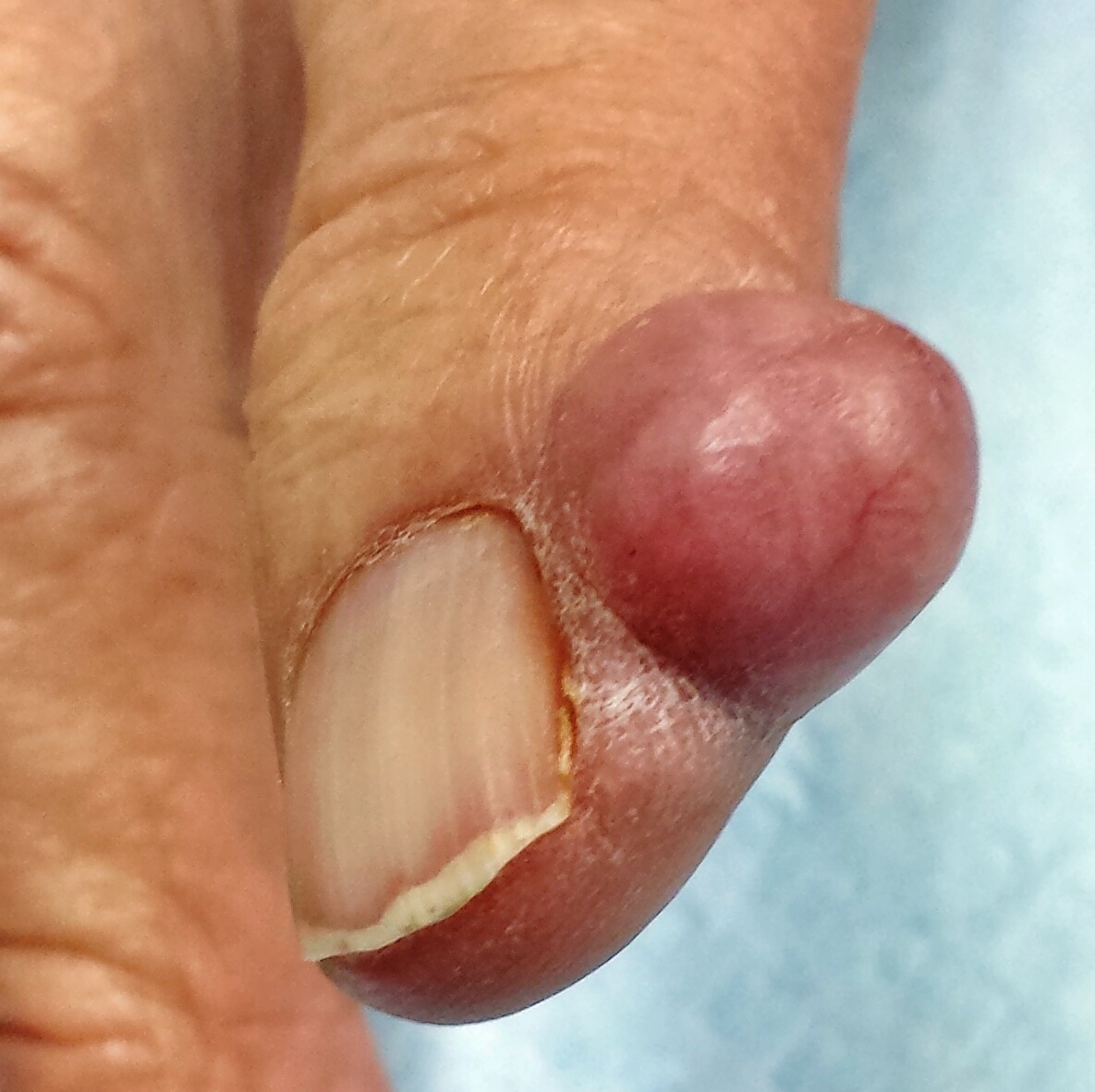
Current Thinking in OTC Analgesia: Patient Considerations and Practical Insights
December 2014
A supplement to Family Practice News® and Internal Medicine News®. This supplement is sponsored by McNeil Consumer Healthcare Division of McNEIL-PPC, Inc.

Topics
- Pain in the United States
- Critical Comorbid Illnesses
- Impact of Pain on Comorbidities and Patient Outcomes
- Pain Management for Patients Taking Concomitant Medications
- Benefits of OTC Analgesics
- Risks of OTC Analgesics
- A Nursing Perspective
- Not All Analgesics Are Appropriate for Every Patient
- Patients’ Safe Use of OTC Analgesics
- Conclusions
Faculty
Chairman
Charles P. Vega, Jr, MD, FAAFP
Clinical Professor, Department of Family Medicine
Director, Patient-Centered Advanced Clinical Education
Director, Program in Medical Education for the Latino Community
University of California, Irvine, School of Medicine
Irvine, California
Faculty
Christopher M. Chappel, MD
Medical Director
Chappel Health and Wellness
Kissimmee, Florida
Brett Badgley Snodgrass, MSN, APRN, FNP-BC
Family Nurse Practitioner
Saint Francis Hospital–Bartlett Interventional Pain Management Clinic
Chief Executive Officer and President
BBS Consultants, Inc.
Bartlett, Tennessee
Copyright © 2014 by Frontline Medical Communications Inc.
December 2014
A supplement to Family Practice News® and Internal Medicine News®. This supplement is sponsored by McNeil Consumer Healthcare Division of McNEIL-PPC, Inc.
Topics
- Pain in the United States
- Critical Comorbid Illnesses
- Impact of Pain on Comorbidities and Patient Outcomes
- Pain Management for Patients Taking Concomitant Medications
- Benefits of OTC Analgesics
- Risks of OTC Analgesics
- A Nursing Perspective
- Not All Analgesics Are Appropriate for Every Patient
- Patients’ Safe Use of OTC Analgesics
- Conclusions
Faculty
Chairman
Charles P. Vega, Jr, MD, FAAFP
Clinical Professor, Department of Family Medicine
Director, Patient-Centered Advanced Clinical Education
Director, Program in Medical Education for the Latino Community
University of California, Irvine, School of Medicine
Irvine, California
Faculty
Christopher M. Chappel, MD
Medical Director
Chappel Health and Wellness
Kissimmee, Florida
Brett Badgley Snodgrass, MSN, APRN, FNP-BC
Family Nurse Practitioner
Saint Francis Hospital–Bartlett Interventional Pain Management Clinic
Chief Executive Officer and President
BBS Consultants, Inc.
Bartlett, Tennessee
Copyright © 2014 by Frontline Medical Communications Inc.