User login
Promoting mental well-being in LGBTQ youth
For many the beginning of a new year is a time to set goals and resolutions for the upcoming year. Often these resolutions are related to health, for example, quit smoking, work out more, lose weight. It is sometimes easy to overlook mental health and well-being as an integral part of overall wellness. This month’s column will focus on how as pediatric providers we can help promote the mental well-being of our patients in practice.
Mental health problems are a significant cause of morbidity and mortality in youth. In 2014, suicide was the second leading cause of death for all youth 10-14 years and 15-24 years.1 While most lesbian, gay, bisexual, transgender, and questioning (LGBTQ) persons live healthy, happy lives, LGBTQ youth are at disproportionate risk for mental illness, probably related to lack of support and to stigma related to their sexual minority and gender minority identities. Studies suggest that LGBTQ youth have suicidality rates two to five times higher than their heterosexual cisgender peers.2,3,4

• Principle 1. A comprehensive diagnostic evaluation should include an age-appropriate assessment of psychosexual development for all youths.
While pediatric providers are unlikely to perform a comprehensive mental health diagnostic evaluation, psychosocial development should regularly be assessed at well visits. It may not be readily apparent which youth are struggling with development of their sexual and gender identity. Nonassuming questions regarding development in theses domains should ideally be integrated into the psychosocial assessment. For example, begin a sexual history by asking, “Are you romantically attracted to males, females, both, or neither?”
• Principle 2. The need for confidentiality in the clinical alliance is a special consideration in the assessment of sexual and gender minority youth.
Confidentiality is important when talking to any youth about their sexual and gender identity. LGBTQ youth in particular may have concerns of family or provider rejection, and they may look for cues that they can safely discuss their sexuality or gender identity without fear of being judged or shamed. Clinicians should be aware of confidentiality practices for minors when discussing these issues. Potential risks of premature disclosure to family and support systems, such as rejection or alienation, also should be considered.
• Principle 3. Family dynamics pertinent to sexual orientation, gender nonconformity, and gender identity should be explored in the context of the cultural values of the youth, family, and community.
Families can have a variety of responses to their child’s sexual minority or gender minority identity, ranging from acceptance to rejection, with some youth being forced to leave home. Many families need to alter their ideas and expectations for a child after their child comes out, and this can lead to feelings of loss and grief accompanied by feelings of anxiety, anger, shame, and guilt.5 Over time, however, the majority of families become affirming and supportive and are not distressed.7 Recognizing that family support reduces negative health outcomes for youth, providers should aim to support and preserve positive family relationships when possible. This may involve education and support for families as well as youth. It is important to be aware that sexual and gender minority youth who are also members of ethnic minorities may face additional challenges.
• Principle 4. Clinicians should inquire about circumstances commonly encountered by youth with sexual and gender minority status that confer increased psychiatric risk.
Providers should recognize that LGBTQ youth are at disproportionate risk of bullying, suicide, substance use, high-risk sexual behaviors, running away, and becoming homeless. Providers should assess for these risks and address them as appropriate.
• Principle 5. Clinicians should aim to foster healthy psychosexual development in sexual and gender minority youth, and protect these individuals’ full capacity for integrated identity formation and functioning.
Providers should support healthy youth development and self-discovery, recognizing that there is a spectrum of sexual and gender identities, with the goal of helping youth achieve their full developmental potential.
• Principle 6. Clinicians should be aware that there is no evidence that sexual orientation can be altered through therapy, and attempts to do so may be harmful.
Therapies targeted at altering sexual orientation or gender identity, often referred to as reparative therapies, can encourage family rejection and decrease self-esteem and connectedness, all of which have been identified as risk factors for suicidality. Providers should educate parents about the potential harm of these types of therapies and ensure that mental health providers to whom patients are being referred are not practicing these potentially harmful therapies.
• Principle 7. Clinicians should be aware of current evidence on the natural course of gender discordance and associated psychopathology in children and adolescents in choosing the treatment goals and modality.
Variation in gender role behavior (for example, dress preference, toy preference, types of play) is typical in early childhood and should be distinguished from gender dysphoria, in which a child expresses distress related to a gender identity that is different from or does not fully align with the child’s sex assigned at birth. Assessing gender development in childhood and the best approach to treatment is best done by professionals with experience and training in gender development, and providers should be familiar with resources in their area. For some, gender identity concerns may not be recognized until adolescence when the onset of puberty and secondary sex characteristics result in increased dysphoria. Best practice guidelines exist for treatment of youth with gender discordance, and there is limited but growing evidence to support best practices. Providers should ensure that the providers and specialists to whom families are referred practice according to current best practices.
• Principle 8. Clinicians should be prepared to consult and act as a liaison with schools, community agencies, and other health care providers, advocating for the unique needs of sexual and gender minority youth and their families.
Pediatric providers can work with mental health professionals to be advocates for their gender and sexual minority patients and raise awareness of issues affecting these special populations such as bullying and suicidality.
• Principle 9. Mental health professionals should be aware of community and professional resources relevant to sexual and gender minority youth.
As medical providers, we have a limited amount of time to see and assess patients, and often are able to best serve our patients and families by connecting them to specialists in the medical community and resources available in the school and community. It is important to know what resources exist in the community to be able to appropriately refer and connect patients.
Resources for providers
• American Academy of Child and Adolescent Psychiatry Practice Parameter on lesbian, gay, bisexual, and transgender youth .
• National LGBT Health Education Center: Training materials and modules with continuing education credits.
Resources for families
• Gay, Lesbian, and Straight Education Network.• Parents, Friends, Families of Lesbians and Gays (PFLAG).
References
1. “10 Leading Causes of Death by Age Group, United States – 2014,” National Center for Injury Prevention and Control, Centers for Disease Control and Prevention.
2. Lesbian, Gay, Bisexual, and Transgender Health: LGBT Youth, Centers for Disease Control and Prevention, Nov. 12, 2014.
3. Am J Public Health. 2001 Aug;91(8):1276-81.
4. Am J Prev Med. 2012 Mar;42(3):221-8.
5. J. Am Acad Child Adolesc Psychiatry. 2012;51(9):957–74.
6. Pediatr Clin North Am. 2016 Dec;63(6):971-83.
7. “Mom, Dad. I’m Gay: How Families Negotiate Coming Out” (Washington, DC: American Psychological Association, 2001).
Dr. Chelvakumar is an attending physician in the division of adolescent medicine at Nationwide Children’s Hospital and an assistant professor of clinical pediatrics at the Ohio State University, both in Columbus.
For many the beginning of a new year is a time to set goals and resolutions for the upcoming year. Often these resolutions are related to health, for example, quit smoking, work out more, lose weight. It is sometimes easy to overlook mental health and well-being as an integral part of overall wellness. This month’s column will focus on how as pediatric providers we can help promote the mental well-being of our patients in practice.
Mental health problems are a significant cause of morbidity and mortality in youth. In 2014, suicide was the second leading cause of death for all youth 10-14 years and 15-24 years.1 While most lesbian, gay, bisexual, transgender, and questioning (LGBTQ) persons live healthy, happy lives, LGBTQ youth are at disproportionate risk for mental illness, probably related to lack of support and to stigma related to their sexual minority and gender minority identities. Studies suggest that LGBTQ youth have suicidality rates two to five times higher than their heterosexual cisgender peers.2,3,4
• Principle 1. A comprehensive diagnostic evaluation should include an age-appropriate assessment of psychosexual development for all youths.
While pediatric providers are unlikely to perform a comprehensive mental health diagnostic evaluation, psychosocial development should regularly be assessed at well visits. It may not be readily apparent which youth are struggling with development of their sexual and gender identity. Nonassuming questions regarding development in theses domains should ideally be integrated into the psychosocial assessment. For example, begin a sexual history by asking, “Are you romantically attracted to males, females, both, or neither?”
• Principle 2. The need for confidentiality in the clinical alliance is a special consideration in the assessment of sexual and gender minority youth.
Confidentiality is important when talking to any youth about their sexual and gender identity. LGBTQ youth in particular may have concerns of family or provider rejection, and they may look for cues that they can safely discuss their sexuality or gender identity without fear of being judged or shamed. Clinicians should be aware of confidentiality practices for minors when discussing these issues. Potential risks of premature disclosure to family and support systems, such as rejection or alienation, also should be considered.
• Principle 3. Family dynamics pertinent to sexual orientation, gender nonconformity, and gender identity should be explored in the context of the cultural values of the youth, family, and community.
Families can have a variety of responses to their child’s sexual minority or gender minority identity, ranging from acceptance to rejection, with some youth being forced to leave home. Many families need to alter their ideas and expectations for a child after their child comes out, and this can lead to feelings of loss and grief accompanied by feelings of anxiety, anger, shame, and guilt.5 Over time, however, the majority of families become affirming and supportive and are not distressed.7 Recognizing that family support reduces negative health outcomes for youth, providers should aim to support and preserve positive family relationships when possible. This may involve education and support for families as well as youth. It is important to be aware that sexual and gender minority youth who are also members of ethnic minorities may face additional challenges.
• Principle 4. Clinicians should inquire about circumstances commonly encountered by youth with sexual and gender minority status that confer increased psychiatric risk.
Providers should recognize that LGBTQ youth are at disproportionate risk of bullying, suicide, substance use, high-risk sexual behaviors, running away, and becoming homeless. Providers should assess for these risks and address them as appropriate.
• Principle 5. Clinicians should aim to foster healthy psychosexual development in sexual and gender minority youth, and protect these individuals’ full capacity for integrated identity formation and functioning.
Providers should support healthy youth development and self-discovery, recognizing that there is a spectrum of sexual and gender identities, with the goal of helping youth achieve their full developmental potential.
• Principle 6. Clinicians should be aware that there is no evidence that sexual orientation can be altered through therapy, and attempts to do so may be harmful.
Therapies targeted at altering sexual orientation or gender identity, often referred to as reparative therapies, can encourage family rejection and decrease self-esteem and connectedness, all of which have been identified as risk factors for suicidality. Providers should educate parents about the potential harm of these types of therapies and ensure that mental health providers to whom patients are being referred are not practicing these potentially harmful therapies.
• Principle 7. Clinicians should be aware of current evidence on the natural course of gender discordance and associated psychopathology in children and adolescents in choosing the treatment goals and modality.
Variation in gender role behavior (for example, dress preference, toy preference, types of play) is typical in early childhood and should be distinguished from gender dysphoria, in which a child expresses distress related to a gender identity that is different from or does not fully align with the child’s sex assigned at birth. Assessing gender development in childhood and the best approach to treatment is best done by professionals with experience and training in gender development, and providers should be familiar with resources in their area. For some, gender identity concerns may not be recognized until adolescence when the onset of puberty and secondary sex characteristics result in increased dysphoria. Best practice guidelines exist for treatment of youth with gender discordance, and there is limited but growing evidence to support best practices. Providers should ensure that the providers and specialists to whom families are referred practice according to current best practices.
• Principle 8. Clinicians should be prepared to consult and act as a liaison with schools, community agencies, and other health care providers, advocating for the unique needs of sexual and gender minority youth and their families.
Pediatric providers can work with mental health professionals to be advocates for their gender and sexual minority patients and raise awareness of issues affecting these special populations such as bullying and suicidality.
• Principle 9. Mental health professionals should be aware of community and professional resources relevant to sexual and gender minority youth.
As medical providers, we have a limited amount of time to see and assess patients, and often are able to best serve our patients and families by connecting them to specialists in the medical community and resources available in the school and community. It is important to know what resources exist in the community to be able to appropriately refer and connect patients.
Resources for providers
• American Academy of Child and Adolescent Psychiatry Practice Parameter on lesbian, gay, bisexual, and transgender youth .
• National LGBT Health Education Center: Training materials and modules with continuing education credits.
Resources for families
• Gay, Lesbian, and Straight Education Network.• Parents, Friends, Families of Lesbians and Gays (PFLAG).
References
1. “10 Leading Causes of Death by Age Group, United States – 2014,” National Center for Injury Prevention and Control, Centers for Disease Control and Prevention.
2. Lesbian, Gay, Bisexual, and Transgender Health: LGBT Youth, Centers for Disease Control and Prevention, Nov. 12, 2014.
3. Am J Public Health. 2001 Aug;91(8):1276-81.
4. Am J Prev Med. 2012 Mar;42(3):221-8.
5. J. Am Acad Child Adolesc Psychiatry. 2012;51(9):957–74.
6. Pediatr Clin North Am. 2016 Dec;63(6):971-83.
7. “Mom, Dad. I’m Gay: How Families Negotiate Coming Out” (Washington, DC: American Psychological Association, 2001).
Dr. Chelvakumar is an attending physician in the division of adolescent medicine at Nationwide Children’s Hospital and an assistant professor of clinical pediatrics at the Ohio State University, both in Columbus.

Letters to the Editor: Your patients are talking: Isn’t it time you take responsibility for your online reputation?
“YOUR PATIENTS ARE TALKING: ISN’T IT TIME YOU TAKE RESPONSIBILITY FOR YOUR ONLINE REPUTATION?”
RON ROMANO AND NEIL H. BAUM, MD (NOVEMBER 2016)
Eschews meaningless Internet obfuscation
As a practicing physician I don’t have time for social media and its accompanying advertising rationale; it’s a wasteland that replaces television. My patients and I go one-on-one, eye-to-eye, and eschew meaningless Internet obfuscation. Don’t we have better things to do with our physician/patient relationship than check online reviews?
Warren Kendall, MD
Grants Pass, Oregon
Share your thoughts! Send your Letter to the Editor to [email protected]. Please include your name and the city and state in which you practice.

Letters to the Editor: Should we change instruments and gloves after closing the uterus?
“PREVENTING INFECTION AFTER CESAREAN DELIVERY: 5 MORE EVIDENCE-BASED MEASURES TO CONSIDER”
KATHRYN E. PATRICK, MD; SARA L. DEATSMAN, MD; AND PATRICK DUFF, MD (DECEMEBER 2016)
Should we change instruments and gloves after closing the uterus?
In reference to the recent article series on preventing infection after cesarean delivery by Drs. Patrick, Deatsman, and Duff, what are the thoughts on using clean instruments and changing gloves after closing the uterus?
Gerrit J. Schipper, MD
Frederick, Maryland
❯❯ Drs. Patrick, Deatsman, and Duff respond:
We appreciate Dr. Schipper’s thoughtful question concerning our recent articles. At present, we are not aware of any rigorous studies that have evaluated the possible protective effect of changing to a different set of surgical instruments after closure of the uterus.
The second part of the question concerning the effect of changing gloves at a certain point in the operation is more intriguing. In an earlier report from our institution, we showed that the dominant hand of the operator becomes heavily contaminated with bacteria during the process of extracting the fetal head from the lower uterine segment.1 The contamination is particularly heavy when the patient has had an extended duration of labor in the presence of ruptured membranes. In a subsequent investigation, we showed that avoidance of manual extraction of the placenta, a process in which the now-contaminated glove of the operator is placed between the placenta and the uterine wall, significantly reduced the frequency of postcesarean endometritis even in patients who already were receiving systemic antibiotic prophylaxis.2 Whether changing gloves after delivery of the baby will further decrease the frequency of postcesarean endometritis, beyond that which can be achieved with systemic antibiotic prophylaxis combined with delivery of the placenta by traction on the cord, has not been studied in a systematic manner.
Given the low frequency of infection that can be achieved with these 2 methods, it would require a very large sample size to show that glove change offered an additional protective effect. Nevertheless, on a practical basis, we think it is very reasonable to change the glove on the dominant hand following a difficult extraction of the presenting part in a patient who has had an extended duration of labor and ruptured membranes. The glove change is particularly important if manual extraction of the placenta is contemplated.
Of note, we would like to acknowledge that the US Food and Drug Administration finalized a ban on the use of powdered surgical gloves effective January 18, 2017.3 The aerosolized glove powder on latex gloves contains proteins that can provoke severe respiratory allergic reactions in patients who are sensitive to latex. Even powdered synthetic gloves can cause airway inflammation, wound inflammation, and postoperative adhesions.
“DOES ONE PARTICULAR CESAREAN TECHNIQUE CONFER BETTER MATERNAL AND NEONATAL OUTCOMES?”
JOHN M. THORP JR, MD (EXAMINING THE EVIDENCE; NOVEMBER 2016)
Choosing a cesarean technique based on “evidence”
I appreciate the commentary by Dr. Thorp concerning cesarean delivery techniques. I have always thought that there was no difference in the outcomes of the various techniques. However, we will continue to waver to the peer pressure of this evidence-based stuff—until we find out later, like now—until things change again. “The more things change, the more they remain the same.”
Dr. Smart Ebinne
Port Harcourt, Nigeria
Share your thoughts! Send your Letter to the Editor to [email protected]. Please include your name and the city and state in which you practice.
- Yancey MK, Clark P, Duff P. The frequency of glove contamination during cesarean delivery. Obstet Gynecol. 1994;83(4):538–542.
- Lasley DS, Eblen A, Yancey MK, Duff P. The effect of placental removal method on the incidence of postcesarean infections. Am J Obstet Gynecol. 1997;176(6):1250–1254.
- US Food and Drug Administration. Banned devices; powdered surgeon’s gloves, powdered patient examination gloves, and absorbable powder for lubricating a surgeon’s glove. Final rule. Fed Regist. 2016;81(243):91722–91731.
“PREVENTING INFECTION AFTER CESAREAN DELIVERY: 5 MORE EVIDENCE-BASED MEASURES TO CONSIDER”
KATHRYN E. PATRICK, MD; SARA L. DEATSMAN, MD; AND PATRICK DUFF, MD (DECEMEBER 2016)
Should we change instruments and gloves after closing the uterus?
In reference to the recent article series on preventing infection after cesarean delivery by Drs. Patrick, Deatsman, and Duff, what are the thoughts on using clean instruments and changing gloves after closing the uterus?
Gerrit J. Schipper, MD
Frederick, Maryland
❯❯ Drs. Patrick, Deatsman, and Duff respond:
We appreciate Dr. Schipper’s thoughtful question concerning our recent articles. At present, we are not aware of any rigorous studies that have evaluated the possible protective effect of changing to a different set of surgical instruments after closure of the uterus.
The second part of the question concerning the effect of changing gloves at a certain point in the operation is more intriguing. In an earlier report from our institution, we showed that the dominant hand of the operator becomes heavily contaminated with bacteria during the process of extracting the fetal head from the lower uterine segment.1 The contamination is particularly heavy when the patient has had an extended duration of labor in the presence of ruptured membranes. In a subsequent investigation, we showed that avoidance of manual extraction of the placenta, a process in which the now-contaminated glove of the operator is placed between the placenta and the uterine wall, significantly reduced the frequency of postcesarean endometritis even in patients who already were receiving systemic antibiotic prophylaxis.2 Whether changing gloves after delivery of the baby will further decrease the frequency of postcesarean endometritis, beyond that which can be achieved with systemic antibiotic prophylaxis combined with delivery of the placenta by traction on the cord, has not been studied in a systematic manner.
Given the low frequency of infection that can be achieved with these 2 methods, it would require a very large sample size to show that glove change offered an additional protective effect. Nevertheless, on a practical basis, we think it is very reasonable to change the glove on the dominant hand following a difficult extraction of the presenting part in a patient who has had an extended duration of labor and ruptured membranes. The glove change is particularly important if manual extraction of the placenta is contemplated.
Of note, we would like to acknowledge that the US Food and Drug Administration finalized a ban on the use of powdered surgical gloves effective January 18, 2017.3 The aerosolized glove powder on latex gloves contains proteins that can provoke severe respiratory allergic reactions in patients who are sensitive to latex. Even powdered synthetic gloves can cause airway inflammation, wound inflammation, and postoperative adhesions.
“DOES ONE PARTICULAR CESAREAN TECHNIQUE CONFER BETTER MATERNAL AND NEONATAL OUTCOMES?”
JOHN M. THORP JR, MD (EXAMINING THE EVIDENCE; NOVEMBER 2016)
Choosing a cesarean technique based on “evidence”
I appreciate the commentary by Dr. Thorp concerning cesarean delivery techniques. I have always thought that there was no difference in the outcomes of the various techniques. However, we will continue to waver to the peer pressure of this evidence-based stuff—until we find out later, like now—until things change again. “The more things change, the more they remain the same.”
Dr. Smart Ebinne
Port Harcourt, Nigeria
Share your thoughts! Send your Letter to the Editor to [email protected]. Please include your name and the city and state in which you practice.
- Yancey MK, Clark P, Duff P. The frequency of glove contamination during cesarean delivery. Obstet Gynecol. 1994;83(4):538–542.
- Lasley DS, Eblen A, Yancey MK, Duff P. The effect of placental removal method on the incidence of postcesarean infections. Am J Obstet Gynecol. 1997;176(6):1250–1254.
- US Food and Drug Administration. Banned devices; powdered surgeon’s gloves, powdered patient examination gloves, and absorbable powder for lubricating a surgeon’s glove. Final rule. Fed Regist. 2016;81(243):91722–91731.
It isn’t over until it’s over
Pediatricians take heart.
Yes, I know it is discouraging when families occasionally ignore our advice and refuse vaccines for their children. It is even worse when political leaders who ought to know better question the safety and value of vaccines.
But let’s not lose perspective. Let me share a quick reminder of why vaccines are (almost) universally considered one of the greatest public health achievements of the 20th century.
Not long ago, I reviewed a clinical case with students as part of a medical microbiology course. A 6-year-old girl presented with fever, headache, and flaccid paralysis of the right arm with areflexia. With little prompting, the students generated a short differential diagnosis. Enterovirus. West Nile virus. “I guess we should include polio,” one student offered. “But who gets that anymore?”
A mere 120 years changes everything. At the dawn of the 20th century, we didn’t even know with certainty what caused polio, although infection was suspected.

On Sept. 9, 1954, the Courier-Journal, a newspaper in my hometown of Louisville, Ky., carried a story about the annual number of polio cases in Jefferson County, noting that they had reached 198 and General Hospital had opened a polio ward usually reserved for epidemics. Concerns about the infection were rippling throughout the state, and the paper reported that at least one high school marching band had elected to withdraw from annual Kentucky State Fair competition because of concerns about infection.
My mom was 10 years old in the summer of 1954, and she recalls that it was a “scary” time. Swimming pools closed. Parents refused to allow their children to go to movie theaters or the local amusement park because of fear that they might come into contact with the virus. My mom said, “Then one of my friends was diagnosed with polio. We had played together the week before she got sick. We worried that we were going to get sick, too. And once you got sick, you didn’t necessarily get better.”
I probably don’t need to remind you that both Dr. Sabin and Dr. Salk did develop successful poliovirus vaccines. Dr. Enders, along with junior colleagues Fred C. Robbins, MD, and Thomas H. Weller, MD, developed the techniques to grow poliovirus and other viruses in culture, making the work of Dr. Sabin and Dr. Salk possible. For this, Dr. Enders, Dr. Robbins, and Dr. Weller received the Nobel Prize in 1954.
Regarding the prediction of long-term protection, I’d say we’re there. According to the Centers for Disease Control and Prevention, wild poliovirus cases have declined more than 99.9% since 1988. According to the Global Polio Eradication Initiative, that means that there are approximately 10 million people walking today who would have otherwise been paralyzed by the disease.
In 2015, there were only 74 cases identified in the world, and these were localized to two countries. Even better, a global commission announced that wild poliovirus type 2 had been eradicated from the world. Eradicated. The last known transmission occurred in India in 1999.
Type 3 poliovirus may not be far behind. The last known case of wildtype poliovirus 3 was detected in 2012.
The complete story of poliovirus eradication efforts could read like a suspense novel: There have been twists and turns, some missed deadlines, and now a bit of irony. Success, in large part, has hinged on the use of trivalent, live attenuated oral poliovirus vaccine (tOPV) throughout much of the world. Now eradication of all polio disease is going to require withdrawal of OPV in countries that still use it.
Rarely, the live attenuated vaccine viruses contained in OPV can cause polio, and since 2012, vaccine-derived cases have exceeded wild poliovirus cases. Vaccine-derived cases include vaccine-associated paralytic polio (VAPP) – paralysis occurs in a vaccine recipient or a close contact – as well as cases of circulating vaccine-derived polioviruses (cVDPVs). Remember that vaccine viruses are shed in the stool, and in communities with low immunization rates, they circulate and acquire mutations that confer the transmissibility and neurovirulence properties of wild viruses. Ultimately, cVDPVs lead to outbreaks.
In 2013, the Global Polio Eradication Initiative published a new “endgame plan” for polio that outlined a stepwise approach for removing OPV from immunization programs. First, it called on all countries to introduce at least one dose of inactivated poliovirus vaccine by the third quarter of 2015, immunizing infants at 14 weeks or at first contact thereafter. Second, it called for all countries to replace tOPV with a bivalent vaccine containing only types 1 and 3 by 2016. Given the eradication of wild poliovirus type 2, keeping type 2 in the oral vaccine just creates risk. An estimated 40% of VAPP cases and 98% of cVDPVs detected since 2012 were caused by poliovirus type 2. The type 2 component of tOPV also interferes with the immune response to the other types. Once poliovirus eradication has been achieved and certified, hopefully no later than 2019, all OPV will be withdrawn.

What’s the role of pediatricians in the United States in polio eradication? For now, our job is to continue to protect all children in the United States against all three types of poliovirus. Current Advisory Committee on Immunization Practices (ACIP) recommendations specify 4 doses of trivalent inactivated poliovirus vaccine (IPV) at ages 2 months, 4 months, 6-18 months, and 4-6 years. Children vaccinated outside the United States with bivalent vaccine, including immigrants and refugees, will need to be revaccinated. Those without appropriate documentation of vaccine (written, dated records that specify trivalent vaccine) also should be revaccinated.
Serologic testing for immunity is no longer recommended. In the past, children without documentation of vaccines could be tested for neutralizing antibodies to poliovirus types 1, 2, and 3. Moving forward, serologic testing for antibodies to poliovirus type 2 won’t be available because it requires live virus, and in accordance with World Health Organization recommendations, laboratories have been destroying supplies of poliovirus type 2.
We also need to make sure that our patients who are traveling internationally receive all recommended vaccines, including a dose of IPV when appropriate. Specific recommendations can be found on the CDC’s pages for travelers.
A 2015 statement from the American Academy of Pediatrics called on pediatricians to consider polio as a potential diagnosis of any child presenting with fever and acute flaccid paralysis (Pediatrics. 2015 Jan;135[1]:196-202). When polio is suspected, public health authorities should be notified and two stool samples collected 24 hours apart, and within 14 days of the onset of paralysis, sent for testing. According to lead author Walter A. Orenstein, MD, “because most polio infections are silent, a case of paralytic polio in the United States may have been acquired from an asymptomatic individual, so a history of travel to a polio-infected area may be absent in the case of paralysis.”
I’ll second what my mom said. Scary.
Dr. Bryant is a pediatrician specializing in infectious diseases at the University of Louisville (Ky.) and Kosair Children’s Hospital, also in Louisville. She said she had no relevant financial disclosures. Email her at [email protected].
Pediatricians take heart.
Yes, I know it is discouraging when families occasionally ignore our advice and refuse vaccines for their children. It is even worse when political leaders who ought to know better question the safety and value of vaccines.
But let’s not lose perspective. Let me share a quick reminder of why vaccines are (almost) universally considered one of the greatest public health achievements of the 20th century.
Not long ago, I reviewed a clinical case with students as part of a medical microbiology course. A 6-year-old girl presented with fever, headache, and flaccid paralysis of the right arm with areflexia. With little prompting, the students generated a short differential diagnosis. Enterovirus. West Nile virus. “I guess we should include polio,” one student offered. “But who gets that anymore?”
A mere 120 years changes everything. At the dawn of the 20th century, we didn’t even know with certainty what caused polio, although infection was suspected.
On Sept. 9, 1954, the Courier-Journal, a newspaper in my hometown of Louisville, Ky., carried a story about the annual number of polio cases in Jefferson County, noting that they had reached 198 and General Hospital had opened a polio ward usually reserved for epidemics. Concerns about the infection were rippling throughout the state, and the paper reported that at least one high school marching band had elected to withdraw from annual Kentucky State Fair competition because of concerns about infection.
My mom was 10 years old in the summer of 1954, and she recalls that it was a “scary” time. Swimming pools closed. Parents refused to allow their children to go to movie theaters or the local amusement park because of fear that they might come into contact with the virus. My mom said, “Then one of my friends was diagnosed with polio. We had played together the week before she got sick. We worried that we were going to get sick, too. And once you got sick, you didn’t necessarily get better.”
I probably don’t need to remind you that both Dr. Sabin and Dr. Salk did develop successful poliovirus vaccines. Dr. Enders, along with junior colleagues Fred C. Robbins, MD, and Thomas H. Weller, MD, developed the techniques to grow poliovirus and other viruses in culture, making the work of Dr. Sabin and Dr. Salk possible. For this, Dr. Enders, Dr. Robbins, and Dr. Weller received the Nobel Prize in 1954.
Regarding the prediction of long-term protection, I’d say we’re there. According to the Centers for Disease Control and Prevention, wild poliovirus cases have declined more than 99.9% since 1988. According to the Global Polio Eradication Initiative, that means that there are approximately 10 million people walking today who would have otherwise been paralyzed by the disease.
In 2015, there were only 74 cases identified in the world, and these were localized to two countries. Even better, a global commission announced that wild poliovirus type 2 had been eradicated from the world. Eradicated. The last known transmission occurred in India in 1999.
Type 3 poliovirus may not be far behind. The last known case of wildtype poliovirus 3 was detected in 2012.
The complete story of poliovirus eradication efforts could read like a suspense novel: There have been twists and turns, some missed deadlines, and now a bit of irony. Success, in large part, has hinged on the use of trivalent, live attenuated oral poliovirus vaccine (tOPV) throughout much of the world. Now eradication of all polio disease is going to require withdrawal of OPV in countries that still use it.
Rarely, the live attenuated vaccine viruses contained in OPV can cause polio, and since 2012, vaccine-derived cases have exceeded wild poliovirus cases. Vaccine-derived cases include vaccine-associated paralytic polio (VAPP) – paralysis occurs in a vaccine recipient or a close contact – as well as cases of circulating vaccine-derived polioviruses (cVDPVs). Remember that vaccine viruses are shed in the stool, and in communities with low immunization rates, they circulate and acquire mutations that confer the transmissibility and neurovirulence properties of wild viruses. Ultimately, cVDPVs lead to outbreaks.
In 2013, the Global Polio Eradication Initiative published a new “endgame plan” for polio that outlined a stepwise approach for removing OPV from immunization programs. First, it called on all countries to introduce at least one dose of inactivated poliovirus vaccine by the third quarter of 2015, immunizing infants at 14 weeks or at first contact thereafter. Second, it called for all countries to replace tOPV with a bivalent vaccine containing only types 1 and 3 by 2016. Given the eradication of wild poliovirus type 2, keeping type 2 in the oral vaccine just creates risk. An estimated 40% of VAPP cases and 98% of cVDPVs detected since 2012 were caused by poliovirus type 2. The type 2 component of tOPV also interferes with the immune response to the other types. Once poliovirus eradication has been achieved and certified, hopefully no later than 2019, all OPV will be withdrawn.
What’s the role of pediatricians in the United States in polio eradication? For now, our job is to continue to protect all children in the United States against all three types of poliovirus. Current Advisory Committee on Immunization Practices (ACIP) recommendations specify 4 doses of trivalent inactivated poliovirus vaccine (IPV) at ages 2 months, 4 months, 6-18 months, and 4-6 years. Children vaccinated outside the United States with bivalent vaccine, including immigrants and refugees, will need to be revaccinated. Those without appropriate documentation of vaccine (written, dated records that specify trivalent vaccine) also should be revaccinated.
Serologic testing for immunity is no longer recommended. In the past, children without documentation of vaccines could be tested for neutralizing antibodies to poliovirus types 1, 2, and 3. Moving forward, serologic testing for antibodies to poliovirus type 2 won’t be available because it requires live virus, and in accordance with World Health Organization recommendations, laboratories have been destroying supplies of poliovirus type 2.
We also need to make sure that our patients who are traveling internationally receive all recommended vaccines, including a dose of IPV when appropriate. Specific recommendations can be found on the CDC’s pages for travelers.
A 2015 statement from the American Academy of Pediatrics called on pediatricians to consider polio as a potential diagnosis of any child presenting with fever and acute flaccid paralysis (Pediatrics. 2015 Jan;135[1]:196-202). When polio is suspected, public health authorities should be notified and two stool samples collected 24 hours apart, and within 14 days of the onset of paralysis, sent for testing. According to lead author Walter A. Orenstein, MD, “because most polio infections are silent, a case of paralytic polio in the United States may have been acquired from an asymptomatic individual, so a history of travel to a polio-infected area may be absent in the case of paralysis.”
I’ll second what my mom said. Scary.
Dr. Bryant is a pediatrician specializing in infectious diseases at the University of Louisville (Ky.) and Kosair Children’s Hospital, also in Louisville. She said she had no relevant financial disclosures. Email her at [email protected].
Charging for medical records: For whom and at what cost?
Do you charge for medical records?
You probably do, and so do I, at times.
Generally, I’m willing to give a patient one copy of their records or transfer them to another doctor for continuation of care, at no charge. People move away. They change insurance or doctors. They have urgent hospital admissions. To me, charging to forward records in these cases is like withholding care.
That’s not to say I don’t lose money on them. It takes a few minutes (or more) of staff time to print them up and fax them. If they need to be mailed, postage costs money. And then there’s paper, printer ink, and so on. I’m sure it adds up to something over the course of the year, although I have no idea how much.

How much you can charge is a more complex issue, with each state setting its own rules. A recent article published in JAMA Internal Medicine noted that a patient in Georgia could pay up to $111.68 for a 100-page record. Hitting someone up for that amount, who’s already having health problems and may be relocating or trying to find a new doctor, seems like making an already difficult situation worse.
But we’re in the digital age now. So how much does it cost to send records? Most files (.doc, .pdf, .jpg, and so on) are interchangeable between Mac and Windows.
Things get iffy here. I mean, it’s easy to send a .pdf file by email, but that’s not particularly secure. And I hate having to sign up and create passwords for the many allegedly safer file-sharing services out there.
Burning records on a CD or DVD certainly saves postage, though takes about the same amount of computer time as printing them up. Not only that, but this seems to be a format that’s on its way out. The last three computers I’ve bought didn’t even have optical drives. CD/DVD’s are starting to resemble VHS tapes in the late 1990s.
Flash drives are the present and immediate future of transferred records. Small, lightweight, and capable of holding a lot. But they still need to be mailed, and are more expensive than paper. They also have security risks that concern me. When a patient hands me one and asks me to plug it in, I never do. There could be a virus or spyware that can compromise the security and privacy of my office, and cost a fortune to reverse the damage.
And so, at the end of that chain of thought, paper still appears to be king. It’s not going to carry ransomware into my office. It can be mailed or faxed, and is easily adaptable to any system (like mine) with a scanner. The paper world may hypothetically no longer exist, but for many things in medicine it still does, and is critical.
Some ultimate solutions, such as a universal database of health care data on all patients or a complete interchangeability between systems, sound great. No one would need to transfer records between doctors and all would have access to their own charts. But at this point in time, while technologically achievable, the privacy concerns and high-stakes security risks make such a thing impossible.
It’s easy to hope that the age of electronic medical records will lead to, as the article states, “easy, inexpensive” reproduction of medical records. But things never seem to be that simple, for some of the reasons I’ve mentioned above.
Dr. Block has a solo neurology practice in Scottsdale, Ariz.
Do you charge for medical records?
You probably do, and so do I, at times.
Generally, I’m willing to give a patient one copy of their records or transfer them to another doctor for continuation of care, at no charge. People move away. They change insurance or doctors. They have urgent hospital admissions. To me, charging to forward records in these cases is like withholding care.
That’s not to say I don’t lose money on them. It takes a few minutes (or more) of staff time to print them up and fax them. If they need to be mailed, postage costs money. And then there’s paper, printer ink, and so on. I’m sure it adds up to something over the course of the year, although I have no idea how much.
How much you can charge is a more complex issue, with each state setting its own rules. A recent article published in JAMA Internal Medicine noted that a patient in Georgia could pay up to $111.68 for a 100-page record. Hitting someone up for that amount, who’s already having health problems and may be relocating or trying to find a new doctor, seems like making an already difficult situation worse.
But we’re in the digital age now. So how much does it cost to send records? Most files (.doc, .pdf, .jpg, and so on) are interchangeable between Mac and Windows.
Things get iffy here. I mean, it’s easy to send a .pdf file by email, but that’s not particularly secure. And I hate having to sign up and create passwords for the many allegedly safer file-sharing services out there.
Burning records on a CD or DVD certainly saves postage, though takes about the same amount of computer time as printing them up. Not only that, but this seems to be a format that’s on its way out. The last three computers I’ve bought didn’t even have optical drives. CD/DVD’s are starting to resemble VHS tapes in the late 1990s.
Flash drives are the present and immediate future of transferred records. Small, lightweight, and capable of holding a lot. But they still need to be mailed, and are more expensive than paper. They also have security risks that concern me. When a patient hands me one and asks me to plug it in, I never do. There could be a virus or spyware that can compromise the security and privacy of my office, and cost a fortune to reverse the damage.
And so, at the end of that chain of thought, paper still appears to be king. It’s not going to carry ransomware into my office. It can be mailed or faxed, and is easily adaptable to any system (like mine) with a scanner. The paper world may hypothetically no longer exist, but for many things in medicine it still does, and is critical.
Some ultimate solutions, such as a universal database of health care data on all patients or a complete interchangeability between systems, sound great. No one would need to transfer records between doctors and all would have access to their own charts. But at this point in time, while technologically achievable, the privacy concerns and high-stakes security risks make such a thing impossible.
It’s easy to hope that the age of electronic medical records will lead to, as the article states, “easy, inexpensive” reproduction of medical records. But things never seem to be that simple, for some of the reasons I’ve mentioned above.
Dr. Block has a solo neurology practice in Scottsdale, Ariz.
How to Ensure a Smooth Transition Into Adult Epilepsy Care
Has this ever happened to you? You are an adult neurologist who has been asked to take on the care of a pediatric neurology patient. The patient who comes to your clinic is a 20-year-old woman with a history of moderate developmental delay and intractable epilepsy. She is on numerous medications, including valproic acid,and has tried the ketogenic diet. You receive a report that she has focal epilepsy, she is having frequent seizures, and her last MRI was performed at age 2.
Prior notes talk about her summer vacations but not much about the future plans for her epilepsy. You see the patient in the clinic, and the family is not happy to be in the adult clinic. They are disappointed that you don’t spend more time with them or fill out myriad forms. You find out that they have not obtained legal guardianship for their daughter and have no plan for work placement after school. She also has various other medical comorbidities that were previously addressed by the pediatric neurologist.

Why does this happen? When patients are simply transferred instead of transitioned between providers when they get too old to be seen by pediatric specialists, the process often does not go smoothly. A true transition of care prepares the patient and the family to understand the underlying disease and everything that goes along with it to be able to successfully seek appropriate care as they move into the adult world.
There is not much evidence on the right way to do this. In 2013, the American Epilepsy Society approved a transition tool that is helpful in outlining the steps for a successful transition. In 2016, the Child Neurology Foundation put forth a consensus statement with eight principles to guide a successful transition. Transitions are an expectation of good care, and they recommend that all offices have a written policy.
Talking about transitioning should start as early as 10 to 12 years of age and should be discussed every year. Thinking about prognosis and a realistic plan for each child as they enter adult life is important. Patients and families should be able to understand how the disease affects them, what their medications are and how to independently obtain them, what comorbidities are associated with their disease, how to stay healthy, how to improve their quality of life, and how to advocate for themselves. As children become teenagers, they should have a concrete plan for ongoing education, work, women’s issues, and an understanding of decision-making capacity and whether legal guardianship or a power of attorney needs to be implemented.
When the pediatric epilepsy patient reaches young adulthood (18 years or older), the adult model of care should be implemented, even if they are still being seen in the pediatric setting. A transition packet should be created that includes a summary of the diagnosis, work-up, previous treatments, and considerations for future treatments and emergency care. Also included is a plan for who will continue to address any non–seizure-related diagnoses the pediatric neurologist may have been managing. The patient and family also have an opportunity to review and contribute to this plan. This packet enables the adult neurologist to easily understand all issues and assume care of the patient, easing this aspect of the transition.

An advance meeting of the patient and family with the adult provider should be arranged whenever possible. To address this, some centers are now creating a transition clinic staffed by both pediatric and adult neurologists and/or nurses. This ideally takes place in the adult setting and is an excellent way to ensure a smooth transition for the patient, family, and providers. Good transition is important to help prevent gaps in care, avoid reinventing the wheel, and improve satisfaction for everyone involved (patient, family, nurses, and neurologists).
The key points are that transition discussions start early, patients and families should be involved and empowered in the process, and the creation of a transition packet for the adult provider is very helpful. Care transitions are something we will be hearing a lot more about in the upcoming years. And hopefully, next time, the patient scenario seen above will go more smoothly!
Suggested Reading
Brown LW, Camfield P, Capers M, et al. The neurologist’s role in supporting transition to adult health care. Neurology. 2016;87(8):835-840.
Has this ever happened to you? You are an adult neurologist who has been asked to take on the care of a pediatric neurology patient. The patient who comes to your clinic is a 20-year-old woman with a history of moderate developmental delay and intractable epilepsy. She is on numerous medications, including valproic acid,and has tried the ketogenic diet. You receive a report that she has focal epilepsy, she is having frequent seizures, and her last MRI was performed at age 2.
Prior notes talk about her summer vacations but not much about the future plans for her epilepsy. You see the patient in the clinic, and the family is not happy to be in the adult clinic. They are disappointed that you don’t spend more time with them or fill out myriad forms. You find out that they have not obtained legal guardianship for their daughter and have no plan for work placement after school. She also has various other medical comorbidities that were previously addressed by the pediatric neurologist.
Why does this happen? When patients are simply transferred instead of transitioned between providers when they get too old to be seen by pediatric specialists, the process often does not go smoothly. A true transition of care prepares the patient and the family to understand the underlying disease and everything that goes along with it to be able to successfully seek appropriate care as they move into the adult world.
There is not much evidence on the right way to do this. In 2013, the American Epilepsy Society approved a transition tool that is helpful in outlining the steps for a successful transition. In 2016, the Child Neurology Foundation put forth a consensus statement with eight principles to guide a successful transition. Transitions are an expectation of good care, and they recommend that all offices have a written policy.
Talking about transitioning should start as early as 10 to 12 years of age and should be discussed every year. Thinking about prognosis and a realistic plan for each child as they enter adult life is important. Patients and families should be able to understand how the disease affects them, what their medications are and how to independently obtain them, what comorbidities are associated with their disease, how to stay healthy, how to improve their quality of life, and how to advocate for themselves. As children become teenagers, they should have a concrete plan for ongoing education, work, women’s issues, and an understanding of decision-making capacity and whether legal guardianship or a power of attorney needs to be implemented.
When the pediatric epilepsy patient reaches young adulthood (18 years or older), the adult model of care should be implemented, even if they are still being seen in the pediatric setting. A transition packet should be created that includes a summary of the diagnosis, work-up, previous treatments, and considerations for future treatments and emergency care. Also included is a plan for who will continue to address any non–seizure-related diagnoses the pediatric neurologist may have been managing. The patient and family also have an opportunity to review and contribute to this plan. This packet enables the adult neurologist to easily understand all issues and assume care of the patient, easing this aspect of the transition.
An advance meeting of the patient and family with the adult provider should be arranged whenever possible. To address this, some centers are now creating a transition clinic staffed by both pediatric and adult neurologists and/or nurses. This ideally takes place in the adult setting and is an excellent way to ensure a smooth transition for the patient, family, and providers. Good transition is important to help prevent gaps in care, avoid reinventing the wheel, and improve satisfaction for everyone involved (patient, family, nurses, and neurologists).
The key points are that transition discussions start early, patients and families should be involved and empowered in the process, and the creation of a transition packet for the adult provider is very helpful. Care transitions are something we will be hearing a lot more about in the upcoming years. And hopefully, next time, the patient scenario seen above will go more smoothly!
Suggested Reading
Brown LW, Camfield P, Capers M, et al. The neurologist’s role in supporting transition to adult health care. Neurology. 2016;87(8):835-840.

CMS makes economics of primary care ACOs more appealing
As you may have read, accountable care organizations have met uneven success over the last several years. But, when they are broken down into categories, physician-sponsored ACOs have done better, particularly those with a strong primary care core.
This is true for several reasons.
In this transition period from a fee-for-service payment system that rewards volume and expensive inpatient care to a pay-for-value system, some ACOs set up by health systems or specialists envisioned the savings coming through lower utilization of their services. They had an inherent impediment to fully committing to keeping people well and avoiding acute care. In contrast, primary care providers are free to be all in with population health value-based programs.

However, because the fee-for-service system has historically left primary care physicians at the bottom of the compensation food chain, we have a “can’t get there from here” dilemma. It is a cruel irony that the group best suited to stretch America’s health care dollar and benefit both professionally and financially usually does not have the capital to create and operate an ACO for the roughly 18 months before shared savings are distributed.
The Centers for Medicare & Medicaid Services has tried to mitigate this by offering financial support for small, non–health system ACOs, particularly those in rural areas. Some of those enrolled ACOs are primary care driven and have been among the most successful in the MSSP.
Nonetheless, the upfront costs, paired with the long delay for the sole economic return on the investment – shared savings – have combined to be deal killers for many promising would-be primary care ACOs.
New upfront payments are game changers
A successful ACO will be assigned one or more patient populations and be given a minimum of 50% of the savings for the overall costs for those populations, if the quality of their health is maintained or improved.
To excise avoidable waste, the ACO looks at gaps in care for those populations – frequent emergency department use for nonemergencies, avoidably high levels of diabetes and obesity, too-high readmission rates, unnecessarily high postacute care costs, etc. They then use evidence-based best team care practices – from patient self care and prevention, to multispecialty coordination and PCMH care management.
Why? Because these proved to give the highest impact on quality and reducing costs. To achieve significant shared savings, the costs are usually measured for a calendar year, then it takes about 6 months for the claims to be reported and paid. Thus, the shared savings check to the ACO will arrive about 18 months after all this is started.
The CMS has also figured out that primary care physician care coordination and management drive quality and savings. The agency knows that incentivizing this type of care, the very type calculated to create ACO success, will net significant savings to the Medicare program.
For example, the pilot project for preventing diabetes will be expanded, because Medicare hopes to save several thousand dollars a year per beneficiary in health care costs.
In a blog entry the day the expanded population health management codes were announced, the CMS acting administrator wrote that, “Over time, if the clinicians qualified to provide these services were to fully provide these services to all eligible beneficiaries, the increase would be as much as $4 billion or more in additional support for care coordination and patient-centered care.”
CMS revenue streams to support ACO success-driving activities include:
• Value-based screening and counseling codes to decrease downstream costs.
• Upward adjustment of evaluation and management reimbursement for assessment of care and care plan development for mobility-impaired patients.
• Annual wellness visits.
• Prolonged E&M services that accrue outside of a patient visit.
• Collaboration with mental health specialists.
• Comprehensive assessment and care planning for patients with cognitive impairment.
• Expansion of the diabetes prevention pilot program; diabetes prevention and diabetes education are two separate services.
• Transitional care management for high-risk patients post discharge.
• Structured obesity management.
The 2017 Medicare fee schedule smoothed some of the bumps in administering and being paid for chronic care management (CCM) services, and it added codes with increased reimbursement aligned with increased complexity of comorbidities/illness.
Perhaps the biggest new payment boost for primary care to engage in ACO high-value activities is actually the Merit-Based Incentive Payment System (MIPS) under MACRA, the Medicare Access and CHIP Reauthorization Act of 2015.
Under MACRA, all Medicare compensation for physicians will be determined by relative delivery of quality and efficient care. Experts are recommending that primary care physicians participate in non–risk-taking ACOs to optimize MIPS value scoring, while also reducing administrative burdens of compliance. Use an ACO’s analytics to support collaborative care and provide the reports required under MIPS.
Let’s be smart about it
According to Gordon Wilhoit, MD, a practicing physician and chief medical officer of an all–primary-care-physician ACO in South Carolina, “This is a no brainer. Start first with your MSSP ACO high-value game plan, then align the complementary care coordination codes, CCM, MIPS, and other revenue stream and reporting activities with it. Now, primary care physicians can finance their ACO and MIPS care coordination efforts with a stream of ongoing payments from these care management codes.
“One of my colleagues saw a 27% increase in revenues in 6 months just from providing and billing for this type of care,” Dr. Wilhoit explained. “And, not counting shared savings or MIPS incentive payments, our office’s reimbursement from these care management codes now exceeds our fee-for-service income, which has not decreased.”
Even with these payments, the CMS will reduce overall net expenditures. Your impact on health care will be more powerful as a manager of the team addressing patients’ overall health than reacting to patient sickness one at a time. The patients you impact the most may be ones you don’t actually see. Your empowerment to practice medicine the right way will continue to grow.
Now, finally, you may start getting compensation that takes away the last big hurdle to creating the infrastructure you need to succeed.
Mr. Bobbitt is a head of the health law group at the Smith Anderson law firm in Raleigh, N.C. He is president of, and Dr. Wilhoit is a consultant with, Value Health Partners, LLC, a health care strategic consulting company. He has years of experience assisting physicians form integrated delivery systems. He has spoken and written nationally to primary care physicians on the strategies and practicalities of forming or joining ACOs. This article is meant to be educational and does not constitute legal advice. For additional information, readers may contact the author at either [email protected] or 919-906-4054.
As you may have read, accountable care organizations have met uneven success over the last several years. But, when they are broken down into categories, physician-sponsored ACOs have done better, particularly those with a strong primary care core.
This is true for several reasons.
In this transition period from a fee-for-service payment system that rewards volume and expensive inpatient care to a pay-for-value system, some ACOs set up by health systems or specialists envisioned the savings coming through lower utilization of their services. They had an inherent impediment to fully committing to keeping people well and avoiding acute care. In contrast, primary care providers are free to be all in with population health value-based programs.
However, because the fee-for-service system has historically left primary care physicians at the bottom of the compensation food chain, we have a “can’t get there from here” dilemma. It is a cruel irony that the group best suited to stretch America’s health care dollar and benefit both professionally and financially usually does not have the capital to create and operate an ACO for the roughly 18 months before shared savings are distributed.
The Centers for Medicare & Medicaid Services has tried to mitigate this by offering financial support for small, non–health system ACOs, particularly those in rural areas. Some of those enrolled ACOs are primary care driven and have been among the most successful in the MSSP.
Nonetheless, the upfront costs, paired with the long delay for the sole economic return on the investment – shared savings – have combined to be deal killers for many promising would-be primary care ACOs.
New upfront payments are game changers
A successful ACO will be assigned one or more patient populations and be given a minimum of 50% of the savings for the overall costs for those populations, if the quality of their health is maintained or improved.
To excise avoidable waste, the ACO looks at gaps in care for those populations – frequent emergency department use for nonemergencies, avoidably high levels of diabetes and obesity, too-high readmission rates, unnecessarily high postacute care costs, etc. They then use evidence-based best team care practices – from patient self care and prevention, to multispecialty coordination and PCMH care management.
Why? Because these proved to give the highest impact on quality and reducing costs. To achieve significant shared savings, the costs are usually measured for a calendar year, then it takes about 6 months for the claims to be reported and paid. Thus, the shared savings check to the ACO will arrive about 18 months after all this is started.
The CMS has also figured out that primary care physician care coordination and management drive quality and savings. The agency knows that incentivizing this type of care, the very type calculated to create ACO success, will net significant savings to the Medicare program.
For example, the pilot project for preventing diabetes will be expanded, because Medicare hopes to save several thousand dollars a year per beneficiary in health care costs.
In a blog entry the day the expanded population health management codes were announced, the CMS acting administrator wrote that, “Over time, if the clinicians qualified to provide these services were to fully provide these services to all eligible beneficiaries, the increase would be as much as $4 billion or more in additional support for care coordination and patient-centered care.”
CMS revenue streams to support ACO success-driving activities include:
• Value-based screening and counseling codes to decrease downstream costs.
• Upward adjustment of evaluation and management reimbursement for assessment of care and care plan development for mobility-impaired patients.
• Annual wellness visits.
• Prolonged E&M services that accrue outside of a patient visit.
• Collaboration with mental health specialists.
• Comprehensive assessment and care planning for patients with cognitive impairment.
• Expansion of the diabetes prevention pilot program; diabetes prevention and diabetes education are two separate services.
• Transitional care management for high-risk patients post discharge.
• Structured obesity management.
The 2017 Medicare fee schedule smoothed some of the bumps in administering and being paid for chronic care management (CCM) services, and it added codes with increased reimbursement aligned with increased complexity of comorbidities/illness.
Perhaps the biggest new payment boost for primary care to engage in ACO high-value activities is actually the Merit-Based Incentive Payment System (MIPS) under MACRA, the Medicare Access and CHIP Reauthorization Act of 2015.
Under MACRA, all Medicare compensation for physicians will be determined by relative delivery of quality and efficient care. Experts are recommending that primary care physicians participate in non–risk-taking ACOs to optimize MIPS value scoring, while also reducing administrative burdens of compliance. Use an ACO’s analytics to support collaborative care and provide the reports required under MIPS.
Let’s be smart about it
According to Gordon Wilhoit, MD, a practicing physician and chief medical officer of an all–primary-care-physician ACO in South Carolina, “This is a no brainer. Start first with your MSSP ACO high-value game plan, then align the complementary care coordination codes, CCM, MIPS, and other revenue stream and reporting activities with it. Now, primary care physicians can finance their ACO and MIPS care coordination efforts with a stream of ongoing payments from these care management codes.
“One of my colleagues saw a 27% increase in revenues in 6 months just from providing and billing for this type of care,” Dr. Wilhoit explained. “And, not counting shared savings or MIPS incentive payments, our office’s reimbursement from these care management codes now exceeds our fee-for-service income, which has not decreased.”
Even with these payments, the CMS will reduce overall net expenditures. Your impact on health care will be more powerful as a manager of the team addressing patients’ overall health than reacting to patient sickness one at a time. The patients you impact the most may be ones you don’t actually see. Your empowerment to practice medicine the right way will continue to grow.
Now, finally, you may start getting compensation that takes away the last big hurdle to creating the infrastructure you need to succeed.
Mr. Bobbitt is a head of the health law group at the Smith Anderson law firm in Raleigh, N.C. He is president of, and Dr. Wilhoit is a consultant with, Value Health Partners, LLC, a health care strategic consulting company. He has years of experience assisting physicians form integrated delivery systems. He has spoken and written nationally to primary care physicians on the strategies and practicalities of forming or joining ACOs. This article is meant to be educational and does not constitute legal advice. For additional information, readers may contact the author at either [email protected] or 919-906-4054.

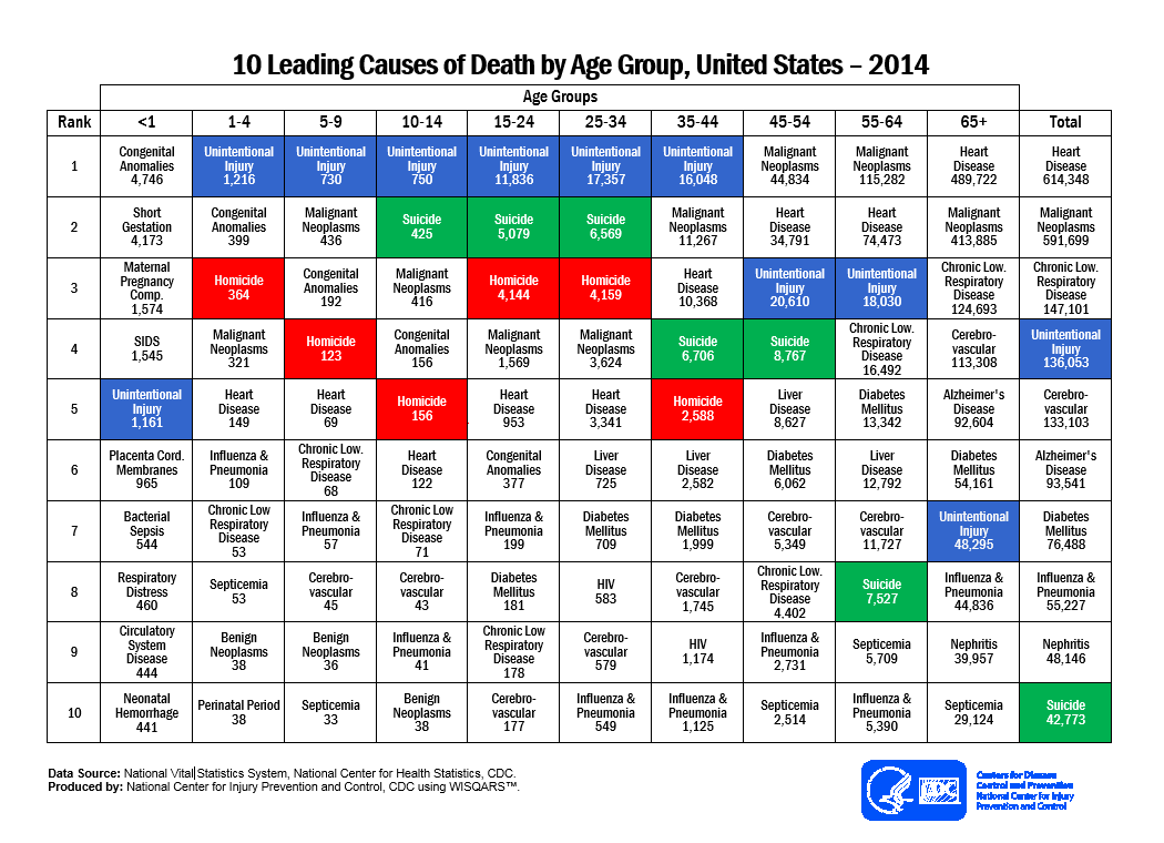
Observe, assess, intervene
On most days when I walk into the exam room for a well-child visit, I find an anxious mom or a fretful father sitting next to a fearless child. I quickly shove aside my unrealistic expectation of finding both parents together holding the child. During the progression of my day, I see a diversity of parents playing their parts in caring for their children. It is sometimes a single mom, strong and robust, surrounded by a firm aura of principles and rules; she is concealing all signs of weakness, to make sure her child doesn’t cross any line that she has so cautiously made. Sometimes it is a single mom who is nervous and scared with a galaxy of fear in her eyes, desperately seeking for reassurance of her parenting. Fathers also come playing many roles, from someone struggling with tears as his child gets immunizations, to someone who has parenting in his bag, and skillfully plays eeny meeny miny moe with the little ones in the waiting room.
They all have one thing in common: the immense love for their children and the pressure of being a single or separated parent. It is indeed a reality that I see in most of the clinic rooms – that 60%-70% of children are not living with both mom and dad in the same house. Please note that these are raw data based entirely on my observation. While I watch each parent struggling as mentioned above, my mind often wanders to how each young child copes with such a situation.

What is our role as pediatricians as we walk into the exam room, as we encounter these different family dynamics? To simplify it for myself, I divide it into three categories: Observe, assess, and intervene. Most of the time as physicians, our gut feelings and instincts guide us to where help is needed. It is important to anticipate the changes a family might go through as we meet a first-time single mom or a family who has recently been separated. As we anticipate and observe, it also is important to ask specific questions of parents who may not feel comfortable volunteering this information:
• “Are you and your child undergoing any sort of stress?”
• “How do you think your child is coping with the separation?”
• “Do you identify any flaws in how things are going now?”
Of course, we need to ask questions about stress and family dynamics of all parents. We also should maintain a high level of sensitivity as we approach such questions. It is important to identify any changes in a child’s emotional and social development as we see them on every visit. And when we deem the need, to intervene and identify resources for the family. We also can help parents with ideas for communication with the child; anger management; helping parents and children understand changes; and encouraging open discussion when possible, instead of bottling up unsaid feelings and emotions. This is especially true for single-parent families, but two-parent families undergo stresses as well, for which pediatricians should keep an eye out.
While it is extremely important for us on every well-child visit to ensure that a child’s physical health is up to par, it is equally important not to ignore their emotional and social well-being as we walk in the room so we can help them flourish into the best version of themselves.
Dr. Fatima is a first-year pediatric resident at Albert Einstein Medical Center, Philadelphia. Email her at [email protected].
On most days when I walk into the exam room for a well-child visit, I find an anxious mom or a fretful father sitting next to a fearless child. I quickly shove aside my unrealistic expectation of finding both parents together holding the child. During the progression of my day, I see a diversity of parents playing their parts in caring for their children. It is sometimes a single mom, strong and robust, surrounded by a firm aura of principles and rules; she is concealing all signs of weakness, to make sure her child doesn’t cross any line that she has so cautiously made. Sometimes it is a single mom who is nervous and scared with a galaxy of fear in her eyes, desperately seeking for reassurance of her parenting. Fathers also come playing many roles, from someone struggling with tears as his child gets immunizations, to someone who has parenting in his bag, and skillfully plays eeny meeny miny moe with the little ones in the waiting room.
They all have one thing in common: the immense love for their children and the pressure of being a single or separated parent. It is indeed a reality that I see in most of the clinic rooms – that 60%-70% of children are not living with both mom and dad in the same house. Please note that these are raw data based entirely on my observation. While I watch each parent struggling as mentioned above, my mind often wanders to how each young child copes with such a situation.
What is our role as pediatricians as we walk into the exam room, as we encounter these different family dynamics? To simplify it for myself, I divide it into three categories: Observe, assess, and intervene. Most of the time as physicians, our gut feelings and instincts guide us to where help is needed. It is important to anticipate the changes a family might go through as we meet a first-time single mom or a family who has recently been separated. As we anticipate and observe, it also is important to ask specific questions of parents who may not feel comfortable volunteering this information:
• “Are you and your child undergoing any sort of stress?”
• “How do you think your child is coping with the separation?”
• “Do you identify any flaws in how things are going now?”
Of course, we need to ask questions about stress and family dynamics of all parents. We also should maintain a high level of sensitivity as we approach such questions. It is important to identify any changes in a child’s emotional and social development as we see them on every visit. And when we deem the need, to intervene and identify resources for the family. We also can help parents with ideas for communication with the child; anger management; helping parents and children understand changes; and encouraging open discussion when possible, instead of bottling up unsaid feelings and emotions. This is especially true for single-parent families, but two-parent families undergo stresses as well, for which pediatricians should keep an eye out.
While it is extremely important for us on every well-child visit to ensure that a child’s physical health is up to par, it is equally important not to ignore their emotional and social well-being as we walk in the room so we can help them flourish into the best version of themselves.
Dr. Fatima is a first-year pediatric resident at Albert Einstein Medical Center, Philadelphia. Email her at [email protected].
How to prepare to care for transgender adolescents
As pediatric and adolescent gynecologists, we are seeing an increasing number of adolescents with gender identity issues and have come to believe that all obstetrician-gynecologists need to have an understanding of varying gender identities, as well as their role in managing these patients’ care.
We had the honor to assist the American College of Obstetricians and Gynecologists’ (ACOG) Committee on Adolescent Health Care in the development of a new Committee Opinion to guide ob.gyns. in caring for transgender adolescents (Obstet Gynecol 2017;129:e11-6). As our culture grows more aware of the nuances and spectrum of gender identity, our health care practices must grow as well. Ob.gyns. are often uniquely positioned as being among the first people transgender adolescents present to – whether it’s signaling disassociation with their gender when answering routine medical questions or directly addressing gender with them as a trusted and private resource. Even when seeing a patient too young to consider hormone therapy, an ob.gyn. can offer vital early behavioral health support, educational and community resources, and specialist referrals.
Transgender adolescent patients have likely faced negative stereotypes and stigmas in social settings or through media that make them cautious and protective of their identity. They are also more likely to face social ostracism such as bullying and/or dissent and rejection from their parents, deepening the vulnerability of their situation. As a result, transgender adolescents can have increased instances of anxiety, depression, sexual harassment, homelessness, and risk-taking behavior. Medical practices can signal to transgender patients that they are safe and welcoming from the start by offering gender neutral forms, brochures, and information for LGBT patients in the waiting room, and having sensitive employees at every step – from the front desk onward.
As we’ve just outlined, transgender adolescent patients face unique challenges, including increased rates of social and mental health risks. In response, ob.gyns. must be prepared to have a comprehensive conversation about health and well-being beyond sexual and reproductive health. They must also be equipped to address the psychosocial issues associated with transgender adolescents. This includes knowledge of what to look for and offering patients resources, education, and referrals to guarantee their health and safety.
It is important that ob.gyns. are aware that transgender men have female reproductive organs and can present with common gynecological problems such as abnormal bleeding, ovarian cysts, and torsion, as well as pregnancy and pregnancy complications. Finally, ob.gyns. can serve a unique role in counseling about fertility and fertility preservation. Thus, not only do we provide essential health care, including ongoing primary care, but we can position ourselves as part of the support network for these adolescents and their families.
Most importantly, when addressing an adolescent transgender patient, we must understand there is no uniform transgender experience. Expressing gender, sexual identity, and behavior patterns will vary from patient to patient. There are a wide range of treatment options available for transgender patients, from hormone to surgical therapies. An ob.gyn.’s responsibility is to help each individual make an informed decision, and help that patient think ahead to the future.
While this all may seem like a lot, it’s important to remember the essential components of our role as health care providers do not change because an adolescent patient is transgender. Care should always include education about their bodies, safe sex, deliberate and thoughtful assessment of symptoms or concerns, and preventive care services such as STI screenings and contraception. We are simply adding more nuanced cultural and medical understanding to those practices.
Dr. Gomez-Lobo is director of pediatric and adolescent obstetrics and gynecology, Medstar Washington Hospital Center/Children’s National Health System, Washington, D.C. Dr. Sokkary is associate professor of ob.gyn. at Navicent Health Center/Mercer School of Medicine in Macon, Ga. They are members of the ACOG Committee on Adolescent Health Care. They reported having no relevant financial disclosures.
Climate change: A call to action for psychiatry
In a recent survey of about 90 psychiatrists attending a national general psychiatry meeting, the overwhelming majority believed that climate change is human caused and the impacts were already or would soon be harming their patients. They expressed an interest in knowing more, reporting that they sometimes had little knowledge about the issues. They also believed that psychiatrists have a role to play in making the case to be better prepared for what our communities are facing. Professional organizations were a desirable choice as a source of information and training.
Because of these responses we, the Climate Psychiatry Alliance, believe that a scientific and well thought out program addressing the mental health impacts of climate disruption with practical advice on how to help our patients and our communities in the face of climate disasters while emphasizing preparedness and prevention, is called for.

As physicians, we are keenly aware of and concerned about the physical harm of extreme weather events causing acute harm, including tornadoes, fires, floods, hurricanes, and so on. We also know about the longer-term chronic conditions, including sea level rise, drought, and permanent and high temperatures.
We also know that the spread of infectious diseases, both old and some new, on us, our children, and even the unborn, is a rising challenge to our communities. The National Academy of Sciences, for example, recently linked the Zika outbreak to a rise in temperature (Proc Nat Acad Sci. 2017;114[1]:119-24). The expanding reach of malaria and the growing incidence of such illnesses as dengue, chikungunya, and Lyme also are linked to higher temperatures.
Though the physical harm from climate disruption is increasingly being discussed, more attention needs to be given to the psychological harm: When the place we call home is burned down, blown away, flooded … when we lose our possessions; maybe our pets, our livelihood; see injuries, illness, and death; the mix of fear, anger, sorrow, and trauma can bring on a full range of psychiatric disorders. Mental health professionals are already seeing posttraumatic stress disorder, depression, and anxiety disorders, as well as an increase in drug and alcohol abuse, violence against women, and child abuse (Ann Glob Health. 2014 Jul-Aug;80[4]:332-44), (Soc Sci Med. 2015;[141]:133-41), (Soc Sci Res. 2013;42[5]:1222-35), (Soc Sci Med. 2017 Jan 6;[175]:161-8).
The link between climate and aggression is clear: For each standard deviation of increased temperature and rainfall, a 4% rise in aggression between individuals and a 14% rise among groups can be expected (Science. 2013 Sep 13;34[6151]:1235367). This is true across all ethnicities and regions. General unrest around the world should come as no surprise to us – nor should its rise in the years ahead, given that temperatures will continue to rise.
The psychological needs and impacts on our communities of exploding numbers of refugees from mounting climate disruption in many areas of the world are of grave concern as the impacts of the upheaval grow ever more profound. The United Nations reports that what we are seeing now and are already unable to address, is just the tip of the iceberg.
Psychiatrists have a special role to play in all of this, because we not only are experts in physical health, we are experts on psychological harm.
We also know that any physical condition or illness carries an attendant emotional toll. Trained in science, we respect the scientific method and the peer-reviewed work that goes into validating what the scientists are telling us.
We know that harm that is entirely accidental is much easier to get over than harm experienced as avoidable. That appropriate action to protect our climate was not taken in time will become more apparent – with fear and anger among patients, families, and communities directed at policy makers and a compounding erosion of trust in our institutions. We must look at how fear, anger, and mistrust drive politics not only in our country but elsewhere since we are now a global village, interconnected.
And we must confront our values: Climate disruption is an issue of social justice, because those who will be hurt the most are from disadvantaged communities, and it is an intergenerational justice issue, because our children will inherit our mistakes. How will we answer these questions?
As experts focused on changing behaviors before it is too late, at confronting denial and resistance in ways that build people up and help influence them to change, and with the standing we have in the community, as psychiatrists we have a unique role to play. We also take seriously that our canon of ethics states our responsibility to serve our communities in ways that enhance their health.
We call upon our professional communities to help us respond to the growing public health crises. Among the many needs: education – putting together trainings for our colleagues and other thought leaders and policy makers that identify the public health burdens and drive action on them; preparedness – advising communities and individuals about how to deal with climate challenges; and prevention – advocating for solutions that reduce our vulnerability with sustainable habits and promote resilience. There is much more for us to uncover and act upon together in the months ahead.
We call upon one another for action, because only collective success will restore our health and keep us safe.
Dr. Van Susteren wrote this commentary on behalf of the Climate Psychiatry Alliance, a professional group dedicated to promoting awareness and action on climate from a mental health perspective. She is a practicing general and forensic psychiatrist in Washington. Dr. Van Susteren serves on the advisory board of the Center for Health and the Global Environment at Harvard T.H. Chan School of Public Health, Boston. She is a former member of the board of directors of the National Wildlife Federation and coauthor of group’s report, “The Psychological Effects of Global Warming on the United States – Why the U.S. Mental Health System is Not Prepared.” In 2006, Dr. Van Susteren sought the Democratic nomination for a U.S. Senate seat in Maryland. Recently, she founded Lucky Planet Foods, a company that provides plant-based, low carbon foods.
In a recent survey of about 90 psychiatrists attending a national general psychiatry meeting, the overwhelming majority believed that climate change is human caused and the impacts were already or would soon be harming their patients. They expressed an interest in knowing more, reporting that they sometimes had little knowledge about the issues. They also believed that psychiatrists have a role to play in making the case to be better prepared for what our communities are facing. Professional organizations were a desirable choice as a source of information and training.
Because of these responses we, the Climate Psychiatry Alliance, believe that a scientific and well thought out program addressing the mental health impacts of climate disruption with practical advice on how to help our patients and our communities in the face of climate disasters while emphasizing preparedness and prevention, is called for.
As physicians, we are keenly aware of and concerned about the physical harm of extreme weather events causing acute harm, including tornadoes, fires, floods, hurricanes, and so on. We also know about the longer-term chronic conditions, including sea level rise, drought, and permanent and high temperatures.
We also know that the spread of infectious diseases, both old and some new, on us, our children, and even the unborn, is a rising challenge to our communities. The National Academy of Sciences, for example, recently linked the Zika outbreak to a rise in temperature (Proc Nat Acad Sci. 2017;114[1]:119-24). The expanding reach of malaria and the growing incidence of such illnesses as dengue, chikungunya, and Lyme also are linked to higher temperatures.
Though the physical harm from climate disruption is increasingly being discussed, more attention needs to be given to the psychological harm: When the place we call home is burned down, blown away, flooded … when we lose our possessions; maybe our pets, our livelihood; see injuries, illness, and death; the mix of fear, anger, sorrow, and trauma can bring on a full range of psychiatric disorders. Mental health professionals are already seeing posttraumatic stress disorder, depression, and anxiety disorders, as well as an increase in drug and alcohol abuse, violence against women, and child abuse (Ann Glob Health. 2014 Jul-Aug;80[4]:332-44), (Soc Sci Med. 2015;[141]:133-41), (Soc Sci Res. 2013;42[5]:1222-35), (Soc Sci Med. 2017 Jan 6;[175]:161-8).
The link between climate and aggression is clear: For each standard deviation of increased temperature and rainfall, a 4% rise in aggression between individuals and a 14% rise among groups can be expected (Science. 2013 Sep 13;34[6151]:1235367). This is true across all ethnicities and regions. General unrest around the world should come as no surprise to us – nor should its rise in the years ahead, given that temperatures will continue to rise.
The psychological needs and impacts on our communities of exploding numbers of refugees from mounting climate disruption in many areas of the world are of grave concern as the impacts of the upheaval grow ever more profound. The United Nations reports that what we are seeing now and are already unable to address, is just the tip of the iceberg.
Psychiatrists have a special role to play in all of this, because we not only are experts in physical health, we are experts on psychological harm.
We also know that any physical condition or illness carries an attendant emotional toll. Trained in science, we respect the scientific method and the peer-reviewed work that goes into validating what the scientists are telling us.
We know that harm that is entirely accidental is much easier to get over than harm experienced as avoidable. That appropriate action to protect our climate was not taken in time will become more apparent – with fear and anger among patients, families, and communities directed at policy makers and a compounding erosion of trust in our institutions. We must look at how fear, anger, and mistrust drive politics not only in our country but elsewhere since we are now a global village, interconnected.
And we must confront our values: Climate disruption is an issue of social justice, because those who will be hurt the most are from disadvantaged communities, and it is an intergenerational justice issue, because our children will inherit our mistakes. How will we answer these questions?
As experts focused on changing behaviors before it is too late, at confronting denial and resistance in ways that build people up and help influence them to change, and with the standing we have in the community, as psychiatrists we have a unique role to play. We also take seriously that our canon of ethics states our responsibility to serve our communities in ways that enhance their health.
We call upon our professional communities to help us respond to the growing public health crises. Among the many needs: education – putting together trainings for our colleagues and other thought leaders and policy makers that identify the public health burdens and drive action on them; preparedness – advising communities and individuals about how to deal with climate challenges; and prevention – advocating for solutions that reduce our vulnerability with sustainable habits and promote resilience. There is much more for us to uncover and act upon together in the months ahead.
We call upon one another for action, because only collective success will restore our health and keep us safe.
Dr. Van Susteren wrote this commentary on behalf of the Climate Psychiatry Alliance, a professional group dedicated to promoting awareness and action on climate from a mental health perspective. She is a practicing general and forensic psychiatrist in Washington. Dr. Van Susteren serves on the advisory board of the Center for Health and the Global Environment at Harvard T.H. Chan School of Public Health, Boston. She is a former member of the board of directors of the National Wildlife Federation and coauthor of group’s report, “The Psychological Effects of Global Warming on the United States – Why the U.S. Mental Health System is Not Prepared.” In 2006, Dr. Van Susteren sought the Democratic nomination for a U.S. Senate seat in Maryland. Recently, she founded Lucky Planet Foods, a company that provides plant-based, low carbon foods.