User login
Prepping for new babies in the family
If I were only to give a few bits of advice to families preparing for a new baby, while physical health problems in the mother or child are the biggest concerns of prospective parents, my coaching also would be aimed at safeguarding relationships in the new family constellation.
I find that having a prenatal visit at least by 34 weeks’ gestation for first-time parents is invaluable to getting to know them so that you are not a stranger after delivery and you can communicate more effectively if there are difficulties.

As for other topics that may not seem like a topic to be raised with a pediatrician, pregnancy fears may need to be prompted. You might say, "Most mothers in the second trimester have scary dreams about their future baby. Has this happened to you?" This gives parents a chance to express concerns, often about birth defects, but sometimes about how they or their partner will be caring for the baby. This can be a chance to ask about how the pregnancy has affected their relationship so far, and how they hope it will change once the baby comes.
The prenatal visit is a time to inform parents about practical matters such as your office’s practices regarding on call, insurance, your website, and the expectation for previsit questionnaires. After taking pregnancy and family histories, the other main topics are plans for circumcision and breast-feeding. This is the time for recommending prenatal labor and delivery classes for both fathers and mothers.
Possibly the most important topic for prospective parents, however, is quite different from these issues so clearly related to health – it is about building relationships. Fathers can be easily engaged on the topic about whether to circumcise or not, but having fathers sign on to supporting breast-feeding may not seem as obviously important. Not only do some couples have low comfort in talking about or exposing the mother’s breasts, but some fathers are even possessive of them and unwilling to share with the baby. A discussion about how the father can be the one to bring the baby from the crib for the middle-of-the-night breast-feeding and then burp, change, and return the infant to the crib is a way to support the (exhausted) mother.
Fathers need to know how important their help and support are to the new mother. Mothers need to be heard by the father (and anyone else who will listen) about their fears and pain during delivery, for as many times as it takes. He needs to tell her how brave she was and how grateful he is. Our son bought his wife a "push present" to acknowledge this marathon achievement!
Fathers also need to understand that things don’t just go back to "normal" once the baby has arrived. The support of the father at this special time is symbolic to the mother of the future of their relationship. I can’t tell you how many mothers, disgruntled with their marriages years into parenting, will call up examples of lack of support in the newborn period as the beginning of the deterioration of their relationship. The mother is exhausted from the well-termed "labor," literally and figuratively "drained" by breast-feeding and the interrupted sleep of the first months. She needs her partner to step up with both hands to help – and express sympathy – to show that he is part of the new parenting team.
I think it is important to emphasize that relationships do change – have to change – when a baby arrives. This can be a coming-together in sharing the chores as well as joys of parenting, or a splintering from lack of the communications co-parenting requires. Egocentricity that sufficed in a marriage without children no longer works when the exponential increase in life demands begins. Lack of social support is the number one risk factor for marital discord and child behavior problems; the main social support in American families is the spouse/partner. A golden rule for each parent to follow is, "Ask what you can do to help."
Other supports in addition to the spouse/partner are important, too. To start this topic you might ask: "How are you going to involve others in and out of the family with this child?" There is a need for both engagement and sometimes limit-setting on others that can be a new kind of task and stress for the couple. The task may involve at first negotiating visits and time with grandparents from each side versus privacy for the parents, then later determining family dietary practices for the new child as she grows; compromising on cultural discipline styles; deciding on how religious practice will be conveyed or not; and even setting limits on toys and gifts.
I encourage parents to engage commitment from other, unrelated adults as "godparents" as an important adjunct to biological family support. This can be especially useful for small or isolated families or those distancing themselves from their own relatives. Such early engagement can begin a lifelong bond that provides both the parents and child significant support over the years. In the case of future divorce (greater than 50%) or death of a parent, a godparent becomes an even more valuable source of stability.
For parents having their first child, the advice is much different than for families having a second child. For second-time parents, I am sometimes asked about when to tell the siblings about sleeping arrangements or how to ease the change when a new baby is coming. But one special opportunity to foster a positive relationship between the siblings occurs in the narrow window between the time of telling the child (second trimester is probably best due to the high rate of early miscarriage) and the birth. This is a time current children can attend so-called "sibling preparation" classes. Along with a strong relationship with the father, expression of empathy, optional involvement in caring for the baby, avoidance of gory details of the delivery and not forcing photos, attendance at these classes has been shown to improve sibling adjustment to the baby.
Parents who can’t take the older child to a sibling class can follow some of the principles themselves. The important points are to tell the sibling that a new baby is coming "because we love children," not as a playmate (since they are not much fun for a long time); that babies cry and sleep and spit up a lot in the beginning (realism), but eventually will be able to smile and play; and especially that "we (the parents) took care of you when you were little, and we will do the same for this baby." A review of the older child’s baby pictures can be a good way to start the conversation.
Siblings who are told in strong ways about the new baby’s point of view (Boy, he sure is hungry! Hungry enough to scream!) have more positive relationships later. While some behavioral regression (50%) and jealousy are common, most children quickly come to care about their new baby, and become loving, protective, and the best playmates and models for new skills a child ever has.
Finally, don’t forget to recommend daily "special time" for each parent with the older child(ren) starting prenatally and continuing forever, to reduce jealousy and provide reassurance that he is still loved no matter who else joins the family!
Dr. Howard is assistant professor of pediatrics at the Johns Hopkins University, Baltimore, and creator of CHADIS (www.CHADIS.com). She has no other relevant disclosures. Dr. Howard’s contribution to this publication was as a paid expert to Frontline. E-mail her at [email protected].
If I were only to give a few bits of advice to families preparing for a new baby, while physical health problems in the mother or child are the biggest concerns of prospective parents, my coaching also would be aimed at safeguarding relationships in the new family constellation.
I find that having a prenatal visit at least by 34 weeks’ gestation for first-time parents is invaluable to getting to know them so that you are not a stranger after delivery and you can communicate more effectively if there are difficulties.
As for other topics that may not seem like a topic to be raised with a pediatrician, pregnancy fears may need to be prompted. You might say, "Most mothers in the second trimester have scary dreams about their future baby. Has this happened to you?" This gives parents a chance to express concerns, often about birth defects, but sometimes about how they or their partner will be caring for the baby. This can be a chance to ask about how the pregnancy has affected their relationship so far, and how they hope it will change once the baby comes.
The prenatal visit is a time to inform parents about practical matters such as your office’s practices regarding on call, insurance, your website, and the expectation for previsit questionnaires. After taking pregnancy and family histories, the other main topics are plans for circumcision and breast-feeding. This is the time for recommending prenatal labor and delivery classes for both fathers and mothers.
Possibly the most important topic for prospective parents, however, is quite different from these issues so clearly related to health – it is about building relationships. Fathers can be easily engaged on the topic about whether to circumcise or not, but having fathers sign on to supporting breast-feeding may not seem as obviously important. Not only do some couples have low comfort in talking about or exposing the mother’s breasts, but some fathers are even possessive of them and unwilling to share with the baby. A discussion about how the father can be the one to bring the baby from the crib for the middle-of-the-night breast-feeding and then burp, change, and return the infant to the crib is a way to support the (exhausted) mother.
Fathers need to know how important their help and support are to the new mother. Mothers need to be heard by the father (and anyone else who will listen) about their fears and pain during delivery, for as many times as it takes. He needs to tell her how brave she was and how grateful he is. Our son bought his wife a "push present" to acknowledge this marathon achievement!
Fathers also need to understand that things don’t just go back to "normal" once the baby has arrived. The support of the father at this special time is symbolic to the mother of the future of their relationship. I can’t tell you how many mothers, disgruntled with their marriages years into parenting, will call up examples of lack of support in the newborn period as the beginning of the deterioration of their relationship. The mother is exhausted from the well-termed "labor," literally and figuratively "drained" by breast-feeding and the interrupted sleep of the first months. She needs her partner to step up with both hands to help – and express sympathy – to show that he is part of the new parenting team.
I think it is important to emphasize that relationships do change – have to change – when a baby arrives. This can be a coming-together in sharing the chores as well as joys of parenting, or a splintering from lack of the communications co-parenting requires. Egocentricity that sufficed in a marriage without children no longer works when the exponential increase in life demands begins. Lack of social support is the number one risk factor for marital discord and child behavior problems; the main social support in American families is the spouse/partner. A golden rule for each parent to follow is, "Ask what you can do to help."
Other supports in addition to the spouse/partner are important, too. To start this topic you might ask: "How are you going to involve others in and out of the family with this child?" There is a need for both engagement and sometimes limit-setting on others that can be a new kind of task and stress for the couple. The task may involve at first negotiating visits and time with grandparents from each side versus privacy for the parents, then later determining family dietary practices for the new child as she grows; compromising on cultural discipline styles; deciding on how religious practice will be conveyed or not; and even setting limits on toys and gifts.
I encourage parents to engage commitment from other, unrelated adults as "godparents" as an important adjunct to biological family support. This can be especially useful for small or isolated families or those distancing themselves from their own relatives. Such early engagement can begin a lifelong bond that provides both the parents and child significant support over the years. In the case of future divorce (greater than 50%) or death of a parent, a godparent becomes an even more valuable source of stability.
For parents having their first child, the advice is much different than for families having a second child. For second-time parents, I am sometimes asked about when to tell the siblings about sleeping arrangements or how to ease the change when a new baby is coming. But one special opportunity to foster a positive relationship between the siblings occurs in the narrow window between the time of telling the child (second trimester is probably best due to the high rate of early miscarriage) and the birth. This is a time current children can attend so-called "sibling preparation" classes. Along with a strong relationship with the father, expression of empathy, optional involvement in caring for the baby, avoidance of gory details of the delivery and not forcing photos, attendance at these classes has been shown to improve sibling adjustment to the baby.
Parents who can’t take the older child to a sibling class can follow some of the principles themselves. The important points are to tell the sibling that a new baby is coming "because we love children," not as a playmate (since they are not much fun for a long time); that babies cry and sleep and spit up a lot in the beginning (realism), but eventually will be able to smile and play; and especially that "we (the parents) took care of you when you were little, and we will do the same for this baby." A review of the older child’s baby pictures can be a good way to start the conversation.
Siblings who are told in strong ways about the new baby’s point of view (Boy, he sure is hungry! Hungry enough to scream!) have more positive relationships later. While some behavioral regression (50%) and jealousy are common, most children quickly come to care about their new baby, and become loving, protective, and the best playmates and models for new skills a child ever has.
Finally, don’t forget to recommend daily "special time" for each parent with the older child(ren) starting prenatally and continuing forever, to reduce jealousy and provide reassurance that he is still loved no matter who else joins the family!
Dr. Howard is assistant professor of pediatrics at the Johns Hopkins University, Baltimore, and creator of CHADIS (www.CHADIS.com). She has no other relevant disclosures. Dr. Howard’s contribution to this publication was as a paid expert to Frontline. E-mail her at [email protected].
In Response to "Cruising With Disaster"
This article is a response to Randy D. Danielsen's editorial "Cruising With Disaster" from the August 2014 issue of Clinician Reviews.
Just my two cents. Have never been on a cruise. My wife thinks it's a bunch of old people eating too much and being entertained by a guy from the Catskills. Although my brother just completed an Alaskan cruise last week—his first. And the good news, he and his wife are still speaking.
Regarding health issues—your friend's wife's death sure was astonishing with regard to rapidity. Don't know that anything would have made a difference. There are going to be rare events like this, and there is no doubt that you do expose yourself to some medical isolation when going on a cruise.
I have to think that the ship physicians have to meet some minimum standards (although I don't know this). A fellow I know in Florida, who's an emergency physician and apparently has the job of supplying the doctors for one of the large cruise lines, thinks it would be relatively easy to find certified emergency physicians.
And all of us on the dole, otherwise known as Medicare, should be immunized against flu, meningococcal infection, and herpes zoster. I would think the safest cruise would be one to Alaska, because you are close to shore lots of the time—which is not necessarily the case in cruises to the tropics.
Would give me grave concern if I had to be taken off a ship and sent to a hospital in some third world country (otherwise known as most of the tropics). I guess if you have some pre-existing significant medical problems, you could opt for the travel insurance that covers this sort of thing.
But the bottom line: Yes, you are assuming some health risks when you go on a cruise, but you could be assuming some similar risks if you go on a vacation to Mexico or most anywhere else outside the United States except, perhaps, major metropolitan areas. Yes, we spend twice as much per capita than anyone else on the planet for health care, but most of the time, if you really need it, we have great access to emergency care on dry land in the US.
Vitamin supplementation
Coming to the end of yet another school physical season, my mind is focused on anticipatory guidance and well child care. Inevitably, nutrition is a main focus of these back to school visits – a very common question is, "Should my child take a vitamin?" While there are relatively clear recommendations for vitamin supplementation in infancy, there seems to be less clear consensus regarding vitamin supplementation in toddlers and school-aged children. Over my years of practice, I have developed a few guiding principles for my recommendations, which have definitely evolved over the years as more evidence becomes available.
• I always ask if a child is taking any kind of vitamin or other supplementation. Routine supplementation with a standard children’s multivitamin is very safe, and I essentially never discourage it. However, high doses of vitamins can be dangerous. Without being asked, most parents don’t think to mention vitamin use, but it is definitely something I want to know.
• For children who are not taking vitamins, but whose parents have concerns about their diet, I recommend a standard chewable multivitamin with iron. The risks of this type of vitamin supplementation are low, and the potential benefits great if a child isn’t getting everything he or she needs through the diet. Of course, I also take the opportunity to discuss strategies for improving diet and nutrition, since that is the best way to get your vitamins and minerals! For children who seem to have healthy well-balanced diets, I suggest it to parents as an option to consider. My recommendation in this situation is less strong, and more driven by parent preference. That said, I do let parents know that even children with seemingly good diets can sometimes not be getting enough of particular vitamins or minerals, most typically vitamin D, iron, or calcium.
• If a child is taking vitamins, or the parent is considering starting the child on vitamins, I recommend standard chewable children’s multivitamins (whatever brand is on sale) rather than gummy vitamins. Many brands of gummy vitamins have lower amounts of vitamin D or other vitamins than do their chewable counterparts. If a child really prefers gummy vitamins, I recommend that parents carefully compare the nutritional information to be sure that if they are going through the expense and trouble of giving a daily vitamin the child is getting all the vitamins and minerals that he or she needs. Lastly, parents should be reminded that vitamins are medications and should be safely stored! Thoughtfully used, vitamin supplements can be an important part of a healthy child’s nutrition.
Dr. Beers is an assistant professor of pediatrics at Children’s National Medical Center and the George Washington University Medical Center, Washington. She is chair of the American Academy of Pediatrics Committee on Residency Scholarships and president of the District of Columbia chapter of the American Academy of Pediatrics. E-mail Dr. Beers at pdnews@ frontlinemedcom.com.

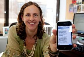
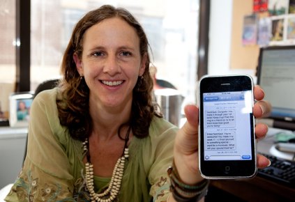
Texting intervention helped smokers quit
Text-messaging programs on phones seem to help smokers quit. The jury’s still out on smartphone apps for smoking cessation when used by patients, but they may help nurses improve screening, several separate studies suggest.
The interactive text-messaging program Text2Quit helped U.S. smokers quit and stay off cigarettes in a 6-month randomized, controlled trial in 503 adults who smoked at least five cigarettes a day, owned a cell phone with an unlimited service plan for short text messaging, and had expressed an interest in quitting cigarettes. Participants already were avid texters, sending or receiving an average of 29 text messages per day before they enrolled in the study.
At the 6-month follow-up, 20% of 241 people in the Text2Quit group said they had not smoked in the prior 7 days, compared with 10% of 262 people in the control group, reported Lorien C. Abroms, Sc.D., and her associates.
Laboratory analyses of saliva from 54 members of the intervention group and 32 in the control group showed that nearly a quarter of participants had lied about quitting. Text2Quit still was effective, however, with biochemically confirmed abstinence from smoking in 11% of the intervention group and 5% of the control group (Am. J. Prev. Med. 2014 June 5 [doi: 10.1016/j.amepre.2014.04.010]). Those biochemical quit rates are similar to rates reported in studies of other text-messaging programs for smoking cessation or of telephone quit-line counseling, she said.
The results support a meta-analysis of five studies that found mobile phone–based interventions (predominantly text messaging) for smoking cessation increased 6-month quit rates (Cochrane Database Syst. Rev. 2012;11:CD006611 [doi: 10.1002/14651858]). Most U.S. studies, however, have been small, uncontrolled pilot trials with shorter follow-up and no biochemical verification of quit rates, Dr. Abroms said.
Text2Quit costs $30 for a 4-month subscription unless you have a code from a sponsoring organization. When U.S. smokers call the national quit-line number (1-800-QUIT-NOW), residents of some states get offered Text2Quit. More than 50,000 smokers have enrolled since the program became available in 2011, reported Dr. Abroms of George Washington University, Washington. Other text-messaging programs are available, such as the free SmokefreeTXT program from the National Cancer Institute’s smokefree.gov site.
People randomized to the control group in Dr. Abroms’ trial initially were given a link to smokefree.gov, but after the launch of SmokefreeTXT in 2011, new control group members instead were given a link to the National Cancer Institute’s "Clearing the Air" guidebook on quitting smoking.
Text2Quit consists of automated, bidirectional text messages, with extra support from e-mails and a web portal. The texts prompt users to track their smoking and report on their cravings and smoking status, with the messages to participants tailored by the reasons for quitting, money saved, and use of medications for smoking cessation. The frequency of texts peaked around the smoker’s chosen quit date, with five texts on that date, approximately two per day in the following week, three per week in the next 2 months, and less than one per week after that.
Considering that an estimated 75% of adults around the world have access to cell phones, text-messaging interventions potentially could puff up the number of quitters, she suggested.
Smartphones aren’t yet as ubiquitous but, still, an estimated 11 million U.S. smokers own a smartphone, Dr. Abroms wrote in a separate study of apps for smoking cessation. We don’t yet know if any of the 400 smoking cessation apps that were available in 2012 work, but her analysis of 98 of the most popular smoking cessation apps found that they seldom include information and strategies that have been proven to help smokers quit.
She and her associates rated 47 apps for iPhones and 51 for Android phones on a 42-point scale, with a top score indicating adherence to the U.S. Public Health Service’s Clinical Practice Guidelines for Treating Tobacco Use and Dependence. The average score was 13. No apps recommended calling a quit line, for example. Only 4% recommended using medications approved for smoking cessation. Only 19% offered practical counseling or advice on how to quit or stay quit (Am. J. Prev. Med. 2013;45:732-36).
On the other hand, it’s possible that national guidelines that were developed for clinical settings don’t apply to mobile apps. If the apps somehow are effective, their reach could magnify any impact, Dr. Abroms suggested. Approximately 779,400 English-language apps for smoking cessation were downloaded per month for Android phones alone during the study period. (The iTunes App Store does not provide numbers for downloads.) That’s more than the approximately 100,000 calls per month to the national quit line in the same time period and 200,000 unique visitors per month to a leading website for smoking cessation, she reported.
"Text messaging programs for smoking cessation have a reasonably good evidence base," according to Dr. Abroms. "The evidence for smartphone apps is still in its early stages. I would not recommend them as a stand-alone intervention, though many with tracking elements may be useful as part of a comprehensive program. Apps that include games could be good as a distraction from smoking."
A separate study found that an experimental "decision support system" app, created for use by nurses on smartphones and tablets, improved rates of screening and counseling for tobacco use by prompting nurses to screen for tobacco use and offered guideline-based treatment recommendations, reported Kenrick Cato, Ph.D. of Columbia University, New York, and his associates.
The 185 registered nurses who were enrolled in advanced practice degree programs handled 14,115 clinic visits, in which they asked patients about smoking status in 84% of visits and offered cessation counseling to 90% of those who expressed a willingness to stop smoking (Oncol. Nurs. Forum 2014;41:145-52).
That compares favorably with federal U.S. data suggesting that tobacco screening happens in approximately 60% of clinic visits, and fewer than 20% of patients get counseling on quitting, Dr. Cato said in a prepared statement released by the university. The federal Healthy People 2020 program aims for tobacco screening in 69% of office visits and counseling rates of 21%, he said.
If further development of the app confirms that it’s useful, it could light a fire under tobacco screening.
Dr. Abroms designed Text2Quit and receives royalties from sales. Dr. Cato reported having no financial disclosures.
On Twitter @sherryboschert
The Medical Roundtable: Type II Pulmonary Hypertension in the Setting of Heart Failure with Preserved Ejection Fraction
DR. GIVERTZ: I’m Michael Givertz. I am the Medical Director of the Heart Transplant and Mechanical Circulatory Support Program at the Brigham and Women’s Hospital in Boston, Massachusetts. I am interested in the management of patients with advanced heart disease and those with heart failure with preserved ejection fraction (HFpEF) as well as those with reduced ejection fraction (EF). In particular, I focus on patients with comorbidities, including pulmonary hypertension (PH) and chronic kidney disease.
DR. LEWIS: I’m Greg Lewis from Massachusetts General Hospital (MGH), Boston. I am a member of the Heart Failure and Transplant Unit, and I direct the cardiopulmonary exercise labs at MGH. Like Dr. Givertz, I am interested in heart failure (HF) with both preserved EF and reduced EF and in patients who have PH complicating both the abovementioned conditions.
DR. PRITZKER: I’m Marc Pritzker. I’m a professor of Cardiovascular Medicine and Surgery at the University of Minnesota and a part of the Advanced Heart Failure Group here. I’m also the Director of the Pulmonary Hypertension Center, and I have a special interest in diastolic dysfunction, which is going to be the leading cause of PH in Western society,1 if it isn’t already.
DR. FRANCIS: Let’s begin with a case and then proceed from there. It’s a short vignette.
An 83-year-old woman is suffering from long-standing diabetes, coronary disease, and hypertension. She’s admitted to the hospital with acute HF. An echocardiogram was performed, which indicates a normal EF of 65%. She has left ventricular (LV) hypertrophy, a small LV cavity, and a large left atrium.
Doppler studies indicate that the left atrial pressure is probably slightly elevated. There is clear diastolic dysfunction, as shown by echocardiography, and enough tricuspid regurgitation to suggest that the pulmonary artery (PA) pressure is approximately 70/30 mm Hg.
She has been given intravenous furosemide, her condition improves, and she is subsequently discharged. She then visits the outpatient clinic where follow-up echocardiogray indicates the persistence of PH. Her estimated PA pressure by echocardiogram is slightly lower (60/30 mm Hg), although her LV filling pressures are probably lower.
Although I’m not exactly sure of the situation at the Brigham or MGH, I suspect that it’s similar to the situation here and around the country: We are now seeing many such patients. They’re often elderly women with many comorbid conditions, and they’re sometimes admitted with atrial fibrillation, which makes the estimation of diastolic function problematic. They’re treated conventionally with diuretics and antihypertensive therapies, but they tend to fare poorly. Often, they return to the hospital and pose a recurrent problem.
Dr. Pritzker, let’s start with you. The patient’s PA pressure is quite high, at least based on the estimation by an echocardiogram. It’s fairly common for these patients to have PH, but this patient’s PA pressure is probably disproportionately high relative to her slightly elevated left atrial pressure.
Granted that we don’t have the hemodynamic data from the catheterization lab; nonetheless, am I right? Are we seeing more of these patients with so-called reactive or disproportionate PH or hypertension that is higher than what you would expect, given the clinical context?
DR. PRITZKER: I agree. We probably encounter 3 or 4 new patients a week who are referred to us because of certain observations during echocardiographic screening. In reality, these patients have diastolic dysfunction, and probably half of the patients in our clinic have PH that appears to be higher than the classical expectation that the PA pressure follows the left atrial pressure.
This situation could be viewed in 2 ways. One is the eyeball method, which many people do and just say, “Well, that seems high to me compared to what the wedge pressure is.” The transpulmonary gradient, which is the mean PA pressure minus the wedge pressure, or, sometimes, the pulmonary diastolic pressure minus the wedge pressure, is above 15 mm Hg.
Based on the information given in this case, the patient’s mean PA pressure is above 40 mm Hg. I’m assuming that modestly elevated pulmonary capillary wedge pressure is approximately 15 to 20 mm Hg, which indicates an elevated transpulmonary gradient by any measurement.
DR. FRANCIS: Dr. Givertz, what is the scope of this problem in your hospital?
DR. GIVERTZ: As Dr. Pritzker was alluding to, we are seeing many such patients. I’m not sure if we’re actually looking for them or if we are looking more closely at the estimates of PA pressure by echocardiogram, which we may have been less attuned to previously.
The problem of HFpEF, which is a broad definition for this patient’s condition, is that it’s not something new: It’s something that we’ve been struggling with for many years, particularly in older patients. As Dr. Francis mentioned, these patients are typically women with a history of hypertension and often, other comorbidities.
This case would seem unusual, mostly due to the estimated PA pressure. I think that’s probably what caught the eye of the clinicians here.
We probably wouldn’t have given it a second thought if the estimated PA pressure was in the 40s or, maybe, 50s. It is only once the pressure increases above 60 mm Hg that we begin to realize that some other factor could be involved or that something was overlooked, which we now need to look at a little more closely.
Since you didn’t mention anything about the mitral valve, let’s assume that there isn’t any mitral valve disease here and we’re not missing significant mitral regurgitation at rest or ischemic mitral regurgitation, which is something you would want to rule out. This is a common problem, and I think we’re seeing it more often because we’re more closely looking for it.
DR. FRANCIS: I agree, Dr. Givertz. But, it turns out that this patient has minimal mitral regurgitation.
Dr. Lewis, as you have a focused laboratory interest in this problem, what do you think of this case, particularly with regard to the scope of the problem?
DR. LEWIS: I think that it is clearly a major problem. I would like to add that these patients can come to subspecialty care through different routes. It’s not uncommon for a patient like the one in question to be hospitalized through the general medical service or to be assessed by a pulmonary specialist or general cardiologist.
Unlike patients who have advanced LV systolic dysfunction and are often routed into the well-oiled machine of an advanced cardiomyopathy clinic, these patients are identified through providers with different scopes of practice. For example, I believe that these patients are often initially referred to a pulmonary physician.
This makes the problem more challenging because there’s a less-inherent ownership within a predefined group of clinicians. Additionally, this case has features that are clearly suggestive of a pre-capillary PH as well as provide evidence of increased left-sided filling pressures. These patients may be directed through different routes of care in the hospital.
It does present a slight problem, particularly when the house staff is presenting the case and people have their own ideas about what we should call it. Dr. Givertz, what should we call this syndrome? What do you refer to it as at the Brigham?
DR. GIVERTZ: That’s an excellent question. We have moved away from the older terminology of diastolic HF, as probably, most have, although there are some strong proponents of this terminology, which assumes that there is some abnormality in diastolic function.
In an 83-year-old woman with long-standing hypertension, I think LV hypertrophy, a small cavity on her echocardiogram report, and a large left atrium are certainly markers of diastolic dysfunction. So, calling this diastolic HF might be justified.
We probably overstep that terminology in patients who have the clinical syndrome of HF, as this patient does, with dyspnea on exertion, volume overload, and a normal or near-normal EF. So, this meets the basic definition of HFpEF, and there are echocardiographic indicators of abnormal diastolic function. So, you could call it diastolic HF.
The key indicator here is the breadth. Although it is just a descriptive term, I would prefer using a term like HFpEF, and we generally use this term on rounds in the hospital or in an ambulatory setting.
The terminology is important because we don’t assume that this is just a problem of diastole or relaxation. There may be other comorbidities and aspects related to the pathophysiology,2 which may have a larger role to play than only causing a simple abnormality in diastolic function.
DR. FRANCIS: I agree, Dr. Givertz, although I have found that there are certain experts and authors who are accustomed to a specific terminology, which is unfortunate because some patients don’t really have diastolic dysfunction, and I don’t think we understand the underlying pathophysiology very well.
DR. PRITZKER: Drs. Givertz and Lewis, is there a certain trigger in echocardiogram reports such that when an elevation in PA pressure is evident, as in this case, the examiners make the diastology a little bit more manifest or look at tricuspid annular plane systolic excursion or PA acceleration time?
I think we all have difficulties with that. I have looked at reports from several different hospitals, and nobody provides a consistent report once an increase in PA pressure has been identified.
DR. GIVERTZ: I think that’s an excellent point. We get comprehensive echocardiogram reports; it’s the way that our laboratory runs. But we don’t have a trigger that leads to the reporting of additional indices of diastolic function. With high PA pressures, a focus on the right ventricle (RV) (in terms of tricuspid annular plane systolic excursion measurements or RV dimensions) would be particularly helpful in terms of having additional information about the etiology of PH.
DR. LEWIS: In our institution, there is no dedicated report type for this constellation of findings. The conclusion of the interpretation would indicate that findings were consistent with diastolic dysfunction.
Dr. Francis, you mentioned in the description that there was some indication of left atrial pressure elevation in the Doppler studies. I know our echocardiographers at MGH generally avoid reporting Doppler measurements such as E/A ratios or estimates of left atrial pressure based on E/E' values.Ultimately, for patient management, it will be important to establish widely accepted echocardiographic criteria that differentiate precapillary PH, postcapillary PH, and mixed PH.
DR. FRANCIS: That’s a good point. As you know, some hospitals, like my former institution the Cleveland Clinic, did attempt to carry out the calculations, but in most places, these calculations are really not done. I personally assessed the echocardiogram as a consequence of my obsessive behavior. I have also talked to the echocardiographers about it and gathered an intuitive sense of impaired filling.
If you were to guess, you would say that the left atrial pressure is somewhat higher than normal. This is a really subjective phrase. It wasn’t actually measured, of course, and it wasn’t calculated.
DR. PRITZKER: I think it would be helpful if each institution could, at least, have a trigger, so that when the patient goes back to a family practitioner, a pulmonologist or another specialist, there is some comprehensive information that can give the practitioner some direction.
As echocardiograms become more capable of calculating these derived values (E/E prime, E to A, etc.), even general cardiologists begin feeling confused. So I like Dr. Lewis’ idea about inserting a comment at the end to at least point people in some direction.
DR. FRANCIS: Let’s focus a little on what’s different about this particular patient, which is something that I’m personally interested in. Dr. Lewis, why is it that some of these patients have higher PA pressures than what you would expect, given the clinical context? Is there something unusual about them? Is there something relevant that we can discuss, which might help identify such a patient?
DR. LEWIS: I think that’s a key question regarding the evaluation of patients like this one. For me, the first thing to do when I see a patient like this is to make sure that there aren’t other factors, other than left heart disease, that may be mediating the PH.
Even if there is a suggestion of elevated left atrial pressure, it’s important to think beyond the left heart, in terms of additional direct insults that may have been sustained by the pulmonary circulation. For example, does the patient have chronic obstructive pulmonary disease that may be contributing to the high pulmonary arterial pressures? Does the patient have sleep apnea or a history of chronic thromboembolic disease in the lung? Even an overlapping syndrome such as scleroderma or sarcoidosis can cause LV dysfunction as well as pulmonary arterial hypertension but would be unusual to detect at an advanced age in terms of relevance to this case.
Epidemiologic studies can also provide clues regarding the type of patients who tend to have higher pulmonary arterial pressures (eg, older individuals or people with higher systolic blood pressure).
I have been surprised by our relative lack of ability to predict who’s going to have a higher transpulmonary gradient, based on the clinical features alone. For a given left atrial pressure, why do some patients have higher transpulmonary gradients than others?
Elevation of transpulmonary gradients is not simply related to degree of elevation in left-sided pressure alone. That is clear from the relatively weak correlation between PA systolic pressure and left-sided filling pressure.3 I don’t think it is related to the duration of HF alone either.
In our patient population, we’ve recorded the time when patients were diagnosed with HF because I commonly hear, “Well, if the patient has HF for a sufficient amount of time, he/she will gradually develop increases in transpulmonary gradient because of high pressure on the left side of the heart.” The duration of HF was not significantly related to the transpulmonary gradient. Therefore, I believe we need to look at new ways of understanding which patients will develop high transpulmonary gradients.
There’s some work being conducted in this area. Investigators at Dr. Francis’s former institution recently published a study about arginine metabolism dysregulation and some circulating markers that were high in patients who had a high burden of precapillary PH and HF.4
Dr. Givertz has been involved in studies evaluating endothelin levels or cyclic guanosine monophosphate-adenosine monophosphate release through the pulmonary circulation as potential indicators of dysregulation in signaling systems within pulmonary circulation. There may also be permissive genotypes that predispose patients to develop precapillary PH in the setting of left heart dysfunction.
So, I think that the question is a good one, but for me, it raises many more questions about why these patients are different. I don’t think we can explain it on the basis of clinical characteristics alone.
DR. FRANCIS: Dr. Lewis has had extensive experience in studying these patients in the laboratory and performing exercise tests on them. Dr. Lewis, from a clinical rather than research standpoint, when would you actually consider conducting more invasive studies like you are currently doing in the research laboratory?
DR. LEWIS: We haven’t talked about invasive phenotyping of patients yet, but to address Dr. Givertz’s point, the question pertains to when we need to carefully study the conditions in the pulmonary circulation. I believe that knowing the left atrial pressure is important for truly understanding the transpulmonary gradient, and I’d like to know whether it’s greater than 15 mm Hg, as Dr. Pritzker indicated; this level serves as a trigger for looking carefully into the pulmonary circulation.
Dr. Francis, I find the exercise status to be particularly informative in patients who exhibit shortness of breath with exertion. However, it’s not entirely clear whether their symptoms are being mediated by left-heart disease or a primary insult to the pulmonary circulation.
Often, even when patients have a resting right-heart catheterization, we see borderline elevations in pulmonary arterial pressure, say, a mean pressure of 20 to 25 mm Hg, and a pulmonary capillary wedge pressure of 10 to 15 mm Hg, but symptoms are present.
In such cases, instead of looking at hemodynamic measurements during a single moment in time, I find it quite helpful to make serial measurements under different loading conditions that are introduced by the very highly relevant physiologic state of exercise. We often encounter patients who have shortness of breath with exertion, which is not entirely explained by the initial diagnostic testing results.
I find it particularly useful to characterize the changes in the pulmonary capillary wedge pressure and transpulmonary gradient when patients are subjected to exercise. We can begin to classify patients based on whether their burden of precapillary PH is high or there is truly a left-sided predominance to their symptoms.
DR. FRANCIS: This gives rise to question of whether therapeutic targets exist. We all know that they exist, but what is the therapy? Perhaps, we’re not so sure about that.
DR. PRITZKER: We’re predominantly using sildenafil because it’s easily available in the hospital and doesn’t require cumbersome pre-approval for acute use like in the case of endothelin antagonists. We have a good experience of using it, and we follow it up with 6-min walks, which perhaps, in this population, is slightly easier to do.
DR. FRANCIS: Dr. Pritzker, what is your threshold for administering sildenafil? Is it lower?
DR. PRITZKER: Yes, because I think there’s more relevant evidence, including the provocative work of an Italian group and Dr. Lewis’ work. We don’t necessarily know why we’re making them better yet, but clearly, people are having fewer symptoms. This is more anecdotal than longitudinal, but patients have fewer symptoms, feel better, and seem to have better function. We also see fewer hospital admissions. I think a previous paper published at the end of last year presented similar findings.
DR. FRANCIS: Dr. Givertz, can you comment about the therapy?
DR. GIVERTZ: I think we’re all gaining experience with using sildenafil or longer-acting phosphodiesterase type 5 (PDE5) inhibitors in this patient population, but that might not be the first thing I would reach for.
This patient was admitted with acute HF. Presumably, there was a fluid problem here as well. I might be initially inclined to make sure I have optimized diuretic therapy. If this patient wasn’t already on nitrates, given the underlying coronary disease and hypertension, I think administering nitrates would be a reasonable next step.
Although there are certainly patients who cannot tolerate nitrate therapy due to its side effects such as headache or light-headedness, I might try administering nitrates before opting for a PDE5 inhibitor.
DR. PRITZKER: I assumed that the patient had been stabilized, and we still had a therapeutic gap after conventional therapy.
DR. FRANCIS: That’s a good point because it is related to the timing of initiation of such therapy; I am quite conservative about it. I don’t have as much experience as the other physicians on the panel, but my tendency would be to go with conventional medical therapy, as bad as it is, and if the patient didn’t improve thereafter, I would think about sending the patient to Dr. Pritzker or beginning sildenafil treatment myself.
Dr. Lewis, you’re actually involved in a National Institute of Health study, a consortium study. Is that right?
DR. LEWIS: That’s right; it’s the Phosphodiesterase-5 Inhibition to Improve Clinical Status and Exercise Capacity in Diastolic Heart Failure (RELAX)trial.5 Michael and Maggie Redfield from the Mayo Clinic are involved as well, and Dr. Redfield is the principal architect of the trial. But, we are adjudicating the primary end point here, which is a cardiopulmonary exercise endpoint.
Dr. Francis, I agree with Drs. Pritzker and Givertz in terms of the therapeutic approach. There certainly have been some cautionary accounts from other pulmonary vasodilators that have been tried in patients who have left heart disease, predominantly those with systolic dysfunction and have not resulted in improved outcomes.6 Therefore, I tend to refrain from using endothelin antagonists as well as the prostacyclins.
There has been some promising work done with PDE5 inhibitors in left-heart disease, and we will know the results from the RELAX trial soon. We completed enrollment of just over 200 patients, and we’re trying to determine whether we can improve exercise capacity over a period of 24 weeks with sildenafil in patients with HFpEF.
The results of PDE5 inhibition in HF with preserved LVEF from the Guazzi group demonstrated that patients with a significant burden of right-heart disease may derive significant improvement in precapillary PH with sildenafil.7 I believe if there is right-heart dysfunction that is largely attributed to high pulmonary vascular resistance or a high transpulmonary gradient, then pulmonary vasodilators like PDE5 inhibitors will have a promising role to play. Potentially, there are also some novel therapies involving the use of some of the soluble guanylate cyclase stimulators and activators in trials.
If patients have a predominant left-heart dysfunction with a modest transpulmonary gradient, I think they are much better served by trying to optimize the diuretic therapy and treat the systemic hypertension.
DR. FRANCIS: Thank you, Dr. Lewis. I think we agree that this is clearly an emerging problem, and it’s prevalence is increasing, partly because of the aging population. It is true that these patients are often admitted to general medicine wards and are sometimes transferred to the cardiology groups when they are faring poorly, and I don’t know what we can do about that.
There does seem to be a subset of patients with disproportionate PH, and there’s a lot of interest in that particular subset. I don’t know if they’re clearly identifiable from the phenoclinical phenotype, but I think that they pose more difficult problems.
Lastly, I’m delighted to know that the RELAX trial is on the verge of completion, and we will probably have some results soon. Management of patients with HFpEF and PH has been a problem for all of us, and as yet, we do not have good, randomized, controlled trials to help us understand how to manage them.
On that note, I’d like to thank the discussants for their participation.
FoxP2 Media LLC is the publisher of The Medical Roundtable.
FoxP2 Media LLC

Connecting with re-creation
I went to the American College of Physicians meeting in Orlando this year and attended a meeting about balancing life and work. Seems a little early in my career for such self-help I know, but I’ve always been a little too self-aware.
The speaker was Linda Clever, an internist and professor at the University of California, San Francisco, who is also a dynamic woman who has written books on living life with a busy career. The 1-hour group discussion was a nice change of pace in a conference full of experts talking about guidelines. I sat down next to an older couple as I settled in for the hour, delighted there was not a PowerPoint in sight. We were quickly asked to break into small groups to talk about strategies for avoiding burnout; my group ended up being me, and the couple.

They must have been in their 70s, maybe even 80s, both retired. He had been a neurologist for more than 40 years (in his day neurology was part of internal medicine, thus his presence at ACP), and she had been a journalist. It seemed fitting that here I was, in my first year as an attending, looking forward into a career and unclear exactly where it was headed, filled with uncertainty; and there they were, an entire career and life behind them, comfortable and confident for the ride they had had. We got started.
I asked them what they had done to try and maintain balance, to have a life amongst a career, or rather a career amongst a life. The wife, short and plump with round thin-rimmed glasses tucked into white hair, could have been plucked right out of a story about the North Pole. She began to recount some of the lessons they had learned. She talked lovingly of her husband and how he was always so engaged and interested in everything and that he was always volunteering to be on this committee or that committee. He sat smiling with big, bushy, white eyebrows, clean-shaven face with a yellow short-sleeve dress shirt and a red and blue tie that hung 2 or 3 inches above his belt. Short like his wife, his legs seemed to swing back and forth under the chair like a child at a playground.
"It always was interesting!" he exclaimed, waving his hands in excitement. "To me it never felt like work. Even in medical school I wanted to be involved in anything."
I thought back to my own days of medical school, involved in or leading multiple activist groups, often to the detriment of grades, and now my already growing list of committees and "nonclinical" activities.

"Once we had children," the wife went on, "I would tell his secretary to schedule school performances and sporting events, right there in his appointment book. If we did that, he never missed an event. In fact, the kids never even knew how busy he was because he was always there. It wasn’t until they grew up that they understood how much he was juggling." His smile just pulsed into an even bigger smile, chuckling here and there as she talked. "So that would be my recommendation to you. Be deliberate about your time with your family; schedule it like you would anything else. "
I made a mental note, arguing briefly with myself that I’m not even very good at scheduling things for work, but then I quickly vowed to do better with both.
I tried to point out that even with scheduling family time, it sure seemed like he worked a lot. How did he stay fresh over such a long career? He paused, raising those animated eyebrows as he deliberated. "Vacations and sabbaticals," he finally determined. "But when you go on vacation it has to be away. In my day you were nearly always available for patient phone calls. The only way to truly be on vacation would be to go far away from phones. Unplugged.
"And then, every 5 years, I would take a 6-month sabbatical. No seeing patients. We would pack up the kids and go to London. Sometimes I would come back at 3 months to take care of a few things, but I think sabbaticals are instrumental in rejuvenating a career. I would work on other projects or write. It made things harder financially, and we had to plan for it, but I came back to my practice refreshed, and the kids learned a lot, too." He settled back into his seat, arms crossed, his smile now looking more pleased and proud than amused.
The small-group time ended and Dr. Clever took polls from the crowd. Among recurring themes were exercise, saying "no," establishing sabbatical programs with your group, and taking time to completely unplug from work – no cell phones, no e-mail.
As the hour came to a close and the crowd shuffled for the exit, I felt that our small-group connection was too important to just say goodbye abruptly. I should get their contact information, I thought, so I can let them know how I am doing with my work-life balance, my sabbaticals, my scheduled time. But I didn’t, and instead watched the couple, hands held, walk down the long corridor, feeling inspired and hopeful that I too could maybe be successful in both career and family.
And so it is that I have written this column many days before it is due (a first for me) so that I can go on vacation. I’m going far away from cell phone access and e-mail. I must admit, there is a little trepidation in the thought of being so disconnected. But, after watching me spend all of Christmas eve working on a research paper, my wife is delighted at any kind of improvement in my work-life balance. That nameless couple would be proud.
Dr. Horton completed his residency in internal medicine and pediatrics at the University of Utah and Primary Children’s Medical Center, both in Salt Lake City, in July 2013, and joined the faculty there. He is sharing his new-career experiences with Hospitalist News.
I went to the American College of Physicians meeting in Orlando this year and attended a meeting about balancing life and work. Seems a little early in my career for such self-help I know, but I’ve always been a little too self-aware.
The speaker was Linda Clever, an internist and professor at the University of California, San Francisco, who is also a dynamic woman who has written books on living life with a busy career. The 1-hour group discussion was a nice change of pace in a conference full of experts talking about guidelines. I sat down next to an older couple as I settled in for the hour, delighted there was not a PowerPoint in sight. We were quickly asked to break into small groups to talk about strategies for avoiding burnout; my group ended up being me, and the couple.
They must have been in their 70s, maybe even 80s, both retired. He had been a neurologist for more than 40 years (in his day neurology was part of internal medicine, thus his presence at ACP), and she had been a journalist. It seemed fitting that here I was, in my first year as an attending, looking forward into a career and unclear exactly where it was headed, filled with uncertainty; and there they were, an entire career and life behind them, comfortable and confident for the ride they had had. We got started.
I asked them what they had done to try and maintain balance, to have a life amongst a career, or rather a career amongst a life. The wife, short and plump with round thin-rimmed glasses tucked into white hair, could have been plucked right out of a story about the North Pole. She began to recount some of the lessons they had learned. She talked lovingly of her husband and how he was always so engaged and interested in everything and that he was always volunteering to be on this committee or that committee. He sat smiling with big, bushy, white eyebrows, clean-shaven face with a yellow short-sleeve dress shirt and a red and blue tie that hung 2 or 3 inches above his belt. Short like his wife, his legs seemed to swing back and forth under the chair like a child at a playground.
"It always was interesting!" he exclaimed, waving his hands in excitement. "To me it never felt like work. Even in medical school I wanted to be involved in anything."
I thought back to my own days of medical school, involved in or leading multiple activist groups, often to the detriment of grades, and now my already growing list of committees and "nonclinical" activities.
"Once we had children," the wife went on, "I would tell his secretary to schedule school performances and sporting events, right there in his appointment book. If we did that, he never missed an event. In fact, the kids never even knew how busy he was because he was always there. It wasn’t until they grew up that they understood how much he was juggling." His smile just pulsed into an even bigger smile, chuckling here and there as she talked. "So that would be my recommendation to you. Be deliberate about your time with your family; schedule it like you would anything else. "
I made a mental note, arguing briefly with myself that I’m not even very good at scheduling things for work, but then I quickly vowed to do better with both.
I tried to point out that even with scheduling family time, it sure seemed like he worked a lot. How did he stay fresh over such a long career? He paused, raising those animated eyebrows as he deliberated. "Vacations and sabbaticals," he finally determined. "But when you go on vacation it has to be away. In my day you were nearly always available for patient phone calls. The only way to truly be on vacation would be to go far away from phones. Unplugged.
"And then, every 5 years, I would take a 6-month sabbatical. No seeing patients. We would pack up the kids and go to London. Sometimes I would come back at 3 months to take care of a few things, but I think sabbaticals are instrumental in rejuvenating a career. I would work on other projects or write. It made things harder financially, and we had to plan for it, but I came back to my practice refreshed, and the kids learned a lot, too." He settled back into his seat, arms crossed, his smile now looking more pleased and proud than amused.
The small-group time ended and Dr. Clever took polls from the crowd. Among recurring themes were exercise, saying "no," establishing sabbatical programs with your group, and taking time to completely unplug from work – no cell phones, no e-mail.
As the hour came to a close and the crowd shuffled for the exit, I felt that our small-group connection was too important to just say goodbye abruptly. I should get their contact information, I thought, so I can let them know how I am doing with my work-life balance, my sabbaticals, my scheduled time. But I didn’t, and instead watched the couple, hands held, walk down the long corridor, feeling inspired and hopeful that I too could maybe be successful in both career and family.
And so it is that I have written this column many days before it is due (a first for me) so that I can go on vacation. I’m going far away from cell phone access and e-mail. I must admit, there is a little trepidation in the thought of being so disconnected. But, after watching me spend all of Christmas eve working on a research paper, my wife is delighted at any kind of improvement in my work-life balance. That nameless couple would be proud.
Dr. Horton completed his residency in internal medicine and pediatrics at the University of Utah and Primary Children’s Medical Center, both in Salt Lake City, in July 2013, and joined the faculty there. He is sharing his new-career experiences with Hospitalist News.

Self-care for the weary
"Try and be nice to people, avoid eating fat, read a good book every now and then get some walking in, and try and live together in peace and harmony with people of all creeds and nations."
–Monty Python, "The Meaning of Life"
1. Set boundaries. This is arguably the most important aspect of the physician-patient relationship. A relationship like this is inherently fertile ground for such Freudian defenses as transference and projection. It’s important to recognize, too, that the process is not necessarily a one-way street. Freud was, after all, flawed like his subjects.
2. Don’t sweat the small stuff. This is self-evident, yet surprisingly difficult to remember.
3. Keep a journal. You’ll want to remember the good times, and keeping track of the bad times just shows you how far you’ve come.
4. Nourish your spirit – church, yoga, meditation. Take your pick. The best life advice I’ve ever received is that the mind needs structured space for quiet because otherwise you are never alone; there is always that voice in your head, constantly narrating your life and probably judging you for it.
5. There is a whole world outside of medicine. How easy is it to let our lives be consumed by our profession? How much happier would it make us to be reminded that music and literature and art exist? For my birthday this year, I asked my sister to give me a book that she thinks I should own.
6. Related to #5: Learn something outside of medicine. There are brilliant podcasts on science, the economy, politics. There are audio courses on great books or philosophy. Pick up a new instrument or a new language.
7. Indulge in the experiences that make you happy, whether that’s going to the beach, or eating out, throwing parties, or traveling. In the arguably dubious field of happiness research, studies show that spending money on experiences leads to happiness much more so than spending money on objects. (Of course, there may be a selection bias problem, in that it is entirely possible that people who are likely to buy experiences are happier at baseline than people who are likely to buy objects. But you get my meaning.)
8. Exercise. Run, take bike rides, walk your dog. Go for hikes. Take rowing lessons.
9. Sleep is crucial. We are not teenagers any longer, and it is a losing proposition to think that you can still get away with barely sleeping. If I get 8 hours of sleep, I am almost guaranteed to not be as grumpy at work. I feel refreshed, my mind is clearer, and I feel better equipped to deal with the challenges of the workday.
10. Forgive yourself – for the grumpiness, for mistakes, for bad outcomes that you could not possibly have done anything about.
11. Bonus: Mental health therapy, if done right, is a fantastic investment in yourself.
Dr. Chan practices rheumatology in Pawtucket, R.I.
Face Time
In this time of evolving medical care, many factors are impinging on the physician-patient relationship. Unfortunately, one of the biggest barriers between our patients and us is a computer screen.
Before I started using an electronic health record (EHR) nearly 4 years ago, I was very anxious about the transition. How would I be able to maintain my patient volume while trying to create a comprehensive electronic document for each patient? Now I love my EHR. It creates a beautiful note quickly and effectively, and I can check all of my medical records from any location. I love e-prescribing, and, best of all, there is never a lost medical record. As I have become more connected to my EHR, however, I think my patients sometimes feel left out. As many of us can attest, it is often hard to maintain good eye contact and communication with patients while completing their electronic record.
Recently, several studies have evaluated the effects of novel technologies on interactions between the patient and health care professionals (HCPs). Montague and Asan1 examined eye gaze patterns between patients and HCPs while EHRs were used to support patient care. Gaze was used because it provides a more objective and measurable indication of attention and communication. In their study, patient visits were recorded using 3 high-resolution video cameras placed at different angles to accurately capture gaze and avoid ambiguity of the gaze direction caused by a single camera angle. One hundred patients and 10 HCPs participated in the study.1
Results indicated that on average HCPs spent nearly one-third of the visit length gazing at the EHR.1 Paper medical records also were used during 79% of the visits with HCPs spending approximately 9% of the visit length gazing at the record. There were times when the patient gaze was undetermined and accounted for more than 28% of the visit length. This unknown gaze might have occurred when the HCP used the computer and the patient disengaged eye contact with the HCP. Another scenario recorded was when patients gazed at the HCP and the HCP gazed at the EHR, an event that accounted for more than 21% of the visit time. The investigators concluded that the patient-physician eye contact dynamic differed with EHRs compared to paper medical records. They also noted that when HCPs spent too much time looking at the computer screen in the examination room, nonverbal cues might have been overlooked. Also, the HCP’s ability to pay attention and communicate with patients was affected.1
Asan et al2 quantitatively examined and compared clinically experienced physician interactions with patients using paper medical records or EHRs in ambulatory primary care settings. Eight experienced family medicine physicians and 80 patients participated in the study. A total of 80 visits—40 with paper and 40 with EHRs—were recorded. The proportion of time physicians spent gazing at medical records was significantly more during EHR visits compared to paper chart visits (35.2% vs 22.1%; P=.001). A significantly smaller proportion of physician time was spent gazing at the patient when using an EHR compared to a paper medical record (52.6% vs 45.6%; P=.041).2
As our use of EHRs increases and evolves, it will be important to factor in these issues to maintain the centrality of the physician-patient rapport. We should attempt to place the computer screen and the patient in physical locations that facilitate the ability to maximally interact with the patient while entering the data. It will be important for those who design the next generation of EHRs to do so with this communication in mind. Until then, I will do my best to give the appropriate attention to both my patient and my EHR.
1. Montague E, Asan O. Dynamic modeling of patient and physician eye gaze to understand the effects of electronic health records on doctor-patient communication and attention. Int J Med Inf. 2014;83:225-234.
2. Asan O, D Smith P, Montague E. More screen time, less face time - implications for EHR design [published online ahead of print May 19, 2014]. J Eval Clin Pract. doi:10.1111/jep.12182.
In this time of evolving medical care, many factors are impinging on the physician-patient relationship. Unfortunately, one of the biggest barriers between our patients and us is a computer screen.
Before I started using an electronic health record (EHR) nearly 4 years ago, I was very anxious about the transition. How would I be able to maintain my patient volume while trying to create a comprehensive electronic document for each patient? Now I love my EHR. It creates a beautiful note quickly and effectively, and I can check all of my medical records from any location. I love e-prescribing, and, best of all, there is never a lost medical record. As I have become more connected to my EHR, however, I think my patients sometimes feel left out. As many of us can attest, it is often hard to maintain good eye contact and communication with patients while completing their electronic record.
Recently, several studies have evaluated the effects of novel technologies on interactions between the patient and health care professionals (HCPs). Montague and Asan1 examined eye gaze patterns between patients and HCPs while EHRs were used to support patient care. Gaze was used because it provides a more objective and measurable indication of attention and communication. In their study, patient visits were recorded using 3 high-resolution video cameras placed at different angles to accurately capture gaze and avoid ambiguity of the gaze direction caused by a single camera angle. One hundred patients and 10 HCPs participated in the study.1
Results indicated that on average HCPs spent nearly one-third of the visit length gazing at the EHR.1 Paper medical records also were used during 79% of the visits with HCPs spending approximately 9% of the visit length gazing at the record. There were times when the patient gaze was undetermined and accounted for more than 28% of the visit length. This unknown gaze might have occurred when the HCP used the computer and the patient disengaged eye contact with the HCP. Another scenario recorded was when patients gazed at the HCP and the HCP gazed at the EHR, an event that accounted for more than 21% of the visit time. The investigators concluded that the patient-physician eye contact dynamic differed with EHRs compared to paper medical records. They also noted that when HCPs spent too much time looking at the computer screen in the examination room, nonverbal cues might have been overlooked. Also, the HCP’s ability to pay attention and communicate with patients was affected.1
Asan et al2 quantitatively examined and compared clinically experienced physician interactions with patients using paper medical records or EHRs in ambulatory primary care settings. Eight experienced family medicine physicians and 80 patients participated in the study. A total of 80 visits—40 with paper and 40 with EHRs—were recorded. The proportion of time physicians spent gazing at medical records was significantly more during EHR visits compared to paper chart visits (35.2% vs 22.1%; P=.001). A significantly smaller proportion of physician time was spent gazing at the patient when using an EHR compared to a paper medical record (52.6% vs 45.6%; P=.041).2
As our use of EHRs increases and evolves, it will be important to factor in these issues to maintain the centrality of the physician-patient rapport. We should attempt to place the computer screen and the patient in physical locations that facilitate the ability to maximally interact with the patient while entering the data. It will be important for those who design the next generation of EHRs to do so with this communication in mind. Until then, I will do my best to give the appropriate attention to both my patient and my EHR.
1. Montague E, Asan O. Dynamic modeling of patient and physician eye gaze to understand the effects of electronic health records on doctor-patient communication and attention. Int J Med Inf. 2014;83:225-234.
2. Asan O, D Smith P, Montague E. More screen time, less face time - implications for EHR design [published online ahead of print May 19, 2014]. J Eval Clin Pract. doi:10.1111/jep.12182.
Can private practice survive?
I ran into Peter the other day. We weren’t close back in high school 50 years ago – Peter was a jock and I wasn’t – but both of us ended up in medicine.
"How’s the radiology business?" I asked him.
"Two more years," he said. "I should be able to hang on."
"That bad?"
"We were taken over by the academic department of a big teaching hospital," Peter said.
"What’s the problem? They want you to publish papers?" I asked.
"They’re not crazy," he said. "They know what kind of papers we’d write. They just want to measure things."
"Measure what?"
"Anything they can," he said. "Productivity, quality. They micromanage everything. We’re not a private practice anymore. We don’t call our own shots."
You hear a lot of talk these days about whether private practice can survive. Judging by anecdotal chats with colleagues in other specialties, I’d say prospects are iffy. A lot of the future is already here.
"I’m going crazy," a local community rheumatologist told me. "There was just one other guy in town. The local hospital knocked on his door and said, ‘Either sell out and work for us on our terms, or you’ll never get another referral.’ So he just closed up and moved out of state."
"How about you?" I asked him. "Have they made you an offer you can’t refuse?"
"Not yet," he said.
Then I met an oncologist from my wife’s family. He is a couple of years younger than I am. "I never thought I’d be working harder than I ever did at my age," he said. "And earning less.
"But we just sold our practice. No choice, really. Reimbursements don’t cover the cost of chemo drugs, so we lose money on every patient we give them to. The hospital that’s buying us can bill for drugs at a higher rate. So I’ll work for them for 5 years, then I’m out."
Then I heard about a neurologist, whose practice his hospital covets. He orders scans and other diagnostic procedures that generate hundreds of thousands of dollars annually, but that of course he can’t bill for. The hospital can. I don’t get how that works, but obviously the hospital does.
The trend toward consolidation proceeds. Doctors become salaried employees of large organizations. Maybe the main reason we dermatologists aren’t being swallowed yet by the big fish is that we’re just not tasty enough. We don’t generate admissions or enough costly procedures to make gulping us down worthwhile.
Switching from being an independent practitioner to a salaried employee means more than a change of location or even lower income; it signals a loss of status. I got a glimpse of how that plays out when I met another friend, an internist who used to be out on his own, but who now works for a big health care organization. "Have you got a quota of patients you have to meet?" I asked him.
"Yes," he said. "They just set that up recently."
"How did you find out?" I asked him.
"They sent out a memo," he said.
In other words, management notified doctors of productivity guidelines the same way that they would tell clerks or auxiliary personnel by which quality metrics their performance and salary would be judged.
"What if you don’t meet your quota because patients cancel or don’t keep their appointments?" I asked him. "You have no control over how patients are booked or reminded to come?"
"They blame the doctors anyway," he said. "Anyway, I’m not full time anymore. I’ll be retiring soon."
To say that hanging around colleagues who talk this way is dispiriting would be an understatement.
Peter the radiologist spoke in similar downbeat tones. "I’m adapting to the new reality fairly well," he said, "because I see the end in sight. But my younger colleagues are having a harder time. They thought they were joining a private practice, and none of them would have signed on for what they’re getting now: harder work, longer hours, no autonomy. They show up every morning cranky, muttering under their breath."
"I can see why," I said. "Whom do they take it out on?"
"Not the administrators," said Peter. "They don’t care. And certainly not the patients. They take it out on their families, I guess."
We agreed that seemed the best strategy.
Dr. Rockoff practices dermatology in Brookline, Mass., and is a longtime contributor to Skin & Allergy News. He serves on the clinical faculty at Tufts University, Boston, and has taught senior medical students and other trainees for 30 years.
Ebola, fear, and us
Recently, two American missionary workers in Liberia made headlines when they became infected with the Ebola virus, which causes a highly fatal hemorrhagic fever. They both received an experimental "secret serum" called ZMapp while in Africa and have since been flown back to the United States to be cared for at Emory University Hospital in Atlanta.
The response to their return has been mixed. While some hail their incredible selflessness and the mission they set out to accomplish, others are lukewarm, indifferent, or even very opposed to their return – so opposed that security was heightened in response to threats. I, of course, am among the former.

I once went on a 5-day missionary trip to Nicaragua. Optimistic about the potential to help the poor and hurting, my biggest challenges were the long plane ride and the hotel accommodations, which were not quite to my liking. The thought of contracting a potentially fatal disease and never returning home to my family never, ever crossed my mind.
Dr. Kent Brantly and Nancy Writebol, both missionary workers, were of a completely different mindset; they had different goals, they faced different challenges. They willingly boarded a plane to fly thousands of miles away from the comfort of their homes, the love of their families, the security of their close friends. They risked everything to care for complete strangers, strangers who posed a real threat to their lives. They never expected to receive any earthly thing in return. Interestingly, after contracting Ebola, Dr. Brantley did receive a unit of blood from a 14-year-old who had survived his fight with the Ebola virus while under his care.
Personally, I can only hope that one day I will have even half of the compassion and servant’s heart that these brave, incredibly selfless individuals possess.
I have to believe that those who object to their presence on American soil are afraid. They do not know what to expect and fear the epidemic spreading to the United States. I remember when I first started treating HIV/AIDS patients in the early ’90s. It was new and it was very, very scary. In those days, we donned protective garments above and beyond what we now know is necessary. Still, at times I, along with countless other health care workers, was overwhelmed by fear.
Like the human immunodeficiency virus, the Ebola virus is spread through direct contact with the blood or other body fluids of an infected person or through exposure to infected objects such as needles. It is not airborne, nor is it spread by contaminated food and water. Unlike with HIV, however, Ebola can be fatal within days. Another difference is that it is believed to be transmissible only if the individual is symptomatic, while countless cases of HIV were the result of exposure to seemingly healthy individuals.
While some in the American public still decry that Dr. Brantley and Mrs. Writebol were brought to a hospital in the United States, I have to believe that the great majority of Americans keep these two incredible individuals in their thoughts and prayers.
Dr. Hester is a hospitalist with Baltimore-Washington Medical Center who has a passion for empowering patients to partner in their health care. She is the creator of the Patient Whiz, a patient-engagement app for iOS. Reach her at [email protected].
Recently, two American missionary workers in Liberia made headlines when they became infected with the Ebola virus, which causes a highly fatal hemorrhagic fever. They both received an experimental "secret serum" called ZMapp while in Africa and have since been flown back to the United States to be cared for at Emory University Hospital in Atlanta.
The response to their return has been mixed. While some hail their incredible selflessness and the mission they set out to accomplish, others are lukewarm, indifferent, or even very opposed to their return – so opposed that security was heightened in response to threats. I, of course, am among the former.
I once went on a 5-day missionary trip to Nicaragua. Optimistic about the potential to help the poor and hurting, my biggest challenges were the long plane ride and the hotel accommodations, which were not quite to my liking. The thought of contracting a potentially fatal disease and never returning home to my family never, ever crossed my mind.
Dr. Kent Brantly and Nancy Writebol, both missionary workers, were of a completely different mindset; they had different goals, they faced different challenges. They willingly boarded a plane to fly thousands of miles away from the comfort of their homes, the love of their families, the security of their close friends. They risked everything to care for complete strangers, strangers who posed a real threat to their lives. They never expected to receive any earthly thing in return. Interestingly, after contracting Ebola, Dr. Brantley did receive a unit of blood from a 14-year-old who had survived his fight with the Ebola virus while under his care.
Personally, I can only hope that one day I will have even half of the compassion and servant’s heart that these brave, incredibly selfless individuals possess.
I have to believe that those who object to their presence on American soil are afraid. They do not know what to expect and fear the epidemic spreading to the United States. I remember when I first started treating HIV/AIDS patients in the early ’90s. It was new and it was very, very scary. In those days, we donned protective garments above and beyond what we now know is necessary. Still, at times I, along with countless other health care workers, was overwhelmed by fear.
Like the human immunodeficiency virus, the Ebola virus is spread through direct contact with the blood or other body fluids of an infected person or through exposure to infected objects such as needles. It is not airborne, nor is it spread by contaminated food and water. Unlike with HIV, however, Ebola can be fatal within days. Another difference is that it is believed to be transmissible only if the individual is symptomatic, while countless cases of HIV were the result of exposure to seemingly healthy individuals.
While some in the American public still decry that Dr. Brantley and Mrs. Writebol were brought to a hospital in the United States, I have to believe that the great majority of Americans keep these two incredible individuals in their thoughts and prayers.
Dr. Hester is a hospitalist with Baltimore-Washington Medical Center who has a passion for empowering patients to partner in their health care. She is the creator of the Patient Whiz, a patient-engagement app for iOS. Reach her at [email protected].