User login
VERTIS CV: Ertugliflozin’s proven benefits fall short of other SGLT2 inhibitors
Further analyses from the cardiovascular outcome trial of the sodium-glucose transporter 2 inhibitor ertugliflozin in patients with type 2 diabetes helped better define positive effects the drug had on preserving renal function, and also gave a tantalizing hint that this drug, and hence possibly the entire SGLT2 inhibitor drug class, may benefit patients with heart failure with reduced ejection fraction.

But the underlying problem for ertugliflozin (Steglatro) – first seen when results from the VERTIS CV trial initially came out in June 2020 at the annual meeting of the American Diabetes Association – was that, while the trial met its primary endpoint of proving noninferiority to placebo for the combined endpoint of cardiovascular death, nonfatal MI, or nonfatal stroke, treatment with ertugliflozin showed no suggestion of benefit, compared with placebo for reducing this endpoint, producing a nonsignificant 3% relative cut in the combined rate of these adverse events, compared with placebo treatment.
‘Somewhat disappointing’ trial performance
Overall, results from VERTIS CV with ertugliflozin were “somewhat disappointing,” commented Melanie J. Davies, MD, who was not involved with the study and chaired a session at the virtual annual meeting of the European Association for the Study of Diabetes that reviewed the main results, put them into perspective, and added a few new exploratory analyses.
Although the results from 8,246-patient VERTIS CV (Evaluation of Ertugliflozin Efficacy and Safety Cardiovascular Outcomes Trial) put ertugliflozin in the same league as other drugs from its class for safety, “we do not see the significant benefits observed in many of the previous cardiovascular outcomes trials” for other drugs in the SGLT2 inhibitor class, specifically canagliflozin (Invokana), dapagliflozin (Farxiga), and empagliflozin (Jardiance), Dr. Davies said in an interview. The upshot, for at least the time being, is that ertugliflozin “is unlikely to receive a label for any new indications,” she predicted. In contrast, the other drugs in the class have, for example, received a U.S. labeled indication to reduce cardiovascular death (empagliflozin) or major cardiovascular disease events (canagliflozin) in adults with type 2 diabetes (T2D) and cardiovascular disease, or to reduce heart failure hospitalizations (dapagliflozin).
The main results from VERTIS CV, posted online in the New England Journal of Medicine after the EASD session, showed a single significant outcome difference between treatment with ertugliflozin and placebo over a median of 3.0 years of follow-up from among 10 reported secondary outcomes: a 30% relative reduction (a 1.1% absolute reduction) in the rate of hospitalization for heart failure, the sole criterion in the report by which ertugliflozin matched the benefits of the other SGLT2 inhibitors.
But the prespecified design of VERTIS CV called for a hierarchical sequence of secondary analyses. The statistically significant noninferiority of the primary endpoint allowed calculation of the initial secondary endpoint, a reduction in the combined rate of cardiovascular death or hospitalization for heart failure. Ertugliflozin treatment cut this outcome by a relative 12%, compared with placebo, a difference that was not significant.
This neutral finding brought to a stop further statistical testing of any of the other secondary endpoints, including impact on hospitalization for heart failure by itself. It also guaranteed that no beneficial effect inferred from the trial’s data would qualify for statistical validity, making it unlikely that ertugliflozin would gain any new label indications from these results. The drug carries a U.S. label that is limited to providing glycemic control.
Choosing among the SGLT2 inhibitors
“What we can say for sure is that there is a glycemic benefit and a heart failure hospitalization benefit” across all four of the SGLT2 inhibitors. “Beyond that, the best we can say today [about using these drugs in practice] is to follow regulatory indications and guidelines recommendations,” commented Javed Butler, MD, a cardiologist and professor and chair of medicine at the University of Mississippi Medical Center, Jackson.

“These results are going to lead to some serious discussions among the research, clinical, and regulatory communities about class effects versus drug effects, and specific trial data versus the totality of evidence,” he said in an interview.
“I think it will influence prescribing ertugliflozin, particularly in patients with established cardiovascular disease, or when the goal is to improve heart failure outcomes of reduce chronic kidney disease,” added Dr. Davies, a professor of diabetes medicine at the University of Leicester (England). “We already have positive benefits [proven for these outcomes] using other agents in the class.”
Perhaps one feature potentially in ertugliflozin’s favor is its price, and whatever impact that might have for payers or patients with inadequate coverage for their drug costs. U.S. websites show a typical retail price for ertugliflozin that is roughly 40% below the three other agents in the class, a difference that can add up to an annual cost savings of about $2,500.
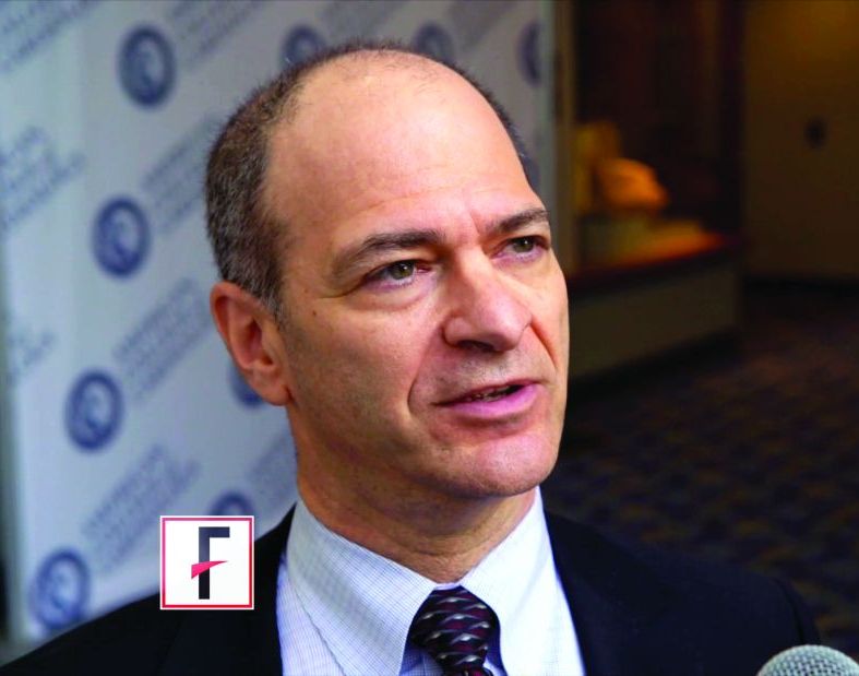
A major consideration for clinicians deciding which SGLT2 inhibitor to prescribe should be “what can the patient afford,” noted Darren K. McGuire, MD, a coinvestigator for VERTIS CV, during discussion of the trial at the EASD virtual meeting.
New analyses show more renal-effect consistency
One surprise in the initial VERTIS CV report was in the study’s key renal outcome, a composite of renal death, need for dialysis, or a doubling of the serum creatinine level, which reflects a cut of at least a 50% in estimated glomerular filtration rate (eGFR). This composite outcome trended toward a significant benefit but fell short, producing a nominal 19% relative reduction. This combined endpoint probably “set the bar too high,” said David Z.I. Cherney, MD, a nephrologist who led the renal assessments run in the trial. He presented several exploratory analyses during the virtual EASD session that provided reassuring evidence that ertugliflozin was not an outlier among the SGLT2 inhibitors when it came to kidney benefits.

Perhaps the most compelling analysis he reported was a slight tweak to the main renal composite endpoint that substituted prevention of a 40% or greater reduction in eGFR for prevention of a 50% or greater reduction. By this somewhat lower bar for efficacy, treatment with ertugliflozin in VERTIS CV linked with a 34% relative risk reduction, compared with placebo (a roughly 1% absolute reduction) that was statistically significant, and importantly came out very close to the effect for this revised endpoint that had been seen for the other three SGLT2 inhibitor drugs.
Focusing on prevention of a 40% or greater drop in eGFR “gives a much more robust measure of renal protection,” Dr. Cherney, a clinician and researcher at the University of Toronto, said in an interview. “The key message is that renal protection is much more uniform” with the rest of the drugs in the class when looked at this way or by some of the other alternative parameters he reported. But the new renal analyses do not address disparities seen among the drugs in the class for several cardiovascular disease effects.
“The overall impression from VERTIS CV is that there was less cardiovascular disease benefit,” except for prevention of heart failure hospitalization, he said.
A teaser for HFpEF
One additional notable new finding discussed during the EASD session stemmed from the investigators ability to mine the medical records of enrolled patients for information about their heart failure history and left ventricular ejection fractions, a data set that was “unique,” compared with the other cardiovascular outcome trials for the drugs in the class, noted Francesco Cosentino, MD, another VERTIS CV coinvestigator and professor of cardiology at the Karolinska Institute in Stockholm.
Roughly a quarter of the enrolled patients had a history of heart failure, and about half of these patients had heart failure with preserved ejection fraction, about 1,000 total patients. In this subgroup treatment with ertugliflozin linked with a 30% relative reduction in hospitalization for heart failure, compared with placebo, a roughly 0.5% absolute reduction. The numbers were small and underpowered for producing convincing evidence, but it provided an intriguing hint of benefit for an unmet need that is currently undergoing further testing in studies designed to specifically explore benefit in this type of heart failure patient, said Dr. Cosentino.
VERTIS CV was sponsored by Merck and Pfizer, the companies that market ertugliflozin. Dr. Davies has been a speaker on behalf of Merck and has had relationships with several other companies. Dr. Butler is a consultant to Merck and several other companies. Dr. McGuire has received honoraria from Merck, nonfinancial support from Pfizer, and has had relationships with several other companies. Dr. Cherney has received honoraria from Merck, nonfinancial research support from Pfizer, and has also had relationships with several other companies. Dr. Cosentino has received fees from Merck and Pfizer, and also from Abbott, AstraZeneca, Bayer, Bristol-Myers Squibb, and Novo Nordisk
Further analyses from the cardiovascular outcome trial of the sodium-glucose transporter 2 inhibitor ertugliflozin in patients with type 2 diabetes helped better define positive effects the drug had on preserving renal function, and also gave a tantalizing hint that this drug, and hence possibly the entire SGLT2 inhibitor drug class, may benefit patients with heart failure with reduced ejection fraction.
But the underlying problem for ertugliflozin (Steglatro) – first seen when results from the VERTIS CV trial initially came out in June 2020 at the annual meeting of the American Diabetes Association – was that, while the trial met its primary endpoint of proving noninferiority to placebo for the combined endpoint of cardiovascular death, nonfatal MI, or nonfatal stroke, treatment with ertugliflozin showed no suggestion of benefit, compared with placebo for reducing this endpoint, producing a nonsignificant 3% relative cut in the combined rate of these adverse events, compared with placebo treatment.
‘Somewhat disappointing’ trial performance
Overall, results from VERTIS CV with ertugliflozin were “somewhat disappointing,” commented Melanie J. Davies, MD, who was not involved with the study and chaired a session at the virtual annual meeting of the European Association for the Study of Diabetes that reviewed the main results, put them into perspective, and added a few new exploratory analyses.
Although the results from 8,246-patient VERTIS CV (Evaluation of Ertugliflozin Efficacy and Safety Cardiovascular Outcomes Trial) put ertugliflozin in the same league as other drugs from its class for safety, “we do not see the significant benefits observed in many of the previous cardiovascular outcomes trials” for other drugs in the SGLT2 inhibitor class, specifically canagliflozin (Invokana), dapagliflozin (Farxiga), and empagliflozin (Jardiance), Dr. Davies said in an interview. The upshot, for at least the time being, is that ertugliflozin “is unlikely to receive a label for any new indications,” she predicted. In contrast, the other drugs in the class have, for example, received a U.S. labeled indication to reduce cardiovascular death (empagliflozin) or major cardiovascular disease events (canagliflozin) in adults with type 2 diabetes (T2D) and cardiovascular disease, or to reduce heart failure hospitalizations (dapagliflozin).
The main results from VERTIS CV, posted online in the New England Journal of Medicine after the EASD session, showed a single significant outcome difference between treatment with ertugliflozin and placebo over a median of 3.0 years of follow-up from among 10 reported secondary outcomes: a 30% relative reduction (a 1.1% absolute reduction) in the rate of hospitalization for heart failure, the sole criterion in the report by which ertugliflozin matched the benefits of the other SGLT2 inhibitors.
But the prespecified design of VERTIS CV called for a hierarchical sequence of secondary analyses. The statistically significant noninferiority of the primary endpoint allowed calculation of the initial secondary endpoint, a reduction in the combined rate of cardiovascular death or hospitalization for heart failure. Ertugliflozin treatment cut this outcome by a relative 12%, compared with placebo, a difference that was not significant.
This neutral finding brought to a stop further statistical testing of any of the other secondary endpoints, including impact on hospitalization for heart failure by itself. It also guaranteed that no beneficial effect inferred from the trial’s data would qualify for statistical validity, making it unlikely that ertugliflozin would gain any new label indications from these results. The drug carries a U.S. label that is limited to providing glycemic control.
Choosing among the SGLT2 inhibitors
“What we can say for sure is that there is a glycemic benefit and a heart failure hospitalization benefit” across all four of the SGLT2 inhibitors. “Beyond that, the best we can say today [about using these drugs in practice] is to follow regulatory indications and guidelines recommendations,” commented Javed Butler, MD, a cardiologist and professor and chair of medicine at the University of Mississippi Medical Center, Jackson.
“These results are going to lead to some serious discussions among the research, clinical, and regulatory communities about class effects versus drug effects, and specific trial data versus the totality of evidence,” he said in an interview.
“I think it will influence prescribing ertugliflozin, particularly in patients with established cardiovascular disease, or when the goal is to improve heart failure outcomes of reduce chronic kidney disease,” added Dr. Davies, a professor of diabetes medicine at the University of Leicester (England). “We already have positive benefits [proven for these outcomes] using other agents in the class.”
Perhaps one feature potentially in ertugliflozin’s favor is its price, and whatever impact that might have for payers or patients with inadequate coverage for their drug costs. U.S. websites show a typical retail price for ertugliflozin that is roughly 40% below the three other agents in the class, a difference that can add up to an annual cost savings of about $2,500.
A major consideration for clinicians deciding which SGLT2 inhibitor to prescribe should be “what can the patient afford,” noted Darren K. McGuire, MD, a coinvestigator for VERTIS CV, during discussion of the trial at the EASD virtual meeting.
New analyses show more renal-effect consistency
One surprise in the initial VERTIS CV report was in the study’s key renal outcome, a composite of renal death, need for dialysis, or a doubling of the serum creatinine level, which reflects a cut of at least a 50% in estimated glomerular filtration rate (eGFR). This composite outcome trended toward a significant benefit but fell short, producing a nominal 19% relative reduction. This combined endpoint probably “set the bar too high,” said David Z.I. Cherney, MD, a nephrologist who led the renal assessments run in the trial. He presented several exploratory analyses during the virtual EASD session that provided reassuring evidence that ertugliflozin was not an outlier among the SGLT2 inhibitors when it came to kidney benefits.
Perhaps the most compelling analysis he reported was a slight tweak to the main renal composite endpoint that substituted prevention of a 40% or greater reduction in eGFR for prevention of a 50% or greater reduction. By this somewhat lower bar for efficacy, treatment with ertugliflozin in VERTIS CV linked with a 34% relative risk reduction, compared with placebo (a roughly 1% absolute reduction) that was statistically significant, and importantly came out very close to the effect for this revised endpoint that had been seen for the other three SGLT2 inhibitor drugs.
Focusing on prevention of a 40% or greater drop in eGFR “gives a much more robust measure of renal protection,” Dr. Cherney, a clinician and researcher at the University of Toronto, said in an interview. “The key message is that renal protection is much more uniform” with the rest of the drugs in the class when looked at this way or by some of the other alternative parameters he reported. But the new renal analyses do not address disparities seen among the drugs in the class for several cardiovascular disease effects.
“The overall impression from VERTIS CV is that there was less cardiovascular disease benefit,” except for prevention of heart failure hospitalization, he said.
A teaser for HFpEF
One additional notable new finding discussed during the EASD session stemmed from the investigators ability to mine the medical records of enrolled patients for information about their heart failure history and left ventricular ejection fractions, a data set that was “unique,” compared with the other cardiovascular outcome trials for the drugs in the class, noted Francesco Cosentino, MD, another VERTIS CV coinvestigator and professor of cardiology at the Karolinska Institute in Stockholm.
Roughly a quarter of the enrolled patients had a history of heart failure, and about half of these patients had heart failure with preserved ejection fraction, about 1,000 total patients. In this subgroup treatment with ertugliflozin linked with a 30% relative reduction in hospitalization for heart failure, compared with placebo, a roughly 0.5% absolute reduction. The numbers were small and underpowered for producing convincing evidence, but it provided an intriguing hint of benefit for an unmet need that is currently undergoing further testing in studies designed to specifically explore benefit in this type of heart failure patient, said Dr. Cosentino.
VERTIS CV was sponsored by Merck and Pfizer, the companies that market ertugliflozin. Dr. Davies has been a speaker on behalf of Merck and has had relationships with several other companies. Dr. Butler is a consultant to Merck and several other companies. Dr. McGuire has received honoraria from Merck, nonfinancial support from Pfizer, and has had relationships with several other companies. Dr. Cherney has received honoraria from Merck, nonfinancial research support from Pfizer, and has also had relationships with several other companies. Dr. Cosentino has received fees from Merck and Pfizer, and also from Abbott, AstraZeneca, Bayer, Bristol-Myers Squibb, and Novo Nordisk
FROM EASD 2020
Nationwide study questions routine long-term beta-blocker post MI
Current American and European guidelines recommending long-term beta-blocker therapy following an acute MI appear to be obsolete in the modern reperfusion era, suggests an analysis of Danish registry data.
Those guidelines are based on old randomized trials of beta-blocker therapy conducted prior to introduction of routine percutaneous coronary intervention and modern multidrug optimal medical therapy for acute MI. There have been no prospective controlled studies in the reperfusion era. And a new Danish national observational study strongly suggests it’s time to reexamine the beta-blocker recommendation, Anders Holt, MD, said at the virtual annual congress of the European Society of Cardiology.
“Stable, optimally treated MI patients do not seem to benefit from beta-blocker treatment exceeding 3 months post hospitalization – bearing in mind this doesn’t apply to patients with other indications for beta-blockers, like heart failure or atrial fibrillation,” said Dr. Holt of Copenhagen University Hospital.
His analysis of Danish national registry data on more than 30,000 patients hospitalized for acute MI during 2003-2018 earned him the annual ESC Young Investigator Award in Population Science.
“This was a crisp and clear presentation of a very creative use of observational epidemiology to try to understand the length of therapy that may or may not be appropriate,” commented award session cochair Paul M. Ridker, MD, director of the Center for Cardiovascular Disease Prevention at Brigham and Women’s Hospital and professor of medicine at Harvard Medical School, both in Boston.
Dr. Holt reported on 30,177 patients optimally treated for a first MI in Danish hospitals during 2003-2018, none of whom had a prior indication or contraindication for beta-blocker therapy. “Optimally treated” meant they underwent percutaneous coronary revascularization and were discharged on a statin and aspirin. As a study requirement, all had to be stable 90 days post hospitalization, at which point 24,770 of the patients were on long-term beta-blocker therapy, and 5,407 (18%) were not. The two groups were comparable in terms of age, sex, comorbidities, and baseline medications. All patients were followed through the registries for a maximum of 3 years, the duration of beta-blocker therapy post MI recommended in American Heart Association/American College of Cardiology guidelines. (The Danish Society of Cardiology recommends 2 years.)
At 3 years post MI, there was no between-group difference in a composite outcome comprising cardiovascular death, recurrent MI, heart failure, stroke, angina, or a cardiac procedure, with a rate of 22.9% in the beta-blocker group and 21.6% in patients not on long-term beta-blocker therapy. The rate of recurrent MI was identical at 6.7% in both groups. Cardiovascular death occurred during 3 years of follow-up in 1.4% of patients on beta-blocker therapy and 1.7% who weren’t, a nonsignificant difference.
“We saw no evidence of any cardioprotective effect, but no increased risk of adverse events resulting in hospitalization, either,” Dr. Holt observed. “I would like to acknowledge that no evidence of effect does not necessarily equal evidence of no effect, but even if there was an effect we can with fair certainty say that it’s probably quite minimal.”
He noted that the Danish registry data indicates that each year since 2012 has shown a growing trend for Danish patients to dispense with long-term beta-blocker therapy after an acute MI.
“This might indicate we are nudging toward a change in practice, where more physicians are thinking that long-term beta-blocker therapy might not be indicated for all MI patients in the reperfusion era,” according to Dr. Holt.
Asked by the four-judge award panel about the possibility of unmeasured confounding in this observational study, Dr Holt responded: “I would be very cautious about asking patients to stop beta-blocker therapy after 3 months just based on this observational data. We can’t speak to causality in an observational study.” But he added that “well-designed observational studies provide valuable data regarding this topic and should not be ignored. They should possibly influence the guidelines and the designs for upcoming randomized trials.”
He conducted several supplementary analyses designed to address the possibility of unevenly distributed unmeasured confounding in the registry study. These analyses proved reassuring. A positive exposure control analysis compared 3-year outcomes in patients who remained on long-term statin therapy and those who didn’t. As expected, outcomes were significantly better in those who did: a 3-year composite outcome rate of 22.1%, compared with 32.1% in patients not on a statin; a cardiovascular death rate of 1.3% with and 2.1% without statin therapy; a recurrent MI rate of 6.6%, compared with 10.1% without a statin; and a 2.8% all-cause mortality with and 5.4% without statin therapy.
In contrast, all-cause mortality was unaffected by whether or not patients were on long-term beta-blocker therapy. And in a negative exposure outcome analysis, no association was found between beta-blocker therapy and the risk of hospitalization for pneumonia, as to be expected if the beta-blocker and no-beta-blocker groups were comparable in key respects.
Dr. Holt reported having no financial conflicts regarding his study.
FROM ESC CONGRESS 2020
Valvular AFib heightens risk in TAVR
Atrial fibrillation has been known to confer an increased risk for poor outcomes after transcatheter aortic valve replacement, but there’s been no evidence of how the etiology of AFib can influence post-TAVR outcomes.

Now, a group of researchers from Bern (Switzerland) University are reporting that valvular AFib almost triples the risk of death or debilitating stroke, compared with patients with no AFib, and significantly increases the risk over nonvalvular AFib.*
“The present findings may have implications for risk stratification in patients undergoing TAVR,” wrote Taishi Okuno, MD, and colleagues in what they said is the first study “to appreciate the combined effect” of AFib and mitral stenosis in TAVR. “The identification of valvular AFib may refine the estimated risk for adverse clinical outcomes in patients undergoing TAVR,” they wrote in JACC: Cardiovascular Interventions.
“The fact that valvular AFib seems to confer a higher risk is an interesting finding,” Fred Welt, MD, professor of cardiology at the University of Utah, Salt Lake City, said in an interview. “I think it helps to a certain extent in prognostication because we can say to patients who have concomitant mitral valve disease that they are at higher risk.” Dr. Welt is also chair of the American College of Cardiology Interventional Council.
The analysis included 1,472 patients with aortic stenosis who had TAVR at Bern University Hospital between August 2007 and June 2018, 32% of whom (465) had atrial fibrillation, subcategorized as nonvalvular (26%, 376) and valvular (6%, 89). The primary endpoint, a composite of cardiovascular death or disabling stroke 1 year after TAVR, occurred in 9.3% of patients with no AFib, 14.5% of those with nonvalvular AFib and 24.2% of patients with valvular AFib.
In terms of hazard ratios, patients with nonvalvular AFib had a 57% greater risk of poor outcomes (P = .009) and those with valvular AFib had a 275% greater risk (P < .001), compared with patients with no AFib. Patients with valvular AFib had a 77% higher rate of cardiovascular death or stroke than those with nonvalvular AFib (P = .027).**
In their analysis, Dr. Okuno and colleagues acknowledged that the definition of valvular AFib used in guidelines and clinical trials isn’t uniform. Valvular atrial fibrillation was defined as AFib with mitral stenosis or a mitral valve prosthesis.
To account for the varying definitions of valvular and nonvalvular AFib, the researchers performed a sensitivity analysis of AFib patients with significant valve disease other than mitral stenosis; 42% of patients in the nonvalvular group fit this definition. Patients with AFib and valvular disease other than mitral stenosis had almost twice the risk of cardiovascular death or disabling stroke at 1 year, compared with patients who had AFib but no significant disease of any valve (20.1% vs. 10.9%, P = .03).
Furthermore, when they excluded patients with mild mitral stenosis from the valvular AFib group, “the effect of an increased risk for cardiovascular death or disabling stroke was no longer statistically significant.”
When the researchers separated out the two elements of the composite endpoint, they found valvular AFib carried a significantly higher risk of cardiovascular death – 21.1% (P < .002) vs. 7% for no AFib and 12.3% (P = .003) for nonvalvular AFib. However, the incidence of cardiovascular events – disabling stroke, nondisabling stroke and transient ischemic attack – showed no significant difference across the three groups, Dr. Okuno and colleagues noted. Specifically, the rates of disabling stroke were 3.8%, 3.7% and 5.7% in the no-AFib, nonvalvular-AFib, and valvular-AFib groups, respectively
In an invited editorial, Bernard Iung, MD, and Vincent Algalarrondo, MD, PhD, noted the problems with the definitions for valvular and nonvalvular AFib. “The term valvular AFib now frequently refers to patients with AFib associated with moderate or severe mitral stenosis or a mechanical heart valve,” they wrote. The definition is justified, they noted, because there’s little evidence on the use of non–vitamin K antagonist oral anticoagulants (NOACs) in patients with mitral stenosis.
They noted the term nonvalvular is “ambiguous” because it doesn’t exclude valvular disease but rather only a subset defined by the restrictive use of a class of anticoagulants. Hence, the definition of valvular AFib “is subject to criticisms and remains not standardized.”
“The individualization of valvular AFib in patients undergoing TAVR is debatable, and the definition used in the present study also included mild mitral stenosis and bioprostheses, thereby highlighting again the lack of a clear and uniform definition of the concept of valvular AFib,” they wrote.
While Dr. Welt said the findings may help in stratifying risk in patients with valvular AFib, he’s not certain how that would influence treatment decisions. “In most cases when we’re considering TAVR in these patients it’s because they have severe symptomatic aortic stenosis,” he said.
Surgery as an alternative is fraught with consequences, he said. “Would it be because you would want to repair the mitral valve as well?” he said. “And once you get into that territory, you’re talking about double-valve surgery, which is a much riskier operation than isolated aortic valve replacement.”
The study raises important questions about patients with valvular AFib, Dr. Welt added. “Why are these patients dying at higher rate? Is it some other arrhythmia or some other hemodynamic problem? Are there other things we can learn about these patients that would help us to better treat patients?”
But exploring these findings further with a randomized clinical trial may not be practical, he added. “The number of patients in whom this is an issue is in the scheme of things rather low: 6%,” he said.
Dr. Okuno has no relevant financial disclosures. Dr. Iung is a consultant for Edwards Lifesciences. Dr. Algalarrondo has been a consultant for Pfizer and Alnylam. Dr. Welt disclosed a relationship with Medtronic.
SOURCE: Okuno T et al. JACC Cardiovasc Interv. 2020 Sep 21. doi: 10.1016/j.jcin.2020.05.049.
Corrections, 9/29/20: An earlier version of this article misstated the increase in risk of (*) death or debilitating stroke and of (**) a poor outcome in those with valvular Afib.
Atrial fibrillation has been known to confer an increased risk for poor outcomes after transcatheter aortic valve replacement, but there’s been no evidence of how the etiology of AFib can influence post-TAVR outcomes.
Now, a group of researchers from Bern (Switzerland) University are reporting that valvular AFib almost triples the risk of death or debilitating stroke, compared with patients with no AFib, and significantly increases the risk over nonvalvular AFib.*
“The present findings may have implications for risk stratification in patients undergoing TAVR,” wrote Taishi Okuno, MD, and colleagues in what they said is the first study “to appreciate the combined effect” of AFib and mitral stenosis in TAVR. “The identification of valvular AFib may refine the estimated risk for adverse clinical outcomes in patients undergoing TAVR,” they wrote in JACC: Cardiovascular Interventions.
“The fact that valvular AFib seems to confer a higher risk is an interesting finding,” Fred Welt, MD, professor of cardiology at the University of Utah, Salt Lake City, said in an interview. “I think it helps to a certain extent in prognostication because we can say to patients who have concomitant mitral valve disease that they are at higher risk.” Dr. Welt is also chair of the American College of Cardiology Interventional Council.
The analysis included 1,472 patients with aortic stenosis who had TAVR at Bern University Hospital between August 2007 and June 2018, 32% of whom (465) had atrial fibrillation, subcategorized as nonvalvular (26%, 376) and valvular (6%, 89). The primary endpoint, a composite of cardiovascular death or disabling stroke 1 year after TAVR, occurred in 9.3% of patients with no AFib, 14.5% of those with nonvalvular AFib and 24.2% of patients with valvular AFib.
In terms of hazard ratios, patients with nonvalvular AFib had a 57% greater risk of poor outcomes (P = .009) and those with valvular AFib had a 275% greater risk (P < .001), compared with patients with no AFib. Patients with valvular AFib had a 77% higher rate of cardiovascular death or stroke than those with nonvalvular AFib (P = .027).**
In their analysis, Dr. Okuno and colleagues acknowledged that the definition of valvular AFib used in guidelines and clinical trials isn’t uniform. Valvular atrial fibrillation was defined as AFib with mitral stenosis or a mitral valve prosthesis.
To account for the varying definitions of valvular and nonvalvular AFib, the researchers performed a sensitivity analysis of AFib patients with significant valve disease other than mitral stenosis; 42% of patients in the nonvalvular group fit this definition. Patients with AFib and valvular disease other than mitral stenosis had almost twice the risk of cardiovascular death or disabling stroke at 1 year, compared with patients who had AFib but no significant disease of any valve (20.1% vs. 10.9%, P = .03).
Furthermore, when they excluded patients with mild mitral stenosis from the valvular AFib group, “the effect of an increased risk for cardiovascular death or disabling stroke was no longer statistically significant.”
When the researchers separated out the two elements of the composite endpoint, they found valvular AFib carried a significantly higher risk of cardiovascular death – 21.1% (P < .002) vs. 7% for no AFib and 12.3% (P = .003) for nonvalvular AFib. However, the incidence of cardiovascular events – disabling stroke, nondisabling stroke and transient ischemic attack – showed no significant difference across the three groups, Dr. Okuno and colleagues noted. Specifically, the rates of disabling stroke were 3.8%, 3.7% and 5.7% in the no-AFib, nonvalvular-AFib, and valvular-AFib groups, respectively
In an invited editorial, Bernard Iung, MD, and Vincent Algalarrondo, MD, PhD, noted the problems with the definitions for valvular and nonvalvular AFib. “The term valvular AFib now frequently refers to patients with AFib associated with moderate or severe mitral stenosis or a mechanical heart valve,” they wrote. The definition is justified, they noted, because there’s little evidence on the use of non–vitamin K antagonist oral anticoagulants (NOACs) in patients with mitral stenosis.
They noted the term nonvalvular is “ambiguous” because it doesn’t exclude valvular disease but rather only a subset defined by the restrictive use of a class of anticoagulants. Hence, the definition of valvular AFib “is subject to criticisms and remains not standardized.”
“The individualization of valvular AFib in patients undergoing TAVR is debatable, and the definition used in the present study also included mild mitral stenosis and bioprostheses, thereby highlighting again the lack of a clear and uniform definition of the concept of valvular AFib,” they wrote.
While Dr. Welt said the findings may help in stratifying risk in patients with valvular AFib, he’s not certain how that would influence treatment decisions. “In most cases when we’re considering TAVR in these patients it’s because they have severe symptomatic aortic stenosis,” he said.
Surgery as an alternative is fraught with consequences, he said. “Would it be because you would want to repair the mitral valve as well?” he said. “And once you get into that territory, you’re talking about double-valve surgery, which is a much riskier operation than isolated aortic valve replacement.”
The study raises important questions about patients with valvular AFib, Dr. Welt added. “Why are these patients dying at higher rate? Is it some other arrhythmia or some other hemodynamic problem? Are there other things we can learn about these patients that would help us to better treat patients?”
But exploring these findings further with a randomized clinical trial may not be practical, he added. “The number of patients in whom this is an issue is in the scheme of things rather low: 6%,” he said.
Dr. Okuno has no relevant financial disclosures. Dr. Iung is a consultant for Edwards Lifesciences. Dr. Algalarrondo has been a consultant for Pfizer and Alnylam. Dr. Welt disclosed a relationship with Medtronic.
SOURCE: Okuno T et al. JACC Cardiovasc Interv. 2020 Sep 21. doi: 10.1016/j.jcin.2020.05.049.
Corrections, 9/29/20: An earlier version of this article misstated the increase in risk of (*) death or debilitating stroke and of (**) a poor outcome in those with valvular Afib.
FROM THE JOURNAL OF THE AMERICAN COLLEGE OF CARDIOLOGY
Smart health devices – promises and pitfalls
What needs to be done before the data deluge hits the office
Hurricane Sally recently crossed the Gulf of Mexico and landed with torrential rainfalls along the Alabama coast. A little rainfall is important for crops; too much leads to devastation. As physicians, we need data in order to help manage patients’ illnesses and to help to keep them healthy. Our fear though is that too much data provided too quickly may have the opposite effect.
Personal monitoring devices

When I bought my first Fitbit 7 years ago, I was enamored with the technology. The Fitbit was little more than a step tracker, yet I proudly wore its black rubber strap on my wrist. It was my first foray into wearable technology, and it felt quite empowering to have an objective way to track my fitness beyond just using my bathroom scale. Now less than a decade later, that Fitbit looks archaic in comparison with the wrist-top technology currently available.
As I write this, the world’s largest technology company is in the process of releasing its sixth-generation Apple Watch. In addition to acting as a smartphone, this new device, which is barely larger than a postage stamp, offers GPS-based movement tracking, the ability to detect falls, continuous heart rate monitoring, a built-in EKG capable of diagnosing atrial fibrillation, and an oxygen saturation sensor. These features weren’t added thoughtlessly. Apple is marketing this as a health-focused device, with their primary advertising campaign claiming that “the future of health is on your wrist,” and they aren’t the only company making this play.
Along with Apple, Samsung, Withings, Fitbit, and other companies continue to bring products to market that monitor our activity and provide new insights into our health. Typically linked to smartphone-based apps, these devices record all of their measurements for later review, while software helps interpret the findings to make them actionable. From heart rate tracking to sleep analysis, these options now provide access to volumes of data that promise to improve our wellness and change our lives. Of course, those promises will only be fulfilled if our behavior is altered as a consequence of having more detailed information. Whether that will happen remains to be seen.
Health system–linked devices
Major advancements in medical monitoring technology are now enabling physicians to get much deeper insight into their patients’ health status. Internet-connected scales, blood pressure cuffs, and exercise equipment offer the ability to upload information into patient portals and integrate that information into EHRs. New devices provide access to information that previously was impossible to obtain. For example, wearable continuous blood glucose monitors, such as the FreeStyle Libre or DexCom’s G6, allow patients and physicians to follow blood sugar readings 24 hours a day. This provides unprecedented awareness of diabetes control and relieves the pain and inconvenience of finger sticks and blood draws. It also aids with compliance because patients don’t need to remember to check their sugar levels on a schedule.
Other compliance-boosting breakthroughs, such as Bluetooth-enabled asthma inhalers and cellular-connected continuous positive airway pressure machines, assist patients with managing chronic respiratory conditions. Many companies are developing technologies to manage acute conditions as well. One such company, an on-demand telemedicine provider called TytoCare, has developed a $299 suite of instruments that includes a digital stethoscope, thermometer, and camera-based otoscope. In concert with a virtual visit, their providers can remotely use these tools to examine and assess sick individuals. This virtual “laying on of hands” may have sounded like science fiction and likely would have been rejected by patients just a few years ago. Now it is becoming commonplace and will soon be an expectation of many seeking care.
But if we are to be successful, everyone must acknowledge that this revolution in health care brings many challenges along with it. One of those is the deluge of data that connected devices provide.
Information overload
There is such a thing as “too much of a good thing.” Described by journalist David Shenk as “data smog” in his 1997 book of the same name, the idea is clear: There is only so much information we can assimilate.
Even after years of using EHRs and with government-implemented incentives that promote “meaningful use,” physicians are still struggling with EHRs. Additionally, many have expressed frustration with the connectedness that EHRs provide and lament their inability to ever really “leave the office.” As more and more data become available to physicians, the challenge of how to assimilate and act on those data will continue to grow. The addition of patient-provided health statistics will only make information overload worse, with clinicians will feeling an ever-growing burden to know, understand, and act on this information.
Unless we develop systems to sort, filter, and prioritize the flow of information, there is potential for liability from not acting on the amount of virtual information doctors receive. This new risk for already fatigued and overburdened physicians combined with an increase in the amount of virtual information at doctors’ fingertips may lead to the value of patient data being lost.
Dr. Notte is a family physician and chief medical officer of Abington (Pa.) Hospital–Jefferson Health. Follow him on Twitter (@doctornotte). Dr. Skolnik is professor of family and community medicine at Sidney Kimmel Medical College, Philadelphia, and associate director of the family medicine residency program at Abington Hospital–Jefferson Health. They have no conflicts related to the content of this piece.
What needs to be done before the data deluge hits the office
What needs to be done before the data deluge hits the office
Hurricane Sally recently crossed the Gulf of Mexico and landed with torrential rainfalls along the Alabama coast. A little rainfall is important for crops; too much leads to devastation. As physicians, we need data in order to help manage patients’ illnesses and to help to keep them healthy. Our fear though is that too much data provided too quickly may have the opposite effect.
Personal monitoring devices
When I bought my first Fitbit 7 years ago, I was enamored with the technology. The Fitbit was little more than a step tracker, yet I proudly wore its black rubber strap on my wrist. It was my first foray into wearable technology, and it felt quite empowering to have an objective way to track my fitness beyond just using my bathroom scale. Now less than a decade later, that Fitbit looks archaic in comparison with the wrist-top technology currently available.
As I write this, the world’s largest technology company is in the process of releasing its sixth-generation Apple Watch. In addition to acting as a smartphone, this new device, which is barely larger than a postage stamp, offers GPS-based movement tracking, the ability to detect falls, continuous heart rate monitoring, a built-in EKG capable of diagnosing atrial fibrillation, and an oxygen saturation sensor. These features weren’t added thoughtlessly. Apple is marketing this as a health-focused device, with their primary advertising campaign claiming that “the future of health is on your wrist,” and they aren’t the only company making this play.
Along with Apple, Samsung, Withings, Fitbit, and other companies continue to bring products to market that monitor our activity and provide new insights into our health. Typically linked to smartphone-based apps, these devices record all of their measurements for later review, while software helps interpret the findings to make them actionable. From heart rate tracking to sleep analysis, these options now provide access to volumes of data that promise to improve our wellness and change our lives. Of course, those promises will only be fulfilled if our behavior is altered as a consequence of having more detailed information. Whether that will happen remains to be seen.
Health system–linked devices
Major advancements in medical monitoring technology are now enabling physicians to get much deeper insight into their patients’ health status. Internet-connected scales, blood pressure cuffs, and exercise equipment offer the ability to upload information into patient portals and integrate that information into EHRs. New devices provide access to information that previously was impossible to obtain. For example, wearable continuous blood glucose monitors, such as the FreeStyle Libre or DexCom’s G6, allow patients and physicians to follow blood sugar readings 24 hours a day. This provides unprecedented awareness of diabetes control and relieves the pain and inconvenience of finger sticks and blood draws. It also aids with compliance because patients don’t need to remember to check their sugar levels on a schedule.
Other compliance-boosting breakthroughs, such as Bluetooth-enabled asthma inhalers and cellular-connected continuous positive airway pressure machines, assist patients with managing chronic respiratory conditions. Many companies are developing technologies to manage acute conditions as well. One such company, an on-demand telemedicine provider called TytoCare, has developed a $299 suite of instruments that includes a digital stethoscope, thermometer, and camera-based otoscope. In concert with a virtual visit, their providers can remotely use these tools to examine and assess sick individuals. This virtual “laying on of hands” may have sounded like science fiction and likely would have been rejected by patients just a few years ago. Now it is becoming commonplace and will soon be an expectation of many seeking care.
But if we are to be successful, everyone must acknowledge that this revolution in health care brings many challenges along with it. One of those is the deluge of data that connected devices provide.
Information overload
There is such a thing as “too much of a good thing.” Described by journalist David Shenk as “data smog” in his 1997 book of the same name, the idea is clear: There is only so much information we can assimilate.
Even after years of using EHRs and with government-implemented incentives that promote “meaningful use,” physicians are still struggling with EHRs. Additionally, many have expressed frustration with the connectedness that EHRs provide and lament their inability to ever really “leave the office.” As more and more data become available to physicians, the challenge of how to assimilate and act on those data will continue to grow. The addition of patient-provided health statistics will only make information overload worse, with clinicians will feeling an ever-growing burden to know, understand, and act on this information.
Unless we develop systems to sort, filter, and prioritize the flow of information, there is potential for liability from not acting on the amount of virtual information doctors receive. This new risk for already fatigued and overburdened physicians combined with an increase in the amount of virtual information at doctors’ fingertips may lead to the value of patient data being lost.
Dr. Notte is a family physician and chief medical officer of Abington (Pa.) Hospital–Jefferson Health. Follow him on Twitter (@doctornotte). Dr. Skolnik is professor of family and community medicine at Sidney Kimmel Medical College, Philadelphia, and associate director of the family medicine residency program at Abington Hospital–Jefferson Health. They have no conflicts related to the content of this piece.
Pesco-Mediterranean diet, fasting ‘ideal’ to reduce CVD risk
A Pesco-Mediterranean diet consisting of plants, legumes, nuts, whole grains, extra-virgin olive oil (EVOO), moderate amounts of dairy products, and fish and/or seafood, together with intermittent fasting (also called time-restricted eating), can reduce risk for cardiovascular disease (CVD), according to a new review.
The authors presented the research and conceptual underpinnings of this approach, which “proposes that following a Pesco-Mediterranean diet with time-restricted eating is evidence-based and ideal for reducing cardiovascular risk,” study coauthor Sarah Smith, PhD, RN, of Saint Luke’s Mid America Heart Institute, Kansas City, Mo., said in an interview.
The review was published online September 14 in the Journal of the American College of Cardiology.
‘Omnivore’s dilemma’
A host of epidemiologic studies and randomized clinical trials support an association between the traditional Mediterranean diet and lower risk for all-cause and CVD mortality, coronary heart disease, metabolic syndrome, neurodegenerative diseases, and other adverse outcome. The diet has been subsequently endorsed by several sets of guidelines, including those from the Department of Health & Human Services and the Department of Agriculture, and the 2019 American Heart Association/American College of Cardiology primary prevention guidelines.
“Although humans are omnivores and can subsist on a myriad of foods, the ideal diet for health remains a dilemma for many people,” lead author James H. O’Keefe, MD, director of preventive cardiology at Saint Luke’s, said in a news release.
“Plant-rich diets reduce CVD risk; however, veganism is difficult to follow and can result in important nutrient deficiencies,” he stated.
On the other hand, “the standard American diet is high in red meat, especially processed meat from animals raised in inhumane conditions, fed unnatural foods, and often treated with hormones and antibiotics,” the authors pointed out.
Together with overconsumption of red meat, sugar and processed food contribute to poor health outcomes, Dr. Smith noted.
The review was designed to present the Pesco-Mediterranean diet as “a solution to the ‘omnivore’s dilemma’ about what to eat,” said Dr. O’Keefe.
Study coauthor Ibrahim M. Saeed, MD, a cardiologist at Saint Luke’s, added that the research “attempts to emphasize the results of landmark prospective trials that highlight good, healthy eating options rather than just [foods that people would] want to avoid.”
Key components
The traditional Mediterranean diet includes “unrestricted use of EVOO,” but the quality of the olive oil is “crucial” and it must be unrefined and cold pressed, the authors emphasized.
The “highly bioactive” polyphenols likely “underlie EVOO’s numerous cardiometabolic benefits,” the researchers wrote, noting that the 2014 PREDIMED trial provided “first-level scientific evidence of [EVOO’s] cardioprotective effects [if used] within the context of the Mediterranean diet.”
The authors recommend “generous use” of EVOO in salad dressings and vegetable dishes, pasta, rice, fish, sauces, or legumes.
They also review the role of tree nuts, noting that they are “nutrient-dense foods rich in unsaturated fats, fiber, protein, polyphenols, phytosterols, tocopherols, and nonsodium minerals” and have been shown beneficial in CVD prevention.
Legumes play a “central role” in the Mediterranean diet and are an “excellent source” of vegetable protein, folate, magnesium, and fiber. Legume consumption is associated with lowered risk for CVD, as well as improved blood glucose, cholesterol, blood pressure, and body weight, the authors stated.
Whole grains like barley, whole oats, brown rice, and quinoa are likewise central components of the traditional Mediterranean diet. The authors warned that refined grain products and commercial precooked pasta or pizza should be “consumed only in small amounts.”
Window of time
In time-restricted eating (which is one type of intermittent fasting), the daily intake of food is limited to a window of time, usually 6-12 hours each day, the authors explained.
When done regularly, this type of eating has been shown to both decrease intra-abdominal adipose tissue and reduce free-radical production. Additionally, it “elicits powerful cellular responses” that may reduce risks for systemic inflammation, diabetes, CVD, cancer, and neurodegenerative diseases.
However, the authors warned, the evidence supporting time-restricted eating is still preliminary.
‘Let food be thy medicine’
Andrew Freeman, MD, cochair of the ACC’s nutrition & lifestyle work group, cautioned that many American plant-based Mediterranean diets often include large amount of feta cheese and lamb and foods are often “heavily doused” in olive oil, while the traditional Mediterranean diet consists primarily of greens and lentils and is plant based.
“The goal would be to have a whole grain and leafy vegetables as the center of the meal, and – if an animal product such as fish is included – it should be limited to as little as possible and used as the garnish rather than the main dish,” he stated.
Moreover, fish are often exposed to large amount of toxins, heavy metals, and microplastics, so “don’t overdo eating fish,” he advised.
Dr. Freeman said that intermittent fasting “has a lot of promise and no harm” and concentrating food consumption during a shorter period in the day instead of “grazing throughout the day” will reduce constant snacking. “But don’t gorge yourself during those hours,” he warned.
Dr. Freeman concluded by citing the guidance of Hippocrates: “Let food be thy medicine.
“There’s some real truth to that,” he added.
No source of funding was listed. Dr. Smith and Dr. Freeman disclosed no relevant financial relationships. Dr. O’Keefe has a major ownership interest in CardioTabs, a supplement company that sells some products containing omega-3 fatty acids.
A version of this article originally appeared on Medscape.com.
‘Cautious’ DOAC underdosing in AFib may push mortality higher
The risk for death goes up for patients with atrial fibrillation (AFib) who are put on direct oral anticoagulants (DOAC) at dosages other than those approved for stroke prevention, whether higher or lower than doses specified in the labeling, suggests a large registry study.
A quarter of more than 10,000 patients in the registry took the drugs at such nonrecommended higher or lower dosages. Overwhelmingly it was the latter, perhaps reflecting caution on the part of some practitioners looking to minimize the risk of bleeding complications.
The risk of major bleeding indeed dropped sharply for those taking DOACs at lower-than-recommended levels, but at the cost of a 25% jump in all-cause mortality over 2 years, report investigators from their analysis of patients in the GARFIELD-AF registry published Sept. 14 in the Journal of the American College of Cardiology.
Risks of major bleeding and of stroke or systemic embolism didn’t climb significantly for patients either under- or overdosed.
In general, “physicians are worried about giving too much anticoagulant, and they tend to favor erring on the low-dose side,” lead author A. John Camm, MD, St. George’s University of London, said in an interview. That’s how it was when an oral anticoagulation meant a vitamin K antagonist (VKA) and underdosing was frequent; and it remains an issue in the DOAC era. “It’s not just a little problem. It’s a very big problem.”
Today, clinicians may prescribe DOACs similar to how they prescribed VKAs, by cautiously choosing a lower dosage for selected patients based on their risk profile, Dr. Camm observed. But in contrast to the VKAs, the DOACs “were studied with different dose-reduction strategies, and their labeling requires them to be prescribed according to different parameters.”
They variously base dosage reductions on age, body weight, renal function, or drug-drug interactions, for example, but some clinicians “tend to think that all of those factors should be applied in every instance, with every drug,” he said.
“So I think there’s some confusion and a lot of caution that physicians use with anticoagulants, and they often forget that the purpose of the anticoagulant is to prevent strokes and adverse outcomes such as mortality,” Dr. Camm said. “But by reducing the dose, they expose their patients to these other major cardiovascular events.”
Numerically, the excess mortality among underdosed patients appeared to be driven by death from heart failure or myocardial infarction. There was little or no contribution from sudden death, fatal strokes, or noncardiovascular death.
The findings “remind clinicians to dose DOACs properly and that there are consequences of dosing errors,” observes Gerald V. Naccarelli, MD, of Penn State University and the Milton S. Hershey Medical Center, Hershey, in an accompanying editorial.
Based on the major clinical trials that established the drugs as mainstream stroke-preventive therapy in AFib, as well as extensive regulatory review, each DOAC’s label-recommended dosing “is a guidance of the truth to achieve the highest efficacy and most safety in our patients,” Dr. Naccarelli wrote. “As clinicians are risk adverse, underdosing might result in lower major bleeding rates, and physicians are blamed for bleeding but not necessarily for allowing embolic strokes to occur. These data raise the issue that underdosing is associated with worse patient outcomes.”
The GARFIELD-AF analysis covered 10,426 adults with nonvalvular AFib in 35 countries who initiated a DOAC from 2013 to 2016. The drugs were prescribed at dosages consistent with recommendations in each respective country’s labeling for stroke prevention in AFib in 72.9% of the cohort. Most full and adjusted dose levels approved by the European Medicines Agency, Food and Drug Administration, and regulators in Japan were the same or similar.
But there were a few exceptions. All dosing criteria across the three regulatory domains were the same for apixaban (Eliquis). But variations included lower dosage options for rivaroxaban (Xarelto) and edoxaban (Savaysa, Lixiana) in Japan, and a uniquely low dabigatran (Pradaxa) dosage option in the United States.
The DOAC used least often was the one most frequently underdosed. More than half of patients prescribed edoxaban were given it at a lower-than-recommended dosage.
The adjusted hazard ratio for all-cause mortality at 2 years for DOAC under- or overdosing, compared with dosing at recommended levels, was 1.24 (95% confidence interval, 1.04-1.48). The difference was driven by underdosing, for which the HR was 1.25 (95% CI, 1.04-1.50). The HR for over-dosing was only 1.19 (95% CI, 0.83-1.71).
Multivariate adjustment accounted for age, sex, and ethnicity; type of AFib; diabetes; hypertension; history of bleeding; prior stroke, transient ischemic attack, or systemic embolism; heart failure; vascular disease; smoking; and heavy alcohol consumption.
The risk of stroke or systemic embolism didn’t go up or down significantly for either overdosed or underdosed patients. Neither group showed an increased risk for major bleeding; however, the HR for major bleeding in underdosed patients fell to 0.50 (95% CI, 0.28-0.88).
Underdosing was more common in some world regions than others. The rate exceeded 30% in all Latin American countries except Argentina, the report stated, and in all Asian countries except Singapore.
Japanese patients have long received oral anticoagulation at lower dosages than are used in the West, Dr. Camm observed. When VKAs were the only choice, for example, international normalized ratio targets were consistently a bit lower in Japan than in, for example, North America or Europe.
“And when [novel] OACs were developed, again, the Japanese took the view that their patients are more vulnerable to bleeding, and therefore a lower dose would be appropriate. In some instances, lower-dose regimens have been specifically studied in the Japanese,” Dr. Camm said. “Having said that, this concept of bleeding being more problematic in Asian patients has expanded well beyond Japan, and therefore in many Asian communities, lower doses of [novel] OACs are chosen.”
Many other factors may contribute to DOAC underdosing, including differences in dosing strategies between primary care practitioners and specialists, or between hospital-based and office-based clinicians, for example.
“It might also be argued that a physician who fails to treat a patient adequately in one arena may also be failing to treat the patient well in other aspects of their care,” Dr. Camm proposed. “Therefore you could have increased mortality due to other cardiovascular causes, or even noncardiovascular events, through absence of good quality care. Our study did not address that specifically. But it might be the case, speculatively.”
The study was supported by a grant from Bayer to the Thrombosis Research Institute, “which sponsors the GARFIELD-AF registry.” Dr. Camm discloses receiving grants and personal fees from Bayer, Boehringer Ingelheim, Pfizer/Bristol-Myers Squibb, and Daiichi Sankyo. Disclosures for the other authors are in the report. Dr. Naccarelli disclosed consulting and participating in research for Janssen and serving as a consultant for Milestone, Sanofi, Omeicos, and Acesion Pharma.
A version of this article originally appeared on Medscape.com.
Observational study again suggests lasting impact of COVID-19 on heart
A new study using cardiac magnetic resonance (CMR) imaging to examine the effects of novel coronavirus infection on the heart showed signs suggestive of myocarditis in 4 out of 26 competitive athletes who recovered from asymptomatic or mild cases of COVID-19.

While these and other similar findings are concerning, commentators are saying the results are preliminary and do not indicate widespread CMR screening is appropriate.
Two of the 4 patients showing signs of myocarditis in this series had no symptoms of COVID-19 but tested positive on routine testing. An additional 12 student athletes (46%) showed late gadolinium enhancement (LGE), of whom 8 (30.8%) had LGE without T2 elevation suggestive of prior myocardial injury.
An additional 12 student athletes (46%) showed late gadolinium enhancement (LGE), of whom 8 (31%) had LGE without T2 elevation suggestive of prior myocardial injury.
This finding, said Saurabh Rajpal, MBBS, MD, the study’s lead author, “could suggest prior myocardial injury or it could suggest athletic myocardial adaptation.”
In a research letter published in JAMA Cardiology, Rajpal and colleagues at Ohio State University in Columbus, described the findings of comprehensive CMR examinations in competitive athletes referred to the sport medicine clinic after testing positive for COVID-19 on reverse transcriptase-polymerase chain reaction between June and August 2020.
The university had made the decision in the spring to use CMR imaging as a screening tool for return to play, said Dr. Rajpal. While CMR is being used for research purposes, the American College of Cardiology’s recent “consensus expert opinion” statement on resumption of sport and exercise after COVID-19 infection does not require CMR imaging for resumption of competitive activity (JAMA Cardiol. 2020 May 13. doi:10.1001/jamacardio.2020.2136).
None of the athletes required hospitalization for their illness, and only 27% reported mild symptoms during the short-term infection, including sore throat, shortness of breath, myalgia, and fever.
On the day of CMR imaging, ECG and transthoracic echocardiography were performed, and serum troponin I was measured. There were no diagnostic ST/T wave changes, ventricular function and volumes were normal, and no athletes showed elevated serum troponin levels.
The updated Lake Louise Criteria were used to assess CMR findings consistent with myocarditis.
“I don’t think this is a COVID-specific issue. We have seen myocarditis after other viral infections; it’s just that COVID-19 is the most studied thus far, and with strenuous activity, inflammation in the heart can be risky,” Dr. Rajpal said in an interview. He added that more long-term and larger studies with control populations are needed.
His group is continuing to follow these athletes and has suggested that CMR “may provide an excellent risk-stratification assessment for myocarditis in athletes who have recovered from COVID-19 to guide safe competitive sports participation.”
Significance still unknown
Matthew Martinez, MD, the director of sports cardiology at Atlantic Health – Morristown (N.J.) Medical Center and the Gagnon Cardiovascular Institute, urged caution in making too much of the findings of this small study.

“We know that viruses cause myocardial damage and myocarditis. What we don’t know is how important these findings are. And in terms of risk, would we find the same phenomenon if we did this, say, in flu patients or in other age groups?” Dr. Martinez said in an interview.
“I haven’t seen all the images, but what I’d want to know is are these very subtle findings? Are these overt findings? Is this part of an active individual with symptoms? I need to know a little more data before I can tell if this influences the increased risk of sudden cardiac death that we often associate with myocarditis. I’m not sure how this should influence making decisions with regards to return to play.”
Dr. Martinez, who is the ACC’s chair of Sports and Exercise but was not an author of their recent guidance on return to sport, said that he is not routinely using CMR to assess athletes post-infection, as per the ACC’s recommendations.
“My approach is to evaluate anybody with a history of COVID infection and, first, determine whether it was an important infection with significant symptoms or not. And then, if they’re participating at a high level or are professional athletes, I would suggest an ECG, echo, and troponin. That’s our recommendation for the last several months and is still an appropriate way to evaluate that group.”
“In the presence of an abnormality or ongoing symptoms, I would ask for an MRI at that point,” said Dr. Martinez.
“We just don’t have much data on athletes with no symptoms to use to interpret these CMR findings and the study didn’t offer any controls. We don’t even know if these findings are new findings or old findings that have just been identified now,” he added.
New, updated recommendations from the ACC are coming soon, said Dr. Martinez. “I do not expect them to include CMR as first line.”
Cardiologists concerned about misinformation
This is at least the fourth study showing myocardial damage post-COVID-19 infection and there is concern in the medical community that the media has overstated the risks of heart damage, especially in athletes, and at the same time overstated the benefits of CMR.
In particular, Puntmann et al reported in July a 100-patient study that showed evidence of myocardial inflammation by CMR in 78% of patients recently recovered from a bout of COVID-19 (JAMA Cardiol. 2020 Jul 27; doi:10.1001/jamacardio.2020.3557).

“That paper is completely problematic,” John Mandrola, MD, of Baptists Medical Associates, Louisville, Ky., said in an interview. “It has the same overarching weaknesses [of other studies] that it’s observational and retrospective, but there were also numerical issues. So to me that paper is an interesting observation, but utterly unconvincing and preliminary,” said Dr. Mandrola.
Those limitations didn’t stop the study from garnering media attention, however. The Altmetric score (an attention score that tracks all mentions of an article in the media and on social media) for the Puntmann et al paper is approaching 13,000, including coverage from 276 news outlets and more than 19,000 tweets, putting it in the 99th percentile of all research outputs tracked by Altmetric to date.
To counter this, an “open letter” posted online just days before the Rajpal study published urging professional societies to “offer clear guidance discouraging CMR screening for COVID-19 related heart abnormalities in asymptomatic members of the general public.” The letter was signed by 51 clinicians, researchers, and imaging specialists from around the world.
Dr. Mandrola, one of the signatories, said: “This topic really scares people, and when it gets in the media like this, I think the leaders of these societies need to come out and say something really clear on major news networks letting people know that it’s just way too premature to start doing CMRs on every athlete that’s gotten this virus.”
“I understand that the current guidelines may be clear that CMR is not a first-line test for this indication, but when the media coverage is so extensive and so overblown, I wonder how much impact the guidelines will have in countering this fear that’s in the community,” he added.
Asked to comment on the letter, Dr. Rajpal said he agrees with the signatories that asymptomatic people from general population do not need routine cardiac MRI. “However, competitive athletes are a different story. Testing depends on risk assessment in specific population and competitive athletes as per our protocol will get enhanced cardiac workup including CMR for responsible and safe start of competitive sports. ... In the present scenario, while we get more data including control data, we will continue with our current protocol.”
Dr. Mandrola is Medscape Cardiology’s Chief Cardiology Consultant. MDedge is part of the Medscape Professional Network.
This article first appeared on Medscape.com.
A new study using cardiac magnetic resonance (CMR) imaging to examine the effects of novel coronavirus infection on the heart showed signs suggestive of myocarditis in 4 out of 26 competitive athletes who recovered from asymptomatic or mild cases of COVID-19.
While these and other similar findings are concerning, commentators are saying the results are preliminary and do not indicate widespread CMR screening is appropriate.
Two of the 4 patients showing signs of myocarditis in this series had no symptoms of COVID-19 but tested positive on routine testing. An additional 12 student athletes (46%) showed late gadolinium enhancement (LGE), of whom 8 (30.8%) had LGE without T2 elevation suggestive of prior myocardial injury.
An additional 12 student athletes (46%) showed late gadolinium enhancement (LGE), of whom 8 (31%) had LGE without T2 elevation suggestive of prior myocardial injury.
This finding, said Saurabh Rajpal, MBBS, MD, the study’s lead author, “could suggest prior myocardial injury or it could suggest athletic myocardial adaptation.”
In a research letter published in JAMA Cardiology, Rajpal and colleagues at Ohio State University in Columbus, described the findings of comprehensive CMR examinations in competitive athletes referred to the sport medicine clinic after testing positive for COVID-19 on reverse transcriptase-polymerase chain reaction between June and August 2020.
The university had made the decision in the spring to use CMR imaging as a screening tool for return to play, said Dr. Rajpal. While CMR is being used for research purposes, the American College of Cardiology’s recent “consensus expert opinion” statement on resumption of sport and exercise after COVID-19 infection does not require CMR imaging for resumption of competitive activity (JAMA Cardiol. 2020 May 13. doi:10.1001/jamacardio.2020.2136).
None of the athletes required hospitalization for their illness, and only 27% reported mild symptoms during the short-term infection, including sore throat, shortness of breath, myalgia, and fever.
On the day of CMR imaging, ECG and transthoracic echocardiography were performed, and serum troponin I was measured. There were no diagnostic ST/T wave changes, ventricular function and volumes were normal, and no athletes showed elevated serum troponin levels.
The updated Lake Louise Criteria were used to assess CMR findings consistent with myocarditis.
“I don’t think this is a COVID-specific issue. We have seen myocarditis after other viral infections; it’s just that COVID-19 is the most studied thus far, and with strenuous activity, inflammation in the heart can be risky,” Dr. Rajpal said in an interview. He added that more long-term and larger studies with control populations are needed.
His group is continuing to follow these athletes and has suggested that CMR “may provide an excellent risk-stratification assessment for myocarditis in athletes who have recovered from COVID-19 to guide safe competitive sports participation.”
Significance still unknown
Matthew Martinez, MD, the director of sports cardiology at Atlantic Health – Morristown (N.J.) Medical Center and the Gagnon Cardiovascular Institute, urged caution in making too much of the findings of this small study.
“We know that viruses cause myocardial damage and myocarditis. What we don’t know is how important these findings are. And in terms of risk, would we find the same phenomenon if we did this, say, in flu patients or in other age groups?” Dr. Martinez said in an interview.
“I haven’t seen all the images, but what I’d want to know is are these very subtle findings? Are these overt findings? Is this part of an active individual with symptoms? I need to know a little more data before I can tell if this influences the increased risk of sudden cardiac death that we often associate with myocarditis. I’m not sure how this should influence making decisions with regards to return to play.”
Dr. Martinez, who is the ACC’s chair of Sports and Exercise but was not an author of their recent guidance on return to sport, said that he is not routinely using CMR to assess athletes post-infection, as per the ACC’s recommendations.
“My approach is to evaluate anybody with a history of COVID infection and, first, determine whether it was an important infection with significant symptoms or not. And then, if they’re participating at a high level or are professional athletes, I would suggest an ECG, echo, and troponin. That’s our recommendation for the last several months and is still an appropriate way to evaluate that group.”
“In the presence of an abnormality or ongoing symptoms, I would ask for an MRI at that point,” said Dr. Martinez.
“We just don’t have much data on athletes with no symptoms to use to interpret these CMR findings and the study didn’t offer any controls. We don’t even know if these findings are new findings or old findings that have just been identified now,” he added.
New, updated recommendations from the ACC are coming soon, said Dr. Martinez. “I do not expect them to include CMR as first line.”
Cardiologists concerned about misinformation
This is at least the fourth study showing myocardial damage post-COVID-19 infection and there is concern in the medical community that the media has overstated the risks of heart damage, especially in athletes, and at the same time overstated the benefits of CMR.
In particular, Puntmann et al reported in July a 100-patient study that showed evidence of myocardial inflammation by CMR in 78% of patients recently recovered from a bout of COVID-19 (JAMA Cardiol. 2020 Jul 27; doi:10.1001/jamacardio.2020.3557).
“That paper is completely problematic,” John Mandrola, MD, of Baptists Medical Associates, Louisville, Ky., said in an interview. “It has the same overarching weaknesses [of other studies] that it’s observational and retrospective, but there were also numerical issues. So to me that paper is an interesting observation, but utterly unconvincing and preliminary,” said Dr. Mandrola.
Those limitations didn’t stop the study from garnering media attention, however. The Altmetric score (an attention score that tracks all mentions of an article in the media and on social media) for the Puntmann et al paper is approaching 13,000, including coverage from 276 news outlets and more than 19,000 tweets, putting it in the 99th percentile of all research outputs tracked by Altmetric to date.
To counter this, an “open letter” posted online just days before the Rajpal study published urging professional societies to “offer clear guidance discouraging CMR screening for COVID-19 related heart abnormalities in asymptomatic members of the general public.” The letter was signed by 51 clinicians, researchers, and imaging specialists from around the world.
Dr. Mandrola, one of the signatories, said: “This topic really scares people, and when it gets in the media like this, I think the leaders of these societies need to come out and say something really clear on major news networks letting people know that it’s just way too premature to start doing CMRs on every athlete that’s gotten this virus.”
“I understand that the current guidelines may be clear that CMR is not a first-line test for this indication, but when the media coverage is so extensive and so overblown, I wonder how much impact the guidelines will have in countering this fear that’s in the community,” he added.
Asked to comment on the letter, Dr. Rajpal said he agrees with the signatories that asymptomatic people from general population do not need routine cardiac MRI. “However, competitive athletes are a different story. Testing depends on risk assessment in specific population and competitive athletes as per our protocol will get enhanced cardiac workup including CMR for responsible and safe start of competitive sports. ... In the present scenario, while we get more data including control data, we will continue with our current protocol.”
Dr. Mandrola is Medscape Cardiology’s Chief Cardiology Consultant. MDedge is part of the Medscape Professional Network.
This article first appeared on Medscape.com.
Role of aspirin explored in primary prevention of CVD in systemic rheumatic diseases
Low-dose aspirin may be considered for the primary prevention of cardiovascular disease (CVD) in patients with autoimmune systemic rheumatic diseases who are at particularly high risk because of their individual cardiovascular risk profile, according to authors of a new review article in the journal Rheumatology who acknowledge the controversial nature of the issue, because while significant cardiovascular benefit from aspirin for secondary prevention is well established, it has not been for primary prevention.
Secondary prevention with daily, low-dose aspirin is part of aggressive, comprehensive risk modification in patients who have experienced an MI or stroke or are considered at high risk for CVD. But when it comes to primary prevention of the onset of disease, the authors, led by Serena Fasano, MD, PhD, of the rheumatology unit at the University of Campania, Naples, Italy, acknowledged the contradictory positions of international guidelines and uncertainty over balancing benefit versus harm – including risk of mortality in the context of excess bleeding. They called for “robust data” from high-quality randomized, controlled trials for subgroups of patients with specific rheumatologic diseases in order to better answer the question of aspirin for primary prevention.
“This review is devoted to reporting the present knowledge on the effectiveness of low-dose [aspirin] in primary CV prevention in a number of autoimmune systemic rheumatic diseases, not a systematic review or meta-analysis,” the authors stated. “We are not claiming to have covered more than a selection of the literature for each disease. Available data are not high-quality data and do not provide firm conclusions.”
The authors focused primarily on accelerated, rather than spontaneous, atherosclerosis or buildup of plaque in artery walls, implicated in ischemic heart diseases such as MI and ischemic cerebrovascular diseases such as stroke. They looked at its association with autoimmune rheumatic diseases, primarily systemic lupus erythematosus (SLE) and RA, but also including antiphospholipid syndrome, systemic sclerosis, mixed connective tissue disease, dermatomyositis/polymyositis, primary Sjögren’s syndrome, and systemic vasculitis.
They shared results from a review of 167 patients with SLE consecutively admitted to their tertiary medical center who had not previously experienced a cardiovascular event and who were prescribed low-dose (100 mg) aspirin on their first visit and followed for 8 years. The cardiovascular event-free rate was higher in the aspirin group and no excess bleeding was noted, although this may be attributable to a younger patient population and routine use of proton pump inhibitors. Subsequently, hydroxychloroquine was added to the aspirin treatment and was associated with further reduction in cardiovascular events.
The research group also conducted a retrospective analysis of 746 patients with RA consecutively admitted to four tertiary medical centers who hadn’t experienced a cardiovascular event previously. Incidence of cardiovascular events was significantly lower in aspirin-treated patients.
Individualized aspirin prescribing with cardiologist comanagement
There may be a modest benefit of using low-dose aspirin on a long-term basis, but that benefit needs to be offset by the risk of bleeds, said M. Elaine Husni, MD, MPH, vice chair of rheumatology and director of the Arthritis and Musculoskeletal Center at the Cleveland Clinic. It’s important to remind clinicians of cardiovascular risk, she said. “But the message for rheumatologists is it needs to be prescribed on an individual basis, rather than based on diagnosis of a rheumatic condition – at least until we have better evidence.”

Dr. Husni recommended keeping an open mind regarding individual approaches – for example, low-dose aspirin plus statins. A composite approach to prevention likely is called for, including attention to lifestyle issues such as smoking cessation, exercise, and weight loss. “That kind of complexity in decision-making highlights the need for comanagement with a cardiologist,” she said. “I’m a big believer in comanagement. At my multidisciplinary medical center, I am able to pick up the phone and talk to a cardiologist with whom our group has a relationship.” If physicians don’t have that kind of relationship with a cardiology group, she suggested reaching out to establish one.
The review paper could give some guidance to rheumatologists for use on an individual case, Michael Nurmohamed, MD, PhD, of the Amsterdam Rheumatology and Immunology Center in the Netherlands commented in an interview. “However, firm recommendations cannot be given as proper investigations are still lacking, as acknowledged by the authors. In addition, the review paper itself has some methodological constraints. Although this is a narrative review, the search strategy should have been specified, and a quality assessment of the individual studies is lacking.”

There is no doubt that the CVD burden in RA and other rheumatologic conditions is substantially increased in comparison to the general population, Dr. Nurmohamed said. That has been assessed by several well-designed, prospective, controlled studies. Other relatively frequent inflammatory arthropathies, including ankylosing spondylitis and psoriatic arthritis, also pose cardiovascular risk.
“Aspirin cannot be recommended for primary CVD prevention in inflammatory arthropathies due to the absence of adequate studies. That’s why the EULAR [European League Against Rheumatism] guidelines did not recommend its use,” he said. Currently, a EULAR task force is developing evidence-based guidelines for primary CVD prevention in the diseases discussed by Fasano et al., where the use of aspirin will be reassessed. “As these guidelines will consider the methodological quality of the underlying studies, they will enable a more refined use of aspirin in daily clinical practice.”

Primary prevention of CVD using aspirin is not currently the standard of care in taking care of patients with rheumatologic disease in the Netherlands, Ronald F. van Vollenhoven, MD, PhD, Dr. Nurmohamed’s colleague and director of the Amsterdam Rheumatology and Immunology Center and the chair of the department of rheumatology and clinical immunology at the Amsterdam University Medical Center, said in an interview.
“One reason may be the limited data, as highlighted in the review by Dr. Fasano and colleagues. However, another consideration is the problem of polypharmacy. Rheumatic diseases usually require chronic treatment, sometimes with multiple medications. This makes it even more of a concern to add an additional medication, even a relatively innocuous one such as low-dose aspirin,” he said.
Dr. Husni, Dr. Nurmohamed, and Dr. van Vollenhoven reported having no relevant disclosures. The authors of the review article had no relevant disclosures.
SOURCE: Fasano S et al. Rheumatology. 2020 Aug 25. doi: 10.1093/rheumatology/keaa335.
Low-dose aspirin may be considered for the primary prevention of cardiovascular disease (CVD) in patients with autoimmune systemic rheumatic diseases who are at particularly high risk because of their individual cardiovascular risk profile, according to authors of a new review article in the journal Rheumatology who acknowledge the controversial nature of the issue, because while significant cardiovascular benefit from aspirin for secondary prevention is well established, it has not been for primary prevention.
Secondary prevention with daily, low-dose aspirin is part of aggressive, comprehensive risk modification in patients who have experienced an MI or stroke or are considered at high risk for CVD. But when it comes to primary prevention of the onset of disease, the authors, led by Serena Fasano, MD, PhD, of the rheumatology unit at the University of Campania, Naples, Italy, acknowledged the contradictory positions of international guidelines and uncertainty over balancing benefit versus harm – including risk of mortality in the context of excess bleeding. They called for “robust data” from high-quality randomized, controlled trials for subgroups of patients with specific rheumatologic diseases in order to better answer the question of aspirin for primary prevention.
“This review is devoted to reporting the present knowledge on the effectiveness of low-dose [aspirin] in primary CV prevention in a number of autoimmune systemic rheumatic diseases, not a systematic review or meta-analysis,” the authors stated. “We are not claiming to have covered more than a selection of the literature for each disease. Available data are not high-quality data and do not provide firm conclusions.”
The authors focused primarily on accelerated, rather than spontaneous, atherosclerosis or buildup of plaque in artery walls, implicated in ischemic heart diseases such as MI and ischemic cerebrovascular diseases such as stroke. They looked at its association with autoimmune rheumatic diseases, primarily systemic lupus erythematosus (SLE) and RA, but also including antiphospholipid syndrome, systemic sclerosis, mixed connective tissue disease, dermatomyositis/polymyositis, primary Sjögren’s syndrome, and systemic vasculitis.
They shared results from a review of 167 patients with SLE consecutively admitted to their tertiary medical center who had not previously experienced a cardiovascular event and who were prescribed low-dose (100 mg) aspirin on their first visit and followed for 8 years. The cardiovascular event-free rate was higher in the aspirin group and no excess bleeding was noted, although this may be attributable to a younger patient population and routine use of proton pump inhibitors. Subsequently, hydroxychloroquine was added to the aspirin treatment and was associated with further reduction in cardiovascular events.
The research group also conducted a retrospective analysis of 746 patients with RA consecutively admitted to four tertiary medical centers who hadn’t experienced a cardiovascular event previously. Incidence of cardiovascular events was significantly lower in aspirin-treated patients.
Individualized aspirin prescribing with cardiologist comanagement
There may be a modest benefit of using low-dose aspirin on a long-term basis, but that benefit needs to be offset by the risk of bleeds, said M. Elaine Husni, MD, MPH, vice chair of rheumatology and director of the Arthritis and Musculoskeletal Center at the Cleveland Clinic. It’s important to remind clinicians of cardiovascular risk, she said. “But the message for rheumatologists is it needs to be prescribed on an individual basis, rather than based on diagnosis of a rheumatic condition – at least until we have better evidence.”
Dr. Husni recommended keeping an open mind regarding individual approaches – for example, low-dose aspirin plus statins. A composite approach to prevention likely is called for, including attention to lifestyle issues such as smoking cessation, exercise, and weight loss. “That kind of complexity in decision-making highlights the need for comanagement with a cardiologist,” she said. “I’m a big believer in comanagement. At my multidisciplinary medical center, I am able to pick up the phone and talk to a cardiologist with whom our group has a relationship.” If physicians don’t have that kind of relationship with a cardiology group, she suggested reaching out to establish one.
The review paper could give some guidance to rheumatologists for use on an individual case, Michael Nurmohamed, MD, PhD, of the Amsterdam Rheumatology and Immunology Center in the Netherlands commented in an interview. “However, firm recommendations cannot be given as proper investigations are still lacking, as acknowledged by the authors. In addition, the review paper itself has some methodological constraints. Although this is a narrative review, the search strategy should have been specified, and a quality assessment of the individual studies is lacking.”
There is no doubt that the CVD burden in RA and other rheumatologic conditions is substantially increased in comparison to the general population, Dr. Nurmohamed said. That has been assessed by several well-designed, prospective, controlled studies. Other relatively frequent inflammatory arthropathies, including ankylosing spondylitis and psoriatic arthritis, also pose cardiovascular risk.
“Aspirin cannot be recommended for primary CVD prevention in inflammatory arthropathies due to the absence of adequate studies. That’s why the EULAR [European League Against Rheumatism] guidelines did not recommend its use,” he said. Currently, a EULAR task force is developing evidence-based guidelines for primary CVD prevention in the diseases discussed by Fasano et al., where the use of aspirin will be reassessed. “As these guidelines will consider the methodological quality of the underlying studies, they will enable a more refined use of aspirin in daily clinical practice.”
Primary prevention of CVD using aspirin is not currently the standard of care in taking care of patients with rheumatologic disease in the Netherlands, Ronald F. van Vollenhoven, MD, PhD, Dr. Nurmohamed’s colleague and director of the Amsterdam Rheumatology and Immunology Center and the chair of the department of rheumatology and clinical immunology at the Amsterdam University Medical Center, said in an interview.
“One reason may be the limited data, as highlighted in the review by Dr. Fasano and colleagues. However, another consideration is the problem of polypharmacy. Rheumatic diseases usually require chronic treatment, sometimes with multiple medications. This makes it even more of a concern to add an additional medication, even a relatively innocuous one such as low-dose aspirin,” he said.
Dr. Husni, Dr. Nurmohamed, and Dr. van Vollenhoven reported having no relevant disclosures. The authors of the review article had no relevant disclosures.
SOURCE: Fasano S et al. Rheumatology. 2020 Aug 25. doi: 10.1093/rheumatology/keaa335.
FROM RHEUMATOLOGY
ECG promising for predicting major depression, treatment response
Individuals with major depressive disorder (MDD) have an increased heart rate – a finding that may have the potential to identify individuals at risk for the disorder and predict treatment response, early research suggests.
Using the rapid-action of the novel antidepressant ketamine and the latest wearable 24-hour electrocardiogram (ECG) technology, investigators found that heart rate could distinguish MDD patients from healthy individuals.
They also found that patients with MDD with the highest resting heart rate had a greater treatment response. In fact, on average, depressed patients had a heart rate that was roughly 10-15 beats per minute higher than healthy controls.
The innovative design of the proof-of-concept study “allowed us to see that average resting heart rate may change quite suddenly to reflect the change in mood,” lead investigator Carmen Schiweck, PhD, Goethe University, Frankfurt am Main, Germany, said in an interview.
These results could have “exciting implications for treatment selection,” and the researchers plan to assess the potential for heart rate to act as an early warning system for depression, they noted.
The findings were presented at the 33rd European College of Neuropsychopharmacology (ECNP) Congress, which was held online this year because of the COVID-19 pandemic.
Identifying trait markers
There have been recent attempts to assess heart rate or heart rate variability (HRV) in patients with MDD to identify trait markers, which are present regardless of the disease phase, or state markers, which are present only during a depressive phase.
However, heart rate and HRV are “highly variable” over a 24-hour cycle, a fact that has been ignored by recent classification efforts, the researchers noted. Moreover, most commonly used antidepressants have a long onset of action, which makes studying their impact on the heart rate challenging.
The researchers’ goal was to determine whether heart rate and HRV could be used as trait markers to distinguish MDD patients from healthy individuals and, through the use of ketamine, whether they can also act as state markers for depression.
For the study, 16 treatment-resistant patients with MDD and 16 age- and sex-matched healthy controls wore a portable ECG device for 4 consecutive days and 3 nights. Heart rate and HRV data were subsequently averaged to obtain a 24-hour ECG.
Participants then received a single infusion of intravenous ketamine for 40 minutes. After waiting for 1 hour, the patients resumed ECG recording for an additional 4 days, with changes in mood assessed using the Hamilton Rating Scale for Depression (HAM-D).
Results showed that, compared with the control group, patients with MDD had a significantly higher 24-hour heart rate (P < .001) and a significantly lower HRV, as measured by the root mean square of successive differences (P < .001).
The investigators also found a reduction in heart rate amplitude, which indicates “significant blunting of circadian rhythm variation throughout the day and less recovery at night.”
Ninety percent accuracy
Harmonic and binary regression showed that heart rate was able to identify those with MDD versus those in the control group, particularly using nighttime readings, with 90.6% accuracy. The data correctly identified 14 (87.5%) patients and 15 (93.8%) members of the control group.
Following treatment, heart rate decreased significantly among the MDD group (P < .001), but there was no significant change in HRV (P = .295).
There was a significant positive association between baseline heart rate and response to treatment on the HAM-D (r = .55; P = .046), which suggested better outcomes in patients with a higher heart rate.
Interestingly, while heart rate was positively correlated with depression severity before treatment (r = .59; P = .03), this relationship disappeared following treatment (r = –0.04; P = .90), suggesting heart rate changes were not linked to depression states.
While heart rate levels may be useful as a trait marker and, potentially, for predicting response to antidepressant treatment, they did not show potential as a state marker, the investigators noted.
They suggested that, while the results need to be confirmed in longitudinal studies, approval of a ketamine nasal spray “may open up new avenues to conceive treatment paradigms, as explored in this study.”
However, “this is a small proof-of-concept study,” the investigators acknowledged. They also point out that only six of the patients with MDD had a reduction in HAM-D scores of at least 30% in response to treatment.
Future research plans
Dr. Schiweck said in an interview that her team was able to identify differences in heart rate and HRV in MDD patients that were not observed in other studies because portable at-home devices allowed them to monitor heart rate continuously over days.
The use of ketamine may also have been advantageous because the Netherlands Study of Depression and Anxiety (NESDA), which was published in 2010, clearly showed that “traditional antidepressants,” in particular tricyclic antidepressants, have a strong influence on heart rate and HRV, Dr. Schiweck said.
because most of the recent studies “just assess patients who are remitted and patients who are currently depressed, but it’s a cross-sectional study design,” said Schiweck.
“If we can follow up the same patients over time then we might really know if it is possible to use heart rate as a state marker for depression,” she added. “That’s what we tried to do with ketamine, but our study was very, very small.”
She noted that the investigators would also like to assess individuals who are “very stressed” and may show some depressive symptoms but don’t yet have a diagnosis of depression.
Mind-body link
Commenting on the findings, Brenda W.J.H. Penninx, MD, PhD, professor of psychiatric epidemiology at the VU University Medical Center in Amsterdam, said the concept of higher heart rate and lower HRV in depression “is indicative of more sympathetic drive and less parasympathetic drive of the autonomic nervous system.”
“That fits with the overall thought that depression is a state with more continuous exposure to stress overactivation of the body, which can be reflected in the [hypothalamic-pituitary-adrenal] axis, leading to higher levels of cortisol stress hormone. But it can also be reflected in the parasympathetic and sympathetic activation of the autonomic nervous system,” she said.
Dr. Penninx, who was also the principal investigator of the NESDA study, was not involved with the current research.
She noted that the NESDA study showed that, when patients with depressive disorder are compared with a healthy controls group, they have a higher heart rate and lower HRV. “But if we then divide people into medicated and nonmedicated people ... then we see that these deviations are only seen in people using medication,” she added.
“Our findings indicate that at least the use of antidepressants is having quite a large impact on autonomic nervous system dysregulation,” Dr. Penninx said. The difference with the current study, she pointed out, is that “it examines the problem over a completely different time scale.”
Although this offers advantages, the current study did not have the large patient numbers that were included in the NESDA study and “they were not clearly able to distinguish the effect of disease from medication,” noted Dr. Penninx.
In addition, this is not an easy area to investigate because there are multiple factors that can mitigate results, including the psychiatric state of the patient, use of medications for both mental illness and cardiometabolic disease, and a patient’s age and gender.
Still, the study clearly illustrates the importance of the interplay between mental health and somatic health and that there is “a very clear indication that we don’t need to separate those,” Dr. Penninx said.
The study was funded by a TGO-IWT Grant from Belgium. The study authors and Dr. Penninx have disclosed no relevant financial relationships.
A version of this article originally appeared on Medscape.com.
For BP screening, shorter rest time yields similar results
Current guidelines recommend a 5-minute rest period before a blood pressure screening measurement, but that might not be necessary for all patients.

In a prospective crossover study, average differences in blood pressure measurements obtained after 0 or 2 minutes of rest were not significantly different than readings obtained after the recommended 5 minutes of rest in adults with systolic blood pressure below 140 mm Hg.
“The average differences in BP by rest period were small, and BPs obtained after shorter rest periods were noninferior to those obtained after 5 minutes when SBP is below 140,” Tammy M. Brady, MD, PhD, Johns Hopkins University, Baltimore, said in an interview.
“This suggests shorter rest times, even 0 minutes, may be reasonable for screening when the initial SBP is below 140,” said Brady.
She presented her research at the joint scientific sessions of the American Heart Association Council on Hypertension, AHA Council on Kidney in Cardiovascular Disease, and American Society of Hypertension..
A challenging recommendation
The 5-minute rest period is “challenging” to implement in busy clinical settings, Dr. Brady said. The researchers therefore set out to determine the effect of no rest and the effect of a shorter rest period (2 minutes) on blood pressure screening.
They recruited 113 adults (mean age, 55; 64% women, 74% Black) with SBP that ranged from below 115 mm Hg to above 145 mm Hg and with diastolic BP that ranged from below 75 mm Hg to above 105 mm Hg. About one-quarter (28%) had SBP in the stage 2 hypertension range (at least 140 mm Hg).
They obtained four sets of automated BP measurements after 5, 2, or 0 minutes of rest. All participants had their BP measured after a second 5-minute rest period as their last measurement to estimate repeatability.
Overall, there was no significant difference in the average BP obtained at any of the rest periods.
After the first and second 5-minute rest period, BPs were 127.5/74.7 mm Hg and 127.0/75.6 mm Hg, respectively. After 2 and 0 minutes of rest, BPs were 126.8/73.7 mm Hg and 126.5/74.0 mm Hg.
When looking just at adults with SBP below 140 mm Hg, there was no more than an average difference of ±2 mm Hg between BPs obtained at the 5-minute resting periods, compared with the shorter resting periods.
However, in those with SBP below 140 mm Hg, BP values were significantly different (defined as more than ±2 mm Hg) with shorter rest periods, “suggesting that shorter rest periods were in fact inferior to resting for 5 minutes in these patients,” Dr. Brady said.
More efficient, economic
“Economics play a significant role in blood pressure screenings, as clinics not as well-funded may find it especially challenging to implement a uniform, 5-minute rest period before testing, which could ultimately reduce the number of patients able to be screened,” Dr. Brady added in a conference statement.
“While our study sample was small, a reasonable approach based on these findings would be to measure blood pressure after minimal to no rest, and then repeat the measurements after 5 minutes only if a patient is found to have elevated blood pressure,” she said.
Weighing in on the results, Karen A. Griffin, MD, who chairs the AHA council on hypertension, said that “reducing the rest period to screen an individual for hypertension may result in faster throughput in the clinic and confer a cost savings.”
“At the present time, in order to maintain the clinic flow, some clinics use a single, often times ‘nonrested’ BP measurement as a screen, reserving the 5-minute rest automated-office BP measurement for patients found to have an elevated screening BP,” noted Dr. Griffin, professor of medicine, Loyola University Medical Center, Maywood, Ill.
“Nevertheless, even if limiting the use of automated-office BP to those who fail the initial screening BP, a cost savings would still be realized by reducing the currently recommended 5-minute rest to 2 minutes and have the most impact in very busy, less well-funded clinics,” said Dr. Griffin.
She cautioned, however, that further studies in a larger population will be needed before making a change to current clinical practice guidelines.
The study had no specific funding. Dr. Brady and Dr. Griffin have no relevant disclosures.
A version of this article originally appeared on Medscape.com.
Current guidelines recommend a 5-minute rest period before a blood pressure screening measurement, but that might not be necessary for all patients.
In a prospective crossover study, average differences in blood pressure measurements obtained after 0 or 2 minutes of rest were not significantly different than readings obtained after the recommended 5 minutes of rest in adults with systolic blood pressure below 140 mm Hg.
“The average differences in BP by rest period were small, and BPs obtained after shorter rest periods were noninferior to those obtained after 5 minutes when SBP is below 140,” Tammy M. Brady, MD, PhD, Johns Hopkins University, Baltimore, said in an interview.
“This suggests shorter rest times, even 0 minutes, may be reasonable for screening when the initial SBP is below 140,” said Brady.
She presented her research at the joint scientific sessions of the American Heart Association Council on Hypertension, AHA Council on Kidney in Cardiovascular Disease, and American Society of Hypertension..
A challenging recommendation
The 5-minute rest period is “challenging” to implement in busy clinical settings, Dr. Brady said. The researchers therefore set out to determine the effect of no rest and the effect of a shorter rest period (2 minutes) on blood pressure screening.
They recruited 113 adults (mean age, 55; 64% women, 74% Black) with SBP that ranged from below 115 mm Hg to above 145 mm Hg and with diastolic BP that ranged from below 75 mm Hg to above 105 mm Hg. About one-quarter (28%) had SBP in the stage 2 hypertension range (at least 140 mm Hg).
They obtained four sets of automated BP measurements after 5, 2, or 0 minutes of rest. All participants had their BP measured after a second 5-minute rest period as their last measurement to estimate repeatability.
Overall, there was no significant difference in the average BP obtained at any of the rest periods.
After the first and second 5-minute rest period, BPs were 127.5/74.7 mm Hg and 127.0/75.6 mm Hg, respectively. After 2 and 0 minutes of rest, BPs were 126.8/73.7 mm Hg and 126.5/74.0 mm Hg.
When looking just at adults with SBP below 140 mm Hg, there was no more than an average difference of ±2 mm Hg between BPs obtained at the 5-minute resting periods, compared with the shorter resting periods.
However, in those with SBP below 140 mm Hg, BP values were significantly different (defined as more than ±2 mm Hg) with shorter rest periods, “suggesting that shorter rest periods were in fact inferior to resting for 5 minutes in these patients,” Dr. Brady said.
More efficient, economic
“Economics play a significant role in blood pressure screenings, as clinics not as well-funded may find it especially challenging to implement a uniform, 5-minute rest period before testing, which could ultimately reduce the number of patients able to be screened,” Dr. Brady added in a conference statement.
“While our study sample was small, a reasonable approach based on these findings would be to measure blood pressure after minimal to no rest, and then repeat the measurements after 5 minutes only if a patient is found to have elevated blood pressure,” she said.
Weighing in on the results, Karen A. Griffin, MD, who chairs the AHA council on hypertension, said that “reducing the rest period to screen an individual for hypertension may result in faster throughput in the clinic and confer a cost savings.”
“At the present time, in order to maintain the clinic flow, some clinics use a single, often times ‘nonrested’ BP measurement as a screen, reserving the 5-minute rest automated-office BP measurement for patients found to have an elevated screening BP,” noted Dr. Griffin, professor of medicine, Loyola University Medical Center, Maywood, Ill.
“Nevertheless, even if limiting the use of automated-office BP to those who fail the initial screening BP, a cost savings would still be realized by reducing the currently recommended 5-minute rest to 2 minutes and have the most impact in very busy, less well-funded clinics,” said Dr. Griffin.
She cautioned, however, that further studies in a larger population will be needed before making a change to current clinical practice guidelines.
The study had no specific funding. Dr. Brady and Dr. Griffin have no relevant disclosures.
A version of this article originally appeared on Medscape.com.
FROM JOINT HYPERTENSION 2020