User login
DNA Alone Inadequate to Identify HPV-Related Cancers
Testing for the presence of human papillomavirus DNA alone, especially using polymerase chain reaction methods, is not adequate to identify which head and neck squamous cell carcinomas are caused by the virus, according to two studies published online Sept. 18 in Cancer Research.
Identifying HPV-driven malignancies is important because they respond better to treatment and have better outcomes than those unrelated to HPV infection. Indeed, treatment of head and neck squamous cell carcinoma (HNSCC) may soon be guided by the tumor’s HPV status, since trials are now underway to determine whether de-escalation of chemo- and radiotherapy is safe and effective in such patients.
At present, however, the biomarkers that are best suited to making this identification are unclear.
Case Series Assesses Biomarkers
In the first study, researchers assessed the usefulness of four biomarkers in determining which HNSCCs in a case series were driven by HPV. They began by examining fresh-frozen tumor biopsy samples from 199 German adults diagnosed as having oropharyngeal squamous cell cancer between 1990 and 2008.
The four biomarkers were HPV-16 viral load, viral oncogene RNA (E6 and E7), p16INK4a, and RNA patterns similar to those characteristic of cervical carcinomas (CxCa RNA), said Dr. Dana Holzinger of the German Cancer Research Center at Heidelberg (Germany) University and her associates.
The simple presence of HPV DNA in a tumor sample was found to be a poor indicator of prognosis, likely because it often signaled past HPV infections or recent oral exposure, rather than active HPV infection that progressed to malignancy, the investigators said (Cancer Res. 2012 Sept. 18 [doi: 10.1158/0008-5472.CAN-11-3934]).
Instead, "we showed that high viral load and a cancer-specific pattern of viral gene expression are most suited to identify patients with HPV-driven tumors among patients with oropharyngeal cancer. Viral expression pattern is a completely new marker in this field, and viral load has hardly been analyzed before," Dr. Holzinger said in a press statement accompanying the publication of these findings.
"Once standardized assays for these markers, applicable in routine clinical laboratories, are established, they will allow precise identification" of cancers that are or are not HPV-driven, which will in turn influence prognosis and treatment, she added.
Results Back Combination Approach
In the second study, Dr. Caihua Liang of Brown University, Providence, R.I., and her associates examined 488 HNSCC samples as well as serum samples collected in a population-based study in the Boston area during 1999-2003.
As in the first study, these investigators found that the mere presence of HPV-16 DNA in these tumors, particularly when detected by PCR analysis, did not accurately predict overall survival or progression-free survival.
Instead, "our study strongly suggests that the combination of detection of HPV-16 DNA in HNSCC tumors [plus] p16 immunostaining with E6/E7 antibodies represents the most clinically valuable surrogate marker for the identification of patients . . . who have a better prognosis," they said (Cancer Res. 2012 Sept. 28 [doi: 10.1158/0008-5472.CAN-11-3277]).
"Assessment of HPV DNA using polymerase chain reaction methods as a biomarker in individual head and neck cancers is a poor predictor of outcome, and is also poorly associated with antibody response indicative of exposure and/or infection by HPV," senior author Dr. Karl T. Kelsey added in the press statement.
"We may not be diagnosing these tumors as accurately and precisely as we need to for adjusting treatments," said Dr. Kelsey, a professor in the department of epidemiology and the department of pathology and laboratory medicine at Brown University.
Dr. Holzinger’s study was funded in part by the European Commission, BMBG/HGAF-Canceropole Grand-Est, and the German Research Foundation. Her associates reported ties to Qiagen and Roche. Dr. Liang’s study was supported by the National Institutes of Health and the Flight Attendant Medical Research Institute, and one associate reported ties to Bristol-Myers Squibb.
Both of these studies demonstrate that the HPV DNA status of head and neck squamous cell carcinomas should be interpreted with caution, said Dr. Eduardo Mendez.
"Further testing to confirm HPV active infection may be warranted, particularly in consideration of de-escalation regimens," he said. In addition, other prognostic factors should be taken into account, such as tumor classification and lymph node status.
Dr. Mendez is at the University of Washington/Fred Hutchinson Cancer Research Center, Seattle. He reported ties to Intuitive Surgical. These remarks were taken from his commentary accompanying Dr. Holzinger’s and Dr. Liang’s reports (Cancer Res. 2012 Sept. 18 [doi: 10.1158/0008-5472.CAN-12-3285]).
Testing for the presence of human papillomavirus DNA alone, especially using polymerase chain reaction methods, is not adequate to identify which head and neck squamous cell carcinomas are caused by the virus, according to two studies published online Sept. 18 in Cancer Research.
Identifying HPV-driven malignancies is important because they respond better to treatment and have better outcomes than those unrelated to HPV infection. Indeed, treatment of head and neck squamous cell carcinoma (HNSCC) may soon be guided by the tumor’s HPV status, since trials are now underway to determine whether de-escalation of chemo- and radiotherapy is safe and effective in such patients.
At present, however, the biomarkers that are best suited to making this identification are unclear.
Case Series Assesses Biomarkers
In the first study, researchers assessed the usefulness of four biomarkers in determining which HNSCCs in a case series were driven by HPV. They began by examining fresh-frozen tumor biopsy samples from 199 German adults diagnosed as having oropharyngeal squamous cell cancer between 1990 and 2008.
The four biomarkers were HPV-16 viral load, viral oncogene RNA (E6 and E7), p16INK4a, and RNA patterns similar to those characteristic of cervical carcinomas (CxCa RNA), said Dr. Dana Holzinger of the German Cancer Research Center at Heidelberg (Germany) University and her associates.
The simple presence of HPV DNA in a tumor sample was found to be a poor indicator of prognosis, likely because it often signaled past HPV infections or recent oral exposure, rather than active HPV infection that progressed to malignancy, the investigators said (Cancer Res. 2012 Sept. 18 [doi: 10.1158/0008-5472.CAN-11-3934]).
Instead, "we showed that high viral load and a cancer-specific pattern of viral gene expression are most suited to identify patients with HPV-driven tumors among patients with oropharyngeal cancer. Viral expression pattern is a completely new marker in this field, and viral load has hardly been analyzed before," Dr. Holzinger said in a press statement accompanying the publication of these findings.
"Once standardized assays for these markers, applicable in routine clinical laboratories, are established, they will allow precise identification" of cancers that are or are not HPV-driven, which will in turn influence prognosis and treatment, she added.
Results Back Combination Approach
In the second study, Dr. Caihua Liang of Brown University, Providence, R.I., and her associates examined 488 HNSCC samples as well as serum samples collected in a population-based study in the Boston area during 1999-2003.
As in the first study, these investigators found that the mere presence of HPV-16 DNA in these tumors, particularly when detected by PCR analysis, did not accurately predict overall survival or progression-free survival.
Instead, "our study strongly suggests that the combination of detection of HPV-16 DNA in HNSCC tumors [plus] p16 immunostaining with E6/E7 antibodies represents the most clinically valuable surrogate marker for the identification of patients . . . who have a better prognosis," they said (Cancer Res. 2012 Sept. 28 [doi: 10.1158/0008-5472.CAN-11-3277]).
"Assessment of HPV DNA using polymerase chain reaction methods as a biomarker in individual head and neck cancers is a poor predictor of outcome, and is also poorly associated with antibody response indicative of exposure and/or infection by HPV," senior author Dr. Karl T. Kelsey added in the press statement.
"We may not be diagnosing these tumors as accurately and precisely as we need to for adjusting treatments," said Dr. Kelsey, a professor in the department of epidemiology and the department of pathology and laboratory medicine at Brown University.
Dr. Holzinger’s study was funded in part by the European Commission, BMBG/HGAF-Canceropole Grand-Est, and the German Research Foundation. Her associates reported ties to Qiagen and Roche. Dr. Liang’s study was supported by the National Institutes of Health and the Flight Attendant Medical Research Institute, and one associate reported ties to Bristol-Myers Squibb.
FROM CANCER RESEARCH
Major Finding: The simple presence of HPV DNA in tumor samples did not accurately identify which cancers were driven by active HPV infection, and thus did not predict which would show the greatest response to treatment.
Data Source: Two analyses were used to assess the prognostic accuracy of various biomarkers in tumor samples from adults with head and neck squamous cell carcinoma.
Disclosures: Dr. Holzinger’s study was funded in part by the European Commission, BMBG/HGAF-Canceropole Grand-Est, and the German Research Foundation. Her associates reported ties to Qiagen and Roche. Dr. Liang’s study was supported by the National Institutes of Health and the Flight Attendant Medical Research Institute, and one associate reported ties to Bristol-Myers Squibb.
Radiation Oncologists Say Medicare Cuts Could Shutter Practices
Radiation oncologists say they could be forced to shut their doors or consolidate their practices, if the proposed cuts to radiation therapy in the 2013 Medicare physician fee schedule are allowed to stay in place.
And this is likely to hasten a shift of services out of the community and into the hospital, according to officials at the American Society for Radiation Oncology (ASTRO), which surveyed its membership in the wake of the Centers for Medicare and Medicaid Services’ proposed rule in early July.
"Some patients will likely receive their care in radiation therapy centers at a far greater distance from their home in both hospital-based facilities and larger freestanding centers that survive these cuts," said Dr. Michael L. Steinberg, president of the ASTRO’s board of directors, in an interview.
With the closure of radiation therapy centers, physicians would seek employment in surviving freestanding or hospital-based facilities, he added,
The CMS is proposing a 15% reduction in payment for IMRT (Intensity Modulated Radiation Therapy) and SBRT (Stereotactic Body Radiation Therapy). The agency also proposed a 19% cut for community-based radiation therapy centers.
IMRT and SBRT account for about a third of Medicare spending on radiation therapy, according to a spokeswoman for the ASTRO. Medicare and Medicaid are the predominant payers for radiation therapy, which is delivered to two-thirds of the nation’s 1.5 million cancer patients annually.
The ASTRO survey, which was conducted online, received about 600 responses. (There are about 4,500 radiation oncologists in the United States.) Of the 599 who participated, almost 60% were from community practices or combined community- and hospital-based practices. The results reported were only for that 60%.
Physicians were not asked about the impact of the almost-29% overall cut in physician pay called for by Medicare’s Sustainable Growth Rate (SGR) formula.
If just the close-to-20% cut for radiation therapy centers goes into effect, 35% of practices said they would probably have to close their doors, and 64% said they would consolidate. Both actions would lead to longer wait times for treatment, said ASTRO CEO Laura Thevenot in a briefing with reporters.
The consequences may also mean that physicians will spend less time with patients – that is, with those patients who would still be able to access a radiation oncologist. Some 70% of respondents said they would limit Medicare patients if the cuts go through, and 49% said they would stop accepting Medicare patients altogether.
Radiation oncologists also said they would cut back on purchases of new technology and lay off support staff and other nonphysician employees.
"The likely end result for limited access to radiation therapy – an integral form of care used in nearly 70% of all cancer patients – would be an erosion in the gains in cure rates, quality of life, and other clinical outcomes that have been achieved in our country over the last 20 years," said Dr. Constantine A. Mantz, a radiation oncologist in Ft. Myers, Fla., during the press briefing.
Ms. Thevenot said that the CMS had used faulty information to calculate the true costs of IMRT and SBRT, and that the agency should revisit the codes for both procedures.
Added Dr. Steinberg in a statement, "ASTRO welcomes a comprehensive review of these procedure codes and supports the necessary sophistication of a process, such as provided by the [American Medical Association’s Relative Value Scale Update Committee (RUC)], to value complex medical procedures including IMRT and SBRT." He is also professor and chairman of radiation oncology at the University of California, Los Angeles.
According to Ms. Thevenot, IMRT was reviewed by AMA’s RUC in fall 2010, and SBRT was reviewed in February 2011.
The ASTRO has also enlisted the support of Congress to overturn the cuts. Representatives Joe Pitts (R-Penn.) and Frank Pallone (D-N.J.), along with Rep. Mike Rogers (R-Mich.), have been circulating a letter decrying the cuts that will eventually be sent to officials at the CMS. The organization says that a "similar bipartisan letter is being drafted in the Senate."
Washington Post Blasts Proliferation of ESAs for Anemia
Anemia drugs sold under the brand names of Procrit, Aranesp, and Epogen come under new and scathing scrutiny in an exclusive report published July 20 in the Washington Post.
The investigative article by Peter Whoriskey alleges that pharmaceutical giants Amgen and Johnson & Johnson "wildly overstated" benefits while understating potentially lethal side effects of these erythropoiesis-stimulating agents (ESAs).
While safety trials required by the Food and Drug Administration lagged for more than a decade, the companies successfully lobbied for a payment system that rewarded physicians for giving large doses of their high-priced drugs, according to the report.
Use of the drugs declined in recent years after studies showed higher mortality rates in patients given ESAs. Epoetin-alfa (Procrit and Epogen) and darbepoetin alfa (Aranesp) are used to treat anemia in patients undergoing cancer chemotherapy or dialysis for chronic kidney disease.
FDA Approves REMS for Long-Acting Opioids
A risk evaluation and mitigation strategy for extended-release and long-acting opioid medications has received Food and Drug Administration approval.
The move has been anticipated as an important element in an attempt to stem the swelling tide of abuse, misuse, and overdose of these prescription drugs while assuring continued access to the highly potent analgesics for patients with moderate to severe persistent pain, Dr. Margaret A. Hamburg said in a media briefing.
The new safety measures, which pertain to approximately 30 currently available medications, will require the manufacturers of the drugs to make FDA-approved education materials available to prescribers no later than March 1, 2013, via development grants to continuing education providers who will develop and deliver the training, said Dr. Hamburg, commissioner of the FDA. In addition, the risk evaluation and mitigation strategy (REMS) will include the distribution of a patient counseling leaflet to prescribers regarding the safe and effective use of the drugs; an updated, single-page medication guide for consumers, an implementation plan, and periodic assessments to evaluate the impact of the program on the safe use of these products, which the FDA will review and use to tweak the program as needed, she said.
"It is important to note that we are focusing on extended-release and long-acting opioid medications ... because these have very specific safety problems that have to be addressed very carefully," according to Dr. John Jenkins, director of the Office of New Drugs within the FDA’s Center for Drug Evaluation and Research. These drugs, including hydromorphone, oxycodone, morphine, oxymorphone, methadone, transdermal fentanyl, and transdermal buprenorphine, "can cause problems even when prescribed appropriately."
The prescriber education component of the REMS will include information regarding the risks and benefits of opioid therapy for individual patients, choosing patients appropriately, patient management and monitoring, and patient counseling, Dr. Jenkins explained. It will also include guidance on the potential for misuse, abuse, and addition, as well as recognizing evidence for these outcomes. The updated medication guide and patient counseling document will include information on how to safely use, store, and dispose of these analgesics, specific instructions for recognizing the signs of potential overdose and advice for preventing accidental exposure to family and household visitors, he said.
With respect to the assessment and auditing component of the program, the FDA has established goals, which the manufacturers are expected to achieve, for the percentage of prescribers who complete the training and for assessing prescribers’ understanding of the risk information. Participation in the educational programs is not mandatory for prescribers, Dr. Hamburg stated. The assessments are also required to evaluate whether the [REMS] adversely affect patient access to these drugs, she said. "Patients in pain must have continued assess to medications they need," she added.
The new REMS program is one component of a multiagency, national strategy unveiled by the White House in 2011 to address prescription drug abuse, "which is this country’s fastest-growing drug problem," said Dr. Hamburg, noting that opioid overdoses in particular were the cause of approximately 15,000 deaths among Americans in 2008 and nearly 16,000 in 2009. Called Epidemic: Responding to America's Prescription Drug Abuse Crisis, the national plan suggests expansion of state-based prescription drug-monitoring programs; seeks creation of recommendations for convenient and environmentally responsible ways to dispose of unused medications; and calls for legislative action to reduce "doctor shopping" and "pill mills."
In the absence of the legislative changes needed to fulfill the comprehensive White House plan, the REMS "is an important and timely step by FDA to supplement prescriber training and consumer information," R. Gil Kerlikowske, director of the White House Office of National Drug Control Policy, said during the telebriefing.
FDA Warns of QT Prolongation with Ondansetron Dose
Preliminary data indicate that a single 32-mg intravenous dose of ondansetron should be avoided because it may increase the risk of QT prolongation, along with the potentially fatal arrhythmia torsades de pointes, the Food and Drug Administration has announced.
GlaxoSmithKline, which manufactures ondansetron (Zofran), is removing the 32-mg single IV dose from the antinausea and vomiting drug’s label, according to an FDA statement.
The updated label will say that ondansetron, a 5-HT3 receptor antagonist, can continue to be used to treat adults and children with chemotherapy-induced nausea and vomiting at the dose of 0.15 mg/kg administered every 4 hours for three doses. "However, no single intravenous dose of ondansetron should exceed 16 mg due to the risk of QT prolongation," the FDA said.
The new data do not affect recommendations for oral doses of ondansetron (including the single 24-mg oral dose) used for chemotherapy-induced nausea and vomiting, the FDA noted. Recommendations on lower IV doses that are used to prevent postoperative nausea and vomiting (the other approved indication for ondansetron) also are unaffected.
Preliminary results of a study conducted by GlaxoSmithKline showed that QT prolongation "occurs in a dose-dependent manner," the FDA said. At the highest dose tested (the single 32-mg IV dose), the maximum mean difference in QTcF from placebo after baseline-correction was 20 msec. At the lower single dose tested (8 mg), the maximum mean difference in QTcF from placebo after baseline correction was 6 msec.
The FDA, which required GlaxoSmithKline to conduct the study, "will evaluate the final study results when available, and will work with GSK to explore an alternative single dose regimen that is both safe and effective for the prevention of chemotherapy-induced nausea and vomiting in adults," the FDA said.
It pointed out that ECG changes, including QT interval prolongation and torsades de pointes, have been reported in patients treated with ondansetron. In September 2011, the agency announced that it was reviewing the potential for QT prolongation with ondansetron.
Patients who have congenital long QT syndrome, heart failure, or bradyarrhythmias, or who are taking other medications that prolong the QT interval "may be at particular risk for QT prolongation" with ondansetron, the FDA warned.
The statement is available at www.fda.gov/Drugs/DrugSafety/ucm310190.htm. Serious adverse events associated with ondansetron should be reported to the FDA at 800-332-1088 or www.fda.gov/medwatch.
Gene Expression Classifier Cuts Some Thyroid Surgery
HOUSTON – A novel gene expression test could eliminate the need for a third of operations done for cytologically indeterminate thyroid nodules by winnowing out low-risk nodules, a large, prospective, double-blind, multicenter study suggests.
The study included fine-needle aspirates of thyroid nodules 1 cm or larger that were classified as indeterminate on cytology and that had corresponding histopathological specimens from excised lesions. A test that measures the expression of 167 genes was applied to 265 cytologically indeterminate nodules, with the patients, physicians, and pathology laboratories blinded to the results of the Afirma gene expression classifier test during management.

In general, cytologically indeterminate thyroid aspirates get classified one of three ways. The negative predictive value of results from the Afirma gene expression classifier test was 95% for the subset cytologically classified as "atypia (or follicular lesion) of undetermined clinical significance," 94% for "follicular neoplasm or lesion suspicious for follicular neoplasm," and 85% for "suspicious cytologic findings," Dr. Erik K. Alexander and his associates reported in a press briefing at the annual meeting of the Endocrine Society.
The Afirma test correctly identified as "suspicious" 78 of the 85 nodules that ultimately were found to be malignant, for a sensitivity of 92% and a specificity of 52%, he said.
A closer look at the seven aspirates that had false negative results from Afirma testing found that six samples lacked a sufficient quantity of thyroid follicular cells to be considered a sufficient sampling of the nodule, said Dr. Alexander of Brigham and Women’s Hospital and Harvard University, Boston.
The results are good enough that clinicians could take a conservative approach to managing most patients whose thyroid nodules are cytologically indeterminate on fine-needle aspiration but benign on the gene expression classifier test, he said.
The New England Journal of Medicine published the study online on June 25 (doi: 10.1056/NEJMoa1203208).
The new gene expression test could eliminate the need for 25,000 of the 75,000 surgeries each year in patients with cytologically indeterminate thyroid aspirates, but at the risk of likely malignancy in 5%-10% of nodules classified as benign, particularly nodules that are cytologically indeterminate but suggestive of cancer, Dr. J. Larry Jameson said in an editorial published simultaneously online by the journal (doi: 10.1056/NEJMe1205893).
In this high-risk group, repeating the fine-needle aspiration biopsy or performing a diagnostic hemithyroidectomy might be reasonable even when the gene expression classifier suggests a benign nodule, said Dr. Jameson of the University of Pennsylvania, Philadelphia.
For patients with indeterminate aspirates and negative Afirma test results who are monitored rather than sent to surgery, clinicians should have a low threshold for repeating fine-needle aspiration if ultrasound tests show rapid nodule growth or characteristics suggestive of cancer, he suggested.
Dr. Jameson called the new gene expression classifier test a "welcome addition" to tools for managing thyroid nodules that could substantially reduce costs by avoiding operations, even when considering the added cost of the test.
One of the strengths of the 49-site study was that 80% of participants came from community-based sites and 20% from academic medical sites, representing a real-world sample, coinvestigator Dr. Bryan R. Haugen said at the press briefing.
He described recent use of the Afirma test at his institution on aspirates from 126 patients with indeterminate cytology results from January 2011 to April 2012. The gene expression classifier results said 56% were benign, 41% were suspicious, and 3% could not produce a result. Seventy patients avoided surgery. Among patients who underwent surgery, 52% of nodes were malignant, said Dr. Haugen, head of endocrinology, metabolism, and diabetes at the University of Colorado, Denver.
Dr. Haugen and Dr. Alexander emphasized that the Afirma test should only be used for aspirates with indeterminate cytology, not for biopsies classified as cytologically benign, because of the 70% specificity when used on benign lesions.
Thyroid nodules are common and can be found in 25%-50% of adults. When fine-needle aspiration biopsy produces indeterminate cytological results, most patients have undergone hemithyroidectomy or total thyroidectomy for diagnosis and treatment.
Veracyte, which makes the gene expression classifier, funded the study and tested the samples in its laboratory. Veracyte employees helped design and supervise the study, conducted the statistical analysis, and made up 6 of the 18 investigators. Dr. Alexander disclosed financial associations with Veracyte, Asuragen, Genzyme, and the Boston Clinical Research Institute. His coinvestigators disclosed financial associations with Veracyte and multiple other companies. Dr. Jameson disclosed financial associations with Quest Diagnostics, Novartis, and Ferring, and said that he knows some of the investigators professionally.
We’re now in an exciting era of molecular diagnostic testing to address the vexing problem of the cytologically indeterminate thyroid nodule, a finding in up to one-third of thyroid nodule patients that in the majority necessitates an operation, which in the majority of patients is unnecessary, in retrospect. The application of both mutational testing and this novel approach of gene expression profiling promises to change the landscape for patients with cytologically indeterminate thyroid nodules.
The name of the game in thyroid nodule assessment is having a sufficiently high negative predictive value from your tests and evaluation to tell the patients they don’t need an operation. A cytologically benign thyroid nodule meets that criterion with a 97% predictive value. In contrast, the negative predictive value of an indeterminate biopsy is only 65% or so. Most patients and most doctors in the United States are not sufficiently reassured by that finding to recommend against surgery.
The gene expression classifier test bridges that gap in cytologically indeterminate thyroid nodules, providing a negative predictive value of 95%, akin to that which currently satisfies us with cytologically benign thyroid nodules.
This test clearly is appropriate only for cytologically indeterminate thyroid nodules, not those already known to be definitively benign or malignant. Within the cytologically indeterminate group, there are two categories for which this test appears to have a sufficiently high negative predictive value: the atypical category and the follicular neoplasm categories. But it’s not good enough for the third category: cytologically suspicious lesions. A cytologically indeterminate thyroid nodule already gives us maybe a 70% negative predictive value. To go to an 85% negative predictive value for suspicious lesions isn’t sufficient.
I see patients with thyroid nodules every single week. The gene expression profile test is something that we currently actively employ in our management of patients with cytologically indeterminate nodules.
We have undertaken formal cost-utility analyses investigating the potential impact of a novel diagnostic such as the one described today. Even with negative predictive values in a range lower than those achieved by this test in the subsets of indeterminate nodules, this is a remarkably cost-saving test. Novel diagnostic innovations that either improve or maintain a comparable quality of life and save money simultaneously are as rare as hens’ teeth in contemporary medicine, so it’s very exciting to see a test that, from a health economics perspective, will be practical to implement.
Dr. Paul W. Ladenson is director of endocrinology and metabolism at Johns Hopkins University, Baltimore. He has been a consultant to Veracyte.
HOUSTON – A novel gene expression test could eliminate the need for a third of operations done for cytologically indeterminate thyroid nodules by winnowing out low-risk nodules, a large, prospective, double-blind, multicenter study suggests.
The study included fine-needle aspirates of thyroid nodules 1 cm or larger that were classified as indeterminate on cytology and that had corresponding histopathological specimens from excised lesions. A test that measures the expression of 167 genes was applied to 265 cytologically indeterminate nodules, with the patients, physicians, and pathology laboratories blinded to the results of the Afirma gene expression classifier test during management.
In general, cytologically indeterminate thyroid aspirates get classified one of three ways. The negative predictive value of results from the Afirma gene expression classifier test was 95% for the subset cytologically classified as "atypia (or follicular lesion) of undetermined clinical significance," 94% for "follicular neoplasm or lesion suspicious for follicular neoplasm," and 85% for "suspicious cytologic findings," Dr. Erik K. Alexander and his associates reported in a press briefing at the annual meeting of the Endocrine Society.
The Afirma test correctly identified as "suspicious" 78 of the 85 nodules that ultimately were found to be malignant, for a sensitivity of 92% and a specificity of 52%, he said.
A closer look at the seven aspirates that had false negative results from Afirma testing found that six samples lacked a sufficient quantity of thyroid follicular cells to be considered a sufficient sampling of the nodule, said Dr. Alexander of Brigham and Women’s Hospital and Harvard University, Boston.
The results are good enough that clinicians could take a conservative approach to managing most patients whose thyroid nodules are cytologically indeterminate on fine-needle aspiration but benign on the gene expression classifier test, he said.
The New England Journal of Medicine published the study online on June 25 (doi: 10.1056/NEJMoa1203208).
The new gene expression test could eliminate the need for 25,000 of the 75,000 surgeries each year in patients with cytologically indeterminate thyroid aspirates, but at the risk of likely malignancy in 5%-10% of nodules classified as benign, particularly nodules that are cytologically indeterminate but suggestive of cancer, Dr. J. Larry Jameson said in an editorial published simultaneously online by the journal (doi: 10.1056/NEJMe1205893).
In this high-risk group, repeating the fine-needle aspiration biopsy or performing a diagnostic hemithyroidectomy might be reasonable even when the gene expression classifier suggests a benign nodule, said Dr. Jameson of the University of Pennsylvania, Philadelphia.
For patients with indeterminate aspirates and negative Afirma test results who are monitored rather than sent to surgery, clinicians should have a low threshold for repeating fine-needle aspiration if ultrasound tests show rapid nodule growth or characteristics suggestive of cancer, he suggested.
Dr. Jameson called the new gene expression classifier test a "welcome addition" to tools for managing thyroid nodules that could substantially reduce costs by avoiding operations, even when considering the added cost of the test.
One of the strengths of the 49-site study was that 80% of participants came from community-based sites and 20% from academic medical sites, representing a real-world sample, coinvestigator Dr. Bryan R. Haugen said at the press briefing.
He described recent use of the Afirma test at his institution on aspirates from 126 patients with indeterminate cytology results from January 2011 to April 2012. The gene expression classifier results said 56% were benign, 41% were suspicious, and 3% could not produce a result. Seventy patients avoided surgery. Among patients who underwent surgery, 52% of nodes were malignant, said Dr. Haugen, head of endocrinology, metabolism, and diabetes at the University of Colorado, Denver.
Dr. Haugen and Dr. Alexander emphasized that the Afirma test should only be used for aspirates with indeterminate cytology, not for biopsies classified as cytologically benign, because of the 70% specificity when used on benign lesions.
Thyroid nodules are common and can be found in 25%-50% of adults. When fine-needle aspiration biopsy produces indeterminate cytological results, most patients have undergone hemithyroidectomy or total thyroidectomy for diagnosis and treatment.
Veracyte, which makes the gene expression classifier, funded the study and tested the samples in its laboratory. Veracyte employees helped design and supervise the study, conducted the statistical analysis, and made up 6 of the 18 investigators. Dr. Alexander disclosed financial associations with Veracyte, Asuragen, Genzyme, and the Boston Clinical Research Institute. His coinvestigators disclosed financial associations with Veracyte and multiple other companies. Dr. Jameson disclosed financial associations with Quest Diagnostics, Novartis, and Ferring, and said that he knows some of the investigators professionally.
FROM THE ANNUAL MEETING OF THE ENDOCRINE SOCIETY
As CT Scans Increase, Concern of Radiation Risk Rises
As the use of CT scans in children – especially multiple scans – is increasing, so is concern about the long-term risks of CT radiation exposure.
There are approximately 7 million CT studies performed in children every year in the United States, and the number is increasing approximately 10% per year, according to the Alliance for Radiation Safety in Pediatric Imaging, a consortium of 63 professional societies representing the fields of radiology, pediatrics, and medical physics and radiation safety.
In a 2012 British study of children from birth to age 21 years who underwent multiple CT scans in 1985-2002, the researchers found a small increased risk of leukemia and brain tumors a decade after the first scan. They estimated that for every 10,000 head CT scans performed on children 10 years of age or younger, one case of leukemia and one brain tumor would occur in the decade following the first CT beyond what would have been expected if the children had no CT scans.
But the physicians involved in this study and others are quick to emphasize the benefits of CT scans: that these scans are extremely important in early diagnosis, clinical decision-making, and for saving lives. But much needs to be done to make sure all CT scans are justified and to reduce the doses of radiation associated with them.
The Food and Drug Administration is working to reduce unnecessary radiation exposure for children on several fronts, through recommending manufacturers design new X-ray imaging devices – including CT scanners – to address pediatric safety issues or by writing new safety training materials that emphasize dose reduction for children on existing equipment.
Read a news article in the Washington Post.
Medullary thyroid cancer: advances in treatment and management of common adverse events associated with therapy
Thyroid cancer is the most common malignancy of the endocrine system. Medullary thyroid cancer (MTC), an intermediate differentiated histotype of thyroid cancer, accounts for approximately 4% of all thyroid cancer cases in the United States. MTC tumors are characterized by increased activation of the proto-oncogene RET, which encodes a receptor tyrosine kinase that promotes cell growth, differentiation, and survival. RET mutations are present in almost all patients with hereditary MTC and in up to 50% of patients with sporadic MTC. MTC tumors also are characterized by overexpression of vascular endothelial growth factor receptors. Until recently, systemic therapy options for MTC treatment were limited. However, based on promising efficacy demonstrated in other solid tumor types, many oral tyrosine kinase inhibitors are being investigated for the treatment of patients with MTC. Recently, vandetanib was approved in the United States for the treatment of patients with symptomatic or progressive MTC with locally advanced or metastatic disease. Common adverse events associated with tyrosine kinase inhibitors under investigation for MTC include diarrhea, rash, hypertension, and QTc prolongation.
*For a PDF of the full article, click on the link to the left of this introduction.
Obesity Linked to More Advanced and More Aggressive Thyroid Cancer
Patients who are obese are more likely than are normal-weight patients to present with advanced papillary thyroid cancer and to have an aggressive subtype of the malignancy, according to a report published online May 21 in the Archives of Surgery.
The reasons for these adverse findings are not yet certain, but the findings are enough to warrant more careful screening for thyroid cancer among obese patients, said Dr. Avital Harari of the section of endocrine surgery, University of California, Los Angeles, and her associates.
Increased body mass index has been linked to an increased incidence of thyroid cancer in several study populations. Higher BMI also has been associated with a more advanced stage of disease at diagnosis in several other types of cancer, including breast and prostate cancers.
To assess a possible relationship between obesity and thyroid cancer, Dr. Harari and her colleagues reviewed the medical records of 443 adults who underwent total thyroidectomy as first-line treatment for papillary thyroid cancer and its variants at their institution during 2004-2011.
The study subjects were categorized as normal weight (18.5-24.9 kg/m2), overweight (25-29.9 kg), obese (30-39.9 kg), or morbidly obese (at least 40 kg). The age range was 18-93 years, with a mean age of 48 years.
Obese and morbidly obese patients were significantly more likely than were thinner patients to have stage III or IV disease at diagnosis. In addition, for the study cohort as a whole, higher BMI was a significant predictor of presenting with stage III or IV disease, with an odds ratio (OR) of 1.94 for overweight subjects, an OR of 2.11 for obese subjects, and an OR of 3.67 for morbidly obese patients, compared with normal-weight patients.
Subgroup analysis showed that the percentages of the most-aggressive subtypes of papillary thyroid cancer were higher among patients in the obese and morbidly obese categories than they were among those in the normal-weight and overweight categories, the investigators said (Arch. Surg. 2012 [doi:10.1001/archsurg.2012.713]).
It was noteworthy that patients with higher BMI did not have higher complication rates than did thinner patients. Rates of wound infection, excessive bleeding, hypocalcemia, respiratory problems, and reintubations were similar across all BMI categories. "However, the number of patients was underpowered to detect a less than 3% complication rate," Dr. Harari and her associates noted.
Obese and morbidly obese patients were significantly more likely to have laryngeal nerve dysfunction after thyroidectomy – a rate of 12%, compared with a 2.6% rate in overweight patients and a 2.0% rate in normal-weight patients. But that was because the obese and morbidly obese patients already had vocal cord dysfunction at presentation, concordant with their more advanced local disease.
"We believe that the cause of [the] increase in aggressive papillary thyroid cancer in the overweight and obese population could be multifactorial," the researchers said.
One such factor may be that diagnosis is delayed in patients with higher BMI because it is more difficult to palpate thyroid nodules in the obese neck, so tumors are more advanced when they are finally detected. Another possibility is that certain biomarkers common in obesity, such as high leptin levels, are associated with cancer development and progression.
It is also likely that obesity and thyroid cancer are both linked to third biological factor, such as diabetes. A recent study of diet and health in older Americans found an increased risk of papillary thyroid cancer among women with diabetes, Dr. Harari and her colleagues said.
"Given our findings, we believe that obese patients are at a higher risk of developing aggressive thyroid cancers and thus should be screened ... by sonography, which has been shown to be more sensitive in detecting thyroid cancer than physical examination alone," they said.
No potential financial conflicts of interest were reported.
The researchers propose that one reason obesity is associated with more advanced thyroid cancer is that it interferes with a thorough physical examination, causing a delay in diagnosis. They thus recommend routine sonographic screening of the thyroid in obese patients, said Dr. Quan-Yang Duh.
But this reasoning is contradicted by the finding that a higher proportion of obese patients than normal-weight patients had micropapillary cancer, with 20%-30% of obese patients having tumors smaller than 1 cm, compared with only 5% of normal-weight patients. "Thus, delay in diagnosis is likely not the answer, or at least not the only answer," he said.
Dr. Duh is chief of the section of endocrine surgery at the University of California, San Francisco. He reported no financial conflicts of interest. These remarks were taken from Dr. Duh’s invited critique of Dr. Harari’s report (Arch. Surg. 2012 [doi:10.1001/archsurg.2012.911]).
Increased body mass index, increased incidence of thyroid cancer, breast cancer, prostate cancer, thyroidectomy, papillary thyroid cancer,
The researchers propose that one reason obesity is associated with more advanced thyroid cancer is that it interferes with a thorough physical examination, causing a delay in diagnosis. They thus recommend routine sonographic screening of the thyroid in obese patients, said Dr. Quan-Yang Duh.
But this reasoning is contradicted by the finding that a higher proportion of obese patients than normal-weight patients had micropapillary cancer, with 20%-30% of obese patients having tumors smaller than 1 cm, compared with only 5% of normal-weight patients. "Thus, delay in diagnosis is likely not the answer, or at least not the only answer," he said.
Dr. Duh is chief of the section of endocrine surgery at the University of California, San Francisco. He reported no financial conflicts of interest. These remarks were taken from Dr. Duh’s invited critique of Dr. Harari’s report (Arch. Surg. 2012 [doi:10.1001/archsurg.2012.911]).
Patients who are obese are more likely than are normal-weight patients to present with advanced papillary thyroid cancer and to have an aggressive subtype of the malignancy, according to a report published online May 21 in the Archives of Surgery.
The reasons for these adverse findings are not yet certain, but the findings are enough to warrant more careful screening for thyroid cancer among obese patients, said Dr. Avital Harari of the section of endocrine surgery, University of California, Los Angeles, and her associates.
Increased body mass index has been linked to an increased incidence of thyroid cancer in several study populations. Higher BMI also has been associated with a more advanced stage of disease at diagnosis in several other types of cancer, including breast and prostate cancers.
To assess a possible relationship between obesity and thyroid cancer, Dr. Harari and her colleagues reviewed the medical records of 443 adults who underwent total thyroidectomy as first-line treatment for papillary thyroid cancer and its variants at their institution during 2004-2011.
The study subjects were categorized as normal weight (18.5-24.9 kg/m2), overweight (25-29.9 kg), obese (30-39.9 kg), or morbidly obese (at least 40 kg). The age range was 18-93 years, with a mean age of 48 years.
Obese and morbidly obese patients were significantly more likely than were thinner patients to have stage III or IV disease at diagnosis. In addition, for the study cohort as a whole, higher BMI was a significant predictor of presenting with stage III or IV disease, with an odds ratio (OR) of 1.94 for overweight subjects, an OR of 2.11 for obese subjects, and an OR of 3.67 for morbidly obese patients, compared with normal-weight patients.
Subgroup analysis showed that the percentages of the most-aggressive subtypes of papillary thyroid cancer were higher among patients in the obese and morbidly obese categories than they were among those in the normal-weight and overweight categories, the investigators said (Arch. Surg. 2012 [doi:10.1001/archsurg.2012.713]).
It was noteworthy that patients with higher BMI did not have higher complication rates than did thinner patients. Rates of wound infection, excessive bleeding, hypocalcemia, respiratory problems, and reintubations were similar across all BMI categories. "However, the number of patients was underpowered to detect a less than 3% complication rate," Dr. Harari and her associates noted.
Obese and morbidly obese patients were significantly more likely to have laryngeal nerve dysfunction after thyroidectomy – a rate of 12%, compared with a 2.6% rate in overweight patients and a 2.0% rate in normal-weight patients. But that was because the obese and morbidly obese patients already had vocal cord dysfunction at presentation, concordant with their more advanced local disease.
"We believe that the cause of [the] increase in aggressive papillary thyroid cancer in the overweight and obese population could be multifactorial," the researchers said.
One such factor may be that diagnosis is delayed in patients with higher BMI because it is more difficult to palpate thyroid nodules in the obese neck, so tumors are more advanced when they are finally detected. Another possibility is that certain biomarkers common in obesity, such as high leptin levels, are associated with cancer development and progression.
It is also likely that obesity and thyroid cancer are both linked to third biological factor, such as diabetes. A recent study of diet and health in older Americans found an increased risk of papillary thyroid cancer among women with diabetes, Dr. Harari and her colleagues said.
"Given our findings, we believe that obese patients are at a higher risk of developing aggressive thyroid cancers and thus should be screened ... by sonography, which has been shown to be more sensitive in detecting thyroid cancer than physical examination alone," they said.
No potential financial conflicts of interest were reported.
Increased body mass index, increased incidence of thyroid cancer, breast cancer, prostate cancer, thyroidectomy, papillary thyroid cancer,
Increased body mass index, increased incidence of thyroid cancer, breast cancer, prostate cancer, thyroidectomy, papillary thyroid cancer,
FROM THE ARCHIVES OF SURGERY
Major Finding: Obese and morbidly obese patients were significantly more likely than were thinner patients to have stage III or IV thyroid cancer at diagnosis, and higher BMI was a significant predictor of presenting with stage III or IV disease, with an OR of 1.94 for overweight subjects, an OR of 2.11 for obese subjects, and an OR of 3.67 for morbidly obese patients, compared with normal-weight patients.
Data Source: A retrospective review of the medical records of 443 adults with papillary thyroid cancer who were treated at a single medical center in 2004-2011.
Disclosures: No potential financial conflicts of interest were reported.
Late Oral Complications After Head/Neck Radiation 'Underreported'
PHOENIX – Late oral effects of head and neck cancer therapy are "multiple, underreported, and underappreciated."
That is the perspective of Joel Epstein, D.M.D., who has worked extensively with head and neck cancer patients experiencing severe dental and other oral problems following radiation therapy.

"The acute complications of head and neck cancer therapy are pretty well known, but the late complications are underappreciated," Dr. Epstein, director of oral medicine at City of Hope National Medical Center, Duarte, Calif., told attendees at the symposium.
As head and neck cancer treatments have advanced and patients are living longer, the spectrum of treatment complications has shifted, he explained. In a 5-year, prospective longitudinal study of 122 patients with oral carcinoma, dry mouth, sticky saliva, speech changes, dental problems, and sleep disturbance were reported by all patients except those treated only with surgery. These complications persisted at 1 and 5 years and affected quality of life (Head Neck 2008;30:461-70).
According to Dr. Epstein, the data illustrate the need for better collaboration between oncologists and dentists. "While people discuss the concept of multidisciplinary [and] interdisciplinary teams for the benefit of our patients, it is unfortunate that dentistry developed separately from physicians and surgeons. So while we need to interact, we’re not really well prepared to do so, particularly in the community," he said.
Clinically, it’s important to evaluate oral care, including brushing, flossing, fluoride, and tobacco abstinence, at all head and neck cancer treatment follow-up visits. Patients should be assessed for xerostomia, speech, swallowing, mucosal sensitivity, and taste. Head and neck and oral exams should include assessments for saliva (wet mucosa), exposed bone, infection, and new lesions or recurrent cancer, and a dental exam (for plaque, caries, and periodontal health), Dr. Epstein recommended.
Dry mouth, in particular, can lead to a host of other chronic problems related to swallowing, eating, sleeping, and dental health. When the 50-item Vanderbilt Head and Neck Symptom Survey was administered to a total of 70 patients, 67 reported having dry mouth at more than 6 months’ follow up (Head Neck 2011 Aug. 24 [doi:10.1002/hed.21816]).
The majority reported that dry mouth makes chewing/swallowing difficult (65) and that it affects their ability to sleep (67) and talk (64). With regard to eating and swallowing, similar majorities reported trouble eating solids (67) and drinking liquids (68), with food getting stuck in their mouth (66) and throat (67).
And, of concern, the same numbers of patients reported the sensation of choking or strangling on solids (66) and liquids (68). "The impact on function from the lack of saliva and the change in quality of saliva are issues we need to be more ready and willing to address," Dr. Epstein commented.
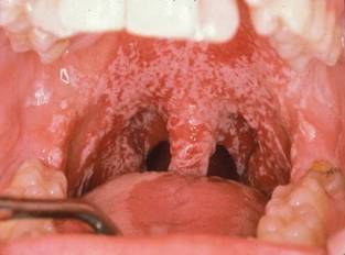
Taste and smell may also be profoundly altered. In the Vanderbilt survey, most patients reported altered taste (68), a decreased desire to eat (68), altered food choices (66), and a decrease in food eaten (66). A change in sense of smell was reported by 69 patients.
Such alterations often result in changes in diet, including decreased consumption of high-fiber food and of vitamins and other nutrients, along with increased consumption of fats, caffeine, and sugar. All of these factors increase the risk for dietary deficiencies, as well as dental caries.
Yet, altered taste sensation is not something patients might think to mention. "Half of patients experience altered taste sensation. But if they think you’re not interested or you don’t ask, you may not know," Dr. Epstein commented.
Periodontal health is often compromised by hyposalivation, which can lead to inflammation, bone/attachment loss, oral infection, and necrosis. Dental demineralization and cavitation may develop as early as 2-3 months after cancer treatment and progress rapidly, leading to fractures of the gum line, tooth loss, and necrosis.
Demineralization appears as a change to white, which may not be recognized as a problem because of the belief that white teeth are healthy. However, recognition at this stage is critical in order to prevent further dental damage, he said.
"The white change near the gum line and the tips of the teeth represent demineralization, and [in] time reversal can be accomplished prior to structural breakdown. Once cavitation has occurred, fillings are needed and prevention must be instituted or the cavities will recur and progress," Dr. Epstein said in an interview.
In the Vanderbilt survey, reported dental problems included difficulty chewing because of teeth/dentures (54 of the 70 patients); tooth sensitivity to hot, cold, or sweet foods (52); teeth feeling looser (51); teeth cracking/chipping (50); and trouble with dentures (24).
Oral candidiasis is another common problem, affecting approximately 39% of head and neck cancer patients during treatment and 33% afterward. One common clinical mistake is prescribing these patients antifungals that contain sugar, such as nystatin. "Nystatin is very high in sugar, and one of the [most commonly] used antifungals. The message is to avoid sugar-sweetened products in dry mouth patients and utilize alternatives," Dr. Epstein said in the interview.
Mucosal sensitivity and pain is also frequent. In a meta-analysis of 22 studies published between 1990 and 2008, the prevalence of trismus was 25.4% in patients who received conventional radiotherapy and 5% for the few intensity-modulated radiation therapy studies that were included, suggesting that the newer radiation modality might diminish the problem (Support. Care Cancer 2010;18:1033-8).
Data suggest that the radiation effect on mandibular movement correlates with the radiation dose to the mastication muscles, with a steep dose-response curve. Onset is typically 2-6 months post treatment and is ongoing. Concurrent chemotherapy may increase the incidence and/or severity of mandibular immobility (Oral Surg. Oral Med. Oral Pathol. Oral Radiol. Endod. 1999;88:365-73).
In the Vanderbilt survey, most patients reported burning in the throat or mouth (69); sensitivity to hot, spicy, or acid food (67); sensitivity to dryness (69); and changes in food intake because of mucosal sensitivity (67); most patients also reported that mucosal sensitivity prevents tooth brushing (63). "Mucosal sensitivity is a quality of life issue," Dr. Epstein said.
The Vanderbilt survey was particularly illuminating, Dr. Epstein commented. Studies that utilize claims data probably underrepresent the problem of long-term oral complications because dental and medical insurance are separate and the data are not easily combined, he added.
"Late oral effects are best diagnosed [and] managed in a multidisciplinary team with close communication between medical and dental providers. ... We really need to come together."
The Multinational Association of Supportive Care in Cancer is developing tools to improve communication between dentistry and medicine. These could be available for beta testing as early as this fall.
Dr. Epstein said he had no relevant financial disclosures.
PHOENIX – Late oral effects of head and neck cancer therapy are "multiple, underreported, and underappreciated."
That is the perspective of Joel Epstein, D.M.D., who has worked extensively with head and neck cancer patients experiencing severe dental and other oral problems following radiation therapy.
"The acute complications of head and neck cancer therapy are pretty well known, but the late complications are underappreciated," Dr. Epstein, director of oral medicine at City of Hope National Medical Center, Duarte, Calif., told attendees at the symposium.
As head and neck cancer treatments have advanced and patients are living longer, the spectrum of treatment complications has shifted, he explained. In a 5-year, prospective longitudinal study of 122 patients with oral carcinoma, dry mouth, sticky saliva, speech changes, dental problems, and sleep disturbance were reported by all patients except those treated only with surgery. These complications persisted at 1 and 5 years and affected quality of life (Head Neck 2008;30:461-70).
According to Dr. Epstein, the data illustrate the need for better collaboration between oncologists and dentists. "While people discuss the concept of multidisciplinary [and] interdisciplinary teams for the benefit of our patients, it is unfortunate that dentistry developed separately from physicians and surgeons. So while we need to interact, we’re not really well prepared to do so, particularly in the community," he said.
Clinically, it’s important to evaluate oral care, including brushing, flossing, fluoride, and tobacco abstinence, at all head and neck cancer treatment follow-up visits. Patients should be assessed for xerostomia, speech, swallowing, mucosal sensitivity, and taste. Head and neck and oral exams should include assessments for saliva (wet mucosa), exposed bone, infection, and new lesions or recurrent cancer, and a dental exam (for plaque, caries, and periodontal health), Dr. Epstein recommended.
Dry mouth, in particular, can lead to a host of other chronic problems related to swallowing, eating, sleeping, and dental health. When the 50-item Vanderbilt Head and Neck Symptom Survey was administered to a total of 70 patients, 67 reported having dry mouth at more than 6 months’ follow up (Head Neck 2011 Aug. 24 [doi:10.1002/hed.21816]).
The majority reported that dry mouth makes chewing/swallowing difficult (65) and that it affects their ability to sleep (67) and talk (64). With regard to eating and swallowing, similar majorities reported trouble eating solids (67) and drinking liquids (68), with food getting stuck in their mouth (66) and throat (67).
And, of concern, the same numbers of patients reported the sensation of choking or strangling on solids (66) and liquids (68). "The impact on function from the lack of saliva and the change in quality of saliva are issues we need to be more ready and willing to address," Dr. Epstein commented.
Taste and smell may also be profoundly altered. In the Vanderbilt survey, most patients reported altered taste (68), a decreased desire to eat (68), altered food choices (66), and a decrease in food eaten (66). A change in sense of smell was reported by 69 patients.
Such alterations often result in changes in diet, including decreased consumption of high-fiber food and of vitamins and other nutrients, along with increased consumption of fats, caffeine, and sugar. All of these factors increase the risk for dietary deficiencies, as well as dental caries.
Yet, altered taste sensation is not something patients might think to mention. "Half of patients experience altered taste sensation. But if they think you’re not interested or you don’t ask, you may not know," Dr. Epstein commented.
Periodontal health is often compromised by hyposalivation, which can lead to inflammation, bone/attachment loss, oral infection, and necrosis. Dental demineralization and cavitation may develop as early as 2-3 months after cancer treatment and progress rapidly, leading to fractures of the gum line, tooth loss, and necrosis.
Demineralization appears as a change to white, which may not be recognized as a problem because of the belief that white teeth are healthy. However, recognition at this stage is critical in order to prevent further dental damage, he said.
"The white change near the gum line and the tips of the teeth represent demineralization, and [in] time reversal can be accomplished prior to structural breakdown. Once cavitation has occurred, fillings are needed and prevention must be instituted or the cavities will recur and progress," Dr. Epstein said in an interview.
In the Vanderbilt survey, reported dental problems included difficulty chewing because of teeth/dentures (54 of the 70 patients); tooth sensitivity to hot, cold, or sweet foods (52); teeth feeling looser (51); teeth cracking/chipping (50); and trouble with dentures (24).
Oral candidiasis is another common problem, affecting approximately 39% of head and neck cancer patients during treatment and 33% afterward. One common clinical mistake is prescribing these patients antifungals that contain sugar, such as nystatin. "Nystatin is very high in sugar, and one of the [most commonly] used antifungals. The message is to avoid sugar-sweetened products in dry mouth patients and utilize alternatives," Dr. Epstein said in the interview.
Mucosal sensitivity and pain is also frequent. In a meta-analysis of 22 studies published between 1990 and 2008, the prevalence of trismus was 25.4% in patients who received conventional radiotherapy and 5% for the few intensity-modulated radiation therapy studies that were included, suggesting that the newer radiation modality might diminish the problem (Support. Care Cancer 2010;18:1033-8).
Data suggest that the radiation effect on mandibular movement correlates with the radiation dose to the mastication muscles, with a steep dose-response curve. Onset is typically 2-6 months post treatment and is ongoing. Concurrent chemotherapy may increase the incidence and/or severity of mandibular immobility (Oral Surg. Oral Med. Oral Pathol. Oral Radiol. Endod. 1999;88:365-73).
In the Vanderbilt survey, most patients reported burning in the throat or mouth (69); sensitivity to hot, spicy, or acid food (67); sensitivity to dryness (69); and changes in food intake because of mucosal sensitivity (67); most patients also reported that mucosal sensitivity prevents tooth brushing (63). "Mucosal sensitivity is a quality of life issue," Dr. Epstein said.
The Vanderbilt survey was particularly illuminating, Dr. Epstein commented. Studies that utilize claims data probably underrepresent the problem of long-term oral complications because dental and medical insurance are separate and the data are not easily combined, he added.
"Late oral effects are best diagnosed [and] managed in a multidisciplinary team with close communication between medical and dental providers. ... We really need to come together."
The Multinational Association of Supportive Care in Cancer is developing tools to improve communication between dentistry and medicine. These could be available for beta testing as early as this fall.
Dr. Epstein said he had no relevant financial disclosures.
EXPERT OPINION FROM A HEAD AND NECK CANCER SYMPOSIUM SPONSORED BY THE AMERICAN SOCIETY FOR RADIATION ONCOLOGY