User login
FDA issues new pregnancy/lactation drug label standards
The U.S. Food and Drug Administration has issued a final rule requiring content and format changes to pregnancy and lactation labeling information for prescription drugs and biologic products.
The long-awaited “Content and Format of Labeling for Human Prescription Drug and Biological Products; Requirements for Pregnancy and Lactation Labeling, or the Pregnancy and Lactation Labeling Rule”(PLLR) is part of broad effort by the FDA to improve the content and format of prescription drug labeling. The PLLR, which finalizes many of the provisions in a proposed rule issued in May 2008 after input from numerous stakeholders, calls for replacement of the current A, B, C, D, and X drug classification system with more detailed information about the risks and benefits of use during pregnancy and breastfeeding.
The rule will take effect June 30, 2015.
Under the PLLR, labels will be required to include three detailed subsections entitled Pregnancy, Lactation, and Females and Males of Reproductive Potential. Each will include a risk summary, a discussion of the supporting data, and relevant information to help providers make prescribing and counseling decisions, according to the FDA. If no data are available to guide decision making, this must be stated.
The Pregnancy subsection combines the existing Pregnancy and Labor and Delivery subsections, and will address use of the drug during pregnancy as well as provide information about relevant registries that collect and maintain data on the use of the product in pregnant women. The Lactation subsection replaces the existing Nursing Mothers subsection, and will include information about use of the product during breastfeeding, including the amount of drug in breast milk and potential effects on the breastfed child. The new Females and Males of Reproductive Potential subsection will address pregnancy testing, contraception, and fertility issues as they relate to use of the product.
The existing A, B, C, D, and X categories were frequently misinterpreted as a grading system, giving an over simplified view of product risk, according to Dr. Sandra Kweder, deputy director of the Office of New Drugs in the FDA’s Center for Drug Evaluation and Research.
The new, more detailed approach to labeling will better address the complex risk-benefit considerations inherent in prescribing decisions during pregnancy and lactation, she said during a press briefing.
“I’m excited because clinicians will, going forward, be able to rely on FDA-approved drug labeling for comprehensive, chronically relevant, and user-friendly information in this part of labeling – something that has been missing for many years,” she said, noting that the changes are particularly important, given that the more than 6 million women who become pregnant each year in the United States take an average of 3-5 different prescription products during the course of their pregnancy and while breastfeeding.
“It is our hope that this new system will help their health care professionals and these women as they discuss treatment options,” she said.
Importantly, the PLLR ensures that more robust and informative data about drugs will be provided than ever before – and in a manner that speaks directly to the concerns that are common among providers, she said.
In addition to the elimination of the letter categories and the addition of the three new subsections, the use of standardized risk statement also was eliminated, as these had the same limitations as the letter categories. A section on inadvertent exposure also was eliminated due to redundancy, as the risk would be the same as with intentional exposure.
The rule also requires that labels be updated as they become outdated.
Much of the information that will be included on the new labels, which will be phased in for existing drugs and required immediately for drugs approved after June 30, 2015, was already included, but was scattered and difficult to find. The new formatting requirements provides for consistency across labels by pulling this information together in one place, Dr. Kweder said.
In an official statement, the American College of Obstetricians and Gynecologists applauded the rule for “taking needed steps to increase understanding about the effect of prescription medicine on women during pregnancy and lactation.”
“The FDA’s updated method of presenting information about both risk and benefit will improve the ability of all physicians to treat their pregnant and breastfeeding patients, as well as women who may become pregnant. It will also help more women to understand and take part in their health care decision making,” according to the ACOG statement, which also noted that the organization hopes the new content on prescription drug and biological product labels will “provide added incentives for clinical research as well as participation in patient registries.”
Christina Chambers, Ph.D., professor of pediatrics and director of clinical research for the department of pediatrics at the University of California, San Diego, also praised the new labeling rule, noting in an interview that “the final rule has been long awaited by many who work in the field of counseling pregnant and breastfeeding women about risks and safety of prescription medications, such as counselors with organizations like MotherToBaby, a service of the Organization of Teratology Information Specialists, which provides information about medication and other exposures during pregnancy and breastfeeding, and which was involved in development of the final rule.
“The MotherToBaby counselors located throughout the United States who answer questions about medication exposures for hundreds of women every day, have struggled for years with trying to explain the not-so-useful A, B, C, D, X pregnancy categories to patients and providers alike who commonly misinterpret their meaning. The new label format is much more content rich and evidence-based, and encompasses the larger picture of the safety data in the context of treatment (or lack of treatment) of the maternal condition. This is a huge step forward – and will make even more clear how critical the need is for more human pregnancy data for all medications likely to be used by women of reproductive age,” she said.
Dr. Chambers is the program director for MotherToBaby California, and director of the MotherToBaby research center at the University of California, San Diego. She reported having no relevant disclosures.

VIDEO: Family physicians can fill rural maternity care gaps
NEW YORK– Rather than relying on more obstetricians to practice in rural settings with limited access to maternity care, family physicians should be trusted to provide “excellent, quality care” to expectant mothers living in less populated areas – including delivering babies by cesarean section.
That’s the recommendation of Dr. Richard A. Young, director of research in family medicine at John Peters Smith Hospital, Fort Worth, Tex.
In a video interview at the annual meeting of the North American Primary Care Research Group, Dr. Young talked about the role family physicians can play in providing quality obstetrical care in underserved areas, and how they can collaborate with local obstetricians to ensure quality care even in complex cases.
The video associated with this article is no longer available on this site. Please view all of our videos on the MDedge YouTube channel
On Twitter @whitneymcknight
AT NAPCRG 2014

Exclusively breastfed neonate weight loss graphed
Losing more than 10% of birthweight early in the postnatal course is common among neonates delivered vaginally and by cesarean section who have been exclusively breastfed, nomograms developed by a team of researchers show. Those graphical depictions of hourly weight loss “may inform clinical care,” Dr. Valerie J. Flaherman and her colleagues reported in Pediatrics.
“The availability of detailed data on weight and feeding for a large cohort allows this study to present the first graphical depiction of hourly weight loss for exclusively breastfed newborns from a large, diverse population,” wrote Dr. Flaherman of the University of California, San Francisco, and her colleagues. “Our curves demonstrate that expected weight loss differs substantially by method of delivery and that this difference persists over time” (Pediatrics 2014 Dec. 1 [doi:10.1542/peds.2014-1532]).

Using available weight data from the births of 108,907 singleton neonates born at 36 weeks’ gestation or later at Northern California Kaiser Permanente hospitals between 2009 and 2013, the researchers created nomograms that estimate percentiles of weight loss among exclusively breastfed infants who were vaginally delivery or delivered by cesarean section.
Dr. Flaherman’s team estimated 50th, 75th, 90th, and 95th percentiles based on analysis of 83,433 vaginally born neonates and 25,474 infants delivered by C-section, all exclusively breastfed. The study excluded infants who received any formula, those weighing less than 2,000 g or more than 5,000 g, and those whose weight, delivery mode, or feeding status was unknown.
Median weight loss for vaginally delivered newborns was 4.2%, 7.1%, and 6.4% at 24, 48, and 72 hours after birth, respectively. Among cesarean section–born newborns, median percent weight loss was 4.9%, 8%, 8.6%, and 5.8% at 24, 48, 72, and 96 hours after birth.
Nearly 5% of vaginally delivered newborns and nearly 10% of newborns delivered by cesarean section had lost at least 10% of their birthweight at 48 hours after birth. Percentile curves began to rise shortly after 48 hours for vaginally delivered newborns and by 72 hours after birth for cesarean section–delivered newborns, although more than 25% of the latter remained at least 10% below their birthweight by the third day.
The research was funded by the U.S. Department of Health & Human Services and the National Institute of Child Health and Human Development. The authors reported no disclosures.
“A major focus of contemporary care during the birth hospitalization is the management of breastfed infants. Although much effort is centered on the provision of optimal instruction and support to mothers initiating breastfeeding, an equally important goal is to appropriately diagnose and manage newborns with breastfeeding difficulties,” Dr. James A. Taylor and Dr. Elizabeth A. Simpson wrote in an accompanying editorial (Pediatrics 2014 Dec. 1 [doi:10.1542/peds.2014-3354]).“The results of the study by Flaherman et al in this issue of Pediatrics provide much needed data for both of these efforts.”
Dr. Flaherman and her colleagues have “normalized weight loss in breastfed infants. The data can be used to allay the anxiety of a new mother. ... More important, using the nomograms, neonates with significant breastfeeding problems may be identified because their weight loss is ≥ 95th percentile for age or because the trajectory of weight loss is atypical.”
Dr. Taylor is at the department of pediatrics at University of Washington in Seattle and Dr. Simpson is at the department of pediatrics at Children’s Mercy Hospital in Kansas City, Mo. Both authors reported no disclosures.
Losing more than 10% of birthweight early in the postnatal course is common among neonates delivered vaginally and by cesarean section who have been exclusively breastfed, nomograms developed by a team of researchers show. Those graphical depictions of hourly weight loss “may inform clinical care,” Dr. Valerie J. Flaherman and her colleagues reported in Pediatrics.
“The availability of detailed data on weight and feeding for a large cohort allows this study to present the first graphical depiction of hourly weight loss for exclusively breastfed newborns from a large, diverse population,” wrote Dr. Flaherman of the University of California, San Francisco, and her colleagues. “Our curves demonstrate that expected weight loss differs substantially by method of delivery and that this difference persists over time” (Pediatrics 2014 Dec. 1 [doi:10.1542/peds.2014-1532]).
Using available weight data from the births of 108,907 singleton neonates born at 36 weeks’ gestation or later at Northern California Kaiser Permanente hospitals between 2009 and 2013, the researchers created nomograms that estimate percentiles of weight loss among exclusively breastfed infants who were vaginally delivery or delivered by cesarean section.
Dr. Flaherman’s team estimated 50th, 75th, 90th, and 95th percentiles based on analysis of 83,433 vaginally born neonates and 25,474 infants delivered by C-section, all exclusively breastfed. The study excluded infants who received any formula, those weighing less than 2,000 g or more than 5,000 g, and those whose weight, delivery mode, or feeding status was unknown.
Median weight loss for vaginally delivered newborns was 4.2%, 7.1%, and 6.4% at 24, 48, and 72 hours after birth, respectively. Among cesarean section–born newborns, median percent weight loss was 4.9%, 8%, 8.6%, and 5.8% at 24, 48, 72, and 96 hours after birth.
Nearly 5% of vaginally delivered newborns and nearly 10% of newborns delivered by cesarean section had lost at least 10% of their birthweight at 48 hours after birth. Percentile curves began to rise shortly after 48 hours for vaginally delivered newborns and by 72 hours after birth for cesarean section–delivered newborns, although more than 25% of the latter remained at least 10% below their birthweight by the third day.
The research was funded by the U.S. Department of Health & Human Services and the National Institute of Child Health and Human Development. The authors reported no disclosures.
FROM PEDIATRICS
Key clinical point: Newborn weight loss of at least 10% of birth weight is common in exclusively breastfed infants.
Major finding: Nearly 5% of vaginally delivered newborns and more than 10% of cesarean section–born newborns lost at least 10% of their birth weight 48 hours after birth.
Data source: Prospective cohort study of 161,471 term, singleton neonates born ≥ 36 weeks gestation at Northern California Kaiser Permanente hospitals in 2009-2013.
Disclosures: The research was funded by the U.S. Department of Health & Human Services and the National Institute of Child Health and Human Development. The authors reported no disclosures.
Large study shows no link between celiac disease and fertility problems
With the exception of those diagnosed between the age of 25 and 29 years, women with celiac disease are no more likely than are women without celiac disease to have fertility problems, according to findings from a large population-based study in the United Kingdom.
Of more than 2.4 million women with prospective primary care records available during their childbearing years (ages 15-49 years) between 1990 and 2013, 6,506 were diagnosed with celiac disease. The women with celiac disease had a similar rate of recorded fertility problems as did those without celiac disease (4.4% vs. 4.1%), Nafeesa N. Dhalwani of the University of Nottingham and City Hospital Nottingham, U.K., and colleagues reported in the December issue of Gastroenterology (doi:10.1053/j.gastro.2014.08.025).
Source: American Gastroenterological Association
Further, the rates of infertility in those with celiac disease were similar to those without celiac disease both before and after diagnosis except in those aged 25-29 years at the time of diagnosis; the rates in those women were 41% higher, compared with those without celiac disease who were the same age (incidence rate ratio, 1.41), the investigators said.
“However, the absolute excess risk [for those diagnosed at age 25-29 years] was only 0.5% (5.2/1,000 person-years), they said.
Women included in the analysis were identified from the Health Improvement Network database. Rates of new clinically recorded fertility problems among those with and without diagnosed celiac disease were stratified by timing of celiac disease diagnosis after adjustment for sociodemographics, comorbidities, and calendar year.
The findings contrast with those from a number of smaller studies that demonstrated an association between infertility and celiac disease, but those studies included small numbers of women, including many who were receiving infertility specialist services, the investigators said, explaining that the women may not have been representative of the general population, and that other small studies found no link between celiac disease and fertility problems.
Celiac disease affects about 1% of the population in North America and Western Europe, and between 60% and 70% of those who are diagnosed are women. Several mechanisms through which celiac disease might affect a woman’s fertility have been described in the literature, but no conclusive evidence exists to support them, they noted.
Despite this lack of evidence and the inconsistent findings from small studies, a number of reviews include infertility as a key nongastrointestinal manifestation of celiac disease. The current findings suggest that most women with celiac disease – whether diagnosed or undiagnosed – do not have a substantially greater likelihood of having clinically recorded fertility problems than do those without celiac disease.
“Therefore, screening when women initially present with fertility problems may not identify a significant number of women with celiac disease, beyond the general population prevalence. This may not always apply to subgroups of women with severe celiac disease. However, in terms of the clinical burden of fertility problems at a population level, these findings should be reassuring for women with celiac disease and all stakeholders involved in their care,” the investigators concluded.
This study was supported by CORE/Coeliac UK, and by a University of Nottingham/Nottingham University Hospitals National Health Service Trust Senior Clinical Research Fellowship. The authors reported having no disclosures.
Key clinical point: Women with celiac disease are not at increased risk of fertility problems.
Major finding: Women with celiac disease had a similar rate of recorded fertility problems as did those without celiac disease (4.4% vs. 4.1%).
Data source: A population-based study of more than 2.4 million women.
Disclosures: This study was supported by CORE/Coeliac UK, and by a University of Nottingham/Nottingham University Hospitals National Health Service Trust Senior Clinical Research Fellowship. The authors reported having no disclosures.
Link between early exposure to acetaminophen and childhood asthma found weak, overstated
The reported link between early life exposure to acetaminophen and the development of asthma in children is “weak” and “overstated” based on currently available evidence, according to a report published by the Archives of Disease in Childhood.
In a review of currently available data culled from Embase and PubMed databases, 1,192 relevant studies conducted between 1967 and 2013 were analyzed, of which 11 were included for analysis. Of these 11 studies, 5 found “increased odds” that exposure to acetaminophen during the first trimester of pregnancy could lead to development of asthma (pooled odds ratio, 1.39); however, there was a high degree of between-study heterogeneity among the trials (I2 = 64.2%, P = .03), reported Dr. M. Cheelo of the University of Melbourne, and associates.

Of those five, only two studies examined the effects of acetaminophen exposure during the second trimester, but attained widely disparate results: Study one reported an OR of 1.06, while the other reported an OR of 2.15, with I2 = 80%. Two studies also tested acetaminophen exposure during the third trimester and found a “weak association,” with a pooled OR of 1.17. Three studies look at acetaminophen exposure through an entire pregnancy, but all had “significant heterogeneity” in their findings (OR = 1.65, 1.22, and 0.74; I2 = 89%). Only one study that was examined adjusted for respiratory tract infections during pregnancy, but according to the authors, “all studies that adjusted for early life respiratory tract infections found a reduction in the association between [acetaminophen] exposure and subsequent childhood asthma” (Arch. Dis. Child. 2014 [doi:10.1136/archdischild-2012-303043]).
The other 6 of the 11 total studies examined acetaminophen exposure over the first 2 years of life. Three of these studies found a “weak positive association,” as did four studies directly comparing children with and without acetaminophen exposure. All but one study adjusted results for respiratory tract infections during pregnancy, which caused a “moderate attenuation of the association between frequency of [acetaminophen] intake and childhood asthma.” Consequently, investigators concluded that “evidence of an association between early life [acetaminophen] and asthma is often overstated, and there is currently insufficient evidence to support changing guidelines in the use of this medicine.”
The authors reported no relevant financial conflicts of interest.
The reported link between early life exposure to acetaminophen and the development of asthma in children is “weak” and “overstated” based on currently available evidence, according to a report published by the Archives of Disease in Childhood.
In a review of currently available data culled from Embase and PubMed databases, 1,192 relevant studies conducted between 1967 and 2013 were analyzed, of which 11 were included for analysis. Of these 11 studies, 5 found “increased odds” that exposure to acetaminophen during the first trimester of pregnancy could lead to development of asthma (pooled odds ratio, 1.39); however, there was a high degree of between-study heterogeneity among the trials (I2 = 64.2%, P = .03), reported Dr. M. Cheelo of the University of Melbourne, and associates.
Of those five, only two studies examined the effects of acetaminophen exposure during the second trimester, but attained widely disparate results: Study one reported an OR of 1.06, while the other reported an OR of 2.15, with I2 = 80%. Two studies also tested acetaminophen exposure during the third trimester and found a “weak association,” with a pooled OR of 1.17. Three studies look at acetaminophen exposure through an entire pregnancy, but all had “significant heterogeneity” in their findings (OR = 1.65, 1.22, and 0.74; I2 = 89%). Only one study that was examined adjusted for respiratory tract infections during pregnancy, but according to the authors, “all studies that adjusted for early life respiratory tract infections found a reduction in the association between [acetaminophen] exposure and subsequent childhood asthma” (Arch. Dis. Child. 2014 [doi:10.1136/archdischild-2012-303043]).
The other 6 of the 11 total studies examined acetaminophen exposure over the first 2 years of life. Three of these studies found a “weak positive association,” as did four studies directly comparing children with and without acetaminophen exposure. All but one study adjusted results for respiratory tract infections during pregnancy, which caused a “moderate attenuation of the association between frequency of [acetaminophen] intake and childhood asthma.” Consequently, investigators concluded that “evidence of an association between early life [acetaminophen] and asthma is often overstated, and there is currently insufficient evidence to support changing guidelines in the use of this medicine.”
The authors reported no relevant financial conflicts of interest.
FROM THE ARCHIVES OF DISEASE IN CHILDHOOD
Key clinical point: Current evidence regarding purported link between early life exposure to acetaminophen and development of childhood asthma is weak and often overstated.
Major finding: Meta-analysis of 11 studies found disparate results linking acetaminophen exposure and childhood asthma, and these results generally were attenuated when adjusted for data related to respiratory tract infections during pregnancy and subsequent childhood asthma.
Data source: Meta-analysis of 11 observational cohort studies.
Disclosures: The authors reported no relevant financial conflicts of interest.

Best practices for the surgical management of adnexal masses in pregnancy
During the 43rd AAGL Global Congress, held November 17–21 in Vancouver, British Columbia, Sarah L. Cohen, MD, MPH, of Brigham and Women’s Hospital in Boston, Massachusetts, stepped attendees through diagnosis and surgical management of adnexal masses in pregnancy, noting the approaches backed by the highest-quality data.
The incidence of adnexal masses in pregnancy is 1 in every 600 live births. A mass can be benign or malignant. Among benign masses found in pregnancy are functional cysts, teratomas, and the corpus luteum.
Work-up
Ultrasound imaging is a valuable component of the work-up, owing to its risk-free nature. Magnetic resonance imaging may be appropriate in selected cases, but gadolinium contrast should be avoided.
In pregnancy, the aim is to limit ionizing radiation to less than 5 to 10 rads to minimize the risk of childhood malignancy/leukemia, with no single imaging study exceeding 5 rads.
Tumor markers may be helpful, but careful interpretation is critical, taking into account the effects of pregnancy itself on CA-125 (which peaks in the first trimester), human chorionic gonadotropin, alpha fetoprotein, inhibin A, and lactate dehydrogenase.
When expectant management may be appropriate
Watchful waiting may be considered for simple cysts less than 6 cm in size, provided the patient is asymptomatic with no signs of malignancy.
Surgery is indicated when the patient is symptomatic, when there is a concern for malignancy, and when a persistent mass exceeds 10 cm in size.
As always, elective surgery is preferable, as emergent surgery in pregnancy is associated with a risk of preterm labor of 22% to 35%.
Optimal timing of surgery
Surgery can be performed safely in any trimester, provided the gynecologist is aware of special concerns. For example, in the first trimester, organogenesis is under way and the corpus luteum is still present. If the corpus luteum is removed, progesterone supplementation is necessary.
When surgery can be postponed to the second trimester, it allows time for possible resolution of the mass.
Mode of surgery
Laparoscopy allows for faster recovery, less pain (and, therefore, lower narcotic exposure to the fetus), and improved maternal ventilation.
Prophylaxis for venous thromboembolism is indicated through the use of pneumatic compression devices and, when appropriate, heparin.
Initial port placement can be performed using a Hassan technique, Veress needle, or optical trocar.
Insufflation pressures of 10 to 15 mm Hg are safe, with intraoperative monitoring of carbon dioxide.
Availability of guidelines
Surgeons should make use of guidelines, when feasible, to guide surgery. For example, the Society of American Gastrointestinal and Endoscopic Surgeons (SAGES) publishes guidelines on surgery during pregnancy. The American College of Obstetricians and Gynecologists also offers guidelines.
Share your thoughts on this article! Send your Letter to the Editor to [email protected]. Please include your name and the city and state in which you practice.
Topiramate
At Motherisk, we receive increasing numbers of queries from women and clinicians about the use of topiramate during pregnancy, mostly related to its use for migraine or seizures.Topiramate is approved for treatment of seizures and for migraine prevention, and in 2012, the Food and Drug Administration approved the combination of extended-release topiramate with the stimulant phentermine (Qsymia) as a chronic treatment for weight management.
When topiramate was initially approved in 1996, human reproductive data were scarce, but animal data suggested that a high dose of topiramate in rats and rabbits may induce some congenital malformations. Since that time, quite a few studies – but not all – have suggested that topiramate may be associated with an increased risk of oral clefts (cleft lip and cleft palate). Studies that have found an increased risk associated with first trimester exposure to topiramate include the North American AED Pregnancy Registry, which found a rate of 14 cases per 1,000 – more than tenfold greater than the rate in the general population.
For seizures and migraines, the FDA has labeled topiramate a pregnancy category D drug (there is evidence of human fetal risk, but “the potential benefits from the use of the drug in pregnant women may be acceptable despite its potential risks.” ) For Qsymia, however, topiramate is contraindicated in pregnancy and is a pregnancy category X, because its use “can cause fetal harm and weight loss offers no potential benefit to a pregnant woman,” the labeling states.
Based on arecent analysis of six controlled studies in the literature of more than 3,000 pregnancies exposed during the first trimester, we determinedthat the risk of oral clefts associated with first trimester exposure was increased by sixfold over controls. To put this into context, in the general population, oral clefts occur in less than 1% (0.07%), whereas in the studies, the rate was 0.36%, about a fivefold increase.
Until the approval of the weight loss indication, women of reproductive age with epilepsy or migraines prescribed topiramate were a relatively small group. But because obesity is so common, we are now in a situation where a drug that is likely a human teratogen, based on strong evidence, will be used by far more women of childbearing age, and an increase in unintended pregnancies exposed to the drug probably will occur. In phase III clinical trials of Qsymia, quite a few women got pregnant, which is not surprising since women who are obese may have more difficulty knowing they are pregnant, for hormonal and other reasons.
Clinicians and women who take the drug for weight loss need to be aware of this risk. It is important to counsel women of childbearing age who are on this medication about the need for contraception during treatment, and to have a pregnancy test before treatment, and every month during treatment. An important factor to keep in mind is that the maximum topiramate dose for weight loss is 92 mg a day, while the typical epilepsy dose is 200-400 mg a day, and for migraines, is 100 mg a day. It may turn out that the same risk may not be evident with the lower dose.
As the labeling states, women who become pregnant while on this drug should stop taking it immediately, and clinicians should counsel them about the possible risks to the fetus. Health care providers and patients should report pregnancies exposed to Qsymia to the Qsymia Pregnancy Surveillance Program, which is monitoring maternal-fetal outcomes of exposed pregnancies, at 888-998-4887, or the FDA’s MedWatch program at 800-332-1088.
Dr. Koren is professor of pediatrics, pharmacology, pharmacy, medicine, and medical genetics at the University of Toronto. He heads the Research Leadership for Better Pharmacotherapy During Pregnancy and Lactation at the Hospital for Sick Children, Toronto, where he is director of the Motherisk Program. He was a consultant to Vivus, the manufacturer of Qsymia. E-mail him at [email protected].

Dr. Robert L. Barbieri’s Editor’s Picks November 2014
Editor in Chief Robert L. Barbieri, MD, provides an overview of three articles appearing in OBG Management’s November 2014 issue. Listen to his take on why these articles are of particular importance to women’s health professionals.
Access all of the articles in the November 2014 issue here.
Editor in Chief Robert L. Barbieri, MD, provides an overview of three articles appearing in OBG Management’s November 2014 issue. Listen to his take on why these articles are of particular importance to women’s health professionals.
Access all of the articles in the November 2014 issue here.
Editor in Chief Robert L. Barbieri, MD, provides an overview of three articles appearing in OBG Management’s November 2014 issue. Listen to his take on why these articles are of particular importance to women’s health professionals.
Access all of the articles in the November 2014 issue here.

Kidney donors at greater risk of preeclampsia, gestational hypertension
Women who donate a kidney are almost two and a half times more likely than are nondonors to have preeclampsia or gestational hypertension in pregnancy, according to a study presented at Kidney Week 2014 and published online simultaneously in the New England Journal of Medicine.
“Information on this potential risk should be included in clinical practice guidelines, shared in the informed-consent processes for potential donors and their recipients when a woman has reproductive potential, and used to guide the care of pregnant donors,” wrote the study authors, led by Dr. Amit X. Garg at the London Kidney Research Unit in London, Ont. (N. Engl. J. Med. 2014 Nov. 14 [doi:10.1056/NEJMoa1408932]).
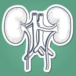
The Canadian retrospective study matched 85 living kidney donors in a 1:6 ratio with 510 healthy nondonors and followed them for almost 11 years. During this time, 131 pregnancies occurred in the donor group and 788 in the nondonor group.
Gestational hypertension or preeclampsia was diagnosed in 15 donors and 38 nondonors (11% vs. 5%, odds ratio for donors, 2.4; 95% confidence interval, 1.2 to 5.0; P = .01), the investigators reported.
No significant differences were observed between groups for other maternal or fetal outcomes, and there were no maternal or perinatal deaths in the study that was part of the Donor Nephrectomy Outcomes Research Network (DONOR).
However, they noted that the study included limitations, such as not recording body mass index, medication use, or the race of study participants.
Confidence intervals for risk estimates also were wide, and physicians used clinical judgment when applying accepted diagnostic criteria for gestational hypertension and preeclampsia.
“It remains possible that gestational hypertension and preeclampsia were more likely to be diagnosed and recorded among donors than nondonors despite similar clinical presentations in two groups,” the investigators wrote.
“There may be a role for government programs to cover the costs of recommended pregnancy care for donors who lack health insurance, including any costs related to the treatment of hypertension,” they added.
The meeting was sponsored by the American Society of Nephrology. The study was supported by a grant from the Canadian Institute of Health Research as well as several other research institutions. Dr. Garg received grants from Astellas and Roche outside this study. Several other authors received grants from a number companies outside this study, while the remainder of the authors had no relevant disclosures.
Women who donate a kidney are almost two and a half times more likely than are nondonors to have preeclampsia or gestational hypertension in pregnancy, according to a study presented at Kidney Week 2014 and published online simultaneously in the New England Journal of Medicine.
“Information on this potential risk should be included in clinical practice guidelines, shared in the informed-consent processes for potential donors and their recipients when a woman has reproductive potential, and used to guide the care of pregnant donors,” wrote the study authors, led by Dr. Amit X. Garg at the London Kidney Research Unit in London, Ont. (N. Engl. J. Med. 2014 Nov. 14 [doi:10.1056/NEJMoa1408932]).
The Canadian retrospective study matched 85 living kidney donors in a 1:6 ratio with 510 healthy nondonors and followed them for almost 11 years. During this time, 131 pregnancies occurred in the donor group and 788 in the nondonor group.
Gestational hypertension or preeclampsia was diagnosed in 15 donors and 38 nondonors (11% vs. 5%, odds ratio for donors, 2.4; 95% confidence interval, 1.2 to 5.0; P = .01), the investigators reported.
No significant differences were observed between groups for other maternal or fetal outcomes, and there were no maternal or perinatal deaths in the study that was part of the Donor Nephrectomy Outcomes Research Network (DONOR).
However, they noted that the study included limitations, such as not recording body mass index, medication use, or the race of study participants.
Confidence intervals for risk estimates also were wide, and physicians used clinical judgment when applying accepted diagnostic criteria for gestational hypertension and preeclampsia.
“It remains possible that gestational hypertension and preeclampsia were more likely to be diagnosed and recorded among donors than nondonors despite similar clinical presentations in two groups,” the investigators wrote.
“There may be a role for government programs to cover the costs of recommended pregnancy care for donors who lack health insurance, including any costs related to the treatment of hypertension,” they added.
The meeting was sponsored by the American Society of Nephrology. The study was supported by a grant from the Canadian Institute of Health Research as well as several other research institutions. Dr. Garg received grants from Astellas and Roche outside this study. Several other authors received grants from a number companies outside this study, while the remainder of the authors had no relevant disclosures.
FROM KIDNEY WEEK 2014
Key clinical point: Information on an increased risk for preeclampsia and gestational hypertension should be included in clinical practice guidelines and in informed-consent processes for potential kidney donors and their recipients.
Major finding: Women who donate a kidney are almost two and a half times more likely than are nondonors to have preeclampsia or gestational hypertension in pregnancy.
Data source: Retrospective cohort study of 85 kidney donors who were matched on a 1:6 ratio with 510 healthy nondonors and followed for a median of 10.9 years.
Disclosures:Dr. Garg received grants from Astellas and Roche outside this study. Several other authors received grants from a number companies outside this study, while the remainder of the authors had no relevant disclosures. The study was supported by a grant from the Canadian Institute of Health Research as well as several other research institutions. The meeting was sponsored by the American Society of Nephrology.
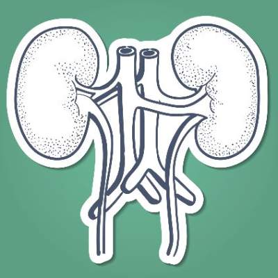
Children born to mothers with RA more likely to be born early
Children born to mothers with rheumatoid arthritis are more likely to be born prematurely and have a lower birth weight than are those born to mothers without the condition, according to a Danish nationwide cohort study.
The reduction in fetal growth, however, is small – a mean difference of 87 g – and unlikely to have any impact on the well being of the child, Ane Rom of Copenhagen University Hospital and her colleagues reported in Arthritis & Rheumatology.
Of 1,917,723 children born in Denmark during 1977-2008, 13,556 children were exposed to either maternal or preclinical RA. Children with mothers who had rheumatoid arthritis (RA) were almost one and half times more likely (odds ratio, 1.48; 95% confidence interval, 1.20-1.84) to be born preterm. The odds of preterm birth were slightly lower, but still elevated, in children with mothers who had preclinical signs of disease, compared with children not exposed to RA (OR, 1.32; 95% CI, 1.07-1.64).
Children exposed to RA had a similar length and head and abdominal circumference at birth, compared with children of mothers without RA, but their birth weight was, on average, 87 g lower (95% CI, –111.23 to –62.84) and their placenta weight was 14 g lower (95% CI, –21.46 to –5.43). The investigators found similar results for children exposed to preclinical RA (Arthritis Rheumatol. 2014 Nov. 13 [doi:10.1002/art.38874]).
Paternal RA was not associated with reduced fetal growth or preterm birth. Maternal RA may affect fetal growth either through fetal programming related to the effect that RA may have on the intrauterine environment, through genetic factors, or through medications taken in pregnancy. “For women with RA, it is reassuring that only a small reduction in fetal growth was found for most of their children, which will have little, if any, impact on perinatal conditions for the child,” Ms. Rom and her associates wrote.
“Clinicians should be aware of the increased risk of preterm birth not only in women diagnosed with RA but also in women with signs of preclinical RA,” they concluded.
Ms. Rom’s work on the study was supported by grants from the National Institutes of Health, the Danish Council for Independent Research, and the Augustinus Foundation. No conflicts of interest were reported.
FROM ARTHRITIS & RHEUMATOLOGY
Key clinical point: Keep aware that pregnant women with RA or signs of preclinical disease may be more likely to give birth preterm and have children with slightly lower birth weight.
Major finding: Children with mothers who had RA were almost one and half times more likely (OR, 1.48; 95% CI, 1.20-1.84) to be born preterm.
Data source: A nationwide cohort study of all singletons born in Denmark during 1977-2008.
Disclosures: Ms. Rom’s work on the study was supported by grants from the National Institutes of Health, the Danish Council for Independent Research, and the Augustinus Foundation. No conflicts of interest were reported.