User login
‘Extremely exciting’ study results guide MM treatment options
CHICAGO – New results from a trial in patients with newly diagnosed multiple myeloma (MM) offer some answers to questions about which treatment route to choose.
Patients who received the triplet of lenalidomide, bortezomib, and dexamethasone (RVD) plus ASCT had a median PFS of 67.5 months, compared with 46.2 months for those who received RVD but did not have a transplant soon after.
However, patients were just as likely to be alive more than 6 years after treatment regardless of whether or not they underwent an immediate stem cell transplant.
In addition, treatment-related adverse events of grade 3 or above were higher in the group that received the transplant immediately after the triplet therapy.
The results were presented during a plenary session at the American Society of Clinical Oncology annual meeting and simultaneously published in the New England Journal of Medicine.
“Our findings confirm the PFS benefit of transplantation as first-line treatment for patients with myeloma and confirms stem cell transplant as a standard of care with certain triplet therapy,” said lead author Paul G. Richardson, MD, professor of medicine, Harvard Medical School, and clinical program leader and director of clinical research at the Jerome Lipper Multiple Myeloma Center at Dana Farber Cancer Institute, Boston.
Another finding from the trial was that the use of maintenance lenalidomide in both groups continuously until progression conferred substantial clinical benefit.
“We can also say that the use of lenalidomide maintenance therapy is also a standard of care,” he added.
Study details
In this trial, Dr. Richardson and colleagues randomly assigned 873 patients newly diagnosed with multiple myeloma to the RVD-alone group (n = 357) or the transplantation group (n = 365). All patients had received one cycle of RVD prior to randomization and then received two additional RVD cycles plus stem-cell mobilization followed by either five additional RVD cycles (the RVD-alone group) or high-dose melphalan plus ASCT followed by two additional RVD cycles (the transplantation group). Lenalidomide was administered to all patients until disease progression, unacceptable side effects, or both.
At a median follow-up of 76.0 months, the risk of disease progression or death was 53% higher among patients who received RVD alone versus the transplantation group (hazard ratio [HR], 1.53; P < .001). The median duration of PFS among patients with a high-risk cytogenetic profile was 55.5 vs. 17.1 months, favoring the transplantation group.
The percentage of patients who were alive without progression at 5 years was 58.4% vs 41.6%, respectively (HR, 1.66) and median duration of response was 56.4 vs 38.9 months, also favoring transplantation (HR, 1.45).
The estimated 5-year overall survival was similar between groups: 80.7% for transplantation and 79.2% for RVD alone (HR for death, 1.10; P > .99). For patients with a high-risk cytogenetic profile, 5-year survival was 63.4% versus 54.3%, respectively.
“This tells us that for patients who had kept transplant in reserve, they had the same overall survival as those who had had a transplant right away, despite there being such impressive initial disease control for the patients in whom transplant was used early,” Dr. Richardson said in a press release from his institution.
Patients who did not undergo immediate transplant received treatment when their disease progressed with newer and active therapies, such as monoclonal antibodies and/or next-generation novel agents, he noted. Only 28% of patients used the reserve option of a transplant.
“It demonstrates the extent to which patients now have options and that we have new data to guide them in balancing the pluses and minuses of each approach,” he added.
When looking at safety, the authors noted that the most common treatment-related adverse events of grade 3 or higher occurred in 279 patients (78.2%) in the RVD-alone group and 344 patients (94.2%) in the transplantation group. Of those patients, 60.5% and 89.9%, respectively, reported hematologic events of grade 3 or higher (P < .001). The 5-year cumulative incidence of invasive second primary cancers was similar in both cohorts (RVD-alone group, 4.9%; transplantation group, 6.5%).
However, while the risk of secondary cancers was similar between groups, Dr. Richardson noted that there was a higher incidence of acute myeloid leukemia and myelodysplastic syndromes in the transplant cohort.
“There was also a significant drop in quality of life across transplant procedures, but the good news is that it was recoverable rapidly,” he said. “What is also really important is that we have prospective, multicenter, national comparative data on toxicity. That’s very important for providing patients with a choice as they move forward with their treatment plan.”
He noted that treatment continues to evolve. “This study was designed in 2009, begun in 2010, and now there is mature data in 2022,” Dr. Richardson said. “This is particularly relevant as we have now further improved the induction treatment for younger patients with newly diagnosed myeloma using quadruplet regimens incorporating monoclonal antibodies and novel next-generation therapies. The results from these studies are extremely exciting.
“Now more than ever, treatment for multiple myeloma can be adapted for each patient,” Dr. Richardson said. “Our study provides important information about the benefits of transplant in the era of highly effective novel therapies and continuous maintenance, as well as the potential risks, to help patients and their physicians decide what approach may be best for them. This is particularly relevant as we have now further improved the induction treatment for younger patients with newly diagnosed myeloma using quadruplet regimens incorporating monoclonal antibodies, such as RVD combined with daratumumab.”
Lack of difference in overall survival
These new results further support an already established role of autologous hematopoietic stem cell transplantation in the management of patients with multiple myeloma, said Samer Al-Homsi, MD, clinical professor of medicine and director of the blood and marrow transplant program at Perlmutter Cancer Center, NYU Langone, New York, who was approached for comment.
“The treatment regimen is applicable to patients who are determined by an expert in transplantation to be fit to receive autologous hematopoietic transplantation,” he added. “Although this study, like many others, establishes hematopoietic stem cell transplantation as part of the standard of care in multiple myeloma, only a fraction of patients are actually offered this important modality of treatment for a variety of reasons, including provider bias,” he noted. “In fact, although improvement in supportive care has enhanced the safety of the procedure, many patients are denied this therapy.”
Dr. Al-Homsi noted that the lack of difference in overall survival might be due to the fact that some patients (28%) in the RVD-alone group did end up undergoing transplantation at the time of progression. “Also, longer follow-up might reveal a difference in overall survival,” he said.
The toxicities are manageable, and the incidence of secondary malignancies was not significantly different between cohorts. “However,” he emphasized, “lenalidomide has been associated in other studies with increased incidence of secondary malignancies and it must be noted that this study used extended administration of lenalidomide until progression.”
Support for this study was provided by grants to the Blood and Marrow Transplant Clinical Trials Network from the National Heart, Lung, and Blood Institute, the National Cancer Institute, R. J. Corman Multiple Myeloma Foundation, Celgene/Bristol Myers Squibb, and Millennium/Takeda Pharmaceutical. Dr. Richardson has reported relationships with Celgene, Janssen, Jazz Pharmaceuticals, Karyopharm Therapeutics, Oncopeptides, Sanofi, Secura Bio, Takeda, and Bristol Myers Squibb. Dr. Al-Homsi has reported no relevant financial relationships.
A version of this article first appeared on Medscape.com.
AT ASCO 2022
Liver transplanted after 3 days outside body
A poor-quality human liver, rejected by all transplant centers, was treated outside the body for 3 days using a perfusion machine that simulated some functions of the human body and has been successfully transplanted into a patient with advanced cirrhosis.
The 62-year-old patient rapidly returned to normal quality of life and at the 1-year follow-up had no signs of liver damage, such as rejection or bile duct injury, according to the report published in Nature Biotechnology.
The study team was led by Pierre-Alain Clavien, MD, PhD, with the department of surgery and transplantation, Swiss Hepato-Pancreato-Biliary and Transplant Center, University Hospital Zürich, and the Wyss Zürich Translational Center, ETH Zürich and University of Zürich.
Expanding the viability window
Livers for transplant are routinely preserved in a static cold solution and implanted within a few hours. Most centers limit the time in the cold solution to 12 hours as the organ’s viability drops quickly after that time.
“This inaugural clinical success opens new horizons in clinical research and promises an extended time window of up to 10 days for assessment of viability of donor organs as well as converting an urgent and highly demanding surgery into an elective procedure,” the authors wrote.
The Liver4Life team, made up of physicians, engineers, and biochemists, developed the complex perfusion machine. Features of the machine, which mimics human body functions, include automated remote control of all key parameters. A pump mimics the heart, an oxygenator replaces the lungs, and a dialysis unit performs as kidneys would. Hormone and nutrient infusions take over the work of the intestines and pancreas. The machine also moves the liver to the rhythm of simulated breathing.
The team had to solve factors that limit viability for any solid organ outside the body over a few hours including hemolysis, hemodynamic stability, glucose control, pathologic glycogen deposition and perfusate quality and dilution.
Additionally, because the organ would be under machine perfusion for several days, the scientists also had to address pressure necrosis.
History behind the procedure
The process started in 2015 with the support of the Wyss Zürich Translational Center, with the goal of long-term ex situ machine perfusion of injured liver grafts.
As part of the agreement from the Swiss regulatory authority (the Federal Office of Public Health) the process would be used only if the organ was rejected by all transplant centers, the recipient had no other options for a donor liver, and if the organ met a rigorous bar for viability.
On May 19, 2021, the team was offered a liver graft from a 29-year-old female donor who had an invasive abdominal desmoid fibromatosis associated with chronic intra-abdominal abscesses and recurrent sepsis episodes from multiresistant bacteria. The donor needed long-term multiple medications and parenteral nutrition. Additionally, there was a 4-cm tumor in segment 1 of the liver.
The liver was refused by all other centers, “primarily because it required diagnostic workup of the liver lesion, which was not immediately possible, and because of the ongoing sepsis in the donor with multiresistant microorganisms,” the authors wrote.
The team removed the liver, and the graft was connected to the Wyss perfusion device for normothermic (37 °C) ex situ perfusion after 4 hours of cold preservation.
A 62-year-old male potential recipient on the official national transplant list, had earlier agreed to be considered for receiving a graft preserved ex situ in the Wyss machine.
The patient was fully informed about the process and the presence of a benign lesion in the graft and accepted the transplantation procedure. The patient had advanced cirrhosis, severe portal hypertension, and multiple and recurrent hepatocellular carcinoma (HCC).
Recipient had ‘near-zero’ chance to get a liver in time
The authors wrote that the patient had “a near-zero chance to receive a graft in time.”
For patients with HCC in Switzerland, the wait for liver transplant is longer than a year and no living-donor options were available.
The transplant operation took 5 hours and 26 minutes and blood loss was limited (600 mL). No transfusion was required. The patient was extubated in the operating room, transferred to the ICU, and discharged 12 days later.
Because a biopsy showed no detectable liver injury or rejection, and based on previous evidence of lower immunogenicity in perfused livers and kidneys, the researchers chose a reduced immunosuppressive regimen with quickly tapering steroids. The steroids were completely discontinued 6 weeks after surgery.
The authors wrote: “In our experience, the absence or very low degree of reperfusion injury seen in our transplant is observed only in living donation, where ‘close-to-perfect’ livers from healthy young donors are transplanted immediately as both donors and recipient are operated in parallel.”
In a press release, the team said the next step is to assess the procedure in other patients in a multicenter study.
Dr. Clavien and several coauthors affiliated with ETH (the Swiss Federal Institute of Technology in Zürich) and the University of Zürich have applied for patents on this new perfusion technology. No other authors have any competing interest.
FROM NATURE BIOTECHNOLOGY
CDC updates guidelines for hepatitis outbreak among children
The Centers for Disease Control and Prevention updated its recommendations for doctors and public health officials regarding the unusual outbreak of acute hepatitis among children.
As of May 5, the CDC and state health departments are investigating 109 children with hepatitis of unknown origin across 25 states and territories.
More than half have tested positive for adenovirus, the CDC said. More than 90% have been hospitalized, and 14% have had liver transplants. Five deaths are under investigation.
This week’s CDC alert provides updated recommendations for testing, given the potential association between adenovirus infection and pediatric hepatitis, or liver inflammation.
“Clinicians are recommended to consider adenovirus testing for patients with hepatitis of unknown etiology and to report such cases to their state or jurisdictional public health authorities,” the CDC said.
Doctors should also consider collecting a blood sample, respiratory sample, and stool sample. They may also collect liver tissue if a biopsy occurred or an autopsy is available.
In November 2021, clinicians at a large children’s hospital in Alabama notified the CDC about five pediatric patients with significant liver injury, including three with acute liver failure, who also tested positive for adenovirus. All children were previously healthy, and none had COVID-19, according to a CDC alert in April.
Four additional pediatric patients with hepatitis and adenovirus infection were identified. After lab testing found adenovirus infection in all nine patients in the initial cluster, public health officials began investigating a possible association between pediatric hepatitis and adenovirus. Among the five specimens that could be sequenced, they were all adenovirus type 41.
Unexplained hepatitis cases have been reported in children worldwide, reaching 450 cases and 11 deaths, according to the latest update from the European Centre for Disease Prevention and Control.
The cases have been reported in more than two dozen countries around the world, with 14 countries reporting more than five cases. The United Kingdom and the United States have reported the largest case counts so far.
In the United Kingdom, officials have identified 163 cases in children under age 16 years, including 11 that required liver transplants.
In the European Union, 14 countries have reported 106 cases collectively, with Italy reporting 35 cases and Spain reporting 22 cases. Outside of the European Union, Brazil has reported 16, Indonesia has reported 15, and Israel has reported 12.
Among the 11 deaths reported globally, the Uniyed States has reported five, Indonesia has reported five, and Palestine has reported one.
The cause of severe hepatitis remains a mystery, according to Ars Technica. Some cases have been identified retrospectively, dating back to the beginning of October 2021.
About 70% of the cases that have been tested for an adenovirus have tested positive, and subtype testing continues to show adenovirus type 41. The cases don’t appear to be linked to common causes, such as hepatitis viruses A, B, C, D, or E, which can cause liver inflammation and injury.
Adenoviruses aren’t known to cause hepatitis in healthy children, though the viruses have been linked to liver damage in children with compromised immune systems, according to Ars Technica. Adenoviruses typically cause respiratory infections in children, although type 41 tends to cause gastrointestinal illness.
“At present, the leading hypotheses remain those which involve adenovirus,” Philippa Easterbrook, a senior scientist at the WHO, said May 10 during a press briefing.
“I think [there’s] also still an important consideration about the role of COVID as well, either as a co-infection or as a past infection,” she said.
WHO officials expect data within a week from U.K. cases, Ms. Easterbrook said, which may indicate whether the adenovirus is an incidental infection or a more direct cause.
A version of this article first appeared on Medscape.com.
Porcine virus a suspect in man’s death after pig heart transplant
A porcine cytomegalovirus (PCMV) in the heart had gone undetected before the operation and may or may not have been instrumental in David Bennett’s death 2 months later, according to a report published in MIT Technology Review.
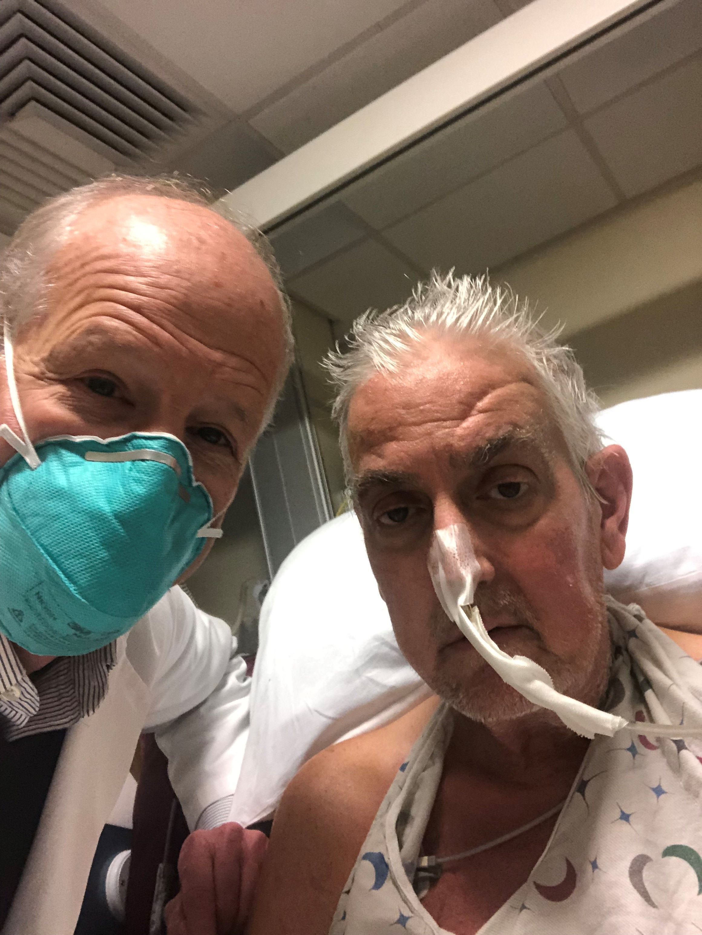
“The issue is now a subject of wide discussion among specialists, who think the infection was a potential contributor to Mr. Bennett’s death and a possible reason why the heart did not last longer,” states the article, written by staff journalist Antonio Regalado.
As described in the story, the xenotransplant saga’s new twist comes from the surgeon who performed the operation, Bartley P. Griffith, MD, University of Maryland, Baltimore, who related the PCMV finding in an April 20 online presentation hosted by the American Society of Transplantation.
Mr. Bennett’s initially promising but later turbulent clinical course, described by his surgeons and widely reported upon his death, included repeated skirmishes with infection and retaliatory adjustments to his immunosuppressant regimen. Those episodes were thought to have contributed to his death, the actual cause of which is undetermined or at least not yet reported.
“We are beginning to learn why he passed on,” Dr. Griffith said in Mr. Regalado’s article, acknowledging further that the porcine virus “maybe was the actor, or could be the actor,” that set off the events leading to Bennett’s death.
Xenotransplant specialists know that PCMV is a potential problem with pig organs and know to test for it before attempting the procedure in animal models, notes the article. It refers to a published series of pig-heart transplants to baboons in Germany. The hearts “lasted only a couple of weeks if the virus was present, while organs free from the infection could survive more than half a year.”
The heart Mr. Bennett received had been extensively screened for bacteria, viruses, and other issues that could have threatened the organ and Mr. Bennett, but the effort apparently fell short. In the MIT Technology Review story, the first author of the German baboon series speculates on how the University of Maryland team might have missed PCMV.
“The U.S. team appears to have tested the pig’s snout for the virus, but often it is lurking deeper in the tissues,” Joachim Denner, PhD, Institute of Virology, Free University of Berlin, said in the article. The virus, he contended, “can be detected and easily removed from pig populations, but unfortunately they didn’t use a good assay and didn’t detect the virus.”
That PCMV escaped detection before the operation “could now factor into some people’s questions over whether the experiment should have taken place at all,” the MIT Technology Review article proposes. “It’s a big red flag,” bioethicist Arthur Caplan, PhD, New York University, said in a quote, adding: “If doctors can’t prevent or control infection, ‘then such experiments are tough to justify.’ ”
A version of this article first appeared on Medscape.com.
A porcine cytomegalovirus (PCMV) in the heart had gone undetected before the operation and may or may not have been instrumental in David Bennett’s death 2 months later, according to a report published in MIT Technology Review.
“The issue is now a subject of wide discussion among specialists, who think the infection was a potential contributor to Mr. Bennett’s death and a possible reason why the heart did not last longer,” states the article, written by staff journalist Antonio Regalado.
As described in the story, the xenotransplant saga’s new twist comes from the surgeon who performed the operation, Bartley P. Griffith, MD, University of Maryland, Baltimore, who related the PCMV finding in an April 20 online presentation hosted by the American Society of Transplantation.
Mr. Bennett’s initially promising but later turbulent clinical course, described by his surgeons and widely reported upon his death, included repeated skirmishes with infection and retaliatory adjustments to his immunosuppressant regimen. Those episodes were thought to have contributed to his death, the actual cause of which is undetermined or at least not yet reported.
“We are beginning to learn why he passed on,” Dr. Griffith said in Mr. Regalado’s article, acknowledging further that the porcine virus “maybe was the actor, or could be the actor,” that set off the events leading to Bennett’s death.
Xenotransplant specialists know that PCMV is a potential problem with pig organs and know to test for it before attempting the procedure in animal models, notes the article. It refers to a published series of pig-heart transplants to baboons in Germany. The hearts “lasted only a couple of weeks if the virus was present, while organs free from the infection could survive more than half a year.”
The heart Mr. Bennett received had been extensively screened for bacteria, viruses, and other issues that could have threatened the organ and Mr. Bennett, but the effort apparently fell short. In the MIT Technology Review story, the first author of the German baboon series speculates on how the University of Maryland team might have missed PCMV.
“The U.S. team appears to have tested the pig’s snout for the virus, but often it is lurking deeper in the tissues,” Joachim Denner, PhD, Institute of Virology, Free University of Berlin, said in the article. The virus, he contended, “can be detected and easily removed from pig populations, but unfortunately they didn’t use a good assay and didn’t detect the virus.”
That PCMV escaped detection before the operation “could now factor into some people’s questions over whether the experiment should have taken place at all,” the MIT Technology Review article proposes. “It’s a big red flag,” bioethicist Arthur Caplan, PhD, New York University, said in a quote, adding: “If doctors can’t prevent or control infection, ‘then such experiments are tough to justify.’ ”
A version of this article first appeared on Medscape.com.
FROM MIT TECHNOLOGY REVIEW
Which solid organ transplant recipients face the highest risk of skin cancer?
BOSTON – .
White patients who meet these criteria should be screening within 2 years after transplant, while Black patients should be screened within 5 years after transplant, Ally-Khan Somani, MD, PhD, said at the annual meeting of the American Academy of Dermatology.
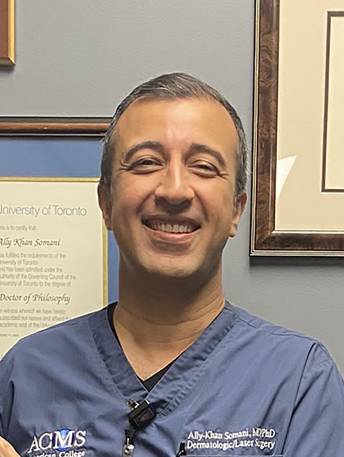
Dr. Somani, director of dermatologic surgery and the division of cutaneous oncology at Indiana University, Indianapolis, based his remarks on consensus screening guidelines assembled from three rounds of Delphi method surveys with 47 dermatologists and 37 transplant physicians, with the goal of establishing skin cancer screening recommendations for SOTRs. Among the dermatologists surveyed, 45% were Mohs surgeons and 55% were general dermatologists.
The panel recommended that the transplant team should perform risk assessment for SOTRs to risk stratify patients for skin cancer screening (high risk vs. low risk). They also proposed that dermatologists perform skin cancer screening by full-body skin examinations, and that SOTRs with a history of skin cancer should continue with routine skin cancer surveillance as recommended by their dermatologists.
Those at low risk for skin cancer include abdominal organ recipients, SOTR age of younger than 50 at time of transplant, and female gender. The guidelines recommend that White, Asian, and Hispanic patients who meet those criteria should be screened within 5 years after transplant, while no consensus was reached for Black patients who meet those criteria.
Based on posttransplant skin cancer incidence rates, risk is increased among males, Whites, thoracic organ recipients, and being age 50 or older, Dr. Somani said. “At our institution, we make sure there’s a good connection between our transplant teams and dermatologists. We recommend rapid referral for suspicious lesions and we educate patients and screen them within 1 year of transplant, or sooner for high-risk patients. Surveillance is increased to every 3 or 4 months for patients with a history of multiple or high-risk cancers or sooner, followed by routine surveillance as recommended by the patient’s dermatologist.”
To risk stratify patients on the development of their first skin cancer post transplantation, researchers developed the Skin and Ultraviolet Neoplasia Transplant Risk Assessment Calculator (SUNTRAC), a prediction tool with a freely available app. Data for the tool were drawn from the Transplant Skin Cancer Network study, a 5-year analysis of 6,340 adult recipients of a first solid organ transplant at 26 transplant centers in the United States. It generates a risk score for SOTRs (low, medium, high, or very high), which informs transplant care providers of a patient’s risk of skin cancer.
Dr. Somani disclosed that he has received grants and funding from Castle Biosciences. He is an adviser to Cook Biotech and a consultant to Sanara MedTech.
BOSTON – .
White patients who meet these criteria should be screening within 2 years after transplant, while Black patients should be screened within 5 years after transplant, Ally-Khan Somani, MD, PhD, said at the annual meeting of the American Academy of Dermatology.
Dr. Somani, director of dermatologic surgery and the division of cutaneous oncology at Indiana University, Indianapolis, based his remarks on consensus screening guidelines assembled from three rounds of Delphi method surveys with 47 dermatologists and 37 transplant physicians, with the goal of establishing skin cancer screening recommendations for SOTRs. Among the dermatologists surveyed, 45% were Mohs surgeons and 55% were general dermatologists.
The panel recommended that the transplant team should perform risk assessment for SOTRs to risk stratify patients for skin cancer screening (high risk vs. low risk). They also proposed that dermatologists perform skin cancer screening by full-body skin examinations, and that SOTRs with a history of skin cancer should continue with routine skin cancer surveillance as recommended by their dermatologists.
Those at low risk for skin cancer include abdominal organ recipients, SOTR age of younger than 50 at time of transplant, and female gender. The guidelines recommend that White, Asian, and Hispanic patients who meet those criteria should be screened within 5 years after transplant, while no consensus was reached for Black patients who meet those criteria.
Based on posttransplant skin cancer incidence rates, risk is increased among males, Whites, thoracic organ recipients, and being age 50 or older, Dr. Somani said. “At our institution, we make sure there’s a good connection between our transplant teams and dermatologists. We recommend rapid referral for suspicious lesions and we educate patients and screen them within 1 year of transplant, or sooner for high-risk patients. Surveillance is increased to every 3 or 4 months for patients with a history of multiple or high-risk cancers or sooner, followed by routine surveillance as recommended by the patient’s dermatologist.”
To risk stratify patients on the development of their first skin cancer post transplantation, researchers developed the Skin and Ultraviolet Neoplasia Transplant Risk Assessment Calculator (SUNTRAC), a prediction tool with a freely available app. Data for the tool were drawn from the Transplant Skin Cancer Network study, a 5-year analysis of 6,340 adult recipients of a first solid organ transplant at 26 transplant centers in the United States. It generates a risk score for SOTRs (low, medium, high, or very high), which informs transplant care providers of a patient’s risk of skin cancer.
Dr. Somani disclosed that he has received grants and funding from Castle Biosciences. He is an adviser to Cook Biotech and a consultant to Sanara MedTech.
AT AAD 22
When CPI fails, HL patients should get timely allo-HCT
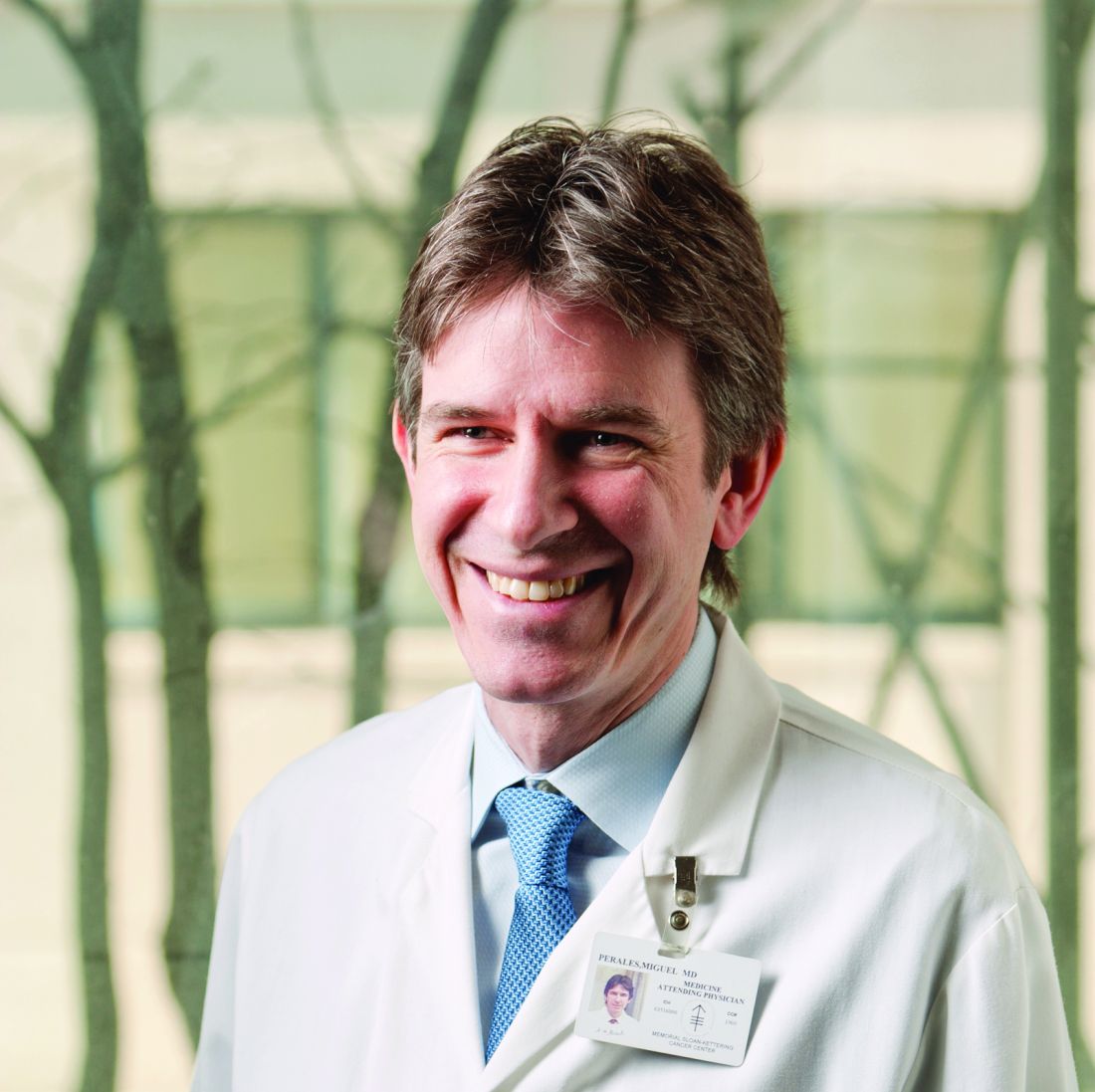
In fact, prior treatment with PD-1–directed therapies nivolumab (Opdivo) and pembrolizumab (Keytruda) appears to improve outcomes in allo-HCT patients, said Miguel-Angel Perales, MD, chief of the adult bone marrow transplant service at Memorial Sloan Kettering Cancer Center in New York.
“The use of allogeneic HCT is decreasing for Hodgkin even though it is a curative option, and we see patients referred after they have had multiple lines of therapy,” Dr. Perales said in an interview. “The lymphoma MDs have a perception that outcomes are poor, and therefore don’t refer.”
To illustrate his point, Dr. Perales shared data from the EBMT database. In 2014, the registry accrued approximately 450 allo-HCT cases; by 2021 this had fallen to fewer than 200 procedures.
Ironically, this declining enthusiasm for transplantation coincides with a steady improvement in transplant outcomes following PD-1 blockade, Dr. Perales noted. For example, an analysis, published in Nature, yielded an 82% overall survival (OS) at 3 years in patients who underwent allo-HCT after CPI treatment (n =209).
“Results of allo-HCT in patients with Hodgkin show a remarkable cure rate,” said Dr. Perales. “Part of that is probably driven by lower relapse due to enhanced graft-versus-lymphoma effect due to long CPI half-life.” (The half-lives of pembrolizumab and nivolumab are 22 and 25 days, respectively.)
At the EBMT meeting, Dr. Perales presented a new retrospective analysis that tested the hypothesis that CPIs might actually improve outcomes for allo-HCT patients. An international team of clinicians from EBMT and the Center for International Blood and Marrow Transplant Research (CIBMTR) compared allo-HCT outcomes with (n = 347) and without (n = 1,382) prior treatment with a checkpoint inhibitor.
They found that prior CPI therapy was, indeed, associated with lower relapse (hazard ratio, 0.53; P = .00023) and longer progression-free survival (PFS) (HR, 0.75; P = .0171).
However, prior PD-1 drugs provided no survival advantage, Dr. Perales said. “The easiest explanation for a study showing a difference in PFS/relapse, not OS, is that we have good treatments that can treat patients who relapse and so their overall survival ends up being the same.”
The researchers also confirmed previous reports that patients who received PD-1 inhibitors prior to transplant had a higher incidence of GVHD. Prevalence of acute grades 2-4 GVHD was significantly higher (P = .027); however, acute grades 3-4 GVHD and chronic GVHD were not significantly different between the two groups.
Dr. Perales speculated that the use of posttransplant cyclophosphamide for GVHD prophylaxis would mitigate the risk of GVHD associated with PD-1 inhibitors, “we have not yet proven that formally ... [we] are still analyzing our data.”
Commenting on the results of the new analysis, Dr. Perales expressed concern that patients are being recruited to early-phase clinical trials after failing on a checkpoint inhibitor, instead of being offered allo-HCT – a potentially curative treatment – because treaters are misinformed about the safety of transplant after these drugs.
The NIH clinical-trials database backs up Dr. Perales’ worries. In the United States, for example, there are currently 19 trials recruiting for relapsed/refractory Hodgkin lymphoma patients prior to transplant. Of these, 15 studies permit enrollment of patients who have failed on CPIs, and 8 are phase 1 or 2 studies.
“The good news is that new drugs, including CPIs, have dramatically changed outcomes in this disease and that fewer patients now need an allo-HCT,” said Dr. Perales. And if a transplant is needed, “it is safe to perform allo-HCT in patients treated with prior CPI.”
However, time is of the essence. “Patients with Hodgkin lymphoma should be referred to allo-HCT if they are not responding or tolerating CPI, rather than go on a series of phase 1 trials,” Dr. Perales said. “Median age is 32, and we should be going for a cure, nothing less.”
Dr. Perales reported receiving honoraria from numerous pharmaceutical companies; serving on the data and safety monitoring boards of Cidara Therapeutics, Medigene, Sellas Life Sciences, and Servier; and serving on the scientific advisory board of NexImmune. He has ownership interests in NexImmune and Omeros, and has received institutional research support for clinical trials from Incyte, Kite/Gilead, Miltenyi Biotec, Nektar Therapeutics, and Novartis.
Survivor’s story foreshadows one of oncology’s greatest successes
At 32 years old, the world was at Larry Unger’s feet. He was vice president at one of Wall Street’s most successful investment management firms, selling mutual funds to more than 1,000 brokers across New York. His clients relied on him for good advice, great jokes, and superlative Yankees tickets. His recent memories included fraternity days at Cornell University and a Harvard law degree. His childhood on the Lower East Side was behind him. He had his own apartment and a beautiful girlfriend.
Then his back started hurting, and he was drenched in sweat at night. His physician suggested it was a basketball injury. Weeks of tests followed, and he changed doctors. Mr. Unger met with an oncologist at Memorial Sloan Kettering Cancer Center who wouldn’t let him go home after the appointment. The next day brought exploratory surgery and an answer to all the questions.
Mr. Unger was diagnosed with stage IIIB Hodgkin lymphoma.
Thirty years later, Mr. Unger credited his survival to the late Subhash Gulati, MD, PhD, then MSKCC’s director of stem cell transplantation. He still recalls Dr. Gulati’s words to him: “Radical situations call for radical solutions.” In 1992, that “radical solution” was an autologous bone-marrow transplant.
“Mr. Unger was a patient pioneer,” said Kenneth Offit, MD, another MSKCC oncologist who also cared for him at that time.
Transplantation for Hodgkin: The early 90s
Hodgkin lymphoma is fairly rare, accounting for just 0.5% of all cancers and 15% of lymphomas. It tends to target young, male adults like Mr. Unger. Today 88% of patients with Hodgkin survive at least 5 years.
When Dr. Gulati offered Mr. Unger his “radical solution” 3 decades ago, the idea of autologous bone marrow transplantation in Hodgkin lymphoma was not new. The first attempt appeared in the literature in the 1950s, but it was still unclear how patients could survive the procedure. It involved destroying the patient’s own immune system prior to the transplant, a huge risk in itself. Worse, the patient was pummeled with chemotherapy and/or radiation to clear out the cancerous bone marrow – a process called “conditioning.”
However, throughout the 1980s, MSKCC had been running clinical trials to perfect the conditioning mix, so by 1992 Dr. Gulati was well-placed to help Mr. Unger.
It is unclear what conditioning Mr. Unger received because his records were not made available. However, around the time that Mr. Unger underwent his transplant, Dr. Gulati and colleagues published the conditioning regimens in use at MSKCC. Patients with refractory or relapsed Hodgkin disease received a conditioning mix of total nodal irradiation (TNI), etoposide (Vepesid) and cyclophosphamide. Patients who had already been through radiotherapy were given carmustine instead of TNI.
In that early publication, Dr. Gulati and the MSKCC team reported 0 “toxic deaths” with the TNI mix, and at the 2-year point 75% of the patients were still alive (n = 28). Patients who had already received radiation treatment did less well, with 55% survival at 2 years, at a cost of 14% toxic deaths (n = 22).
Mr. Unger’s experience, 30 years ago
According to Mr. Unger, the initial treatment for his stage IIIB Hodgkin lymphoma was MOPP (mechlorethamine hydrochloride, vincristine sulfate, procarbazine hydrochloride, and prednisone) plus ABVD (doxorubicin hydrochloride, bleomycin sulfate, vinblastine sulfate, and dacarbazine).
“They wanted to give me two chemo programs at once because they said I was very sick,” Mr. Unger recalled. “I wound up staying in the hospital quite a bit because every time I got these [treatments] I’d get a fever. This went on for month after month after month. Finally, they said: ‘The tumors are starting to shrink. ... I want you to meet Dr. Gulati.’ ”
Mr. Unger said that Dr. Gulati told him: “There is another procedure called the bone marrow transplant which we’ve been doing. This would be like hitting it with a nuclear weapon. We would really wipe it out and make sure that you never come back.”
The alternative was high-dose radiotherapy. However, Dr. Gulati shared MSKCC’s hard-won knowledge that an autologous transplant was less successful after radiation. Dr. Gulati also told Mr. Unger that surgery was needed before the transplant: a laparotomy to restage his tumors.
After discussing the situation with his father, Mr. Unger decided to undergo the transplant.
The night before treatment started, he was laughing and joking with a friend in his room at MSKCC. The next day, the laughing stopped. The conditioning, he said, “was harrowing beyond belief ... 100 times worse than the chemo.”
Chemotherapy came first, followed 2 days later by radiation, presumably TNI. Mr. Unger experienced constant vomiting, intraocular bleeding and high fevers; the soft tissue of his throat “fell apart,” he said.
“I couldn’t move. It was like being dead,” he said. “Finally, maybe after a month or so, I could finally have a little water.” Mr. Unger said his immune system took 6-8 weeks to recover. He concluded, with heroic understatement, “it was rough.”
The battle against Hodgkin was over, but fallout from the chemotherapy lingered. Although Mr. Unger was able to return to his family and the job he loved, in the following years he was never entirely well. He contracted shingles soon after his transplant, then diabetes within 15 years. A heart attack followed in 2008 then, in 2015, an autoimmune disease that still affects his mobility.
However, Mr. Unger remains grateful: “The fact that we did these cutting-edge techniques with me got me to the point where – although I had some problems afterward, and I have problems now – it gave me well over 30 years of a really great life.”
“There are a lot of good doctors out there,” he added. “Some of them go to extraordinary lengths to help people. I try to do the same with the extra 30 years I’ve been given, try to be nice to people and make people feel good. I don’t really see any other reason to be on earth.”
Treatment for Hodgkin lymphoma: 2022
For a comparison of Mr. Unger’s experience with the current approach to Hodgkin lymphoma, this news organization spoke to Miguel-Angel Perales, MD, current chief of the adult bone marrow transplant service at MSKCC. Although Dr. Perales could not comment specifically on Mr. Unger’s case without his records, Dr. Perales was able to review the revolutions in treatment for all patients over the past 30 years.
Certainly, physicians no longer need to inflict a laparotomy on patients just to stage the disease, Dr. Perales said. “This sounds barbaric today. Nowadays we have PET scans.”
Another key change, Dr. Perales said, is in the up-front management of the disease.
For example, MOPP “is going back to the prehistory of chemotherapy,” Dr. Perales said. He was not surprised to learn that Mr. Unger later developed complications such as diabetes and heart disease.
“We’ve completely revolutionized the treatment,” Dr. Perales said. “We [now] use combinations that are much less toxic than MOPP, [and] we’re curing more patients up front.” Treatment is tailored by stage and the likelihood of response to therapy. Aggressive approaches are reserved for patients more likely to fail treatment.
Pretransplant conditioning has also changed for the better, with less toxicity and fewer long-term complications. Total body irradiation has “fallen by the wayside,” said Dr. Perales. Instead, patients get BEAM, a combination of carmustine, etoposide, cytarabine (Cytosar-U, Ara-C), and melphalan (Alkeran), 1 week before the transplant.
Perhaps the most profound change, which began in the 1990s shortly after Larry’s transplant, was that peripheral-blood stem cells gradually replaced bone marrow for both autologous and allogeneic transplant. In 2022, nearly all autologous transplants use peripheral-blood stem cells.
Instead of onerous bone-marrow aspiration in the operating room, the stem cells are collected from the patient’s blood. First, the patient’s bone marrow is hyperstimulated with high doses of filgrastim (G-CSF, Neupogen, Granix) for several days. Stem cells spill into the patient’s blood. Once blood is collected from the patient, the stem cells are separated and stored ready for the transplant. (In theory, stem cell products are “cancer free”; in practice there may be some contaminating cells, said Dr. Perales.)
Nowadays “transplanting” the stem cells back into the body bears no relation to what happened in 1992. The stem-cell infusion is typically an outpatient procedure, and one-third of patients may never be admitted to the hospital at all.
In contrast to Mr. Unger’s excruciating 8-week hospital stay, immune recovery currently takes 12-14 days, often entirely in the patient’s own home, with the option of extra filgrastim to speed things up.
Despite these profound changes, said Dr. Perales, the real quantum leap has occurred post transplant.
In 2015, a multinational team led by MSKCC’s Dr. Craig Moskowitz published a trial in the Lancet showing that brentuximab vedotin halved the risk of relapse after autologous transplantation in high-risk Hodgkin lymphoma patients versus placebo (hazard ratio, 0.57; P = .0013; n = 329). The CD30-directed antibody-drug conjugate was so successful that the placebo patients were encouraged to cross over into the treatment group; many of them were salvaged.
As a result, Dr. Perales said, brentuximab vedotin has now become the standard in high-risk Hodgkin patients following a transplant.
The checkpoint inhibitors nivolumab (Opdivo) and pembrolizumab (Keytruda) have also been “transformational” in Hodgkin lymphoma, Dr. Perales said. He explained that Hodgkin lymphoma is “exquisitely sensitive” to these therapies because the disease expresses high levels of the binding proteins for these drugs. This allows the immunotherapies to hit both the immune system and the disease.
Most cancers have response rates for checkpoint inhibitors below 40%, according to a recent analysis by Anas Younes, former chief of lymphoma at MSKCC, and his colleague Eri Matsuki, then a visiting fellow. By contrast, in Hodgkin lymphoma response to these drugs is 66%-87%.
Dr. Perales said: “It tells you how effective these drugs are, that we could move from somebody getting MOPP, which is like throwing a nuclear bomb at somebody, to a combination of two drugs that can easily be given out-patient and that have very little, if any, side effects.”
The future: No chemo, no transplants?
“One of the holy grails in Hodgkin would be if we could treat patients with the combination of a checkpoint inhibitor and brentuximab and what is being termed the ‘chemotherapy-free’ approach to Hodgkin disease,” said Dr. Perales.
What else remains to be done in the world of transplants for Hodgkin lymphoma?
Dr. Perales didn’t hesitate: “To eliminate the need for them. If we can have better targeted therapy up front that cures more patients, then we never even have to consider transplant. Basically, to put me out of work. I’m sure I’ll find other things to do.”
The current treatment of Hodgkin lymphoma “is really what we all consider one of the successes in oncology,” said Dr. Perales. “It’s a beautiful story.”
Dr. Perales reported receiving honoraria from numerous pharmaceutical companies; serves on data and safety monitoring boards for Cidara Therapeutics, Medigene, Sellas Life Sciences, and Servier; and serves on the scientific advisory board of NexImmune. He has ownership interests in NexImmune and Omeros, and has received institutional research support for clinical trials from Incyte, Kite/Gilead, Miltenyi Biotec, Nektar Therapeutics, and Novartis.
Surgeons in China ‘are the executioners,’ procuring organs before brain death
In a deep dive into obscure Chinese language transplant journals, a pair of researchers from Australia and Israel have added a new layer of horror to what’s already known about forced organ harvesting in China.
Searching for documentation that vital organs are being harvested from nonconsenting executed prisoners, a practice that the China Tribunal confirmed “beyond any reasonable doubt” in 2020, Jacob Lavee, MD, an Israeli heart transplant surgeon, and Matthew Roberston, a PhD student at Australian National University, uncovered something even more shocking: that vital organs are being explanted from patients who are still alive.
“We have shown for the first time that the transplant surgeons are the executioners – that the mode of execution is organ procurement. These are self-admissions of executing the patient,” Dr. Lavee told this news organization. “Up until now, there has been what we call circumstantial evidence of this, but our paper is what you’d call the smoking gun, because it’s in the words of the physicians themselves that they are doing it. In the words of these surgeons, intubation was done only after the beginning of surgery, which means the patients were breathing spontaneously up until the moment the operation started ... meaning they were not brain dead.”
The research, published in the American Journal of Transplantation, involved intricate analysis of thousands of Chinese language transplant articles and identified 71 articles in which transplant surgeons describe starting organ procurement surgery before declaring their patients brain dead.
“What we found were improper, illegitimate, nonexistent, or false declarations of brain death,” Mr. Robertson said in an interview. He explained that this violates what’s known as the dead donor rule, which is fundamental in transplant ethics. “The surgeons wrote that the donor was brain dead, but according to everything we know about medical science, they could not possibly have been brain dead because there was no apnea test performed. Brain death is not just something you say, there’s this whole battery of tests, and the key is the apnea test, [in which] the patient is already intubated and ventilated, they turn the machine off, and they’re looking for carbon dioxide in the blood above a certain level.”
Mr. Robertson and Dr. Lavee have painstakingly documented “incriminating sentences” in each of the 71 articles proving that brain death had not occurred before the organ explantation procedure began. “There were two criteria by which we claimed a problematic brain death declaration,” said Mr. Robertson, who translated the Chinese. “One was where the patient was not ventilated and was only intubated after they were declared brain dead; the other was that the intubation took place immediately prior to the surgery beginning.”
“It was mind-boggling,” said Dr. Lavee, from Tel Aviv University. “When I first started reading, my initial reaction is, ‘This can’t be.’ I read it once, and again, and I insisted that Matt get another independent translation of the Chinese just to be sure. I told him, ‘There’s no way a physician, a surgeon could write this – it doesn’t make sense.’ But the more of these papers we read, we saw it was a pattern – and they didn’t come out of a single medical center, they are spread all over China.”
For the analysis, Mr. Robertson wrote code and customized an algorithm to examine 124,770 medical articles from official Chinese databases between 1980 and 2020. The 71 articles revealing cases involving problematic brain death came from 56 hospitals (of which 12 were military) in 33 cities across 15 provinces, they report. In total, 348 surgeons, nurses, anesthesiologists, and other medical workers or researchers were listed as authors of these publications.
Why would these medical personnel write such self-incriminating evidence? The researchers say it’s unclear. “They don’t think anyone’s reading this stuff,” Mr. Robertson suggests. “Sometimes it’s revealed in just five or six characters in a paper of eight pages.” Dr. Lavee wonders if it’s also ignorance. “If this has been a practice for 20 or 30 years in China, I guess nobody at that time was aware they were doing something wrong, although how to declare brain death is something that is known in China. They’ve published a lot about it.”
The article is “evidence that this barbarity continues and is a very valuable contribution that continues to bring attention to an enormous human rights violation,” said Arthur Caplan, PhD, head of the Division of Medical Ethics at New York University’s Grossman School of Medicine. “What they’ve reported has been going on for many, many years, the data are very clear that China’s doing many more transplants than they have cadaver organ donors,” he said, adding that the country’s well-documented and lucrative involvement in transplant tourism “means you have to have a donor ready when the would-be recipient appears; you have to have a matched organ available, and that’s hard to do waiting on a cadaver donor.”
Although the researchers found no incriminating publications after 2015, they speculate that this is likely due to growing awareness among Chinese surgeons that publishing the information would attract international condemnation. “We think these practices are continuing to go on,” said Dr. Lavee. He acknowledged that a voluntary organ donation program is slowly developing in parallel to this. He said, given China’s place as the world’s second largest transplant country behind the U.S., as well as its low rate of voluntary donation, it’s reasonable to conclude that the main source of organs remains prisoners on death row.
Dr. Caplan and the researchers have called for academic institutions and medical journals to resume their previous boycotts of Chinese transplant publications and speakers, but as long as China denies the practices, economic and political leaders will turn a blind eye. “In the past, I don’t think the question of China’s medical professional involvement in the execution of donors has been taken as seriously as it should have,” said Mr. Robertson. “I certainly hope that with the publication of this paper in the leading journal in the field, this will change.”
The study was supported by the Google Cloud Research Credits program, the Australian Government Research Training Program Scholarship, and the Victims of Communism Memorial Foundation. Mr. Robertson, Dr. Lavee, and Dr. Caplan have disclosed no relevant financial relationships.
A version of this article first appeared on Medscape.com.
Live-donor liver transplants for patients with CRC liver mets
These patients usually have a poor prognosis, and for many, palliative chemotherapy is the standard of care.
“For the first time, we have been able to demonstrate [outside of Norway] that liver transplantation for patients with unresectable liver metastases is feasible with good outcomes,” lead author Gonzalo Sapisochin, MD, PhD, an assistant professor of surgery at the University of Toronto, said in an interview.
“Furthermore, this is the first time we are able to prove that living donation may be a good strategy in this setting,” Dr. Sapisochin said of the series of 10 cases that they published in JAMA Surgery.
The series showed “excellent perioperative outcomes for both donors and recipients,” noted the authors of an accompanying commentary. They said the team “should be commended for adding liver-donor live transplantation to the armamentarium of surgical options for patients with CRC liver metastases.”
However, they express concern about the relatively short follow-up of 1.5 years and the “very high” recurrence rate of 30%.
Commenting in an interview, lead editorialist Shimul Shah, MD, an associate professor of surgery and the chief of solid organ transplantation at the University of Cincinnati, said: “I agree that overall survival is an important measure to look at, but it’s hard to look at overall survival with [1.5] years of follow-up.”
Other key areas of concern are the need for more standardized practices and for more data on how liver transplantation compares with patients who just continue to receive chemotherapy.
“I certainly think that there’s a role for liver transplantation in these patients, and I am a big fan of this,” Dr. Shah emphasized, noting that four patients at his own center have recently received liver transplants, including three from deceased donors.
“However, I just think that as a community, we need to be cautious and not get too excited too early,” he said. “We need to keep studying it and take it one step at a time.”
Moving from deceased to living donors
Nearly 70% of patients with CRC develop liver metastases, and when these are unresectable, the prognosis is poor, with 5-year survival rates of less than 10%.
The option of liver transplantation was first reported in 2015 by a group in Norway. Their study included 21 patients with CRC and unresectable liver tumors. They reported a striking improvement in overall survival at 5 years (56% vs. 9% among patients who started first-line chemotherapy).
But with shortages of donor livers, this approach has not caught on. Deceased-donor liver allografts are in short supply in most countries, and recent allocation changes have further shifted available organs away from patients with liver tumors.
An alternative is to use living donors. In a recent study, Dr. Sapisochin and colleagues showed viability and a survival advantage, compared with deceased-donor liver transplantation.
Building on that work, they established treatment protocols at three centers – the University of Rochester (N.Y.) Medical Center, the Cleveland Clinic, , and the University Health Network in Toronto.
Of 91 evaluated patients who were prospectively enrolled with liver-confined, unresectable CRC liver metastases, 10 met all inclusion criteria and received living-donor liver transplants between December 2017 and May 2021. The median age of the patients was 45 years; six were men, and four were women.
These patients all had primary tumors greater than stage T2 (six T3 and four T4b). Lymphovascular invasion was present in two patients, and perineural invasion was present in one patient.
The median time from diagnosis of the liver metastases to liver transplant was 1.7 years (range, 1.1-7.8 years).
At a median follow-up of 1.5 years (range, 0.4-2.9 years), recurrences occurred in three patients, with a rate of recurrence-free survival, using Kaplan-Meier estimates, of 62% and a rate of overall survival of 100%.
Rates of morbidity associated with transplantation were no higher than those observed in established standards for the donors or recipients, the authors noted.
Among transplant recipients, three patients had no Clavien-Dindo complications; three had grade II, and four had grade III complications. Among donors, five had no complications, four had grade I, and one had grade III complications.
All 10 donors were discharged from the hospital 4-7 days after surgery and recovered fully.
All three patients who experienced recurrences were treated with palliative chemotherapy. One died of disease after 3 months of treatment. As of the time of publication of the study, the other two had survived for 2 or more years following their live donor liver transplant.
Patient selection key
The authors are now investigating tumor subtypes, responses in CRC liver metastases, and other factors, with the aim of developing a novel screening method to identify appropriate candidates more quickly.
In the meantime, they emphasized that indicators of disease biology, such as the Oslo Score, the Clinical Risk Score, and sustained clinical response to systemic therapy, “remain the key filters through which to select patients who have sufficient opportunity for long-term cancer control, which is necessary to justify the risk to a living donor.”
Dr. Sapisochin reported receiving grants from Roche and Bayer and personal fees from Integra, Roche, AstraZeneca, and Novartis outside the submitted work. Dr. Shah disclosed no relevant financial relationships.
A version of this article first appeared on Medscape.com.
FROM JAMA SURGERY
Repurposed drug could revolutionize stem cell transplantation
When the Food and Drug Administration approved abatacept in December 2021 as prophylaxis for acute graft-versus-host disease (aGVHD) in adults and children 2 years and older who are undergoing hematopoietic stem cell transplantation (HSCT), the announcement was notable for couple of key reasons.
Abatacept was approved based on results from the ABA2 trial, which evaluated 142 adults and children with hematologic malignancies who received a four-dose regimen of abatacept in addition to standard of care – a calcineurin inhibitor (CNI) plus methotrexate (MTX) – prior to undergoing an 8/8 HLA-matched, unrelated donor (URD) HSCT, or standard of care alone.
Another arm of the trial examined 43 recipients of a 7/8 HLA-mismatched URD HSCT who received abatacept plus standard of care, compared with a prespecified registry cohort group provided by the Center for International Blood and Marrow Transplant Research, who received CNI and MTX.
Results published in the Journal of Clinical Oncology showed the proportion of patients in the 8/8 group with severe aGVHD in the abatacept group 100 days after HSCT was not significantly lower, compared with the standard of care group (6.8% vs. 14.8%; P = .13), but there was a significant improvement in severe aGVHD–free survival (SGFS) 180 days after HSCT in the abatacept group, compared with the group that received standard of care (93.2% vs. 80%; P = .05).
Among patients in the 7/8 group, there was a significant difference in the proportion of patients with severe aGVHD favoring the abatacept group (2.3% vs. 30.2%; P < .001), and significantly improved SGFS, compared with the CIBMTR registry cohort (97.7% vs. 58.7%; P < .001)
A post hoc analysis of ABA2 published as a research letter in Blood Advances assessed abatacept using real-world data from CIBMTR. Researchers compared the 8/8 group that received standard of care with the 7/8 group that received abatacept plus standard of care and found no significant differences between relapse-free survival and overall survival for patients in the 8/8 group (adjusted hazard ratio, 0.60; 95% confidence interval, 0.28-1.28; P = .19) and 7/8 group (aHR, 0.77; 95% CI, 0.34-1.71; P = .51).
The results suggest “abatacept may eliminate that risk of a mismatched transplant in the setting of that analysis and that small cohort that was assessed there, which is good news for patients that may not have a fully matched donor on the registry,” said Stephen Spellman, vice president at Be The Match Research (operated by the National Marrow Donor Program), and senior scientific director of CIBMTR. The findings from ABA2 “were even more impressive than necessarily expected, especially in the 7/8 arm. This is a truly substantial reduction in acute GVHD risk in that patient population,” he said in an interview.
Could abatacept fuel greater use of mismatched, unrelated donors?
One downside of using an HLA-mismatched donor is the potential risk of developing aGVHD, Doris M. Ponce, MD, a hematologic oncologist with Memorial Sloan Kettering Cancer Center in New York, who was not involved with the research, said in an interview.
Potential risk factors for aGVHD include “having a female multiparous donor, HLA-mismatched donor, unrelated donor, donor and recipient age (>40 years), [peripheral blood stem cell] stem cell graft, recipient [cytomegalovirus] serostatus (recipient/donor), myeloablative conditioning, [total body irradiation]–based conditioning, [and] gut microbiome dysbiosis,” Dr. Ponce explained.
Abatacept’s approval may have particular relevance for people of color. “It’s been understood for a long time that the likelihood of finding an 8/8 well-matched, volunteer unrelated donor varies by race or ethnicity,” Steven Devine, MD, a board-certified oncologist who is chief medical officer of Be The Match and associate scientific director at CIBMTR, said in an interview.
Mr. Spellman noted that, of the more than 35 million donors on worldwide registries accessible through the National Marrow Donor Program’s Be The Match Registry, “the match rates differ quite substantially by race and ethnicity.” Approximately 29% of African Americans find a full match on the registry, compared with 81% of Whites, 49% of Hispanics, and 47% Asian/Pacific Islanders.
“Being able to utilize a 7/8 match in a safe, effective manner using abatacept, which abatacept has been approved for, does increase those match rates quite substantially,” he explained. Among African Americans, this means the match rate increases to 84%, among Hispanics and Asian/Pacific Islanders to approximately 90%, and among Whites to about 98%.
That kind of improvement in the match rate is “the equivalent of adding more than more than 10 million ethnically diverse donors to the registry in 1 day,” Dr. Devine said. “The availability of abatacept could really level the playing field for patients in need of a lifesaving transplant.”
Further study of abatacept
With abatacept, “I think the results are really encouraging, and I think that further studies [are needed] to better define how the drug would work and whether it can later prevent chronic graft versus host disease,” Dr. Devine said. He said the ABA3 trial has been designed around this question, with the hypothesis that extending abatacept to an eight-dose regimen may help with chronic GVHD.
Although the FDA’s approval of abatacept was recent, Mr. Spellman said, Be The Match has seen early indications that mismatched donors in the registry are being used, which may point to an increased utilization of abatacept. “Through October to December of 2021, there was a pretty substantial increase in the use of mismatched, unrelated donors in that time frame.”
Dr. Devine noted that he is seeing a lot of interest in using abatacept. “I think people are still learning how best to incorporate it into their standard of care right now.”
Meanwhile, Memorial Sloan Kettering Cancer Center is already planning to use abatacept, Dr. Ponce noted. “We have abatacept in our formulary for adult and children, and are planning on using it for patients receiving an unmodified graft from a [matched unrelated donor] or 1-allele [mismatched unrelated donor] using CNI and MTX-based GVHD prophylaxis.”
Dr. Devine and Mr. Spellman are employees of Be The Match and CIBMTR, which provided the registry control group for the ABA2 trial. Dr. Devine also reported that he has been a scientific advisory board member for Bristol-Myers Squibb. Dr. Ponce reports no relevant conflicts of interest.