User login
We Asked 7 Doctors: How Do You Get Patients to Exercise?
We know exercise can be a powerful medical intervention. Now scientists are finally starting to understand why.
A recent study in rats found that exercise positively changes virtually every tissue in the body. The research was part of a large National Institutes of Health initiative called MoTrPAC (Molecular Transducers of Physical Activity Consortium) to understand how physical activity improves health and prevents disease. As part of the project, a large human study is also underway.
“What was mind-blowing to me was just how much every organ changed,” said cardiologist Euan A. Ashley, MD, professor of medicine at Stanford University, Stanford, California, and the study’s lead author. “You really are a different person on exercise.”
The study examined hundreds of previously sedentary rats that exercised on a treadmill for 8 weeks. Their tissues were compared with a control group of rats that stayed sedentary.
Your patients, unlike lab animals, can’t be randomly assigned to run on a treadmill until you switch the machine off.
So how do you persuade your patients to become more active?
We asked seven doctors what works for them. They shared 10 of their most effective persuasion tactics.
1. Focus on the First Step
“It’s easy to say you want to change behavior,” said Jordan Metzl, MD, a sports medicine specialist at the Hospital for Special Surgery in New York City who instructs medical students on how to prescribe exercise. “It’s much more difficult to do it.”
He compares it with moving a tractor tire from point A to point B. The hardest part is lifting the tire off the ground and starting to move it. “Once it’s rolling, it takes much less effort to keep it going in the same direction,” he said.
How much exercise a patient does is irrelevant until they’ve given that tire its first push.
“Any amount of exercise is better than nothing,” Dr. Ashley said. “Let’s just start with that. Making the move from sitting a lot to standing more has genuine health benefits.”
2. Mind Your Language
Many patients have a deep-rooted aversion to words and phrases associated with physical activity.
“Exercise” is one. “Working out” is another.
“I often tell them they just have to start moving,” said Chris Raynor, MD, an orthopedic surgeon based in Ottawa, Ontario. “Don’t think about it as working out. Think about it as just moving. Start with something they already like doing and work from there.”
3. Make It Manageable
This also applies to patients who’re injured and either waiting for or recovering from surgery.
“Joints like motion,” said Rachel M. Frank, MD, an orthopedic surgeon at the University of Colorado Sports Medicine, Denver, Colorado. “The more mobile you can be, the easier your recovery’s going to be.”
That can be a challenge for a patient who wasn’t active before the injury, especially if he or she is fixed on the idea that exercise doesn’t matter unless they do it for 30-45 minutes at a time.
“I try to break it down into manageable bits they can do at home,” Dr. Frank said. “I say, ‘Look, you brush your teeth twice a day, right? Can you do these exercises for 5 or 10 minutes before or after you brush your teeth?’ ”
4. Connect Their Interests to Their Activity Level
Chad Waterbury, DPT, thought he knew how to motivate a postsurgical patient to become more active and improve her odds for a full recovery. He told her she’d feel better and have more energy — all the usual selling points.
None of it impressed her.
But one day she mentioned that she’d recently become a grandmother for the first time. Dr. Waterbury, a physical therapist based in Los Angeles, noticed how she lit up when she talked about her new granddaughter.
“So I started giving her scenarios, like taking her daughter to Disneyland when she’s 9 or 10. You have to be somewhat fit to do something like that.”
It worked, and Dr. Waterbury learned a fundamental lesson in motivation. “You have to connect the exercise to something that’s important in their life,” he said.
5. Don’t Let a Crisis Go to Waste
“There are very few things more motivating than having a heart attack,” Dr. Ashley said. “For the vast majority of people, that’s a very sobering moment where they reassess everything in their lives.”
There’ll never be a better time to persuade a patient to become more active. In his cardiology practice, Dr. Ashley has seen a lot of patients make that switch.
“They really do start to prioritize their health in a way they never did before,” he said.
6. Emphasize the Practical Over the Ideal
Not all patients attach negative feelings to working out. For some, it’s the goal.
Todd Ivan, MD, calls it the “ ’I need to get to the gym’ lament”: Something they’ve aspired to but rarely if ever done.
“I tell them I’d welcome a half-hour walk every day to get started,” said Dr. Ivan, a consultation-liaison psychiatrist at Summa Health in Akron, Ohio. “It’s a way to introduce the idea that fitness begins with small adjustments.”
7. Go Beneath the Surface
“Exercise doesn’t generally result in great weight loss,” said endocrinologist Karl Nadolsky, DO, an obesity specialist and co-host of the Docs Who Lift podcast.
But a lot of his patients struggle to break that connection. It’s understandable, given how many times they’ve been told they’d weigh less if they moved more.
Dr. Nadolsky tells them it’s what’s on the inside that counts. “I explain it as very literal, meaning their physical health, metabolic health, and mental health.”
By reframing physical activity with an internal rather than external focus — the plumbing and wiring vs the shutters and shingles — he gives them permission to approach exercise as a health upgrade rather than yet another part of their lifelong struggle to lose weight.
“A significant number of our patients respond well to that,” he said.
8. Appeal to Their Intellect
Some patients think like doctors: No matter how reluctant they may be to change their mind about something, they’ll respond to evidence.
Dr. Frank has learned to identify these scientifically inclined patients. “I’ll flood them with data,” she said. “I’ll say, ‘These studies show that if you do x, y, z, your outcome will be better.’ ”
Dr. Ashley takes a similar approach when his patients give him the most common reason for not exercising: “I don’t have time.”
He tells them that exercise doesn’t take time. It gives you time.
That’s according to a 2012 study of more than 650,000 adults that associated physical activity with an increased lifespan.
As one of the authors said in an interview, a middle-aged person who gets 150 minutes a week of moderate exercise will, on average, gain 7 more minutes of life for each minute of exercise, compared with someone who doesn’t get any exercise.
The strategy works because it brings patients out of their day-to-day lives and into the future, Dr. Ashley said.
“What about your entire life?” he asks them. “You’re actually in this world for 80-plus years, you hope. How are you going to spend that? You have to think about that when you’re in your 40s and 50s.”
9. Show Them the Money
Illness and injury, on top of everything else, can be really expensive.
Even with good insurance, a health problem that requires surgery and/or hospitalization might cost thousands of dollars out of pocket. With mediocre insurance, it might be tens of thousands.
Sometimes, Dr. Frank said, it helps to remind patients of the price they paid for their treatment. “I’ll say, ‘Let’s get moving so you don’t have to pay for this again.’ ”
Protecting their investment can be a powerful motivation.
10. Make It a Team Effort
While the doctors we interviewed have a wide range of specialties — cardiology, sports medicine, psychiatry, endocrinology, orthopedics, and physical therapy — their patients have one thing in common.
They don’t want to be in a doctor’s office. It means they have something, need something, or broke something.
It might be a treatable condition that’s merely inconvenient or a life-threatening event that’s flat-out terrifying.
Whatever it is, it pulls them out of their normal world. It can be a lonely, disorienting experience.
Sometimes the best thing a doctor can do is stay connected with the patient. “This is like a team sport,” Dr. Frank tells her patients. “I’m going to be your coach, but you’re the captain of the team.”
In some cases, she’ll ask the patient to message her on the portal after completing the daily or weekly exercises. That alone might motivate the patient — especially when she responds to their messages.
After all, nobody wants to let the coach down.
A version of this article first appeared on Medscape.com.
Unseen Toll: Cancer Patients’ Spouses Face Higher Suicide Risk
TOPLINE:
Spouses of patients with cancer face a significantly higher risk for suicide attempts and deaths, especially within the first year after their spouse is diagnosed with cancer, according to an analysis based in Denmark.
METHODOLOGY:
- A growing body of evidence has revealed higher levels of psychological distress and an increased risk for psychiatric disorders among spouses of patients with cancer, but less is known about suicidal behaviors among spouses.
- In a recent analysis, researchers assessed the risk for suicide attempts and suicide deaths among the spouses of patients with cancer in a nationwide cohort based in Denmark.
- Researchers collected registry-based data from 1986 to 2016, comparing suicide attempts and deaths between individuals with a spouse diagnosed with cancer and those without. Suicide attempts were identified through The Danish National Patient Register and The Danish Psychiatric Central Research Register, and suicide deaths were identified through The Danish Register of Causes of Death.
- A total of 409,338 spouses of patients with cancer (exposed group) were compared with 2,046,682 matched control participants (unexposed group). The participants were followed from cohort entry until a first suicide attempt, suicide death, death from other causes, emigration, or December 31, 2016, whichever came first.
TAKEAWAY:
- Spouses of patients with cancer had an increased risk for suicide attempts (hazard ratio [HR], 1.28) and suicide deaths (HR, 1.47), especially within the first year after a cancer diagnosis (HR for attempts, 1.45; HR for deaths, 2.56).
- The increased risk for suicide attempts was more pronounced among men (HR, 1.42), those with a lower household income (HR, 1.39), and those with a history of cancer themselves (HR, 1.57).
- Among those who attempted suicide, researchers observed positive associations for most, but not all, cancer types and for cancers diagnosed at regional spread or an advanced stage (HR, 1.66) or an unknown stage (HR, 1.28), as well as following the death of the spouse to cancer (HR, 1.57).
- Researchers also observed an increased risk for suicide death for most, but not all, cancer types and greater increases for cancers diagnosed at more advanced stages (HR, 1.61) or unknown stages (HR, 1.52), as well as following the spouse’s death (HR, 1.70).
IN PRACTICE:
“To our knowledge, this nationwide cohort study is the first to show that spouses of patients with cancer have an elevated risk of both suicide attempt and suicide death,” the authors concluded. “These findings suggest a need for clinical and societal awareness to prevent suicidal behaviors among spouses of patients with cancer, particularly during the first year following the cancer diagnosis.”
In an accompanying editorial, experts noted that “the mental health impacts may well be higher in countries that have more restricted healthcare access,” given that Denmark has universal healthcare. The editorialists also noted the “pressing need to integrate spousal health more fully into cancer survivorship care.
“Psychosocial distress should no longer be a hidden and unaddressed cause of suffering in spouses of patients with cancer,” they wrote.
SOURCE:
The study, led by Qianwei Liu, MD, PhD, Institute of Environmental Medicine, Karolinska Institutet in Stockholm, Sweden, and the accompanying editorial were published online in JAMA Oncology.
LIMITATIONS:
Residual confounding was one potential limitation, though the researchers tried to control for several important confounders. The result may not be generalizable to other countries with different healthcare systems, cultural contexts, or burdens of cancer and suicidal behaviors.
DISCLOSURES:
One coauthor reported receiving grants from Forte during the conduct of the study. Another coauthor
disclosed receiving grants from the Swedish Cancer Society. Additional disclosures are noted in the original article.
This article was created using several editorial tools, including AI, as part of the process. Human editors reviewed this content before publication. A version of this article first appeared on Medscape.com.
Could Baseline MRIs Reshape Prostate Cancer Risk Assessment?
The multicenter, real-world trial showed that men with low-risk or favorable intermediate-risk disease who had higher Prostate Imaging Reporting and Data System (PI-RADS) scores at baseline were more likely to be reclassified with more aggressive disease on a future biopsy, wrote lead author Kiran R. Nandalur, MD and colleagues. The study was published in The Journal of Urology.
This means that without MRI, some cases of prostate cancer are being labeled as lower-risk than they actually are.
The investigators noted that MRI is increasingly being used to choose patients who are appropriate for active surveillance instead of treatment, but related clinical data are scarce.
Although PI-RADS is the preferred metric for characterizing prostate tumors via MRI, “most previous studies on the prognostic implications of baseline PI-RADS score included smaller populations from academic centers, limited inclusion of clinical and pathologic data into models, and/or [are] ambiguous on the implications of PI-RADS score,” they wrote.
These knowledge gaps prompted the present study.
How Were Baseline MRI Findings Related to Prostate Cancer Disease Risk?
The dataset included 1491 men with prostate cancer that was diagnosed at 46 hospital-based, academic, or private practice urology groups. All had low-risk or favorable intermediate-risk disease and had undergone MRI within 6 months before or after initial biopsy, along with enrollment in active surveillance.
“A novel aspect of this study was that the MRIs were not read by dedicated prostate MRI experts at academic institutions, but rather a mix of community and academic radiologists,” Dr. Nandalur, medical director of Corewell Health East Radiology, Royal Oak, Michigan, said in an interview.
After traditional risk factors were accounted for, baseline PI-RADS (four or more lesions) was significantly associated with increased likelihood of biopsy reclassification to high-grade prostate cancer on surveillance biopsy (hazard ratio, 2.3; 95% CI 1.6-3.2; P < .001).
“These patients with suspicious lesions on their initial MRI were more than twice as likely to have higher-grade disease within 5 years,” Nandalur noted. “This result was not only seen in the low-risk group but also in the favorable intermediate-risk group, which hasn’t been shown before.”
Grade group 2 vs 1 and increasing age were also associated with significantly increased risk for reclassification to a more aggressive cancer type.
How Might These Findings Improve Outcomes in Patients With Prostate Cancer?
Currently, 60%-70% of patients with low-risk disease choose active surveillance over immediate treatment, whereas 20% with favorable intermediate-risk disease choose active surveillance, according to Dr. Nandalur.
For low-risk patients, PI-RADS score is unlikely to change this decision, although surveillance intervals could be adjusted in accordance with risk. More notably, those with favorable intermediate-risk disease may benefit from considering PI-RADS score when choosing between active surveillance and immediate treatment.
“Most of the management strategies for prostate cancer are based on just your lab values and your pathology,” Dr. Nandalur said, “but this study shows that maybe we should start taking MRI into account — into the general paradigm of management of prostate cancer.”
Ideally, he added, prospective studies will confirm these findings, although such studies can be challenging to perform and similar data have historically been sufficient to reshape clinical practice.
“We are hoping that [baseline PI-RADS score] will be adopted into the NCCN [National Comprehensive Cancer Network] guidelines,” Dr. Nandalur said.
How Likely Are These Findings to Reshape Clinical Practice?
“The study’s large, multicenter cohort and its focus on the prognostic value of baseline MRI in active surveillance make it a crucial contribution to the field, providing evidence that can potentially refine patient management strategies in clinical practice,” Ismail Baris Turkbey, MD, FSAR, head of MRI Section, Molecular Imaging Branch, National Cancer Institute, Rockville, Maryland, said in a written comment.
“The findings from this study are likely to have a significant impact on clinical practice and potentially influence future guidelines in the management of localized prostate cancer, particularly in the context of active surveillance,” Dr. Turkbey said. “MRI, already a commonly used imaging modality in prostate cancer management, may become an even more integral part of the initial assessment and ongoing monitoring of patients with low or favorable-intermediate risk prostate cancer.”
Dr. Turkbey noted several strengths of the study.
First, the size and the diversity of the cohort, along with the variety of treatment centers, support generalizability of findings. Second, the study pinpoints a “critical aspect” of active surveillance by uncovering the link between baseline MRI findings and later risk reclassification. Finally, the study also showed that increasing age was associated with higher likelihood of risk reclassification, “further emphasizing the need for personalized risk assessment” among these patients.
What Were Some Limitations of This Study?
“One important limitation is the lack of inter-reader agreement for PI-RADS evaluations for baseline MRIs,” Dr. Turkbey said. “Variation of PI-RADS is quite known, and centralized evaluations could have made this study stronger. Same applies for centralized quality evaluation of MRIs using The Prostate Imaging Quality (PI-QUAL) score. These items are difficult to do in a multicenter prospective data registry, and maybe authors will consider including these additional analyses in their future work.”
How Does This New Approach to Prostate Cancer Risk Assessment Compare With Recent Advances in AI-Based Risk Assessment?
Over the past few years, artificial intelligence (AI)–assisted risk assessment in prostate cancer has been gaining increasing attention. Recently, for example, Artera, a self-styled “precision medicine company,” released the first AI tool to help patients choose between active surveillance and active treatment on the basis of analysis of digital pathology images.
When asked to compare this approach with the methods used in the present study, Dr. Nandalur called the AI model “a step forward” but noted that it still relies on conventional risk criteria.
“Our data show imaging with MRI has independent prognostic information for prostate cancer patients considering active surveillance, over and above these traditional factors,” he said. “Moreover, this predictive ability of MRI was seen in low and favorable intermediate risk groups, so the additive value is broad.”
Still, he predicted that the future will not involve a binary choice, but a combination approach.
“The exciting aspect is that MRI results can eventually be added to this novel AI model and further improve prediction models for patients,” Dr. Nandalur said. “The combination of recent AI models and MRI will likely represent the future paradigm for prostate cancer patients considering active surveillance versus immediate treatment.”
The study was supported by Blue Cross and Blue Shield of Michigan. The investigators and Dr. Turkbey reported no conflicts of interest.
A version of this article first appeared on Medscape.com.
FROM THE JOURNAL OF UROLOGY
Liposomal Irinotecan May Be Effective in Later Lines for Pancreatic Cancer
“The selection of later lines of chemotherapy regimens should be based on the differential safety profile, patient status, the cost of treatment, and health‐related quality of life,” reported lead author Amol Gupta, MD, from the Sidney Kimmel Comprehensive Cancer Center at the Johns Hopkins Hospital in Baltimore.
The findings, published in Cancer on July 10, 2024, are “noteworthy with respect to calling attention to this important situation and the role of a new drug in the PDAC pharmacologic armamentarium,” Vincent J. Picozzi, MD from the Division of Hematology‐Oncology, Virginia Mason Medical Center, Seattle, wrote in an accompanying editorial.
Treatment of PDAC remains a challenge, the authors noted, as most diagnoses occur at a metastatic or locally advanced stage at which treatments provide only modest benefits and significant toxicities.
The introduction of the first‐line FOLFIRINOX regimen (fluorouracil [5‐FU], leucovorin [LV], irinotecan [IRI], and oxaliplatin) and gemcitabine plus nanoparticle albumin‐bound (nab)‐paclitaxel “has improved the survival of patients with advanced PDAC and increased the number of patients eligible for subsequent therapies” beyond the first-line setting, according to the authors. But survival benefits with FOLFIRINOX and gemcitabine‐based therapy remain poor, with median overall survival (OS) of 6 and 7.6 months, respectively, they noted.
Timing Liposomal Irinotecan
The liposomal formulation of IRI has resulted in improved pharmacokinetics and decreased toxicity, but use of this new formulation in later lines of therapy is sometimes limited by concerns that prior exposure to conventional IRI in FOLFIRINOX might produce cross-resistance and reduce the benefit of nal-IRI, the authors explained.
To examine this question, the researchers included eight retrospective chart reviews published up until April 2023 that included a total of 1368 patients who were treated with nal-IRI for locally advanced or metastatic PDAC (five studies) or locally advanced or metastatic PDAC (three studies). Patients ranged in age from 57.8 to 65 years, with the proportion of male patients ranging from 45.7% to 68.6%. The sample sizes of the studies ranged from 29 to 675 patients, with a follow-up range of 6-12.9 months.
Between 84.5% and 100% of patients had received two or more prior lines of therapy, with prior IRI exposure ranging from 20.9% to 100%. In total, 499 patients had prior IRI exposure, mostly in the first-line setting. When reported, most patients gave the reason for IRI discontinuation as disease progression.
Across all patients, the pooled progression-free survival (PFS) and OS were 2.02 months and 4.26 months, respectively, with comparable outcomes in patients with prior IRI exposure compared with those without: PFS (hazard ratio [HR], 1.17; 95% CI, 0.94-1.47; P = .17) and OS (HR, 1.16; 95% CI, 0.95-1.42; P = .16), respectively. This held true regardless of whether patients had experienced progressive disease on conventional IRI. Specifically, the PFS and OS of patients who discontinued conventional IRI because of progressive disease were comparable (HR, 1.50 and HR, 1.70) with those of patients who did not have progressive disease.
The authors reported there was substantial variation in predictors of nal-IRI outcomes among the studies examined. One study reported a numerical but not statistically significant better PFS and OS associated with longer IRI exposure and a higher cumulative dose of prior IRI. Two studies suggested worse PFS and OS associated with later treatment lines of nal-IRI, although adjusted HR did not reflect this. Sequence of treatment was a significant predictor of outcome, as were surgery and metastatic disease, bone and liver metastases, serum albumin < 40 g/L, a neutrophil‐to-lymphocyte ratio > 5, and an elevated baseline carbohydrate antigen 19‐9 level.
“There are several reasons to consider why nal‐IRI may be effective after standard IRI has failed to be so,” said Dr. Picozzi, suggesting pharmacokinetics or an improved tumor tissue/normal tissue exposure ratio as possible explanations. “However, as the authors point out, the results from this pooled, retrospective review could also simply be the result of methodological bias (eg, selection bias) or patient selection,” he added.
“Perhaps the best way of considering the results of the analysis (remembering that the median PFS was essentially the same as the first restaging visit) is that nal‐IRI may be just another in a list of suboptimal treatment options in this situation, he wrote. “If its inherent activity is very low in third‐line treatment (like all other agents to date), the use and response to prior standard IRI may be largely irrelevant.”
Consequently, he concurs with the authors that the selection of third-line treatment options and beyond for advanced APDAC should not be influenced by prior IRI exposure.
First-Line Liposomal Irinotecan Approved
In February, the US Food and Drug Administration (FDA) approved nal-IRI (Onivyde) as part of a new first-line regimen for first-line metastatic PDAC. In the new regimen (NALIRIFOX), nal-IRI is substituted for the conventional IRI found in FOLFIRINOX, boosting the cost more than 15-fold from around $500-$7800 per cycle.
The FDA approval was based on the results of the NAPOLI-3 study, which did not compare outcomes of NALIRIFOX with FOLFIRINOX, but rather, compared first-line nal-IRI with the combination of nab‐paclitaxel and gemcitabine. The study showed longer OS (HR, 0.83; 95% CI, 0.70-0.99; P = .04) and PFS (HR, 0.69; 95% CI, 0.58-0.83; P < .001), with first-line nal-IRI.
The absence of a head-to-head comparison has some oncologists debating whether the new regimen is a potential new standard first-line treatment or whether the cost outweighs the potential benefits.
One study author reported grants/contracts from AbbVie, Bristol Myers Squibb, Curegenix, Medivir, Merck, and Nouscom; personal/consulting fees from Bayer, Catenion, G1 Therapeutics, Janssen Pharmaceuticals, Merck, Merus, Nouscom, Regeneron, Sirtex Medical Inc., Tango Therapeutics, and Tavotek Biotherapeutics; and support for other professional activities from Bristol Myers Squibb and Merck outside the submitted work. Another study author reported personal/consulting fees from Astellas Pharma, AstraZeneca, IDEAYA Biosciences, Merck, Merus, Moderna, RenovoRx, Seattle Genetics, and TriSalus Life Sciences outside the submitted work. The remaining authors and Picozzi disclosed no conflicts of interest.
A version of this article first appeared on Medscape.com.
FROM CANCER
Elinzanetant Reduces Menopausal Symptoms
TOPLINE:
. The drug also improved sleep disturbances and menopause-related quality of life, with a favorable safety profile.
METHODOLOGY:
- Researchers conducted two randomized, double-blind, placebo-controlled phase 3 trials (OASIS 1 and 2) across 77 sites in the United States, Europe, Canada, and Israel.
- A total of 796 postmenopausal participants aged 40-65 years experiencing moderate to severe vasomotor symptoms were included.
- Participants received either 120 mg of elinzanetant or a placebo once daily for 12 weeks, followed by elinzanetant for an additional 14 weeks.
- Primary outcomes measured were changes in frequency and severity of vasomotor symptoms from baseline to weeks 4 and 12, using an electronic hot flash daily diary.
- Secondary outcomes included changes in sleep disturbances and menopause-related quality of life, assessed using the Patient-Reported Outcomes Measurement Information System Sleep Disturbance–Short Form 8b (PROMIS SD SF 8b) and Menopause-Specific Quality of Life (MENQOL) questionnaires.
TAKEAWAY:
- Elinzanetant significantly reduced the frequency of vasomotor symptoms by week 4 (OASIS 1: −3.3 [95% CI, −4.5 to −2.1]; OASIS 2: −3.0 [95% CI, −4.4 to −1.7]; P < .001).
- By week 12, elinzanetant further reduced vasomotor symptom frequency (OASIS 1: −3.2 [95% CI, −4.8 to −1.6]; OASIS 2: −3.2 [95% CI, −4.6 to −1.9]; P < .001).
- Elinzanetant improved sleep disturbances, with significant reductions in PROMIS SD-SF 8b total T scores at week 12 (OASIS 1: −5.6 [95% CI, −7.2 to −4.0]; OASIS 2: −4.3 [95% CI, −5.8 to −2.9]; P < .001).
- Menopause-related quality of life also improved significantly with elinzanetant, as indicated by reductions in MENQOL total scores at week 12 (OASIS 1: −0.4 [95% CI, −0.6 to −0.2]; OASIS 2: − 0.3 [95% CI, −0.5 to − 0.1]; P = .0059).
IN PRACTICE:
“These results have clinically relevant implications because vasomotor symptoms often pose significant impacts on menopausal individual’s overall health, everyday activities, sleep, quality of life, and work productivity,” wrote the study authors.
SOURCE:
The studies were led by JoAnn V. Pinkerton, MD, MSCP, University of Virginia Health in Charlottesville, and James A. Simon, MD, MSCP, George Washington University in Washington, DC. The results were published online in JAMA.
LIMITATIONS:
The OASIS 1 and 2 trials included only postmenopausal individuals, which may limit the generalizability of the findings to other populations. The study relied on patient-reported outcomes, which can be influenced by subjective perception and may introduce bias. The placebo response observed in the trials is consistent with that seen in other vasomotor symptom studies, potentially affecting the interpretation of the results. Further research is needed to assess the long-term safety and efficacy of elinzanetant beyond the 26-week treatment period.
DISCLOSURES:
Dr. Pinkerton received grants from Bayer Pharmaceuticals to the University of Virginia and consulting fees from Bayer Pharmaceutical. Dr. Simon reported grants from Bayer Healthcare, AbbVie, Daré Bioscience, Mylan, and Myovant/Sumitomo and personal fees from Astellas Pharma, Ascend Therapeutics, California Institute of Integral Studies, Femasys, Khyra, Madorra, Mayne Pharma, Pfizer, Pharmavite, Scynexis Inc, Vella Bioscience, and Bayer. Additional disclosures are noted in the original article.
This article was created using several editorial tools, including AI, as part of the process. Human editors reviewed this content before publication. A version of this article first appeared on Medscape.com.
Untreated Hypertension Tied to Alzheimer’s Disease Risk
TOPLINE:
Older adults with untreated hypertension have a 36% increased risk for Alzheimer’s disease (AD) compared with those without hypertension and a 42% increased risk for AD compared with those with treated hypertension.
METHODOLOGY:
- In this meta-analysis, researchers analyzed the data of 31,250 participants aged 60 years or older (mean age, 72.1 years; 41% men) from 14 community-based studies across 14 countries.
- Mean follow-up was 4.2 years, and blood pressure measurements, hypertension diagnosis, and antihypertensive medication use were recorded.
- Overall, 35.9% had no history of hypertension or antihypertensive medication use, 50.7% had a history of hypertension with antihypertensive medication use, and 9.4% had a history of hypertension without antihypertensive medication use.
- The main outcomes were AD and non-AD dementia.
TAKEAWAY:
- In total, 1415 participants developed AD, and 681 developed non-AD dementia.
- Participants with untreated hypertension had a 36% increased risk for AD compared with healthy controls (hazard ratio [HR], 1.36; P = .041) and a 42% increased risk for AD (HR, 1.42; P = .013) compared with those with treated hypertension.
- Compared with healthy controls, patients with treated hypertension did not show an elevated risk for AD (HR, 0.961; P = .6644).
- Patients with both treated (HR, 1.285; P = .027) and untreated (HR, 1.693; P = .003) hypertension had an increased risk for non-AD dementia compared with healthy controls. Patients with treated and untreated hypertension had a similar risk for non-AD dementia.
IN PRACTICE:
“These results suggest that treating high blood pressure as a person ages continues to be a crucial factor in reducing their risk of Alzheimer’s disease,” the lead author Matthew J. Lennon, MD, PhD, said in a press release.
SOURCE:
This study was led by Matthew J. Lennon, MD, PhD, School of Clinical Medicine, UNSW Sydney, Sydney, Australia. It was published online in Neurology.
LIMITATIONS:
Varied definitions for hypertension across different locations might have led to discrepancies in diagnosis. Additionally, the study did not account for potential confounders such as stroke, transient ischemic attack, and heart disease, which may act as mediators rather than covariates. Furthermore, the study did not report mortality data, which may have affected the interpretation of dementia risk.
DISCLOSURES:
This research was supported by the National Institute on Aging of the National Institutes of Health. Some authors reported ties with several institutions and pharmaceutical companies outside this work. Full disclosures are available in the original article.
This article was created using several editorial tools, including AI, as part of the process. Human editors reviewed this content before publication. A version of this article first appeared on Medscape.com.
Veterans Found Relief From Chronic Pain Through Telehealth Mindfulness
TOPLINE:
METHODOLOGY:
- Researchers conducted a randomized clinical trial of 811 veterans who had moderate to severe chronic pain and were recruited from three Veterans Affairs facilities in the United States.
- Participants were divided into three groups: Group MBI (270), self-paced MBI (271), and usual care (270), with interventions lasting 8 weeks.
- The primary outcome was pain-related function measured using a scale on interference from pain in areas like mood, walking, work, relationships, and sleep at 10 weeks, 6 months, and 1 year.
- Secondary outcomes included pain intensity, anxiety, fatigue, sleep disturbance, participation in social roles and activities, depression, and posttraumatic stress disorder (PTSD).
TAKEAWAY:
- Pain-related function significantly improved in participants in both the MBI groups versus usual care group, with a mean difference of −0.4 (95% CI, −0.7 to −0.2) for group MBI and −0.7 (95% CI, −1.0 to −0.4) for self-paced MBI (P < .001).
- Compared with the usual care group, both the MBI groups had significantly improved secondary outcomes, including pain intensity, depression, and PTSD.
- The probability of achieving 30% improvement in pain-related function was higher for group MBI at 10 weeks and 6 months and for self-paced MBI at all three timepoints.
- No significant differences were found between the MBI groups for primary and secondary outcomes.
IN PRACTICE:
“The viability and similarity of both these approaches for delivering MBIs increase patient options for meeting their individual needs and could help accelerate and improve the implementation of nonpharmacological pain treatment in health care systems,” the study authors wrote.
SOURCE:
The study was led by Diana J. Burgess, PhD, of the Center for Care Delivery and Outcomes Research, VA Health Systems Research in Minneapolis, Minnesota, and published online in JAMA Internal Medicine.
LIMITATIONS:
The trial was not designed to compare less resource-intensive MBIs with more intensive mindfulness-based stress reduction programs or in-person MBIs. The study did not address cost-effectiveness or control for time, attention, and other contextual factors. The high nonresponse rate (81%) to initial recruitment may have affected the generalizability of the findings.
DISCLOSURES:
The study was supported by the Pain Management Collaboratory–Pragmatic Clinical Trials Demonstration. Various authors reported grants from the National Center for Complementary and Integrative Health and the National Institute of Nursing Research.
This article was created using several editorial tools, including AI, as part of the process. Human editors reviewed this content before publication. A version of this article first appeared on Medscape.com.
Commentary: Targeted Therapies in PsA, September 2024

The question of whether effective targeted therapies for psoriasis reduce the incidence or "prevent" psoriatic arthritis (PsA) has increasingly become a topic of interest. Also of interest is whether there are differences between different drug classes for treating psoriasis and PsA. To evaluate whether there is a difference between patients treated with interleukin (IL)-23 vs IL-12/23 inhibitors, Tsai and colleagues conducted a retrospective cohort study that included the propensity score–matched data of patients with psoriasis from the TriNetX database who were treated with either IL-23 inhibitors (n = 2142) or IL-12/23 inhibitors (n = 2142). Patients treated with IL-23 inhibitors vs IL-12/23 inhibitors demonstrated no significant difference in the risk for PsA (hazard ratio 0.96; P = .812) and cumulative incidence of PsA (P = .812). Given the many drawbacks of administrative database-based retrospective studies, I would ideally like to see prospective studies conducted to evaluate the differential risk for PsA between targeted therapies for psoriasis. However, patients can be assured that the beneficial effect, if any, is likely to be similar between these two drug classes in regard to PsA prevention.
One important question when treating patients with PsA with biologic therapies is whether treatment with methotrexate needs to be continued. In a post hoc analysis of phase 3 trials (BE OPTIMAL, BE COMPLETE, and BE VITAL) that included patients with PsA who were biologic-naive (n = 852) or had an incomplete response to a tumor necrosis factor (TNF) inhibitor (n = 400), McInnes and colleagues evaluated the efficacy and safety of bimekizumab in patients with active PsA with or without concomitant methotrexate treatment at baseline. They demonstrated that through week 52, nearly half of the patients receiving bimekizumab with or without methotrexate achieved a ≥50% improvement in American College of Rheumatology response (biologic-naive ~55%; TNF inhibitor ~48-56%) and minimal disease activity (biologic-naive ~55%; TNF inhibitor ~47%). Thus, bimekizumab demonstrated similar sustained efficacy for 52 weeks, regardless of concomitant methotrexate use. Therefore, concomitant treatment with methotrexate may not be necessary when treating PsA patients with bimekizumab.
Nonpharmacologic interventions, such as diet and exercise, are likely to be of benefit to PsA patients, but studies on such therapies are lacking. In a cross-sectional study that enrolled 279 patients with PsA and 76 patients with psoriasis, Katsimbri and colleagues showed that patients reporting high vs low levels of exercise had significantly lower median values of Disease Activity Index for PsA and erythrocyte sedimentation rate, and fewer tender and swollen joints. Similarly, high vs low adherence to the Mediterranean diet was associated with a lower Psoriasis Area and Severity Index and body surface area affected by psoriasis. Thus, exercise and a Mediterranean diet may improve disease activity outcomes in PsA, and may be an important adjunct to immunomodulatory therapy. However, prospective interventional trials are required.
Finally, a study evaluated whether the initiation of targeted therapies, such as biologics, led to a decrease in the use of other arthritis-related treatments and healthcare use in PsA. Using data from the French health insurance database, Pina Vegas and colleagues evaluated the difference in the proportion of users of associated treatments, hospitalizations, and sick leaves between 6 months before and 3-9 months after treatment initiation. In a cohort of 9793 patients, they found that first-line targeted therapy significantly reduced the use of nonsteroidal anti-inflammatory drugs (NSAID; −15%), prednisone (−9%), methotrexate (−15%), and mood disorder treatments (−2%), and lowered the rate of hospitalizations (−12%) and sick leave (−4%; all P < 10-4). TNF inhibitors showed greater reductions in NSAID and prednisone use compared with IL-17 inhibitors, with similar outcomes for IL-12/23 inhibitors.
The Next Frontier of Antibiotic Discovery: Inside Your Gut
Scientists at Stanford University and the University of Pennsylvania have discovered a new antibiotic candidate in a surprising place: the human gut.
In mice, the antibiotic — a peptide known as prevotellin-2 — showed antimicrobial potency on par with polymyxin B, an antibiotic medication used to treat multidrug-resistant infections. Meanwhile, the peptide mainly left commensal, or beneficial, bacteria alone. The study, published in Cell, also identified several other potent antibiotic peptides with the potential to combat antimicrobial-resistant infections.
The research is part of a larger quest to find new antibiotics that can fight drug-resistant infections, a critical public health threat with more than 2.8 million cases and 35,000 deaths annually in the United States. That quest is urgent, said study author César de la Fuente, PhD, professor of bioengineering at the University of Pennsylvania, Philadelphia.
“The main pillars that have enabled us to almost double our lifespan in the last 100 years or so have been antibiotics, vaccines, and clean water,” said Dr. de la Fuente. “Imagine taking out one of those. I think it would be pretty dramatic.” (Dr. De la Fuente’s lab has become known for finding antibiotic candidates in unusual places, like ancient genetic information of Neanderthals and woolly mammoths.)
The first widely used antibiotic, penicillin, was discovered in 1928, when a physician studying Staphylococcus bacteria returned to his lab after summer break to find mold growing in one of his petri dishes. But many other antibiotics — like streptomycin, tetracycline, and erythromycin — were discovered from soil bacteria, which produce variations of these substances to compete with other microorganisms.
By looking in the gut microbiome, the researchers hoped to identify peptides that the trillions of microbes use against each other in the fight for limited resources — ideally, peptides that wouldn’t broadly kill off the entire microbiome.
Kill the Bad, Spare the Good
Many traditional antibiotics are small molecules. This means they can wipe out the good bacteria in your body, and because each targets a specific bacterial function, bad bacteria can become resistant to them.
Peptide antibiotics, on the other hand, don’t diffuse into the whole body. If taken orally, they stay in the gut; if taken intravenously, they generally stay in the blood. And because of how they kill bacteria, targeting the membrane, they’re also less prone to bacterial resistance.
The microbiome is like a big reservoir of pathogens, said Ami Bhatt, MD, PhD, hematologist at Stanford University in California and one of the study’s authors. Because many antibiotics kill healthy gut bacteria, “what you have left over,” Dr. Bhatt said, “is this big open niche that gets filled up with multidrug-resistant organisms like E coli [Escherichia coli] or vancomycin-resistant Enterococcus.”
Dr. Bhatt has seen cancer patients undergo successful treatment only to die of a multidrug-resistant infection, because current antibiotics fail against those pathogens. “That’s like winning the battle to lose the war.”
By investigating the microbiome, “we wanted to see if we could identify antimicrobial peptides that might spare key members of our regular microbiome, so that we wouldn’t totally disrupt the microbiome the way we do when we use broad-spectrum, small molecule–based antibiotics,” Dr. Bhatt said.
The researchers used artificial intelligence to sift through 400,000 proteins to predict, based on known antibiotics, which peptide sequences might have antimicrobial properties. From the results, they chose 78 peptides to synthesize and test.
“The application of computational approaches combined with experimental validation is very powerful and exciting,” said Jennifer Geddes-McAlister, PhD, professor of cell biology at the University of Guelph in Ontario, Canada, who was not involved in the study. “The study is robust in its approach to microbiome sampling.”
The Long Journey from Lab to Clinic
More than half of the peptides the team tested effectively inhibited the growth of harmful bacteria, and prevotellin-2 (derived from the bacteria Prevotella copri)stood out as the most powerful.
“The study validates experimental data from the lab using animal models, which moves discoveries closer to the clinic,” said Dr. Geddes-McAlister. “Further testing with clinical trials is needed, but the potential for clinical application is promising.”
Unfortunately, that’s not likely to happen anytime soon, said Dr. de la Fuente. “There is not enough economic incentive” for companies to develop new antibiotics. Ten years is his most hopeful guess for when we might see prevotellin-2, or a similar antibiotic, complete clinical trials.
A version of this article first appeared on Medscape.com.
FROM CELL
Cancer Treatment 101: A Primer for Non-Oncologists
The remaining 700,000 or so often proceed to chemotherapy either immediately or upon cancer recurrence, spread, or newly recognized metastases. “Cures” after that point are rare.
I’m speaking in generalities, understanding that each cancer and each patient is unique.
Chemotherapy
Chemotherapy alone can cure a small number of cancer types. When added to radiation or surgery, chemotherapy can help to cure a wider range of cancer types. As an add-on, chemotherapy can extend the length and quality of life for many patients with cancer. Since chemotherapy is by definition “toxic,” it can also shorten the duration or harm the quality of life and provide false hope. The Table summarizes what chemotherapy can and cannot achieve in selected cancer types.
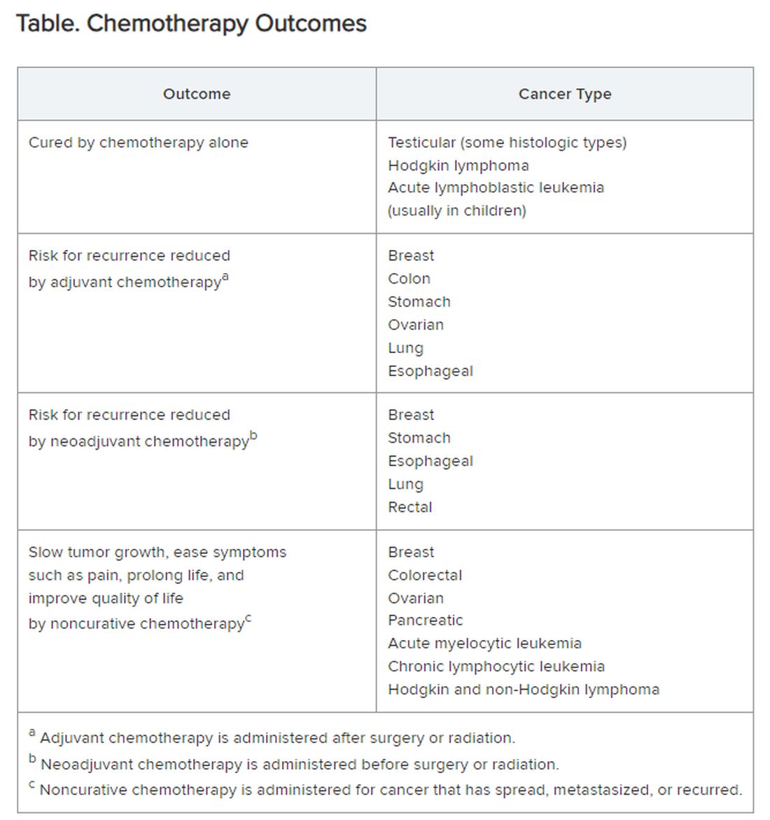
Careful, compassionate communication between patient and physician is key. Goals and expectations must be clearly understood.
Organized chemotherapeutic efforts are further categorized as first line, second line, and third line.
First-line treatment. The initial round of recommended chemotherapy for a specific cancer. It is typically considered the most effective treatment for that type and stage of cancer on the basis of current research and clinical trials.
Second-line treatment. This is the treatment used if the first-line chemotherapy doesn’t work as desired. Reasons to switch to second-line chemo include:
- Lack of response (the tumor failed to shrink).
- Progression (the cancer may have grown or spread further).
- Adverse side effects were too severe to continue.
The drugs used in second-line chemo will typically be different from those used in first line, sometimes because cancer cells can develop resistance to chemotherapy drugs over time. Moreover, the goal of second-line chemo may differ from that of first-line therapy. Rather than chiefly aiming for a cure, second-line treatment might focus on slowing cancer growth, managing symptoms, or improving quality of life. Unfortunately, not every type of cancer has a readily available second-line option.
Third-line treatment. Third-line options come into play when both the initial course of chemo (first line) and the subsequent treatment (second line) have failed to achieve remission or control the cancer’s spread. Owing to the progressive nature of advanced cancers, patients might not be eligible or healthy enough for third-line therapy. Depending on cancer type, the patient’s general health, and response to previous treatments, third-line options could include:
- New or different chemotherapy drugs compared with prior lines.
- Surgery to debulk the tumor.
- Radiation for symptom control.
- Targeted therapy: drugs designed to target specific vulnerabilities in cancer cells.
- Immunotherapy: agents that help the body’s immune system fight cancer cells.
- Clinical trials testing new or investigational treatments, which may be applicable at any time, depending on the questions being addressed.
The goals of third-line therapy may shift from aiming for a cure to managing symptoms, improving quality of life, and potentially slowing cancer growth. The decision to pursue third-line therapy involves careful consideration by the doctor and patient, weighing the potential benefits and risks of treatment considering the individual’s overall health and specific situation.
It’s important to have realistic expectations about the potential outcomes of third-line therapy. Although remission may be unlikely, third-line therapy can still play a role in managing the disease.
Navigating advanced cancer treatment is very complex. The patient and physician must together consider detailed explanations and clarifications to set expectations and make informed decisions about care.
Interventions to Consider Earlier
In traditional clinical oncology practice, other interventions are possible, but these may not be offered until treatment has reached the third line:
- Molecular testing.
- Palliation.
- Clinical trials.
- Innovative testing to guide targeted therapy by ascertaining which agents are most likely (or not likely at all) to be effective.
I would argue that the patient’s interests are better served by considering and offering these other interventions much earlier, even before starting first-line chemotherapy.
Molecular testing. The best time for molecular testing of a new malignant tumor is typically at the time of diagnosis. Here’s why:
- Molecular testing helps identify specific genetic mutations in the cancer cells. This information can be crucial for selecting targeted therapies that are most effective against those specific mutations. Early detection allows for the most treatment options. For example, for non–small cell lung cancer, early is best because treatment and outcomes may well be changed by test results.
- Knowing the tumor’s molecular makeup can help determine whether a patient qualifies for clinical trials of new drugs designed for specific mutations.
- Some molecular markers can offer information about the tumor’s aggressiveness and potential for metastasis so that prognosis can be informed.
Molecular testing can be a valuable tool throughout a cancer patient’s journey. With genetically diverse tumors, the initial biopsy might not capture the full picture. Molecular testing of circulating tumor DNA can be used to monitor a patient’s response to treatment and detect potential mutations that might arise during treatment resistance. Retesting after metastasis can provide additional information that can aid in treatment decisions.
Palliative care. The ideal time to discuss palliative care with a patient with cancer is early in the diagnosis and treatment process. Palliative care is not the same as hospice care; it isn’t just about end-of-life. Palliative care focuses on improving a patient’s quality of life throughout cancer treatment. Palliative care specialists can address a wide range of symptoms a patient might experience from cancer or its treatment, including pain, fatigue, nausea, and anxiety.
Early discussions allow for a more comprehensive care plan. Open communication about all treatment options, including palliative care, empowers patients to make informed decisions about their care goals and preferences.
Specific situations where discussing palliative care might be appropriate are:
- Soon after a cancer diagnosis.
- If the patient experiences significant side effects from cancer treatment.
- When considering different treatment options, palliative care can complement those treatments.
- In advanced stages of cancer, to focus on comfort and quality of life.
Clinical trials. Participation in a clinical trial to explore new or investigational treatments should always be considered.
In theory, clinical trials should be an option at any time in the patient’s course. But the organized clinical trial experience may not be available or appropriate. Then, the individual becomes a de facto “clinical trial with an n of 1.” Read this brief open-access blog post at Cancer Commons to learn more about that circumstance.
Innovative testing. The best choice of chemotherapeutic or targeted therapies is often unclear. The clinician is likely to follow published guidelines, often from the National Comprehensive Cancer Network.
These are evidence based and driven by consensus of experts. But guideline-recommended therapy is not always effective, and weeks or months can pass before this ineffectiveness becomes apparent. Thus, many researchers and companies are seeking methods of testing each patient’s specific cancer to determine in advance, or very quickly, whether a particular drug is likely to be effective.
Read more about these leading innovations:
SAGE Oncotest: Entering the Next Generation of Tailored Cancer Treatment
Alibrex: A New Blood Test to Reveal Whether a Cancer Treatment is Working
PARIS Test Uses Lab-Grown Mini-Tumors to Find a Patient’s Best Treatment
Using Live Cells from Patients to Find the Right Cancer Drug
Other innovative therapies under investigation could even be agnostic to cancer type:
Treating Pancreatic Cancer: Could Metabolism — Not Genomics — Be the Key?
High-Energy Blue Light Powers a Promising New Treatment to Destroy Cancer Cells
All-Clear Follow-Up: Hydrogen Peroxide Appears to Treat Oral and Skin Lesions
Cancer is a tough nut to crack. Many people and organizations are trying very hard. So much is being learned. Some approaches will be effective. We can all hope.
Dr. Lundberg, editor in chief, Cancer Commons, has disclosed no relevant financial relationships.
A version of this article appeared on Medscape.com.
The remaining 700,000 or so often proceed to chemotherapy either immediately or upon cancer recurrence, spread, or newly recognized metastases. “Cures” after that point are rare.
I’m speaking in generalities, understanding that each cancer and each patient is unique.
Chemotherapy
Chemotherapy alone can cure a small number of cancer types. When added to radiation or surgery, chemotherapy can help to cure a wider range of cancer types. As an add-on, chemotherapy can extend the length and quality of life for many patients with cancer. Since chemotherapy is by definition “toxic,” it can also shorten the duration or harm the quality of life and provide false hope. The Table summarizes what chemotherapy can and cannot achieve in selected cancer types.
Careful, compassionate communication between patient and physician is key. Goals and expectations must be clearly understood.
Organized chemotherapeutic efforts are further categorized as first line, second line, and third line.
First-line treatment. The initial round of recommended chemotherapy for a specific cancer. It is typically considered the most effective treatment for that type and stage of cancer on the basis of current research and clinical trials.
Second-line treatment. This is the treatment used if the first-line chemotherapy doesn’t work as desired. Reasons to switch to second-line chemo include:
- Lack of response (the tumor failed to shrink).
- Progression (the cancer may have grown or spread further).
- Adverse side effects were too severe to continue.
The drugs used in second-line chemo will typically be different from those used in first line, sometimes because cancer cells can develop resistance to chemotherapy drugs over time. Moreover, the goal of second-line chemo may differ from that of first-line therapy. Rather than chiefly aiming for a cure, second-line treatment might focus on slowing cancer growth, managing symptoms, or improving quality of life. Unfortunately, not every type of cancer has a readily available second-line option.
Third-line treatment. Third-line options come into play when both the initial course of chemo (first line) and the subsequent treatment (second line) have failed to achieve remission or control the cancer’s spread. Owing to the progressive nature of advanced cancers, patients might not be eligible or healthy enough for third-line therapy. Depending on cancer type, the patient’s general health, and response to previous treatments, third-line options could include:
- New or different chemotherapy drugs compared with prior lines.
- Surgery to debulk the tumor.
- Radiation for symptom control.
- Targeted therapy: drugs designed to target specific vulnerabilities in cancer cells.
- Immunotherapy: agents that help the body’s immune system fight cancer cells.
- Clinical trials testing new or investigational treatments, which may be applicable at any time, depending on the questions being addressed.
The goals of third-line therapy may shift from aiming for a cure to managing symptoms, improving quality of life, and potentially slowing cancer growth. The decision to pursue third-line therapy involves careful consideration by the doctor and patient, weighing the potential benefits and risks of treatment considering the individual’s overall health and specific situation.
It’s important to have realistic expectations about the potential outcomes of third-line therapy. Although remission may be unlikely, third-line therapy can still play a role in managing the disease.
Navigating advanced cancer treatment is very complex. The patient and physician must together consider detailed explanations and clarifications to set expectations and make informed decisions about care.
Interventions to Consider Earlier
In traditional clinical oncology practice, other interventions are possible, but these may not be offered until treatment has reached the third line:
- Molecular testing.
- Palliation.
- Clinical trials.
- Innovative testing to guide targeted therapy by ascertaining which agents are most likely (or not likely at all) to be effective.
I would argue that the patient’s interests are better served by considering and offering these other interventions much earlier, even before starting first-line chemotherapy.
Molecular testing. The best time for molecular testing of a new malignant tumor is typically at the time of diagnosis. Here’s why:
- Molecular testing helps identify specific genetic mutations in the cancer cells. This information can be crucial for selecting targeted therapies that are most effective against those specific mutations. Early detection allows for the most treatment options. For example, for non–small cell lung cancer, early is best because treatment and outcomes may well be changed by test results.
- Knowing the tumor’s molecular makeup can help determine whether a patient qualifies for clinical trials of new drugs designed for specific mutations.
- Some molecular markers can offer information about the tumor’s aggressiveness and potential for metastasis so that prognosis can be informed.
Molecular testing can be a valuable tool throughout a cancer patient’s journey. With genetically diverse tumors, the initial biopsy might not capture the full picture. Molecular testing of circulating tumor DNA can be used to monitor a patient’s response to treatment and detect potential mutations that might arise during treatment resistance. Retesting after metastasis can provide additional information that can aid in treatment decisions.
Palliative care. The ideal time to discuss palliative care with a patient with cancer is early in the diagnosis and treatment process. Palliative care is not the same as hospice care; it isn’t just about end-of-life. Palliative care focuses on improving a patient’s quality of life throughout cancer treatment. Palliative care specialists can address a wide range of symptoms a patient might experience from cancer or its treatment, including pain, fatigue, nausea, and anxiety.
Early discussions allow for a more comprehensive care plan. Open communication about all treatment options, including palliative care, empowers patients to make informed decisions about their care goals and preferences.
Specific situations where discussing palliative care might be appropriate are:
- Soon after a cancer diagnosis.
- If the patient experiences significant side effects from cancer treatment.
- When considering different treatment options, palliative care can complement those treatments.
- In advanced stages of cancer, to focus on comfort and quality of life.
Clinical trials. Participation in a clinical trial to explore new or investigational treatments should always be considered.
In theory, clinical trials should be an option at any time in the patient’s course. But the organized clinical trial experience may not be available or appropriate. Then, the individual becomes a de facto “clinical trial with an n of 1.” Read this brief open-access blog post at Cancer Commons to learn more about that circumstance.
Innovative testing. The best choice of chemotherapeutic or targeted therapies is often unclear. The clinician is likely to follow published guidelines, often from the National Comprehensive Cancer Network.
These are evidence based and driven by consensus of experts. But guideline-recommended therapy is not always effective, and weeks or months can pass before this ineffectiveness becomes apparent. Thus, many researchers and companies are seeking methods of testing each patient’s specific cancer to determine in advance, or very quickly, whether a particular drug is likely to be effective.
Read more about these leading innovations:
SAGE Oncotest: Entering the Next Generation of Tailored Cancer Treatment
Alibrex: A New Blood Test to Reveal Whether a Cancer Treatment is Working
PARIS Test Uses Lab-Grown Mini-Tumors to Find a Patient’s Best Treatment
Using Live Cells from Patients to Find the Right Cancer Drug
Other innovative therapies under investigation could even be agnostic to cancer type:
Treating Pancreatic Cancer: Could Metabolism — Not Genomics — Be the Key?
High-Energy Blue Light Powers a Promising New Treatment to Destroy Cancer Cells
All-Clear Follow-Up: Hydrogen Peroxide Appears to Treat Oral and Skin Lesions
Cancer is a tough nut to crack. Many people and organizations are trying very hard. So much is being learned. Some approaches will be effective. We can all hope.
Dr. Lundberg, editor in chief, Cancer Commons, has disclosed no relevant financial relationships.
A version of this article appeared on Medscape.com.