User login
Sickle cell patients suffer discrimination, poor care – and shorter lives
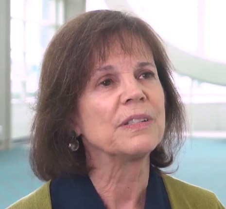
For more than a year, NeDina Brocks-Capla avoided one room in her large, brightly colored San Francisco house – the bathroom on the second floor.
“It was really hard to bathe in here, and I found myself not wanting to touch the walls,” she explained. The bathroom is where Ms. Brocks-Capla’s son Kareem Jones died in 2013 at age 36, from sickle cell disease.
It’s not just the loss of her son that upsets Ms. Brocks-Capla; she believes that if Mr. Jones had gotten the proper medical care, he might still be alive today.
Sickle cell disease is an inherited disorder that causes some red blood cells to bend into a crescent shape. The misshapen, inflexible cells clog the blood vessels, preventing blood from circulating oxygen properly, which can cause chronic pain, multiorgan failure, and stroke. About 100,000 people in the United States have sickle cell disease, and most of them are African American.
Patients and experts alike say it’s no surprise then that while life expectancy for almost every major malady is improving, patients with sickle cell disease can expect to die younger than they did 20 years ago. In 1994, life expectancy for sickle cell patients was 42 for men and 48 for women. By 2005, life expectancy had dipped to 38 for men and 42 for women.
Sickle cell disease is “a microcosm of how issues of race, ethnicity and identity come into conflict with issues of health care,” said Keith Wailoo, PhD, a professor at Princeton University who writes about the history of the disease.
It is also an example of the broader discrimination experienced by African Americans in the medical system. Nearly a third report that they have experienced discrimination when going to the doctor, according to a poll by NPR, Robert Wood Johnson Foundation, and Harvard T.H. Chan School of Public Health.
“One of the national crises in health care is the care for adult sickle cell,” said leading researcher and physician Elliott Vichinsky, MD, who started the sickle cell center at UCSF Benioff Children’s Hospital Oakland in 1978. “This group of people can live much longer with the management we have, and they’re dying because we don’t have access to care.”
Indeed, with the proper care, Dr. Vichinsky’s center and the handful of other specialty clinics like it across the country have been able to increase life expectancy for sickle cell patients well into their 60s.
Dr. Vichinsky’s patient Derek Perkins, 45, knows he has already beaten the odds. He sits in an exam room decorated with cartoon characters at Children’s Hospital Oakland, but this is the adult sickle cell clinic. He’s been Dr. Vichinsky’s patient since childhood.
“Without the sickle cell clinic here in Oakland, I don’t know what I would do. I don’t know anywhere else I could go,” Mr. Perkins said.
When Mr. Perkins was 27, he once ended up at a different hospital where doctors misdiagnosed his crisis. He went into a coma and was near death before his mother insisted he be transferred.
“Dr. Vichinsky was able to get me here to Children’s Hospital, and he found out what was wrong and within 18 hours – all I needed was an emergency blood transfusion and I was awake,” Mr. Perkins recalled.
Kareem Jones lived just across the bay from Mr. Perkins, but he had a profoundly different experience.
Mr. Jones’ mother, Ms. Brocks-Capla, said her son received excellent medical care as a child, but once he turned 18 and aged out of his pediatric program, it felt like falling off a cliff. Mr. Jones was sent to a clinic at San Francisco General Hospital, but it was open only for a half-day, one day each week. If he was sick any other day, he had two options: leave a voicemail for a clinic nurse or go to the emergency room. “That’s not comprehensive care – that’s not consistent care for a disease of this type,” said Ms. Brocks-Capla.
Ms. Brocks-Capla is a retired supervisor at a worker’s compensation firm. She knew how to navigate the health care system, but she couldn’t get her son the care he needed. Like most sickle cell patients, Mr. Jones had frequent pain crises. Usually he ended up in the emergency department where, Ms. Brocks-Capla said, the doctors didn’t seem to know much about sickle cell disease.
When she tried to explain her son’s pain to the doctors and nurses, she recalled, “they say have a seat. ‘He can’t have a seat! Can’t you see him?’ ”
Studies have found that sickle cell patients have to wait up to 50% longer for help in the emergency department than do other pain patients. The opioid crisis has made things even worse, Dr. Vichinsky added, as patients in terrible pain are likely to be seen as drug seekers with addiction problems rather than patients in need.
Despite his illness, Mr. Jones fought to have a normal life. He lived with his girlfriend, had a daughter, and worked as much as he could between pain crises. He was an avid San Francisco Giants fan.
For years, he took hydroxyurea, but it had side effects, and after a while Mr. Jones had to stop taking it. “And that was it, because you know there isn’t any other medication out there,” said Ms. Brocks-Capla.
Indeed, hydroxyurea, which the Food and Drug Administration first approved in 1967 as a cancer drug, was the only drug on the market to treat sickle cell during Mr. Jones’ lifetime. In July, the FDA approved a second drug, Endari (L-glutamine oral powder), specifically to treat patients with sickle cell disease.
Funding by the federal government and private foundations for the disease pales in comparison to other disorders. Cystic fibrosis offers a good comparison. It is another inherited disorder that requires complex care and most often occurs in Caucasians. Cystic fibrosis gets 7-11 times more funding per patient than does sickle cell disease, according to a 2013 study in the journal Blood. From 2010 to 2013 alone, the FDA approved five new drugs for the treatment of cystic fibrosis.
“There’s no question in my mind that class and color are major factors in impairing their survival. Without question,” Dr. Vichinsky said of sickle cell patients. “The death rate is increasing. The quality of care is going down.”
Without a new medication, Mr. Jones got progressively worse. At 36, his kidneys began to fail, and he had to go on dialysis. He ended up in the hospital, with the worst pain of his life. The doctors stabilized him and gave him pain meds but did not diagnose the underlying cause of the crisis. He was released to his mother’s care, still in incredible pain.
At home, Ms. Brocks-Capla ran him a warm bath to try to soothe his pain and went downstairs to get him a change of clothes. As she came back up the stairs, she heard loud banging against the bathroom walls.
“So I run into the bathroom and he’s having a seizure. And I didn’t know what to do. I was like, ‘Oh come on, come on. Don’t do this. Don’t do this to me.’ ”
She called 911. The paramedics came but couldn’t revive him. “He died here with me,” she said.
It turned out Mr. Jones had a series of small strokes. His organs were in failure, something Ms. Brocks-Capla said the hospital missed. She believes his death could have been prevented with consistent care – the kind he got as a child. Dr. Vichinsky thinks she is probably right.
“I would say 40% or more of the deaths I’ve had recently have been preventable – I mean totally preventable,” he said, but he got to the cases too late. “It makes me so angry. I’ve spent my life trying to help these people, and the harder part is you can change this – this isn’t a knowledge issue. It’s an access issue.”
Dr. Vichinsky’s center and others like it have made major advances in screening patients for the early signs of organ failure and intervening to prevent premature death. Patients at these clinics live 2 decades longer than the average sickle cell patient.
Good care for sickle cell requires time and training for physicians, but it often doesn’t pay well, because many patients are on Medicaid or other government insurance programs. The result is that most adult sickle cell patients still struggle even to access treatments that have been around for decades, Dr. Vichinsky said.
The phenomenon is nothing new — the disease that used to be known as sickle cell anemia has had a long and sordid past. It was first identified in 1910 and helped launch the field of molecular biology. But most of the research was used to study science rather than improving care for sickle cell patients, Dr. Vichinsky said.
In the 1960s and 1970s, sickle cell became a lightning rod for the civil rights movement. At the time, the average patient died before age 20. The Black Panther Party took up the cause and began testing people at its “survival conferences” across the country.
“I’m sure we tested over four-and-a-half-thousand people for sickle cell anemia last night – and I think that the voter registration is running neck and neck with it,” Black Panther Party Chairman Bobby Seale told news crews at an event in Oakland in 1972.
The movement grew, and Washington listened. “It is a sad and shameful fact that the causes of this disease have been largely neglected throughout our history,” President Richard Nixon told Congress in 1971. “We cannot rewrite this record of neglect, but we can reverse it. To this end, this administration is increasing its budget for research and treatment of sickle cell disease.”
For a while, funding did increase, newborn screening took hold, and by the 1990s, life expectancy had doubled, with patients living into their 40s. But over time, funding waned, clinics closed, and life expectancy started dropping again.
Dr. Vichinsky pushes against that trend for patients like Derek Perkins. The father of four looks healthy and robust, but like most sickle cell patients, he has episodes of extreme pain and has problems with his kidneys, heart, hips, and breathing. Keeping him thriving requires regular checkups and constant monitoring for potential problems.
“The program Dr. Vichinsky is running here, I feel I owe my life to [it],” said Mr. Perkins. “If it wasn’t for him and the things that he did for me, my family wouldn’t have me.”
Kaiser Health News is a national health policy news service that is part of the nonpartisan Henry J. Kaiser Family Foundation. KHN’s coverage of children’s health care issues is supported in part by a grant from The Heising-Simons Foundation.
Weight recidivism after bariatric surgery: What constitutes failure?
NATIONAL HARBOR, MD. – A standard definition of bariatric surgery failure based on weight regain is needed to assess long-term outcomes in place of the seemingly arbitrary thresholds now in use, according to discussion generated by long-term outcome studies presented at Obesity Week 2017.

In another study, presented by Colin Martyn, MD, a general surgery resident at Texas Tech University Health Sciences Center, El Paso, the bariatric surgery failure rate at 11 years was characterized as an “alarming” 33.9%. In this study, bariatric surgery was considered a failure if the patient did not maintain excess weight loss (EWL) of 50% or greater.
The problem with this definition, like many others, is that “it fails to recognize that there could be significant health benefits and improvements in quality of life with less weight loss,” according to Philip Schauer, MD, director of the Cleveland Clinic Bariatric and Metabolic Institute. As the invited discussant for the data presented by Dr. Martyn, Dr. Schauer acknowledged that 50% EWL has been used by others as the dividing line between success and failure, but he called it “obsolete.”
This definition was one of several applied to weight recidivism in the study presented by Dr. Morell. Others included weight regain of more than 25% EWL over the postoperative nadir, an increase in body mass index to more than 35 kg/m2 after achieving a lower BMI, and a postsurgical BMI increase of more than 5 mg/m2. Not surprisingly, weight recidivism “varied widely with regard to the definitions used,” Dr. Morell reported.
Dr. Morell’s study involved evaluation of 1,766 patients with at least 1 year of follow-up after bariatric procedure. Most (1,490 patients) underwent laparoscopic Roux-en-y gastric bypass. After 2 years of follow-up, 93% achieved at least the 50% EWL threshold of treatment success, but Dr. Morell reported that the proportion above this or any threshold progressively diminished over time. For a definition of treatment success, Dr. Morell favors maintenance of at least 20% total weight loss as a threshold of long-term clinical success, a threshold met by 75% of patients at 5 years, in his analysis.

As has been shown in these studies and reported previously, the regaining of weight over time after bariatric surgery is common and progressive, but both studies ignited controversy about what measure is meaningful for declaring that bariatric surgery has failed over the long term. None of the current thresholds for failure are based on evidence that clinical benefit has been lost. Rather, it appears that these are simply accepted conventions.
“It bothers me to hear the word failure in these presentations, because I think the paradigm is changing from success and failure to that of treating chronic disease,” said Stacy Brethauer, MD, a staff surgeon in the Cleveland Clinic Digestive Disease Institute. Dr. Brethauer, the moderator of the session at Obesity Week where both long-term follow-up papers were presented, agreed that the at least 50% EWL benchmark is “flawed.” He suggested that more clinically meaningful methods of evaluating long-term outcome are needed for both clinical and research purposes.
The discussant of Dr. Morell’s paper, Samer G. Mattar, MD, a bariatric surgeon at the Swedish Medical Center, Seattle, also called for metrics based on clinical benefit rather than on weight alone.
“I would caution against this overall emphasis that we seem to place on weight gain and weight loss as a benchmark and predominant objective for what we do,” he said. “Our nonsurgeon colleagues have repeatedly demonstrated clinical benefits from total body weight loss of 10% or even 5%. So let’s not beat up ourselves over trying to maintain a greater than 50% EWL in all our patients.”
AGA created the Obesity Practice Guide to help gastroenterologists integrate and operationalize obesity management in their practice for financial success. Learn more at www.gastro.org/obesity.
NATIONAL HARBOR, MD. – A standard definition of bariatric surgery failure based on weight regain is needed to assess long-term outcomes in place of the seemingly arbitrary thresholds now in use, according to discussion generated by long-term outcome studies presented at Obesity Week 2017.
In another study, presented by Colin Martyn, MD, a general surgery resident at Texas Tech University Health Sciences Center, El Paso, the bariatric surgery failure rate at 11 years was characterized as an “alarming” 33.9%. In this study, bariatric surgery was considered a failure if the patient did not maintain excess weight loss (EWL) of 50% or greater.
The problem with this definition, like many others, is that “it fails to recognize that there could be significant health benefits and improvements in quality of life with less weight loss,” according to Philip Schauer, MD, director of the Cleveland Clinic Bariatric and Metabolic Institute. As the invited discussant for the data presented by Dr. Martyn, Dr. Schauer acknowledged that 50% EWL has been used by others as the dividing line between success and failure, but he called it “obsolete.”
This definition was one of several applied to weight recidivism in the study presented by Dr. Morell. Others included weight regain of more than 25% EWL over the postoperative nadir, an increase in body mass index to more than 35 kg/m2 after achieving a lower BMI, and a postsurgical BMI increase of more than 5 mg/m2. Not surprisingly, weight recidivism “varied widely with regard to the definitions used,” Dr. Morell reported.
Dr. Morell’s study involved evaluation of 1,766 patients with at least 1 year of follow-up after bariatric procedure. Most (1,490 patients) underwent laparoscopic Roux-en-y gastric bypass. After 2 years of follow-up, 93% achieved at least the 50% EWL threshold of treatment success, but Dr. Morell reported that the proportion above this or any threshold progressively diminished over time. For a definition of treatment success, Dr. Morell favors maintenance of at least 20% total weight loss as a threshold of long-term clinical success, a threshold met by 75% of patients at 5 years, in his analysis.
As has been shown in these studies and reported previously, the regaining of weight over time after bariatric surgery is common and progressive, but both studies ignited controversy about what measure is meaningful for declaring that bariatric surgery has failed over the long term. None of the current thresholds for failure are based on evidence that clinical benefit has been lost. Rather, it appears that these are simply accepted conventions.
“It bothers me to hear the word failure in these presentations, because I think the paradigm is changing from success and failure to that of treating chronic disease,” said Stacy Brethauer, MD, a staff surgeon in the Cleveland Clinic Digestive Disease Institute. Dr. Brethauer, the moderator of the session at Obesity Week where both long-term follow-up papers were presented, agreed that the at least 50% EWL benchmark is “flawed.” He suggested that more clinically meaningful methods of evaluating long-term outcome are needed for both clinical and research purposes.
The discussant of Dr. Morell’s paper, Samer G. Mattar, MD, a bariatric surgeon at the Swedish Medical Center, Seattle, also called for metrics based on clinical benefit rather than on weight alone.
“I would caution against this overall emphasis that we seem to place on weight gain and weight loss as a benchmark and predominant objective for what we do,” he said. “Our nonsurgeon colleagues have repeatedly demonstrated clinical benefits from total body weight loss of 10% or even 5%. So let’s not beat up ourselves over trying to maintain a greater than 50% EWL in all our patients.”
AGA created the Obesity Practice Guide to help gastroenterologists integrate and operationalize obesity management in their practice for financial success. Learn more at www.gastro.org/obesity.
AT OBESITY WEEK 2017
Key clinical point: Many patients regain weight after bariatric surgery, but experts argue over the definition of long-term treatment failure, for which there is no standard.
Major finding: After 5 or more years of follow-up, failure rates range from 25% to 70% depending on definition of unacceptable weight regain.
Data source: A retrospective review.
Disclosures: Dr. Morell and Dr. Martyn reported no financial relationships relevant to this topic.
Methotrexate holiday linked to better flu vaccine immunogenicity
SAN DIEGO – Patients with well-controlled rheumatoid arthritis (RA) fared well during a 2-week holiday from methotrexate after flu vaccination and later showed signs of boosted immunity against the flu in comparison with patients who had not stopped the drug, according to results from a randomized controlled trial.
The research doesn’t confirm that vaccinated patients who take a break from methotrexate actually have lower rates of flu. Still, the findings suggest that brief holidays from methotrexate could be feasible in a variety of situations, such as after vaccinations and prior to surgery, said Jin Kyun Park, MD, of Seoul (South Korea) National University Hospital, lead author of the study presented at the annual meeting of the American College of Rheumatology.
The study notes that RA patients are especially prone to infections for two reasons: dysfunctional immune systems and immunity-weakening treatments. According to Dr. Park, methotrexate reduces the effectiveness of flu vaccines by 15%-20%.
In a previous study, Dr. Park and his colleagues found no statistically significant sign of increased flares in patients who went without methotrexate for 2 weeks before and 2 weeks after vaccination, 4 weeks after vaccination, and 4 weeks before vaccination (Ann Rheum Dis. 2017 Sep;76[9]:1559-65).
The earlier findings also suggested that flu vaccine uptake is highest in those who stop methotrexate after vaccination.
For the new study, a randomized controlled trial, researchers recruited patients with well-controlled RA. They assigned 159 to continue weekly doses of methotrexate after flu vaccination and 161 to stop it for 2 weeks.
The groups in the final analysis (156 and 160 subjects, respectively) were similar – about 85% women, average age of 52-53 years, and about half took glucocorticoids. Their methotrexate dose per week was about 13 mg.
At 4 weeks, just over three-quarters of the patients who had briefly stopped methotrexate showed at least a fourfold increase in hemagglutination inhibition antibody titer against two or more vaccine strains. Of those who continued the medication, just 54.5% showed this level of response, which the researchers considered to be satisfactory.
The researchers reported that there was no appreciable increase in RA disease activity.
Dr. Park cautioned that vaccine titers don’t directly reflect immunoprotection levels. Patients who took a break from methotrexate were less likely to develop a flulike illness, but the difference wasn’t statistically significant.
The research raises questions about whether methotrexate could be stopped a week or two before surgery to lower the risk of infections, Dr. Park said.
Dr. Park said that future research should focus on whether stopping methotrexate briefly affects whether patients go on to develop the flu. He would also like to look at whether a break from the medication will boost the immune response in RA patients who get herpes zoster (shingles) vaccines.
Paul Sufka, MD, of HealthPartners and Regions Hospital in St. Paul, Minn., praised the research. The 2-week break from methotrexate is “a fairly pragmatic approach,” said Dr. Sufka, who moderated a press conference where Dr. Park presented his research.
“You can actually pull this off,” he said, versus telling patients to stop the medication for the 2 weeks before they get vaccinated. He cautioned, however, that “these people have a fairly low disease activity. You may not be able to pull this off with those who have high disease activity.”
Dr. Park and Dr. Sufka reported no relevant disclosures. A study author reported consulting for Pfizer and receiving research grants from Green Cross Corp. and Hanmi Pharmaceutical. The study was funded by Green Cross.
AT ACR 2017
Key clinical point:
Major finding: More than three-quarters of patients who had briefly stopped methotrexate and 54.5% of patients who kept using methotrexate showed at least a fourfold increase in hemagglutination inhibition antibody titer against two or more vaccine strains at 4 weeks.
Data source: A randomized controlled trial of 320 patients with RA who were taking methotrexate.
Disclosures: The study was funded by Green Cross Corp. The presenter reported no relevant disclosures. A study author reported consulting for Pfizer and receiving research grants from Green Cross and Hanmi Pharmaceutical.
VIDEO: Biologic use during pregnancy had no impact on serious infection risks in infants
SAN DIEGO – Researchers found no evidence of increased risk of serious or opportunistic infections in infants born to pregnant women who were treated with biologic medication for their rheumatoid arthritis, according to a cohort study.
“These data add to what we’re beginning to learn about these medications that are so commonly used in women of reproductive age, and who have concerns about whether they can use them safely or not during pregnancy,” lead study author Christina D. Chambers, PhD, MPH, said during a press briefing at the annual meeting of the American College of Rheumatology. To date, theoretical concern exists that the use of biologics could interfere with postnatal immune function in the infant, said Dr. Chambers, a perinatal epidemiologist and teratologist at the University of California, San Diego. “The theory has been that because of the size of the molecule, little placental transfer is thought to take place early in pregnancy, but later in pregnancy, more placental transfer may be possible,” she said.
In an effort to investigate the risk of serious or opportunistic infections for infants whose mothers used biologics during pregnancy, the researchers conducted an observational cohort study from pregnant women participating in the Organization of Teratology Information Specialists (OTIS) Autoimmune Diseases in Pregnancy Project from 2004 through 2016. Mothers fell into one of three groups: 502 pregnancies where the mother with RA was treated with a biologic with or without other disease modifying anti-rheumatic medications during her pregnancy (group A); 231 pregnancies where the mother had RA but did not use any biologics during pregnancy (group B), and 423 pregnancies where the mother had no chronic diseases at all (group C). The investigators defined the serious or opportunistic infections as a list of 16 infections that included X-ray proven pneumonia, septic arthritis, osteomyelitis, tuberculosis, herpes, listeria, legionella, mycobacteria, systemic cytomegalovirus and abscess. The one-year follow-up data was collected from medical records and corroborated with maternal reports.
Among the pregnant mothers in group A, 43% took their last dose in the first or second trimester, and 57% percent took their last dose in the third trimester. Dr. Chambers reported that 20 of the 502 infants in group A developed at least one serious or opportunistic infection, for a rate of 4%, while the rates among infants in groups B and C were 2.6% and 2.1%, respectively. The most common infections seen were X-ray proven pneumonia, sepsis, bacteremia, meningitis, and abscess. Between 11% and 19% of infants had more than one infection over the one-year period.
In a subset analysis of 285 women in group A who had third trimester exposure to one of the biologics, 10 infants had at least one serious or opportunistic infection, for a rate of 3.5%, which was statistically similar to that of groups B and C (2.6% and 2.1%, respectively).
“These data provide some reassurance for clinicians who are concerned that their patients need to be treated with a biologic late in pregnancy rather than take them off the drug during that period of time,” Dr. Chambers said. She acknowledged certain limitations of the study, including the fact that the researchers did not examine risk of less serious infections, such as more frequent colds or ear infections in the infants, and they did not have any direct measure of their immune function.
Dr. Chambers disclosed having received research support from AbbVie, Amgen, Bristol Myers Squibb, Celgene, Janssen Pharmaceutica Products, L.P., Pfizer Inc, Roche Pharmaceuticals, Seqirus, GSK, UCB, and Sanofi-Aventis.
The video associated with this article is no longer available on this site. Please view all of our videos on the MDedge YouTube channel
AT ACR 2017
Key clinical point:
Major finding: Infections occurred in 4% of infants born to mothers with RA treated with a biologic with or without other disease modifying anti-rheumatic medications during her pregnancy.
Study details: An observational cohort study of 1,156 pregnant women with RA.
Disclosures: Dr. Chambers disclosed having received research support from AbbVie, Amgen, Bristol Myers Squibb, Celgene, Janssen Pharmaceutica Products, L.P., Pfizer Inc, Roche Pharmaceuticals, Seqirus, GSK, UCB, and Sanofi-Aventis.
MACRA Monday: Poor HbA1c control
If you haven’t started reporting quality data for the Merit-Based Incentive Payment System (MIPS), there’s still time to avoid a 4% cut to your Medicare payments.
Under the Pick Your Pace approach being offered this year, the Centers for Medicare & Medicaid Services allows clinicians to test the system by reporting on one quality measure for one patient through paper-based claims. Be sure to append a Quality Data Code (QDC) to the claim form for care provided up to Dec. 31, 2017, in order to avoid a penalty in payment year 2019.
Consider this measure:
The video associated with this article is no longer available on this site. Please view all of our videos on the MDedge YouTube channel
Measure #1: Diabetes: HbA1c Poor Control
The measure is aimed at capturing the percentage of patients aged 18-75 years with diabetes who had a hemoglobin A1c greater than 9.0%. For this inverse measure, a lower performance rate indicates better clinical care.
What you need to do: Document the patient’s most recent HbA1c level that was performed during the last 12 months.
Eligible cases include patients aged 18-75 years on the date of the encounter who had a documented diagnosis of diabetes. One of the following services must be performed at the visit (CPT or HCPCS): 97802, 97803, 97804, 99201, 99202, 99203, 99204, 99205, 99211, 99212, 99213, 99214, 99215, 99217, 99218, 99219, 99220, 99221, 99222, 99223, 99231, 99232, 99233, 99238, 99239, 99281, 99282, 99283, 99284, 99285, 99291, 99304, 99305, 99306, 99307, 99308, 99309, 99310, 99315, 99316, 99318, 99324, 99325, 99326, 99327, 99328, 99334, 99335, 99336, 99337, 99341, 99342, 99343, 99344, 99345, 99347, 99348, 99349, 99350, G0270, G0271, G0402, G0438, G0439.
To get credit under MIPS, be sure to include a QDC that shows that you successfully performed the measure or that you had a good reason for not doing so. For instance, CPT II 3046F indicates that the most recent hemoglobin A1c level was greater than 9.0%, CPT II 3044F indicates that the most recent HbA1c level was less than 7.0%, and CPT II 3045F indicates that the most recent HbA1c level was between 7.0% and 9.0%.
CMS has a full list of measures available for claims-based reporting at qpp.cms.gov. The American Medical Association also has created a step-by-step guide for reporting on one quality measure.
Certain clinicians are exempt from reporting and do not face a penalty under MIPS:
• Those who enrolled in Medicare for the first time during a performance period.
• Those who have Medicare Part B allowed charges of $30,000 or less.
• Those who have 100 or fewer Medicare Part B patients.
• Those who are significantly participating in an Advanced Alternative Payment Model (APM).

VIDEO: Obesity linked to worse outcomes in axial spondyloarthropathy
SAN DIEGO – Among patients with axial spondyloarthropathy, higher BMI and obesity independently predicts worse disease outcomes, according to results from a registry study.
“Obesity is one of the biggest public health challenges facing us in the 21st century,” lead study author Gillian Fitzgerald, MD, said in an interview in advance of the annual meeting of the American College of Rheumatology.
“Traditionally, we have a perception of patients with axial SpA being of normal or even thin body habitus. However, recent studies have indicated that this is not the case and that obesity is prevalent in axial SpA patients. The negative consequences of obesity in the general population are well documented, with affected patients suffering greater morbidity and mortality.”
Dr. Fitzgerald, of St. James’s Hospital, Dublin, Ireland, noted that research to date in axial SpA indicates that disease outcomes may be worse in obese patients. However, existing literature looking at obesity in axial SpA is relatively sparse. In an effort to clarify this issue, she and her associates evaluated 683 patients from the Ankylosing Spondylitis Registry of Ireland (ASRI), which is designed to provide descriptive epidemiological data on the Irish axSpA population via standardized clinical assessments and structured interviews. The mean age of the 683 patients enrolled as of June 2017 was 46, the majority (77%) were men, their mean disease duration was 19 years, and their mean delay to diagnosis was nine years. Most (79%) fulfilled Modified New York modified criteria, their mean Bath Ankylosing Spondylitis Disease Activity Index (BASDAI) was 3.9, their mean Bath Ankylosing Spondylitis Metrology Index (BASMI) was 3.6, their mean Bath Ankylosing Spondylitis Functional Index (BASFI) was 3.6 and their mean Health Assessment Questionnaire (HAQ) was 0.52.
Based on WHO criteria, the cohort’s mean BMI was 27.8 kg/m2. Of these, 38.9% were overweight and 28.4% were obese. “Indeed, only 32% of the cohort have a healthy BMI,” Dr. Fitzgerald commented. “When we looked at the relationship between BMI and disease outcomes, we found that obese patients had more severe disease than their normal weight and overweight counterparts, with higher measures of disease activity, quality of life, disability and function, as well as worse spinal mobility.”
The researchers also observed that the prevalence of smoking was lower in obese patients, compared with normal weight patients (18% vs. 38, respectively). In univariable linear regression, BMI and obesity were associated with higher BASDAI, BASMI, BASFI and HAQ scores. In multivariable regression analysis, only obesity remained an independent predictor of higher disease activity and worse function (P less than .01).
“As clinicians, we are always looking for ways to reduce the burden of disease that patients carry and to improve outcomes,” Dr. Fitzgerald said. “In this study, we demonstrated that over two-thirds of our axial SpA patients are either overweight or obese, and that these patients have more severe disease. Further research is needed to clarify this relationship between obesity and disease severity; in particular, the effect of losing weight on disease outcomes needs to be clarified. However, when devising treatment plans for axial SpA patients, this study provides rheumatologists with a strong rationale to include strategies to actively control weight.”
She acknowledged that the study’s cross-sectional design is a limitation. “This means cause and effect can’t be determined exclusively from this study; therefore, prospective studies are required to further clarify this relationship that we have noted between obesity and disease outcomes.”
ASRI is funded by an unrestricted grant from AbbVie and Pfizer. Dr. Fitzgerald disclosed having received research support from AbbVie.
The video associated with this article is no longer available on this site. Please view all of our videos on the MDedge YouTube channel
AT ACR 2017
Key clinical point: Obese patients with axial spondyloarthropathy have worse disease outcomes.
Major finding: In multivariable regression analysis, only obesity remained an independent predictor of higher disease activity and worse function (P less than .01).
Study details: A cross-sectional study of 683 patients with axial spondyloarthropathy.
Disclosures: ASRI is funded by an unrestricted grant from AbbVie and Pfizer. Dr. Fitzgerald disclosed having received research support from AbbVie Hopkins.

Cancer drug costs increasing despite competition

Cancer drug costs in the US increase substantially after launch, regardless of competition, according to a study published in the Journal of Clinical Oncology.*
Researchers studied 24 cancer drugs approved over the last 20 years and found a mean cumulative cost increase of about 37%, or 19% when adjusted for inflation.
Among drugs approved to treat hematologic malignancies, the greatest inflation-adjusted price increases were for arsenic trioxide (57%), nelarabine (55%), and rituximab (49%).
The lowest inflation-adjusted price increases were for ofatumumab (8%), clofarabine (8%), and liposomal vincristine (18%).
For this study, Daniel A. Goldstein, MD, of Emory University in Atlanta, Georgia, and his colleagues measured the monthly price trajectories of 24 cancer drugs approved by the US Food and Drug Administration. This included 10 drugs approved to treat hematologic malignancies between 1997 and 2011.
To account for discounts and rebates, the researchers used the average sales prices published by the Centers for Medicare and Medicaid Services and adjusted to general and health-related inflation rates. For each drug, the researchers calculated the cumulative and annual drug cost changes.
Results
The mean follow-up was 8 years. The mean cumulative cost increase for all 24 drugs was +36.5% (95% CI, 24.7% to 48.3%).
The general inflation-adjusted increase was +19.1% (95% CI, 11.0% to 27.2%), and the health-related inflation-adjusted increase was +8.4% (95% CI, 1.4% to 15.4%).
Only 1 of the 24 drugs studied had a price decrease over time. That drug is ziv-aflibercept, which was approved to treat metastatic colorectal cancer in 2012.
Ziv-aflibercept was launched with an annual price exceeding $110,000. After public outcry, the drug’s manufacturer, Sanofi, cut the price in half. By the end of the study’s follow-up period in 2017, the cost of ziv-aflibercept had decreased 13% (inflation-adjusted decrease of 15%, health-related inflation-adjusted decrease of 20%).
Cost changes for the drugs approved to treat hematologic malignancies are listed in the following table.
| Drug (indication, approval date, years of follow-up) | Mean monthly cost at launch | Mean annual cost change (SD) | Cumulative cost change | General and health-related inflation-adjusted change, respectively |
| Arsenic trioxide (APL, 2000, 12) | $11,455 | +6% (4) | +95% | +57%, +39% |
| Bendamustine (CLL, NHL, 2008, 8) | $6924 | +5% (5) | +50% | +32%, +21% |
| Bortezomib (MM, MCL, 2003, 12) | $5490 | +4% (3) | +63% | +31%, +16% |
| Brentuximab (lymphoma, 2011, 4) | $19,482 | +8% (0.1) | +35% | +29%, +22% |
| Clofarabine (ALL, 2004, 11) | $56,486 | +3% (3) | +31% | +8%, -4% |
| Liposomal vincristine (ALL, 2012, 3) | $34,602 | +8% (0.5) | +21% | +18%, +14% |
| Nelarabine (ALL, lymphoma, 2005, 10) | $18,513 | +6% (2) | +83% | +55%, +39% |
| Ofatumumab (CLL, 2009, 6) | $4538 | +3% (2) | +17% | +8%, -0.5% |
| Pralatrexate (lymphoma, 2009, 6) | $31,684 | +6% (4) | +43% | +31%, +21% |
| Rituximab (NHL, CLL, 1997, 12) | $4111 | +5% (0.5) | +85% | +49%, +32% |
Abbreviations: ALL, acute lymphoblastic leukemia; APL, acute promyelocytic leukemia; CLL, chronic lymphocytic leukemia; MCL, mantle cell lymphoma; MM, multiple myeloma; NHL, non-Hodgkin lymphoma; SD, standard deviation.
The researchers noted that there was a steady increase in drug costs over the study period, regardless of whether a drug was granted a new supplemental indication, the drug had a new off-label indication, or a competitor drug was approved.
The only variable that was significantly associated with price change was the amount of time that had elapsed from a drug’s launch.
This association was significant in models in which the researchers used prices adjusted to inflation (P=0.002) and health-related inflation (P=0.023). However, it was not significant when the researchers used the actual drug price (P=0.085). ![]()
*Data in the abstract differ from data in the body of the JCO paper. This article includes data from the body of the JCO paper.
Rib fracture diagnosis in the panscan era
Clinical question: Do rib fractures observed on chest CT carry the same morbidity and mortality risk as those observed in chest radiograph?
Background: Traditionally studies have shown that first and second rib fractures on chest radiograph after blunt trauma are associated with substantial morbidity and mortality. With growing frequency of CT imaging in the “panscan” era, it is unknown whether similar rib fractures found on CT carry the same meaning.
Setting: 10 level I trauma centers.
Synopsis: Data from the National Emergency X-Radiography Utilization Study showed that, of the 8,661 patients who suffered blunt trauma and received both chest radiograph and chest CT, 23.9% had rib fractures. Rib fractures were observed in 66.1% of chest CT–only cases. Patients with rib fractures had a higher admission rate (88.7% versus 45.8%) and higher mortality (5.6% versus 2.7%) than patients without rib fractures. Mortality rate and great-vessel injury were higher in those with first or second rib fractures. The mortality of patients with rib fractures observed only on chest CT was not statistically different from those whose fractures were also seen in chest radiograph. The study included patients who were more severely injured and may have been more likely to receive a CT, which may have led to an overestimation of fractures found. The actual causes of admission and death were not reviewed.
Bottom line: CT in trauma-imaging protocol can identify patients with rib fractures well, compared with combined CT with chest radiograph. Rib fractures are associated with higher rates of admission and mortality risk than those without rib fractures. Specifically, first or second rib fractures are found to have greater risk for mortality and great-vessel injury.
Citation: Murphy CE 4th, Raja AS, Baumann BM, et al. Rib fracture diagnosis in the panscan era. Ann Emerg Med. 2017. doi: 10.1016/j.annemergmed.2017.04.011.
Dr. Xu is assistant professor and hospitalist, Icahn School of Medicine of the Mount Sinai Health System, New York.
The Tempest Within
On the heels of Hurricane Harvey, which devastated the city of Houston and other communities in Texas and Louisiana, Hurricane Irma ravaged several islands in the Caribbean—and then headed for the states. In the days before she made landfall in the US, the media offered seemingly minute-to-minute updates on her progress. Each new forecast seemed to contradict the previous, demonstrating the unpredictability of natural disasters. But as the hurricane crept closer, one thing was evident: Florida was going to take a hard hit.
Keeping up with the fluctuating weather report was like watching a tennis match: East Coast … nope, not the East Coast … probably the middle of the state. We breathed periodic sighs of relief but remained leery. Then, several of the spaghetti plots (may I never hear that term again!) showed Irma veering west—right over our heads. Hysteria set in. One meteorologist sounded absolutely frantic as she warned people to GET READY!!!!
Now, to be clear: My purpose in writing is not to disparage media coverage or governmental response, nor to minimize anyone else’s struggles. Rather, I want to share how the hurricane affected my neighbors, friends, family, and myself—and continues to do so, weeks afterward.
Once Irma’s course was set, we swept into action. Our emergency plan included hurricane-proofing our home—protective awnings placed over the windows; outside decorations put away; grill stored properly; palm trees trimmed—and laying in sufficient supplies (gallon jugs of water, needed medications, bread and peanut butter). We gathered important documents, filled the car with gas, and made sure to have cash on hand. This flurry of activity got the adrenaline pumping, but there was something satisfying about checking off each item on our list. Before you knew it, we were set.
Then Governor Scott took the proactive step of declaring a state of emergency, before Irma was even in striking distance. This was beneficial for all Floridians, since it positioned us to receive federal assistance if needed and allowed local officials to act quickly, without the burden of bureaucracy or red tape.
However, as this news spread, our phones began to ring, buzz, and ping. Friends around the country wanted to know, “Are you okay?” and to offer us a place to stay if we needed to get out. These well-intentioned messages were appreciated—but each expression of concern reminded us that we were facing something big. We were fine, though. Prepared. And the storm could still bypass us or at least hit in a weakened state.
As Irma moved closer, the nervous energy in our little community began to rise. Some neighbors headed north several days before the predicted arrival, spending hours in traffic. We were glad we’d decided to shelter in place instead! Our decision was met with worried looks and wringing hands, which perplexed us. After all, we live about two miles from the Gulf of Mexico and 40-plus feet above sea level. My parents had moved to the area in 1978, and Dad always told us it was where people evacuated to, not from. No problem!
Except … then the mandatory evacuation notice was given. Uh oh! Time to revisit our “shelter in place” plan. What were the options again?
Plan A: Call a friend who lives three towns away in a non-evacuation zone. But her daughter gets evacuated every storm, so she, her husband, and their three dogs had already claimed the guest room.
Plan B: Call another friend in the next town over. She was happy to accommodate us! We planned to arrive the night before the predicted hit and wait out the storm there. The plan was foolproof ... until she also got a mandatory evacuation notice.
Plan C: Find a hotel in a safe area. No luck—all booked.
Admittedly, with each snag in our plans, our stress and anxiety increased. We began to question our initial decision to stay put. Had we missed our opportunity to get out of town?
Then, thankfully, the phone rang. Our friend’s daughter had miraculously secured us hotel rooms about 30 miles from our home.
The strangest thing about riding out a hurricane is that you have days of anticipation and action—you prepare—and then you just have to wait. In the confines of our “bunker,” we had naught to do but track the storm. For three days, local television stations aired only the weather; there was no respite from the red tracking markers. The endless barrage of information added to our already heightened stress levels. We wondered what we would face once we returned home. The thought of major damage sickened us.
Three days after Irma visited, we were cleared to head home. We were admittedly nervous to see what she had left behind—but incredibly, we found our house exactly as we had left it. We hugged each other, in tears. The surrounding damage ranged from minor to major, but thankfully, no one was injured. Neighbors who had fled town asked us to send pictures of their homes so they could see for themselves. The consensus: What was broken can be fixed; we are all thankful to have survived. In a way, I expected that to be the end of the story. But I was wrong.
In my February 2010 editorial, I addressed the aftermath of the earthquake that had struck Haiti the month before.1 Seven years later, 2.5 million Haitians are still in need of humanitarian aid, and 55,000 people are in camps and makeshift camps.2 You might be thinking, “An earthquake in Haiti is much different from a hurricane in the United States.” That may be true, but some victims of Hurricane Sandy are still trying to repair damage to their homes, five years later.3 So while Hurricanes Irma and Harvey—let alone Hurricane Sandy—are already off the front pages, the despair, emotional impact, and disbelief associated with the disasters endure. As one woman described, “You’re in complete shock. You’re trying to figure out, ‘Is this happening to me? Am I in some sort of dream I can’t wake up from?’”3
We are not people who are easily discouraged or quick to worry about things we can’t control. But this experience was very different: The sensory overload was unnerving, and the anxiety and stress linger. As a result, I have an entirely new perspective on the effect of disaster on mental health. The worst we suffered was four days without power, yet as I write this weeks later, I find myself in tears, reliving the fear and anxiety we felt during Irma’s wrath. We were ready for a hurricane, but not for the emotional turmoil that has followed.
Have you experienced a disaster, natural or otherwise, that left you shaken? Share your experience, and any advice you may have, with me at [email protected].
1. Onieal ME. When helping hands are tied. Clinician Reviews. 2010;20(2):C2, 18-20.
2. Cook J. 7 years after Haiti’s earthquake, millions still need aid. www.huffingtonpost.com/entry/haiti-earthquake-anniversary_us_5875108de4b02b5f858b3f9c. Accessed October 6, 2017.
3. Sandoval E, Marcius CR, Durkin E, Dillon N. Hurricane Sandy victims’ homes still uninhabitable nearly five years later. www.nydailynews.com/new-york/hurricane-sandy-victims-living-limbo-years-article-1.3463866. Accessed October 6, 2017.

On the heels of Hurricane Harvey, which devastated the city of Houston and other communities in Texas and Louisiana, Hurricane Irma ravaged several islands in the Caribbean—and then headed for the states. In the days before she made landfall in the US, the media offered seemingly minute-to-minute updates on her progress. Each new forecast seemed to contradict the previous, demonstrating the unpredictability of natural disasters. But as the hurricane crept closer, one thing was evident: Florida was going to take a hard hit.
Keeping up with the fluctuating weather report was like watching a tennis match: East Coast … nope, not the East Coast … probably the middle of the state. We breathed periodic sighs of relief but remained leery. Then, several of the spaghetti plots (may I never hear that term again!) showed Irma veering west—right over our heads. Hysteria set in. One meteorologist sounded absolutely frantic as she warned people to GET READY!!!!
Now, to be clear: My purpose in writing is not to disparage media coverage or governmental response, nor to minimize anyone else’s struggles. Rather, I want to share how the hurricane affected my neighbors, friends, family, and myself—and continues to do so, weeks afterward.
Once Irma’s course was set, we swept into action. Our emergency plan included hurricane-proofing our home—protective awnings placed over the windows; outside decorations put away; grill stored properly; palm trees trimmed—and laying in sufficient supplies (gallon jugs of water, needed medications, bread and peanut butter). We gathered important documents, filled the car with gas, and made sure to have cash on hand. This flurry of activity got the adrenaline pumping, but there was something satisfying about checking off each item on our list. Before you knew it, we were set.
Then Governor Scott took the proactive step of declaring a state of emergency, before Irma was even in striking distance. This was beneficial for all Floridians, since it positioned us to receive federal assistance if needed and allowed local officials to act quickly, without the burden of bureaucracy or red tape.
However, as this news spread, our phones began to ring, buzz, and ping. Friends around the country wanted to know, “Are you okay?” and to offer us a place to stay if we needed to get out. These well-intentioned messages were appreciated—but each expression of concern reminded us that we were facing something big. We were fine, though. Prepared. And the storm could still bypass us or at least hit in a weakened state.
As Irma moved closer, the nervous energy in our little community began to rise. Some neighbors headed north several days before the predicted arrival, spending hours in traffic. We were glad we’d decided to shelter in place instead! Our decision was met with worried looks and wringing hands, which perplexed us. After all, we live about two miles from the Gulf of Mexico and 40-plus feet above sea level. My parents had moved to the area in 1978, and Dad always told us it was where people evacuated to, not from. No problem!
Except … then the mandatory evacuation notice was given. Uh oh! Time to revisit our “shelter in place” plan. What were the options again?
Plan A: Call a friend who lives three towns away in a non-evacuation zone. But her daughter gets evacuated every storm, so she, her husband, and their three dogs had already claimed the guest room.
Plan B: Call another friend in the next town over. She was happy to accommodate us! We planned to arrive the night before the predicted hit and wait out the storm there. The plan was foolproof ... until she also got a mandatory evacuation notice.
Plan C: Find a hotel in a safe area. No luck—all booked.
Admittedly, with each snag in our plans, our stress and anxiety increased. We began to question our initial decision to stay put. Had we missed our opportunity to get out of town?
Then, thankfully, the phone rang. Our friend’s daughter had miraculously secured us hotel rooms about 30 miles from our home.
The strangest thing about riding out a hurricane is that you have days of anticipation and action—you prepare—and then you just have to wait. In the confines of our “bunker,” we had naught to do but track the storm. For three days, local television stations aired only the weather; there was no respite from the red tracking markers. The endless barrage of information added to our already heightened stress levels. We wondered what we would face once we returned home. The thought of major damage sickened us.
Three days after Irma visited, we were cleared to head home. We were admittedly nervous to see what she had left behind—but incredibly, we found our house exactly as we had left it. We hugged each other, in tears. The surrounding damage ranged from minor to major, but thankfully, no one was injured. Neighbors who had fled town asked us to send pictures of their homes so they could see for themselves. The consensus: What was broken can be fixed; we are all thankful to have survived. In a way, I expected that to be the end of the story. But I was wrong.
In my February 2010 editorial, I addressed the aftermath of the earthquake that had struck Haiti the month before.1 Seven years later, 2.5 million Haitians are still in need of humanitarian aid, and 55,000 people are in camps and makeshift camps.2 You might be thinking, “An earthquake in Haiti is much different from a hurricane in the United States.” That may be true, but some victims of Hurricane Sandy are still trying to repair damage to their homes, five years later.3 So while Hurricanes Irma and Harvey—let alone Hurricane Sandy—are already off the front pages, the despair, emotional impact, and disbelief associated with the disasters endure. As one woman described, “You’re in complete shock. You’re trying to figure out, ‘Is this happening to me? Am I in some sort of dream I can’t wake up from?’”3
We are not people who are easily discouraged or quick to worry about things we can’t control. But this experience was very different: The sensory overload was unnerving, and the anxiety and stress linger. As a result, I have an entirely new perspective on the effect of disaster on mental health. The worst we suffered was four days without power, yet as I write this weeks later, I find myself in tears, reliving the fear and anxiety we felt during Irma’s wrath. We were ready for a hurricane, but not for the emotional turmoil that has followed.
Have you experienced a disaster, natural or otherwise, that left you shaken? Share your experience, and any advice you may have, with me at [email protected].
1. Onieal ME. When helping hands are tied. Clinician Reviews. 2010;20(2):C2, 18-20.
2. Cook J. 7 years after Haiti’s earthquake, millions still need aid. www.huffingtonpost.com/entry/haiti-earthquake-anniversary_us_5875108de4b02b5f858b3f9c. Accessed October 6, 2017.
3. Sandoval E, Marcius CR, Durkin E, Dillon N. Hurricane Sandy victims’ homes still uninhabitable nearly five years later. www.nydailynews.com/new-york/hurricane-sandy-victims-living-limbo-years-article-1.3463866. Accessed October 6, 2017.
New, persistent opioid use more common after bariatric surgery
SAN DIEGO – Bariatric patients are nearly 50% more likely than general surgery patients to start using opioids after their procedures and continue taking the painkillers for a year, a new study finds.
It’s not clear why bariatric patients are at higher risk of continued opioid use, nor whether they are more likely to become addicted. Still, bariatric patients are a target for “intervention, enhanced education, early referral to specialists, protocols minimizing inpatient and outpatient narcotics, opioid-free operations, system-based interventions and prescribing guidelines,” says study lead author Sanjay Mohanty, MD, a surgery resident with the Henry Ford Health System, who spoke in a presentation at the annual clinical congress of the American College of Surgeons.
There’s been little research into opioid use among bariatric patients, said Dr. Mohanty. In 2013, a retrospective study found that 8% of 11,719 bariatric patients were chronic opioid users, and more than three-quarters of those remained so after 1 year. However, that study was completed in 2010 before the height of the opioid epidemic (JAMA. 2013;310(13):1369-76).
More recently, a 2017 study found that opioid use among 1,892 bariatric patients who weren’t using at baseline grew from 5.8% at 6 months to 14.2% at 7 years. The study tracked patients until January 2015 (Surg Obes Relat Dis. 2017 Aug;13 (8):1337-46).

For the new study, researchers tracked 14,063 bariatric patients in the Michigan Bariatric Surgery Collaborative, a group of Michigan hospitals and health systems, from 2006-2017.
Of the patients, 73% were opioid-naive at baseline and 27% were users. At 1 year after procedure, overall use dropped slightly to 24%. However, 905 patients – 8.8% of the initial opioid-native group – were new and persistent opioid users.
According to Dr. Carlin, this is almost 50% higher than in patients after general surgical procedures.
These users were significantly more likely to be black (OR 1.67), less likely to have private insurance (0.76 OR), more likely to have income under $25,000 (OR 1.43), and more likely to have a mobility limitation (OR 1.78).
The researchers also found evidence linking a higher risk of new and persistent opioid use to lack of unemployment, depression, musculoskeletal disorders, tobacco use and gastric bypass procedures.
Why might bariatric patients in general be more susceptible to new and persistent opioid use? “We don’t know that answer,” Dr. Carlin said. “Maybe there’s some addiction transfer. Or maybe it’s something physiologic. We’re doing an operation on the gut, and that could have an impact on absorption.”
As for solutions, Dr. Carlin says “prescribe less, prescribe differently, be more patient-specific. We’re looking at different modalities to treat the pain such as nerve blocks during surgery, anti-inflammatories and muscle relaxants.”
And if patients aren’t using opioids in the hospital and not having that much pain, he said, physicians don’t send any pills home with them.
The next steps should include research into links between opioids and perioperative complications and surgical outcomes, the researchers suggested.
Dr. Carlin and Dr. Mohanty report no relevant disclosures.
SAN DIEGO – Bariatric patients are nearly 50% more likely than general surgery patients to start using opioids after their procedures and continue taking the painkillers for a year, a new study finds.
It’s not clear why bariatric patients are at higher risk of continued opioid use, nor whether they are more likely to become addicted. Still, bariatric patients are a target for “intervention, enhanced education, early referral to specialists, protocols minimizing inpatient and outpatient narcotics, opioid-free operations, system-based interventions and prescribing guidelines,” says study lead author Sanjay Mohanty, MD, a surgery resident with the Henry Ford Health System, who spoke in a presentation at the annual clinical congress of the American College of Surgeons.
There’s been little research into opioid use among bariatric patients, said Dr. Mohanty. In 2013, a retrospective study found that 8% of 11,719 bariatric patients were chronic opioid users, and more than three-quarters of those remained so after 1 year. However, that study was completed in 2010 before the height of the opioid epidemic (JAMA. 2013;310(13):1369-76).
More recently, a 2017 study found that opioid use among 1,892 bariatric patients who weren’t using at baseline grew from 5.8% at 6 months to 14.2% at 7 years. The study tracked patients until January 2015 (Surg Obes Relat Dis. 2017 Aug;13 (8):1337-46).
For the new study, researchers tracked 14,063 bariatric patients in the Michigan Bariatric Surgery Collaborative, a group of Michigan hospitals and health systems, from 2006-2017.
Of the patients, 73% were opioid-naive at baseline and 27% were users. At 1 year after procedure, overall use dropped slightly to 24%. However, 905 patients – 8.8% of the initial opioid-native group – were new and persistent opioid users.
According to Dr. Carlin, this is almost 50% higher than in patients after general surgical procedures.
These users were significantly more likely to be black (OR 1.67), less likely to have private insurance (0.76 OR), more likely to have income under $25,000 (OR 1.43), and more likely to have a mobility limitation (OR 1.78).
The researchers also found evidence linking a higher risk of new and persistent opioid use to lack of unemployment, depression, musculoskeletal disorders, tobacco use and gastric bypass procedures.
Why might bariatric patients in general be more susceptible to new and persistent opioid use? “We don’t know that answer,” Dr. Carlin said. “Maybe there’s some addiction transfer. Or maybe it’s something physiologic. We’re doing an operation on the gut, and that could have an impact on absorption.”
As for solutions, Dr. Carlin says “prescribe less, prescribe differently, be more patient-specific. We’re looking at different modalities to treat the pain such as nerve blocks during surgery, anti-inflammatories and muscle relaxants.”
And if patients aren’t using opioids in the hospital and not having that much pain, he said, physicians don’t send any pills home with them.
The next steps should include research into links between opioids and perioperative complications and surgical outcomes, the researchers suggested.
Dr. Carlin and Dr. Mohanty report no relevant disclosures.
AT THE ACS CLINICAL CONGRESS
Key clinical point: It’s common for opioid-naive bariatric patients to begin using opioids after surgery and continuing at 1 year.
Major finding: At 1 year after surgery, 8.8% of the 73% of bariatric patients who were opioid-naive were new and persistent opioid users.
Data source: 14,063 Michigan bariatric patients tracked from 2006-2017.
Disclosures: The study authors report no relevant disclosures.