User login
Protein may be target for enhancing HSC transplants

Targeting the protein Del-1 could potentially improve hematopoietic stem cell (HSC) transplants, according to researchers.
The team found that Del-1 promoted engraftment in murine transplant recipients, but the protein also promoted the retention of hematopoietic progenitors in the bone marrow of mice that received granulocyte colony-stimulating factor (G-CSF).
The researchers therefore believe that enhancing Del-1 in HSC transplant recipients might improve engraftment.
And inhibiting Del-1 could increase progenitor mobilization in transplant donors.
The researchers detailed these findings and theories in The Journal of Clinical Investigation.
“Because the hematopoietic stem cell niche is so important for the creation of bone marrow and blood cells and because Del-1 is a soluble protein and easily manipulated, one can see that it could be a target in many potential applications,” said study author George Hajishengallis, DDS, PhD, of Penn Dental Medicine in Philadelphia, Pennsylvania.
For Dr Hajishengallis, the route to studying Del-1 in the bone marrow began in his field of dental medicine.
Dr Hajishengallis and Triantafyllos Chavakis, Dr med, of Technische Universität Dresden in Germany, identified Del-1 as a potential drug target for gum disease. (Del-1 prevents inflammatory cells from moving into the gums.)
Both of the researchers’ labs also discovered that Del-1 is expressed in bone marrow, so the groups began following up to examine the protein’s function there.
Their work revealed that Del-1 is a key regulator of the HSC niche.
Del-1 is expressed by cells that promote HSC maintenance under steady-state conditions. This includes arteriolar endothelial cells, CXCL12-abundant reticular cells, and cells of the osteoblastic lineage.
The researchers also found that Del-1 regulates long-term HSC proliferation and differentiation toward the myeloid lineage.
In mice that received HSC transplants, Del-1 promoted progenitor engraftment and the generation of both progenitors and mature myeloid cells. The researchers noted that this was dependent upon β3 integrin expression in hematopoietic cells.
The team also found that Del-1 promotes hematopoietic progenitors’ response to systemic inflammation (induced by lipopolysaccharide). And the protein promotes the retention of hematopoietic progenitors in the bone marrow after G-CSF administration.
Taken together, these findings suggest that manipulating Del-1 might enhance HSC transplants.
“It’s easy to think of practical applications,” Dr Hajishengallis said. “Now, we need to find out whether it works in practice, so our studies continue.” ![]()
Docs to receive slightly better Medicare pay bump than originally proposed
Physicians will see a 0.41% increase to their payments under the Medicare physician fee schedule in 2018, a slight increase from the proposed 0.31% uptick but still short of the 0.5% increase promised under the Medicare Access and CHIP Reauthorization Act (MACRA).
Officials at the Centers for Medicare & Medicaid Services were unable to find adequate funding in so-called misvalued codes to back the larger increase, as required by law, according to the final version of the 2018 physician fee schedule, released Nov. 2 and scheduled for publication in the Federal Register on Nov. 15.
“We finalized these changes based on stakeholder feedback and to better align with the MIPS data submission requirements for the quality performance category,” CMS said in a fact sheet detailing the provisions of the final rule.
CMS also is delaying the start of the appropriate use criteria (AUC) for imaging services, a program that would deny payments for imaging services unless the ordering physician consulted appropriate use criteria. The program will begin with an educational and operational testing year in 2020. Physicians will be required to start using AUCs and reporting this information on claims, but CMS will pay claims regardless of whether they correctly contain the required AUC data.
“This allows both clinicians and the agency to prepare for this new program,” the agency said in the fact sheet, CMS had proposed 2019 be the educational and operational testing year.
In response to comments submitted to the agency, CMS is changing its policy on billing codes for biosimilars administered under Medicare Part B.
“Effective January 1, 2018, newly approved biosimilar products with a common reference product will no longer be grouped in the same billing code,” the agency said in the fact sheet. “By encouraging innovation and greater manufacturer participation in the marketplace, we believe that this policy change will result in the licensing of more biosimilar products, thus creating a stable and robust market, driving market competition, and decreasing uncertainty about access and payment.”
The final rule implements proposed expansion of the Medicare Diabetes Prevention Program from a demonstration project to a nationwide program in 2018, however the implementation will be delayed for three months until April 1, 2018, rather than at the beginning of the year. The program provides payments to physicians based on performance goals being met by patients, including meeting certain numbers of service and maintenance sessions with the program and achieving specific weight loss goals.
CMS also finalized a number of new telemedicine payment codes.
Mike Nelson, MD, FCCP, comments: One need not “google” too long to find that the United States performs quite poorly in overall health care when compared with other nations, despite spending more than any of the comparators…we’re 37th this year. This information from Avalere Health portends a further drop in our ranking next year. The privilege of good health is a responsibility of the individual, but the right to affordable health care is a responsibility of the government. It is time for our legislators to stop playing partisan politics and start communicating to propose a workable and affordable solution.

Mike Nelson, MD, FCCP, comments: One need not “google” too long to find that the United States performs quite poorly in overall health care when compared with other nations, despite spending more than any of the comparators…we’re 37th this year. This information from Avalere Health portends a further drop in our ranking next year. The privilege of good health is a responsibility of the individual, but the right to affordable health care is a responsibility of the government. It is time for our legislators to stop playing partisan politics and start communicating to propose a workable and affordable solution.
Mike Nelson, MD, FCCP, comments: One need not “google” too long to find that the United States performs quite poorly in overall health care when compared with other nations, despite spending more than any of the comparators…we’re 37th this year. This information from Avalere Health portends a further drop in our ranking next year. The privilege of good health is a responsibility of the individual, but the right to affordable health care is a responsibility of the government. It is time for our legislators to stop playing partisan politics and start communicating to propose a workable and affordable solution.
Physicians will see a 0.41% increase to their payments under the Medicare physician fee schedule in 2018, a slight increase from the proposed 0.31% uptick but still short of the 0.5% increase promised under the Medicare Access and CHIP Reauthorization Act (MACRA).
Officials at the Centers for Medicare & Medicaid Services were unable to find adequate funding in so-called misvalued codes to back the larger increase, as required by law, according to the final version of the 2018 physician fee schedule, released Nov. 2 and scheduled for publication in the Federal Register on Nov. 15.
“We finalized these changes based on stakeholder feedback and to better align with the MIPS data submission requirements for the quality performance category,” CMS said in a fact sheet detailing the provisions of the final rule.
CMS also is delaying the start of the appropriate use criteria (AUC) for imaging services, a program that would deny payments for imaging services unless the ordering physician consulted appropriate use criteria. The program will begin with an educational and operational testing year in 2020. Physicians will be required to start using AUCs and reporting this information on claims, but CMS will pay claims regardless of whether they correctly contain the required AUC data.
“This allows both clinicians and the agency to prepare for this new program,” the agency said in the fact sheet, CMS had proposed 2019 be the educational and operational testing year.
In response to comments submitted to the agency, CMS is changing its policy on billing codes for biosimilars administered under Medicare Part B.
“Effective January 1, 2018, newly approved biosimilar products with a common reference product will no longer be grouped in the same billing code,” the agency said in the fact sheet. “By encouraging innovation and greater manufacturer participation in the marketplace, we believe that this policy change will result in the licensing of more biosimilar products, thus creating a stable and robust market, driving market competition, and decreasing uncertainty about access and payment.”
The final rule implements proposed expansion of the Medicare Diabetes Prevention Program from a demonstration project to a nationwide program in 2018, however the implementation will be delayed for three months until April 1, 2018, rather than at the beginning of the year. The program provides payments to physicians based on performance goals being met by patients, including meeting certain numbers of service and maintenance sessions with the program and achieving specific weight loss goals.
CMS also finalized a number of new telemedicine payment codes.
Novel metabolite may be key in NAFLD
WASHINGTON – A newly identified metabolite of the gut microbiome may be a potentially useful biomarker in determining the severity of nonalcoholic fatty liver disease (NAFLD) and may provide a new treatment target, according to results of an analysis of serum metabolites isolated from 100 pairs of twins presented at the annual meeting of the American Association for the Study of Liver Diseases.
The researchers at the NAFLD Research Center at the University of California at San Diego isolated a metabolite derived from the gut microbiome, known as 3-(4-hydroxyphenyl)lactate, from 713 serum metabolites they analyzed, 440 of which were identified as heritable, said Cyrielle Caussy, MD, PhD. The researchers further winnowed that pool down to 94 associated with fibrosis alone and 170 associated with hepatic steatosis alone, 56 of which overlapped to have a shared gene effect with both hepatic steatosis and fibrosis, six of which derived from the gut microbiome.

Of the four heritable serum metabolites the researchers found to be significantly associated with NAFLD after adjustment for age, sex and Hispanic ethnicity, 3-(4-hydroxyphenyl)lactate had the highest odds ratio (95% confidence interval): 4.29 (1.87-9.81, P = .0006) vs. phenyllactate (OR 2.12, 1.09-4.10, P = .0258), palmitic acid (2.58, 1.31-5.17, P = .0065) and gamma-glutamylisoleucine (2.98, 1.36-6.51, P = .0062).
Dr. Caussy noted that previous studies have found a strong correlation between bacterial species in the gut and advanced fibrosis in NAFLD (Cell Metab. 2017;25:1054-62). This latest research takes those findings to the next level, Dr. Caussy said. “This metabolite could be a useful biomarker of the severity of NAFLD and may be a target for future treatment of NAFLD and could be used to monitor a treatment response,” she added.
The goal of the study was to determine if any serum metabolites have a shared genetic effect with hepatic steatosis and fibrosis, Dr. Caussy said. “The heritability of serum metabolites associated with NAFLD and their shared gene effect with hepatic steatosis and fibrosis have not been assessed yet,” she noted. The researchers isolated the serum metabolites from a cohort of 100 pairs of twins and 56 other relatives in the Southern California Twins Register, and validated the data in a cohort of 156 patients who had biopsy-proven NAFLD.
Dr. Caussy reported having no financial disclosures.
WASHINGTON – A newly identified metabolite of the gut microbiome may be a potentially useful biomarker in determining the severity of nonalcoholic fatty liver disease (NAFLD) and may provide a new treatment target, according to results of an analysis of serum metabolites isolated from 100 pairs of twins presented at the annual meeting of the American Association for the Study of Liver Diseases.
The researchers at the NAFLD Research Center at the University of California at San Diego isolated a metabolite derived from the gut microbiome, known as 3-(4-hydroxyphenyl)lactate, from 713 serum metabolites they analyzed, 440 of which were identified as heritable, said Cyrielle Caussy, MD, PhD. The researchers further winnowed that pool down to 94 associated with fibrosis alone and 170 associated with hepatic steatosis alone, 56 of which overlapped to have a shared gene effect with both hepatic steatosis and fibrosis, six of which derived from the gut microbiome.
Of the four heritable serum metabolites the researchers found to be significantly associated with NAFLD after adjustment for age, sex and Hispanic ethnicity, 3-(4-hydroxyphenyl)lactate had the highest odds ratio (95% confidence interval): 4.29 (1.87-9.81, P = .0006) vs. phenyllactate (OR 2.12, 1.09-4.10, P = .0258), palmitic acid (2.58, 1.31-5.17, P = .0065) and gamma-glutamylisoleucine (2.98, 1.36-6.51, P = .0062).
Dr. Caussy noted that previous studies have found a strong correlation between bacterial species in the gut and advanced fibrosis in NAFLD (Cell Metab. 2017;25:1054-62). This latest research takes those findings to the next level, Dr. Caussy said. “This metabolite could be a useful biomarker of the severity of NAFLD and may be a target for future treatment of NAFLD and could be used to monitor a treatment response,” she added.
The goal of the study was to determine if any serum metabolites have a shared genetic effect with hepatic steatosis and fibrosis, Dr. Caussy said. “The heritability of serum metabolites associated with NAFLD and their shared gene effect with hepatic steatosis and fibrosis have not been assessed yet,” she noted. The researchers isolated the serum metabolites from a cohort of 100 pairs of twins and 56 other relatives in the Southern California Twins Register, and validated the data in a cohort of 156 patients who had biopsy-proven NAFLD.
Dr. Caussy reported having no financial disclosures.
AT THE LIVER MEETING 2017
Key clinical point: Researchers have identified a novel serum metabolite that may be the key to linking the role of the gut microbiome with the presence of nonalcoholic fatty liver disease (NAFLD)–related fibrosis.
Major finding: An analysis of 713 serum metabolites identified a novel, gut microbiome–derived serum metabolite known as 3-(4-hydroxyphenyl)lactate that had an odds ratio of 4.29 for NAFLD.
Data source: Cross-sectional analysis of a prospective cohort of 156 subjects in the Southern California Twin Study Cohort.
Disclosures: Dr. Caussy reported having no financial disclosures.
Recommend high CBD, low THC products to marijuana-using patients with psychosis
NEW ORLEANS – Marijuana and psychosis don’t mix, according to Erica Rapp, MD, an assistant professor of psychiatry at the University of Colorado Anschutz Medical Campus, Aurora.
Regular use is associated with increased positive psychotic symptoms, a wide-range of poor psycho-social outcomes, reduced medication adherence, and a higher rate of relapse that’s not entirely explained by reduced adherence. One study even found an increased risk of suicide, she said at the American Psychiatric Association’s Institute on Psychiatric Services meeting.
Epidemiological studies, meanwhile, have found a “robust” association between regular marijuana use and an increased risk of schizophrenia, about two-times higher in the general population and about four-times higher in people who are predisposed to psychosis. It’s possible that marijuana doesn’t actually increase the rate of psychotic disorders, but just makes them come on sooner. “While that is almost certainly true, it doesn’t exclude the overall increased risk,” Dr. Rapp said.
Researchers are working to unravel the cross-talk between marijuana and psychosis. It seems that with heavy, regular use, you “are basically loading your brain with dopamine; [perhaps] people become more sensitized to dopamine-induced perceptual and cognitive problems,” she said.
“Luckily, if we can get people to stop use early in the course of their psychotic illness, they do much better. There are improvements in mood, anxiety, positive psychotic symptoms, medication adherence, and global functioning,” she said.
It’s not easy to get people to stop, however.
The notion of “marijuana as a potent cause of schizophrenia ... is something that users do not like to hear.” Many consider marijuana “a medicine, and that if they smoke it, they are going to be healthier. If you suggest that” marijuana might do “some bad things in addition to all the great things they think it’s doing, they immediately think you are a narc, and shut down. It’s hard to have an open conversion,” Dr. Rapp said.
At this point, it seems that it’s the tetrahydrocannabinol (THC) in marijuana that causes problems for people who have or who are prone to psychosis, and that it’s the cannabidiol (CBD) component that’s responsible for the therapeutic effects. CBD appears to be a dopamine D2 receptor antagonist, and some small pilot studies have found anti-psychotic effects. “CBD actually has some potential as a treatment” for psychosis, especially for negative symptoms, she said.
So, when abstinence isn’t an option, “I try to steer people towards CBD, rather than THC. Labels in dispensaries tell you what the THC and CBD content are. Those can be really unreliable, but it’s something.” Staff can point out high CBD products. “I also usually caution against smoking. I tell people that smoking anything has bad effects on your health,” she said. Edibles are among the many alternative formulations.
Patients who are truly interested in therapeutic benefits appreciate the message. “If they say, ‘oh, that doesn’t sound like fun,’ it tells me they are really looking for the psychoactive THC high,” she said.
Dr. Rapp did not have any industry disclosures.
AT IPS 2017
No benefit found in pre-bariatric surgery weight loss programs
NATIONAL HARBOR, MD – Many third-party payers require candidates for bariatric surgery to complete weight loss programs in order to qualify for reimbursement, but two new studies presented at Obesity Week 2017 have found no identifiable justification for the delay in treatment.
“When comparing those who did or did not participate in a weight management program, there was no significant benefit in regard to surgery complications, patient rate of followup, or percent excess weight loss at 12 months,” reported Andrew Schneider, MD, who is completing his residency in general surgery in the Greenville Health Systems, Greenville, South Carolina.
No significant differences were observed in a long list of procedural and outcome variables including operating time, length of hospital stay, and excess weight loss (EWL) at 3, 6, and 12 months, according to Dr. Schneider, who emphasized that no differences even approached significance.
A second study, evaluating the effect of presurgical weight management programs from a different perspective, drew the same conclusion. In this study, the goal was to correlate the number of preoperative weight loss sessions with change in multiple outcomes including EWL, according Genna Hymowitz, PhD, a psychologist at the Stony Brook Medicine Bariatric and Metabolic Weight Loss Center, Stony Brook, New York.
No correlation was observed between number of presurgical weight management program visits and any outcome evaluated in followup out to 12 months, according to Dr. Hymowitz. There was one exception.
“The number of visits attended and weight loss 3 weeks after surgery was a negative correlation, suggesting that the number of sessions attended was associated with lower excess weight loss,” Dr. Hymowitz reported.
Insurance company requirements for presurgical weight management programs vary widely, but the American Society for Metabolic and Bariatric Surgery (ASMBS) concluded in a position statement issued in 2011 that they are unsupported by controlled evidence. According to this statement, which referenced several clinical studies, “there is no evidence of any kind that insurance mandated preoperative weight loss…has any clear impact on postoperative outcomes or weight loss.”
In the ASBMS statement, the objection is directed at specific requirements for medically supervised weight loss program. These can demand six or more months of participation before reimbursement for surgery will be granted. In the ASBMS statement, mandated treatment required by insurance companies is distinguished from Medicare policy. Medicare reimbursement requires patients to fail medical treatment prior to bariatric surgery but providers are allowed to define failure. In contrast, specified periods of medical management required by insurance companies can have the effect of delaying treatment with proven efficacy in appropriate candidates.
Asked to speculate why insurance companies mandate supervised weight loss program for bariatric surgery eligibility, Dr. Schneider suggested that it might be considered a method to evaluate patient motivation and compliance. However, he also acknowledged that the requirement is likely to provide a barrier for some individuals thereby reducing surgical costs for the third-party payers.
While there are now several studies, including those cited in the ASBMS position statement, arguing that these mandates should be eliminated, longer followup is needed, according to Maher El Chaar, MD, Co-Medical Director, Bariatric surgery, St. Luke’s University Hospital, Allentown, Pennsylvania. One of the moderators for the Obesity Week session in which the two latest studies were presented, Dr. El Chaar said that insurance company representatives with whom he has spoken insist that longer-term studies are needed.
“When I point out that there is no data supporting mandated weight management programs, they tell me that there is very little data beyond 12 months,” Dr. El Chaar explained. He suggested data beyond 12 months could be helpful in the effort to get these requirements waived.
Dr. Schneider and Dr. Hymowitz reported no relevant financial relationships.
AT OBESITY WEEK 2017
Key clinical point: Two studies concluded mandated weight loss programs prior to bariatric surgery offer no clinical value.
Major finding: When compared for weight loss at 3, 6, or 12 months after surgery, there was no difference in weight change for participants versus non-participants.
Data source: Retrospective and prospective analyses.
Disclosures: Dr. Schneider and Dr. Hymowitz reported no relevant financial relationships.
New BACE1 study launches in the shadow of verubecestat’s demise
BOSTON – Undeterred by a failed BACE inhibitor study with worrisome adverse events, an ambitious new clinical trial will investigate a different BACE-inhibiting molecule as an Alzheimer’s preventive in people at high genetic risk of Alzheimer’s disease.
The global Generation 2 trial intends to recruit about 3,300 cognitively normal subjects aged 60-75 years, who have either one or two copies of the apolipoprotein e4 (ApoE4) allele, said Pierre Tariot, MD, who announced the new study during the Clinical Trials on Alzheimer’s Disease 2017 meeting.
The Alzheimer Preventive Initiative and industry partners Novartis and Amgen are sponsoring the trial, which is a sister study to Generation 1. Generation 1, now ongoing, targets cognitively normal ApoE4 subjects only. It is a four-armed trial, comparing placebo to both CNP520 and an active immunotherapy called CAD106. Together, the studies comprise the Generation Program.
Both trials are event-driven, and will run 5-8 years. Generation 2 has a dual primary endpoint – a successful trial will find either a delay in progression to mild cognitive impairment or dementia relative to placebo, or significant differences from baseline in cognitive change as measured by the Alzheimer’s Preclinical Composite Cognitive test, or both.
The study partners are enthusiastic about CNP520, which performed well in its safety and tolerability studies. In an interview, Dr. Tariot called the CNP520 “likely a best-in-class molecule.”
“The only adverse events we saw in the entire safety program were a few cases of itchy skin,” said Dr. Tariot, director of the Banner Alzheimer Institute in Phoenix. “One of the reasons we chose Novartis as a study partner is that this drug of theirs looked remarkably clean – so clean that when we were deciding what doses to look at, the decision related only to what degree of BACE inhibition we wanted, not safety.”
The drug seemed very well-tolerated at every dose tested (1 mg, 10 mg, 25 mg, and 75 mg). Generation 1 employs a 50-mg oral dose per day. Generation 2 will investigate both 50 mg and 15 mg.
Site investigators are solidly behind the choice, said Anton Porsteinsson, MD, a Generation investigator at the University of Rochester (N.Y.) Medical Center, despite the unveiling at CTAD of worrisome adverse events seen in Merck’s failed EPOCH trial.
“I am even more convinced now that BACE inhibitors are drugs that need to be used early and that are probably highly indicated for this population,” he said in an interview. Dr. Porsteinsson wasn’t overly disturbed by data that Merck released during the meeting on its BACE inhibitor, verubecestat. In February, the company pulled the plug on its EPOCH trial investigating verubecestat in patients with mild-moderate AD. An interim analysis determined that there was no chance of success with the molecule.
Despite the general disappointment of yet another rainy-day parade in late-stage AD drug trials, researchers who heard the EPOCH post-mortem during CTAD expressed considerable concern over the unexpected, wide-ranging, and serious adverse events the trial accumulated.
Although there were no cases of Amyloid-Related Imaging Abnormalities (ARIA), a number of adverse events occurred significantly more often in the active groups than the placebo group. These included rash, falls and injuries, insomnia, headache, anxiety, suicidal ideation, diarrhea, dizziness, and weight loss.
At least some of these were hinted at in the bench science that brought verubecestat to late-stage clinical development. BACE is important for proper muscle function, and some BACE-knockout mice displayed a decrease in muscle spindles, receptors that sense changes in the length of muscle fibers. Other peculiarities in the mice have included axon targeting errors, reduced myelination, memory impairment, neurochemical abnormalities, alterations in neurogenesis and astrogenesis, increased age-related neurodegeneration, reduced spine density, retinal pathology, endophenotypes of schizophrenia, and seizures.
But the verubecestat findings aren’t a show-stopper for the more target-specific CNP520, Dr. Porsteinsson said. Verubecestat inhibited both BACE1 and BACE2; CNP520, only BACE1. And Dr. Porsteinsson, like most researchers, believes that preventing the accumulation of neurotoxic AB species will probably be much more effective clinically than trying to dissolve large stubborn brain plaques, which have already wreaked cognitive havoc.
In EPOCH, “even in people with advanced disease and a high load of insoluble plaques; they saw an 80% reduction in the production of AB and a 4% decrease in plaque burden. To me that signals this is not the class to use in moderate patients, or even mild, but in these very early stages where you don’t have full saturation, it might just be perfect,” he said.
“Do the side effects give me pause? Obviously and maybe mostly because we don’t know exactly what the driver was. But what we do is keep a close eye on everyone, maybe do extra safety monitoring, Most importantly, we picked a drug that shows less issues with any of these problems.”
Cognitively normal ApoE4 carriers – especially homozygotes – are an extremely important population to study, both in terms of clinical and scientific need. The gene is the single largest genetic risk factor for Alzheimer’s; those who carry two copies are 60% more likely than the general population to develop Alzheimer’s.
“These people truly need an effective intervention,” said Dr. Porsteinsson. “At the preclinical stage, they don’t truly have a disease yet; they are living their lives unimpaired. But something is percolating, and the results won’t be good. Even early on, there are fairly significant changes going on in the brain: a buildup of amyloid and tau, excess oxidative damage, inflammation. And if you don’t deal with it early on, it’s like trying to cure cancer once it’s metastasized.”
In the larger scientific picture, success in a primary prevention trial would finally put to rest questions about the amyloid cascade hypothesis and explain the long string of anti-amyloid drug trial failures, all of which were targeted at people with more advanced disease.
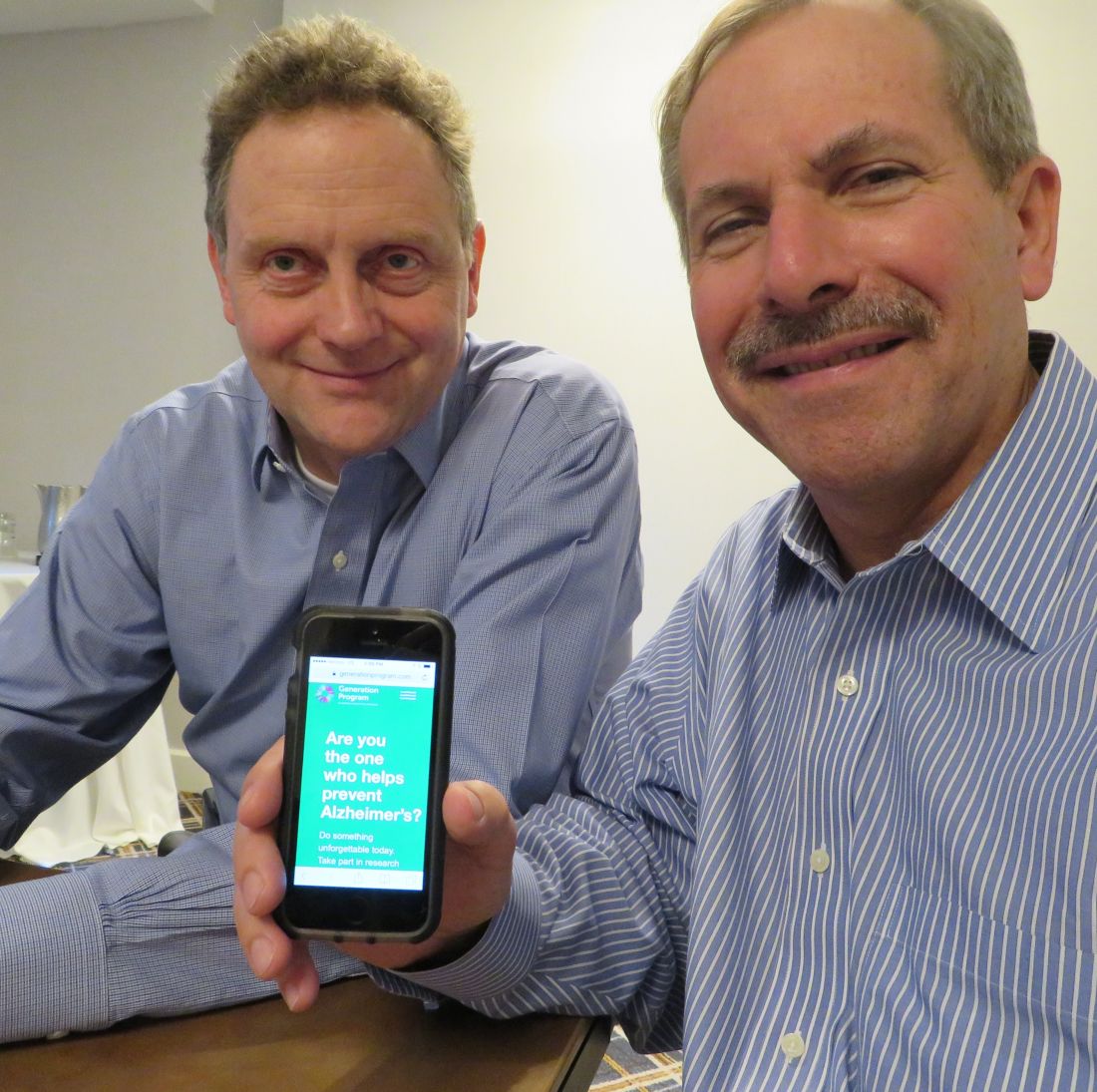
To enroll the entire cohort, Generation 2 will need to screen about 30,000 people. This sounds like a daunting task, but a new digital platform makes it eminently do-able, Dr. Tariot said. Both Generation studies are enrolling online. The consumer-friendly website offers detailed information about Alzheimer’s disease in general, ApoE4 risk, the studies’ structure, and the investigational medications. Most importantly, Dr. Tariot said, the website has a direct link to GeneMatch. Hosted by Banner Alzheimer Institute, GeneMatch prescreens potential trial participants (U.S. residents aged 55-75 years) by taking baseline demographic information, and mailing out free, simple-to-use cheek swab kits for genetic analysis. The program then stores and sorts the information, matching volunteers with appropriate trials according to location, age, interest, and genetic status. People don’t necessarily learn their ApoE4 status, unless they are recruited into a genetics-driven trial.
Launched 2 years ago, GeneMatch has already accrued more than 280,000 volunteers – a pretty remarkable achievement in itself, Dr. Tariot said. “People are very motivated to help find a cure for Alzheimer’s. Many of them have a family member who has the disease. Others are concerned about themselves, and many people just want to be part of something important.”
Dr. Porsteinsson agreed, relaying a startling interaction he had with a patient, who found out he was not qualified for a Generation study, meaning, of course, that he was ApoE4 negative.
“He told me he was actually kind of disappointed, because he believes so much in this study, and wanted to be part of doing something important for humanity,” Dr. Porsteinsson said. “Of course, I didn’t let him off the hook. I directed him to a bunch of other studies he was qualified for. We need everyone’s help to solve this.”
Both Dr. Tariot and Dr. Porsteinsson have reported financial relationships with numerous pharmaceutical companies.
* This story was updated 11/7/17.
[email protected]
On Twitter @Alz_Gal
BOSTON – Undeterred by a failed BACE inhibitor study with worrisome adverse events, an ambitious new clinical trial will investigate a different BACE-inhibiting molecule as an Alzheimer’s preventive in people at high genetic risk of Alzheimer’s disease.
The global Generation 2 trial intends to recruit about 3,300 cognitively normal subjects aged 60-75 years, who have either one or two copies of the apolipoprotein e4 (ApoE4) allele, said Pierre Tariot, MD, who announced the new study during the Clinical Trials on Alzheimer’s Disease 2017 meeting.
The Alzheimer Preventive Initiative and industry partners Novartis and Amgen are sponsoring the trial, which is a sister study to Generation 1. Generation 1, now ongoing, targets cognitively normal ApoE4 subjects only. It is a four-armed trial, comparing placebo to both CNP520 and an active immunotherapy called CAD106. Together, the studies comprise the Generation Program.
Both trials are event-driven, and will run 5-8 years. Generation 2 has a dual primary endpoint – a successful trial will find either a delay in progression to mild cognitive impairment or dementia relative to placebo, or significant differences from baseline in cognitive change as measured by the Alzheimer’s Preclinical Composite Cognitive test, or both.
The study partners are enthusiastic about CNP520, which performed well in its safety and tolerability studies. In an interview, Dr. Tariot called the CNP520 “likely a best-in-class molecule.”
“The only adverse events we saw in the entire safety program were a few cases of itchy skin,” said Dr. Tariot, director of the Banner Alzheimer Institute in Phoenix. “One of the reasons we chose Novartis as a study partner is that this drug of theirs looked remarkably clean – so clean that when we were deciding what doses to look at, the decision related only to what degree of BACE inhibition we wanted, not safety.”
The drug seemed very well-tolerated at every dose tested (1 mg, 10 mg, 25 mg, and 75 mg). Generation 1 employs a 50-mg oral dose per day. Generation 2 will investigate both 50 mg and 15 mg.
Site investigators are solidly behind the choice, said Anton Porsteinsson, MD, a Generation investigator at the University of Rochester (N.Y.) Medical Center, despite the unveiling at CTAD of worrisome adverse events seen in Merck’s failed EPOCH trial.
“I am even more convinced now that BACE inhibitors are drugs that need to be used early and that are probably highly indicated for this population,” he said in an interview. Dr. Porsteinsson wasn’t overly disturbed by data that Merck released during the meeting on its BACE inhibitor, verubecestat. In February, the company pulled the plug on its EPOCH trial investigating verubecestat in patients with mild-moderate AD. An interim analysis determined that there was no chance of success with the molecule.
Despite the general disappointment of yet another rainy-day parade in late-stage AD drug trials, researchers who heard the EPOCH post-mortem during CTAD expressed considerable concern over the unexpected, wide-ranging, and serious adverse events the trial accumulated.
Although there were no cases of Amyloid-Related Imaging Abnormalities (ARIA), a number of adverse events occurred significantly more often in the active groups than the placebo group. These included rash, falls and injuries, insomnia, headache, anxiety, suicidal ideation, diarrhea, dizziness, and weight loss.
At least some of these were hinted at in the bench science that brought verubecestat to late-stage clinical development. BACE is important for proper muscle function, and some BACE-knockout mice displayed a decrease in muscle spindles, receptors that sense changes in the length of muscle fibers. Other peculiarities in the mice have included axon targeting errors, reduced myelination, memory impairment, neurochemical abnormalities, alterations in neurogenesis and astrogenesis, increased age-related neurodegeneration, reduced spine density, retinal pathology, endophenotypes of schizophrenia, and seizures.
But the verubecestat findings aren’t a show-stopper for the more target-specific CNP520, Dr. Porsteinsson said. Verubecestat inhibited both BACE1 and BACE2; CNP520, only BACE1. And Dr. Porsteinsson, like most researchers, believes that preventing the accumulation of neurotoxic AB species will probably be much more effective clinically than trying to dissolve large stubborn brain plaques, which have already wreaked cognitive havoc.
In EPOCH, “even in people with advanced disease and a high load of insoluble plaques; they saw an 80% reduction in the production of AB and a 4% decrease in plaque burden. To me that signals this is not the class to use in moderate patients, or even mild, but in these very early stages where you don’t have full saturation, it might just be perfect,” he said.
“Do the side effects give me pause? Obviously and maybe mostly because we don’t know exactly what the driver was. But what we do is keep a close eye on everyone, maybe do extra safety monitoring, Most importantly, we picked a drug that shows less issues with any of these problems.”
Cognitively normal ApoE4 carriers – especially homozygotes – are an extremely important population to study, both in terms of clinical and scientific need. The gene is the single largest genetic risk factor for Alzheimer’s; those who carry two copies are 60% more likely than the general population to develop Alzheimer’s.
“These people truly need an effective intervention,” said Dr. Porsteinsson. “At the preclinical stage, they don’t truly have a disease yet; they are living their lives unimpaired. But something is percolating, and the results won’t be good. Even early on, there are fairly significant changes going on in the brain: a buildup of amyloid and tau, excess oxidative damage, inflammation. And if you don’t deal with it early on, it’s like trying to cure cancer once it’s metastasized.”
In the larger scientific picture, success in a primary prevention trial would finally put to rest questions about the amyloid cascade hypothesis and explain the long string of anti-amyloid drug trial failures, all of which were targeted at people with more advanced disease.
To enroll the entire cohort, Generation 2 will need to screen about 30,000 people. This sounds like a daunting task, but a new digital platform makes it eminently do-able, Dr. Tariot said. Both Generation studies are enrolling online. The consumer-friendly website offers detailed information about Alzheimer’s disease in general, ApoE4 risk, the studies’ structure, and the investigational medications. Most importantly, Dr. Tariot said, the website has a direct link to GeneMatch. Hosted by Banner Alzheimer Institute, GeneMatch prescreens potential trial participants (U.S. residents aged 55-75 years) by taking baseline demographic information, and mailing out free, simple-to-use cheek swab kits for genetic analysis. The program then stores and sorts the information, matching volunteers with appropriate trials according to location, age, interest, and genetic status. People don’t necessarily learn their ApoE4 status, unless they are recruited into a genetics-driven trial.
Launched 2 years ago, GeneMatch has already accrued more than 280,000 volunteers – a pretty remarkable achievement in itself, Dr. Tariot said. “People are very motivated to help find a cure for Alzheimer’s. Many of them have a family member who has the disease. Others are concerned about themselves, and many people just want to be part of something important.”
Dr. Porsteinsson agreed, relaying a startling interaction he had with a patient, who found out he was not qualified for a Generation study, meaning, of course, that he was ApoE4 negative.
“He told me he was actually kind of disappointed, because he believes so much in this study, and wanted to be part of doing something important for humanity,” Dr. Porsteinsson said. “Of course, I didn’t let him off the hook. I directed him to a bunch of other studies he was qualified for. We need everyone’s help to solve this.”
Both Dr. Tariot and Dr. Porsteinsson have reported financial relationships with numerous pharmaceutical companies.
* This story was updated 11/7/17.
[email protected]
On Twitter @Alz_Gal
AT CTAD
Seven years after bariatric surgery, more than 40% still off insulin
NATIONAL HARBOR, MD – Forty-four percent of insulin-dependent patients with type 2 diabetes mellitus (DM2) were at their glycemic target without insulin a median of seven years after surgery. The data from the largest study to evaluate long-term outcomes in this population were presented at Obesity Week 2017.
“These data confirm that the impressive metabolic effects of bariatric surgery in patients with type 2 diabetes are sustained beyond five years,” reported Ali Aminian, MD, a surgeon who specializes in bariatric procedures at the Cleveland Clinic, Cleveland, Ohio. He said that long-term efficacy has not been well characterized previously.

Reaching the glycemic target, defined as less than 7% HbA1c, without insulin was only one of the primary endpoints. The other was diabetes remission, which was defined as HbA1c less than 6.5%, fasting blood glucose less than 126 mg/dL, and being off all diabetes medications. This was observed in 15% of the patients after a median of 7 years followup.
Contrasting short-term results, defined as outcomes one to two years after bariatric surgery with the long-term followup, Dr. Aminian was able to show that declines were relatively modest over time. For example, 51% were at the glycemic target off insulin at the short-term mark, which translates into an absolute decline of only 7% relative to the 44% observed at the long-term followup assessment.
Similarly, 70% had achieved the American Diabetes Association (ADA) goal of less than 7% within the first two years of surgery, while 59% remained at this goal at the most recent followup. The proportion taking insulin at the short-term mark was 36% rising only to 40% long-term.
When data were stratified by procedure, results favored RYGB over sleeve gastrectomy. For example, 47% of the RYGB patients versus 33% of the sleeve gastrectomy patients were able to reach the ADA goal without insulin at the end of the study. The proportions in diabetes remission were 17% and 10%, respectively. RYGB was also associated with greater improvement in BMI (median -12 vs. - 8 kg/m2) and reduced late weight gain (median 20% vs. 31%).
However, Dr. Aminian, who did not provide statistical calculations for these differences, cautioned that higher risk patients might have been preferentially selected for sleeve gastrectomy. He noted that difference in median HbA1c levels was significantly lower in the RYGB group two years after surgery (P less than .001) but the numerical advantage had lost significance at the last followup (P = .32).
In an evaluation of predictors for glycemic control, a shorter duration of diabetes (less than 10 years) and good glycemic control prior to surgery were both predictors of achieving the primary outcomes on the basis of a multivariate analysis, according to Dr. Aminian. Younger age was a marginal predictor, but Dr. Aminian said that neither type of procedure nor presurgical BMI predicted outcomes from the multivariate analysis.
Relative to baseline, there were significant improvements in median LDL (P = .001). In addition, HDL, triglyceride levels, systolic, and diastolic blood pressure measurements were all significantly improved, both short-term and long-term after bariatric surgery (all P values less than .001), according to Dr. Aminian. When expressed as ADA goals, 82% of participants had blood pressure less than 140/90 mm Hg 7 years after surgery relative to 44% at baseline (P less than .001). The proportion with LDL less 100 mg/dL approached, but did not reach clinical significance (61% vs. 70%; P=0.06).
“When you consider all three parameters [ADA targets for glycemic control, blood pressure control, and lipid control], only 3% of patients met all three targets at baseline but 32% [P< less than .001] were at these targets at long-term followup,” Dr. Aminian reported.
Dr. Aminian reported having no relevant financial relationships.
As the invited discussant on these data, Raul Rosenthal, MD, Director, Bariatric and Metabolic Institute, Cleveland Clinic Florida, Weston, Florida, reiterated that time with diabetes prior to bariatric surgery may be an important predictor of postsurgical control of metabolic parameters.
“I published a paper about 10 years ago on outcomes in patients with diabetes, and in our experience 5 years was the limit. If you have a history of 5 years or less with diabetes, the chance of going into remission were 80%, and if it was more than 5 years, the likelihood dropped dramatically,” Dr. Rosenthal noted. He indicated duration of diabetes deserves further evaluation for its potential relevance to the optimal timing of bariatric surgery.
NATIONAL HARBOR, MD – Forty-four percent of insulin-dependent patients with type 2 diabetes mellitus (DM2) were at their glycemic target without insulin a median of seven years after surgery. The data from the largest study to evaluate long-term outcomes in this population were presented at Obesity Week 2017.
“These data confirm that the impressive metabolic effects of bariatric surgery in patients with type 2 diabetes are sustained beyond five years,” reported Ali Aminian, MD, a surgeon who specializes in bariatric procedures at the Cleveland Clinic, Cleveland, Ohio. He said that long-term efficacy has not been well characterized previously.
Reaching the glycemic target, defined as less than 7% HbA1c, without insulin was only one of the primary endpoints. The other was diabetes remission, which was defined as HbA1c less than 6.5%, fasting blood glucose less than 126 mg/dL, and being off all diabetes medications. This was observed in 15% of the patients after a median of 7 years followup.
Contrasting short-term results, defined as outcomes one to two years after bariatric surgery with the long-term followup, Dr. Aminian was able to show that declines were relatively modest over time. For example, 51% were at the glycemic target off insulin at the short-term mark, which translates into an absolute decline of only 7% relative to the 44% observed at the long-term followup assessment.
Similarly, 70% had achieved the American Diabetes Association (ADA) goal of less than 7% within the first two years of surgery, while 59% remained at this goal at the most recent followup. The proportion taking insulin at the short-term mark was 36% rising only to 40% long-term.
When data were stratified by procedure, results favored RYGB over sleeve gastrectomy. For example, 47% of the RYGB patients versus 33% of the sleeve gastrectomy patients were able to reach the ADA goal without insulin at the end of the study. The proportions in diabetes remission were 17% and 10%, respectively. RYGB was also associated with greater improvement in BMI (median -12 vs. - 8 kg/m2) and reduced late weight gain (median 20% vs. 31%).
However, Dr. Aminian, who did not provide statistical calculations for these differences, cautioned that higher risk patients might have been preferentially selected for sleeve gastrectomy. He noted that difference in median HbA1c levels was significantly lower in the RYGB group two years after surgery (P less than .001) but the numerical advantage had lost significance at the last followup (P = .32).
In an evaluation of predictors for glycemic control, a shorter duration of diabetes (less than 10 years) and good glycemic control prior to surgery were both predictors of achieving the primary outcomes on the basis of a multivariate analysis, according to Dr. Aminian. Younger age was a marginal predictor, but Dr. Aminian said that neither type of procedure nor presurgical BMI predicted outcomes from the multivariate analysis.
Relative to baseline, there were significant improvements in median LDL (P = .001). In addition, HDL, triglyceride levels, systolic, and diastolic blood pressure measurements were all significantly improved, both short-term and long-term after bariatric surgery (all P values less than .001), according to Dr. Aminian. When expressed as ADA goals, 82% of participants had blood pressure less than 140/90 mm Hg 7 years after surgery relative to 44% at baseline (P less than .001). The proportion with LDL less 100 mg/dL approached, but did not reach clinical significance (61% vs. 70%; P=0.06).
“When you consider all three parameters [ADA targets for glycemic control, blood pressure control, and lipid control], only 3% of patients met all three targets at baseline but 32% [P< less than .001] were at these targets at long-term followup,” Dr. Aminian reported.
Dr. Aminian reported having no relevant financial relationships.
As the invited discussant on these data, Raul Rosenthal, MD, Director, Bariatric and Metabolic Institute, Cleveland Clinic Florida, Weston, Florida, reiterated that time with diabetes prior to bariatric surgery may be an important predictor of postsurgical control of metabolic parameters.
“I published a paper about 10 years ago on outcomes in patients with diabetes, and in our experience 5 years was the limit. If you have a history of 5 years or less with diabetes, the chance of going into remission were 80%, and if it was more than 5 years, the likelihood dropped dramatically,” Dr. Rosenthal noted. He indicated duration of diabetes deserves further evaluation for its potential relevance to the optimal timing of bariatric surgery.
AT OBESITY WEEK 2017
Key clinical point: In the largest study to follow insulin-dependent patients after bariatric surgery, substantial benefits persist after median 7 years of followup.
Major finding: Among 252 insulin-dependent patients followed for a minimum of 5 years, 44% remain off insulin and 15% are off all anti-diabetic medications.
Data source: Retrospective single-center analysis.
Disclosures: Dr. Aminian reported having no relevant financial relationships.
Nebulized glycopyrrolate improves lung function in COPD
TORONTO – Glycopyrrolate, a novel nebulized long-acting muscarinic antagonist (LAMA) in development, was well-tolerated and significantly improved lung function and health status in COPD patients regardless of baseline lung function or age, according to a subgroup analysis of pooled results from two randomized trials.*
There are currently no nebulized LAMAs approved for use in the U.S.
Jill Ohar, MD, from Wake Forest University School of Medicine (Winston-Salem, N.C.), presented this secondary analysis of the GOLDEN-3 and GOLDEN-4 trials at the CHEST annual meeting. She and her colleagues evaluated the efficacy and safety of glycopyrrolate in patients with a forced expiratory volume 1(FEV1) % predicted of less than 50 and an FEV1 % predicted of greater than or equal to 50, in age ranges of less than 65 years, greater than or equal to 65 years and at least 75 years, as measured by trough FEV1.
Similarly, both glycopyrrolate doses produced significant (P less than .05) and clinically meaningful lung function improvements vs. placebo in participants less than 65 years of age, at least 65 years, and greater than or equal to 75 years.
Glycopyrrolate use for 12 weeks led to greater improvements over placebo in St. George’s Respiratory Questionnaire (SGRQ) total score, in patients in both lung function classes. There were a higher percentage of SGRQ responders in the treatment arms compared to placebo arms.
The highest SGRQ improvement in SGRQ (−6.287) was seen in the 47 patients that comprised the at-least-75 years of age subgroup receiving glycopyrrolate 25 mcg BID. “It’s a small number of people, but I think it’s [valuable] to see if the very aged act in any way differently than the entire greater than or equal to 65-year-old group,” said Dr. Ohar.
Adverse event rates were similar for placebo and both glycopyrrolate doses, with no safety signals seen according to baseline lung function or age. Few cardiovascular events of special interest were seen.
“Looking at major adverse cardiovascular events, such as fatal MIs, other cardiovascular deaths, arrhythmias, etc., we see nothing that would suggest that the drug overall is associated with an undue number of these versus placebo,” reported Dr. Ohar.
GOLDEN 3 and 4 were replicate, 12-week, phase 3, randomized, double-blind, placebo-controlled studies that evaluated glycopyrrolate solution administered by an investigational eFlow Close System (eFLOW CS) nebulizer in individuals with moderate-to-very severe COPD, including those with continued background use of a long-acting beta2-agonist (LABA), with or without an inhaled corticosteroid (ICS). In each of the trials, about 30% of patients were on LABA ICS, noted Dr. Ohar in her presentation. A total of 653 subjects were randomized in GOLDEN 3 and 641 in GOLDEN 4.
Its manufacturer, Sunovion Pharmaceuticals, resubmitted the product to the FDA in June 2017 in response to a Complete Response Letter received from the FDA in May 2017. The FDA is expected to act on the new submission on December 15, 2017. The novel agent is being considered for the long-term, maintenance treatment of airflow obstruction in people with COPD, including chronic bronchitis and/or emphysema.
Dr. Ohar reported that she serves on the advisory boards of several pharmaceutical companies. The other three authors are employees of Sunovion Pharmaceuticals Inc.
*This article was updated on Nov. 6, 2017.
Eric Gartman, MD, FCCP, comments: If approved, this would represent the first nebulized LAMA available in the U.S. – so in the small population of patients that is unable to utilize standard delivery devices, this would provide an option. It is unclear if this medication must be administered via the proprietary nebulizer that was used in the study – but if so, this would certainly add to the already extremely high cost of respiratory medications and further limit access for many patients.

Eric Gartman, MD, FCCP, comments: If approved, this would represent the first nebulized LAMA available in the U.S. – so in the small population of patients that is unable to utilize standard delivery devices, this would provide an option. It is unclear if this medication must be administered via the proprietary nebulizer that was used in the study – but if so, this would certainly add to the already extremely high cost of respiratory medications and further limit access for many patients.
Eric Gartman, MD, FCCP, comments: If approved, this would represent the first nebulized LAMA available in the U.S. – so in the small population of patients that is unable to utilize standard delivery devices, this would provide an option. It is unclear if this medication must be administered via the proprietary nebulizer that was used in the study – but if so, this would certainly add to the already extremely high cost of respiratory medications and further limit access for many patients.
TORONTO – Glycopyrrolate, a novel nebulized long-acting muscarinic antagonist (LAMA) in development, was well-tolerated and significantly improved lung function and health status in COPD patients regardless of baseline lung function or age, according to a subgroup analysis of pooled results from two randomized trials.*
There are currently no nebulized LAMAs approved for use in the U.S.
Jill Ohar, MD, from Wake Forest University School of Medicine (Winston-Salem, N.C.), presented this secondary analysis of the GOLDEN-3 and GOLDEN-4 trials at the CHEST annual meeting. She and her colleagues evaluated the efficacy and safety of glycopyrrolate in patients with a forced expiratory volume 1(FEV1) % predicted of less than 50 and an FEV1 % predicted of greater than or equal to 50, in age ranges of less than 65 years, greater than or equal to 65 years and at least 75 years, as measured by trough FEV1.
Similarly, both glycopyrrolate doses produced significant (P less than .05) and clinically meaningful lung function improvements vs. placebo in participants less than 65 years of age, at least 65 years, and greater than or equal to 75 years.
Glycopyrrolate use for 12 weeks led to greater improvements over placebo in St. George’s Respiratory Questionnaire (SGRQ) total score, in patients in both lung function classes. There were a higher percentage of SGRQ responders in the treatment arms compared to placebo arms.
The highest SGRQ improvement in SGRQ (−6.287) was seen in the 47 patients that comprised the at-least-75 years of age subgroup receiving glycopyrrolate 25 mcg BID. “It’s a small number of people, but I think it’s [valuable] to see if the very aged act in any way differently than the entire greater than or equal to 65-year-old group,” said Dr. Ohar.
Adverse event rates were similar for placebo and both glycopyrrolate doses, with no safety signals seen according to baseline lung function or age. Few cardiovascular events of special interest were seen.
“Looking at major adverse cardiovascular events, such as fatal MIs, other cardiovascular deaths, arrhythmias, etc., we see nothing that would suggest that the drug overall is associated with an undue number of these versus placebo,” reported Dr. Ohar.
GOLDEN 3 and 4 were replicate, 12-week, phase 3, randomized, double-blind, placebo-controlled studies that evaluated glycopyrrolate solution administered by an investigational eFlow Close System (eFLOW CS) nebulizer in individuals with moderate-to-very severe COPD, including those with continued background use of a long-acting beta2-agonist (LABA), with or without an inhaled corticosteroid (ICS). In each of the trials, about 30% of patients were on LABA ICS, noted Dr. Ohar in her presentation. A total of 653 subjects were randomized in GOLDEN 3 and 641 in GOLDEN 4.
Its manufacturer, Sunovion Pharmaceuticals, resubmitted the product to the FDA in June 2017 in response to a Complete Response Letter received from the FDA in May 2017. The FDA is expected to act on the new submission on December 15, 2017. The novel agent is being considered for the long-term, maintenance treatment of airflow obstruction in people with COPD, including chronic bronchitis and/or emphysema.
Dr. Ohar reported that she serves on the advisory boards of several pharmaceutical companies. The other three authors are employees of Sunovion Pharmaceuticals Inc.
*This article was updated on Nov. 6, 2017.
AT CHEST 2017
Key clinical point: Nebulized glycopyrrolate improved lung function and was well tolerated irrespective of baseline lung function or age.
Major finding: Statistically and clinically meaningful improvements in trough FEV1 at 12 weeks were seen in individuals, regardless of their baseline FEV1 % predicted.
Data source: Pooled findings from 2 RCTs, GOLDEN 3 and GOLDEN 4, that together included 1,294 moderate-to-very severe COPD patients.
Disclosures: Dr. Ohar reported that she serves on the advisory boards of several pharmaceutical companies. The other three authors are employees of Sunovion Pharmaceuticals Inc.
Remimazolam surpasses midazolam for bronchoscopy sedation
TORONTO – An investigational sedative, remimazolam, that’s similar to midazolam but with faster onset and offset, resulted in significantly better procedural success compared with midazolam in a multicenter, phase III trial with 431 patients.
The results also showed that remimazolam was as safe as midazolam (Versed), with a very similar adverse event profile, said Gerard A. Silvestri, MD, FCCP, at the CHEST annual meeting.
The bronchoscopy trial enrolled patients at any of 15 U.S. centers with an American Society of Anesthesiologists (ASA) physical status classification of I-III and scheduled for diagnostic or therapeutic bronchoscopy. The enrolled patients averaged 62 years of age, and 38% were in ASA class III.
All patients received initial sedation treatment with fentanyl, followed by a three-to-one randomization to blinded remimazolam, blinded placebo that included midazolam rescue, or open-label midazolam. The study’s primary efficacy endpoint was procedural success, defined as patients who underwent the complete procedure without need for an alternative sedative and without need for more than five doses of the patient’s assigned medication within any 15 minute period during the procedure or need for more than three midazolam doses within any 12-minute period in the patients randomized to receive midazolam.
This primary endpoint occurred in 83% of 303 patients in the remimazolam arm, 5% of 59 patients in the placebo arm, and 34% of 69 patients in the midazolam arm, a statistically significant difference between the remimazolam patients and each of the comparator groups, reported Dr. Silvestri, a professor of medicine and a lung cancer pulmonologist at the Medical University of South Carolina in Charleston.
The results also demonstrated the faster inset and offset of remimazolam. Treatment achieved adequate sedation to start the procedure after a median of 5 minutes with remimazolam, a median of 15.5 minutes with midazolam, and a median of 17 minutes among patients in the placebo group. Once sedation finished, patients returned to being fully alert after a median of 6 minutes with remimazolam, a median of 12 minutes with midazolam, and a median of 13.5 minutes for patients in the placebo arm.
“What’s nice about remimazolam is that the adverse event profile is exactly the same as with placebo and midazolam, and you have a reversal agent,” the same as what’s used for midazolam, he said.
Dr. Silvestri suggested several additional studies he would like to see run on remimazolam to better understand its clinical performance and role. These include studying the drug in the elderly, patients with an ASA classification of IV, obese patients, and those on high narcotic doses. He also suggested comparing remimazolam directly with propofol, testing remimazolam as a stand-alone agent without fentanyl co-administration, and trying the drug during other pulmonary procedures such as pleural-catheter placement and other invasive procedures, and in ICU patients.
The trial was funded by Paion, the company developing remimazolam. Dr. Silvestri and Dr. Stanbrook had no relevant disclosures.
[email protected]
On Twitter @mitchelzoler
AT CHEST 2017
Key clinical point:
Major finding: The rate of successful bronchoscopies was 83% with remimazolam, 34% with midazolam, and 5% with placebo.
Data source: A multicenter, phase III trial with 431 patients.
Disclosures: The trial was funded by Paion, the company developing remimazolam. Dr. Silvestri and Dr. Stanbrook had no relevant disclosures.
Hispanics bear brunt of NAFLD disease burden
WASHINGTON — Significant racial and ethnic disparities exist in nonalcoholic fatty liver disease prevalence and severity in the United States, with Hispanics at highest risk and blacks at the lowest, but the risk of death from NAFLD is highest in whites, according to a meta-analysis of 34 studies presented at the annual meeting of the American Association for the Study of Liver Diseases.

The findings are based on a meta-analysis of studies in the MEDLINE, EMBASE, and Cochrane databases from inception through August 2016 that included 368,569 unique patients that characterized disparities in NAFLD prevalence, severity, or prognosis, Dr. Rich said.
When the researchers drilled down into the data, they found the disparities dissipated somewhat. “When we looked at the severity of NAFLD we looked at two things: whether there was NASH (nonalcoholic steatohepatitis) present, or if there was the presence of advanced fibrosis,” Dr. Rich said. “We found there was no significant difference in the risk of NASH in Hispanic patients, compared to white patients; however, the proportion of Hispanic NAFLD patients that had NASH was 45.4%, compared to 32.2% in whites.” The pooled relative risk was 1.09.
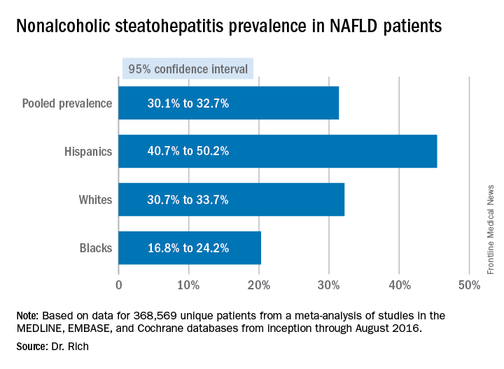
The meta-analysis tended to attenuate the disparities found in disease severity, compared with the disparities the researchers found in prevalence, Dr. Rich said. “Data are limited and discordant on racial and ethnic differences in NAFLD prognosis and outcomes,” she said. “In the current literature the studies have notable limitations, highlighting the need for future high-quality data in this area, and further studies are needed to determine the need and pathways to reduce NAFLD disparities in the future,” she said.
Dr. Rich had no financial relationships to disclose.
WASHINGTON — Significant racial and ethnic disparities exist in nonalcoholic fatty liver disease prevalence and severity in the United States, with Hispanics at highest risk and blacks at the lowest, but the risk of death from NAFLD is highest in whites, according to a meta-analysis of 34 studies presented at the annual meeting of the American Association for the Study of Liver Diseases.
The findings are based on a meta-analysis of studies in the MEDLINE, EMBASE, and Cochrane databases from inception through August 2016 that included 368,569 unique patients that characterized disparities in NAFLD prevalence, severity, or prognosis, Dr. Rich said.
When the researchers drilled down into the data, they found the disparities dissipated somewhat. “When we looked at the severity of NAFLD we looked at two things: whether there was NASH (nonalcoholic steatohepatitis) present, or if there was the presence of advanced fibrosis,” Dr. Rich said. “We found there was no significant difference in the risk of NASH in Hispanic patients, compared to white patients; however, the proportion of Hispanic NAFLD patients that had NASH was 45.4%, compared to 32.2% in whites.” The pooled relative risk was 1.09.
The meta-analysis tended to attenuate the disparities found in disease severity, compared with the disparities the researchers found in prevalence, Dr. Rich said. “Data are limited and discordant on racial and ethnic differences in NAFLD prognosis and outcomes,” she said. “In the current literature the studies have notable limitations, highlighting the need for future high-quality data in this area, and further studies are needed to determine the need and pathways to reduce NAFLD disparities in the future,” she said.
Dr. Rich had no financial relationships to disclose.
AT THE LIVER MEETING 2017
Key clinical point: Hispanics have a much higher incidence of fatty liver disease than other ethnic groups.
Major finding: NAFLD prevalence is highest in Hispanics and lower in blacks, compared with whites, although differences between groups are smaller in high-risk cohorts (range, 47.6%-55.5%) than in population-based cohorts (range, 13.0%-22.9%).
Data source: Meta-analysis of 34 studies in the MEDLINE, EMBASE, and Cochrane databases from inception through August 2016 that involved 368,569 patients.
Disclosures: Dr. Rich had no financial relationships to disclose.