User login
Modifiable Factors May Protect Memory Despite Genetic Risk for Alzheimer’s Disease
TORONTO—Modifiable risk factors predict memory resilience in aging adults with genetic risk for Alzheimer’s disease, according to data presented at the Alzheimer’s Association International Conference. Several factors predict memory resilience in both sexes. A greater number of sex-specific factors apply to females than to males, however. The predictive factors are similar, regardless of whether risk results from APOE or CLU.
Kirstie McDermott, graduate student at the University of Alberta in Edmonton, Canada, and colleagues sought to determine whether longitudinal memory trajectories suggest a distinct phenotype of memory resilience, which they defined as high memory performance despite genetic risk for Alzheimer’s disease. The researchers also looked for predictors of memory resilience.
Ms. McDermott and colleagues followed 642 cognitively normal adults between ages 53 and 95 who were participating in the Victoria Longitudinal Study. Maximum follow-up was nine years. Carriers of the APOE ε4 allele or the CLU CC genotype were considered to be at genetic risk for Alzheimer’s disease. The investigators tested 22 predictors of resilience from five nongenetic risk domains (ie, demographic, functional, health, mobility, and lifestyle). They stratified their analyses by sex to compare the effect of these predictors in males and females.
Memory was stable in 67.6% of females and in 52.8% of males. Growth mixture modeling enabled the investigators to identify two similar classes of longitudinal memory trajectories among males and females. Random forest analysis identified younger age, higher education, stronger grip, and more everyday novel cognitive activity (such as playing bridge or filing taxes) as important predictors of memory resilience for both sexes.
For females, living with another person; being married; lower pulse pressure; higher peak expiratory flow; better subjective health rating; faster walking and turning time; more social visits with family, friends, and neighbors; and volunteering more often predicted memory resilience. The only predictor of memory resilience specific to males was a low number of depressive symptoms. The predictors were similar for both genotypes of Alzheimer’s disease risk.
“The more we know about both common and gender-specific factors that may protect against Alzheimer’s risk, the better we can create tailored and appropriate interventions that promote functional maintenance and delay cognitive decline,” said Ms. McDermott.
—Erik Greb
How Are Migraine and Stress Related?
SAN DIEGO—Stress is a fact of life. More than 40 years of data have established the relationship between stress and migraine. Biobehavioral treatments such as relaxation training, biofeedback, and cognitive behavioral therapy may help to modify patients’ response to stress and reduce the likelihood of a migraine attack, according to an overview presented at the 58th Annual Scientific Meeting of the American Headache Society.
“It can be difficut or impossible to change many factors in our life that create stress, but we can modify how we think about and react to them. That is what we help our patients do using biobehavioral treatments such as cognitive behavioral therapy,” said Dawn Buse, PhD, Director of Behavioral Medicine at Montefiore Headache Center and Associate Professor of Neurology at Albert Einstein College of Medicine in New York. For example, neurologists can teach patients better coping strategies, reduce catastrophizing, encourage the use of social support, and create healthy lifestyle behaviors and other potentially protective factors.

Stress can play many roles in migraine. It can exacerbate migraine attack frequency or be a premonitory feature, prognostic factor, or a consequence of an attack. In addition, stress can be a risk factor for the onset of migraine and is a common trigger.
In a study by Kelman of 1,750 patients diagnosed with migraine, 76% of the participants reported having triggers. Nine percent of patients experienced triggers very frequently, 27% experienced triggers frequently, and 40% of patients encountered triggers occasionally. In addition, 80% of respondents reported stress as a trigger for migraine, making it the most common trigger. In a study by Wöber et al published in 2007 in Cephalalgia, “stress in private life” and “psychic tension” were associated with an increased risk of migraine attacks. A study published in the European Journal of Pain in 2014 reported strong associations between migraine and job stress, which researchers defined as low job control, high job demands, and low social support.
Other data suggest that migraine attacks may result from a combination of triggers, rather than from one trigger. A study of the association between stress and menstruation in migraine published in the International Journal of Women’s Health in 2014 included 307 participants with migraine without aura. Participants kept a headache diary for 14 months. The data suggested that women were more susceptible to stress in the premenstrual period. The researchers also found a statistically significant relationship between alcohol and migraine attacks only during stressful periods.
Martin and colleagues conducted three studies comparing people with migraine or tension-type headache and controls without headache. Participants attempted to solve difficult anagrams for 35 minutes while receiving negative feedback. They found that the stress condition was associated with increased head pain, compared with controls.
Sleep duration may have a relationship with stress. Data published in Pain in 2012 suggested that two consecutive days of high stress levels or low sleep were strongly predictive of headache. In contrast, two consecutive days of low stress or adequate sleep were considered protective factors that may help raise the headache attack threshold. Information like this may help give patients a sense of control, said Dr. Buse.
Two Types of Stressors
Investigators generally categorize stressors as daily hassles or major life events. According to research published in 1992 in Pain, people with recurrent headache experience more hassles than controls do. Daily hassles are also associated with an increased frequency of headache attacks.
Adverse childhood experiences such as emotional neglect, emotional abuse, sexual abuse, and other forms of abuse are considered major life events and appear comorbid with migraine incidence and attack frequency. Data published in Neurology showed that people who experienced more types of traumatic events in early childhood were more likely to have a higher frequency of severe headache, including chronic migraine.
Appraisal and Self-Efficacy
Years of research support the thesis that modifiable factors such as appraisal and self-efficacy influence migraine, said Dr. Buse. Studies have found that people with tension-type headache appraise stressful life events more negatively than controls do. This result raises the possibility that appraising stressful events less negatively could reduce headache pain or frequency.
People with tension-type headache also perceive themselves as having less control (ie, low self-efficacy). Self-efficacy helps to moderate the impact of stressful events on headache. According to a 1984 electromyography biofeedback study published in the Journal of Consulting and Clinical Psychology, a greater sense of self-efficacy resulted in a reduction in headache.
Coping and Social Support
Improving their coping skills and seeking greater social support also may benefit patients with headache. Migraineurs may respond to stress with more wishful thinking, self-criticism, social withdrawal, and catastrophizing, compared with controls. A study published in the Journal of Behavioral Sciences reported that patients with tension-type headache practiced less effective coping strategies, were more likely to blame themselves, and made less use of social support than did controls. Researchers also found that people with migraine tend to engage in catastrophizing while thinking about their condition.
A study in the European Journal of Pain found that migraine was associated with low social support. In addition to poor coping skills, lack of social support can be both a stressor and a consequence of severe headache. A patient’s social support may decrease if migraine causes him or her to miss events and avoid making social commitments. Furthermore, data indicate that people with headache are significantly less satisfied with the support available to them. Compared with controls, they scored lower in all types of functional support.
Dr. Buse suggested that physicians can help patients modify their response to stressors through behavioral therapies as well as by assessing and improving self-efficacy, enhancing social support, and improving the sense of control. Physicians can improve patients’ psychologic well-being by modifying dysfunctional coping styles, negative attributions, and catastrophizing.
Future Directions in Stress and Migraine Research
“Future directions in migraine treatment may include disseminating behavioral treatments at low cost to large numbers of people via the Internet, smartphones, apps, and wearable technology,” said Dr. Buse. “Technology can be used to collect data related to migraine attacks and deliver messages, including healthy reminders, coping strategies, and relaxtion exercises, in real time. There is an expanding body of literature testing the efficacy of delivering behavioral treatments using these modalties. The possibilites and potential benefits for our patients are very exciting.”
—Erica Robinson
Suggested Reading
Kelman L. The triggers or precipitants of the acute migraine attack. Cephalalgia. 2007;27(5):394-402.
Martin PR, Milech D, Nathan PR. Towards a functional model of chronic headaches: investigation of antecedents and consequences. Headache. 1993;33(9):461-470.
Santos IS, Griep RH, Alves MG, et al. Job stress is associated with migraine in current workers: the Brazilian longitudinal study of adult health (ELSA-Brasil). Eur J Pain. 2014;18(9):1290-1297.
Tietjen GE. Childhood maltreatment and headache disorders. Curr Pain Headache Rep. 2016;20(4):26.
Wildeman C, Emanuel N, Leventhal JM, et al. The prevalence of confirmed maltreatment among US children, 2004 to 2011. JAMA Pediatr. 2014;168(8):706-713.
SAN DIEGO—Stress is a fact of life. More than 40 years of data have established the relationship between stress and migraine. Biobehavioral treatments such as relaxation training, biofeedback, and cognitive behavioral therapy may help to modify patients’ response to stress and reduce the likelihood of a migraine attack, according to an overview presented at the 58th Annual Scientific Meeting of the American Headache Society.
“It can be difficut or impossible to change many factors in our life that create stress, but we can modify how we think about and react to them. That is what we help our patients do using biobehavioral treatments such as cognitive behavioral therapy,” said Dawn Buse, PhD, Director of Behavioral Medicine at Montefiore Headache Center and Associate Professor of Neurology at Albert Einstein College of Medicine in New York. For example, neurologists can teach patients better coping strategies, reduce catastrophizing, encourage the use of social support, and create healthy lifestyle behaviors and other potentially protective factors.
Stress can play many roles in migraine. It can exacerbate migraine attack frequency or be a premonitory feature, prognostic factor, or a consequence of an attack. In addition, stress can be a risk factor for the onset of migraine and is a common trigger.
In a study by Kelman of 1,750 patients diagnosed with migraine, 76% of the participants reported having triggers. Nine percent of patients experienced triggers very frequently, 27% experienced triggers frequently, and 40% of patients encountered triggers occasionally. In addition, 80% of respondents reported stress as a trigger for migraine, making it the most common trigger. In a study by Wöber et al published in 2007 in Cephalalgia, “stress in private life” and “psychic tension” were associated with an increased risk of migraine attacks. A study published in the European Journal of Pain in 2014 reported strong associations between migraine and job stress, which researchers defined as low job control, high job demands, and low social support.
Other data suggest that migraine attacks may result from a combination of triggers, rather than from one trigger. A study of the association between stress and menstruation in migraine published in the International Journal of Women’s Health in 2014 included 307 participants with migraine without aura. Participants kept a headache diary for 14 months. The data suggested that women were more susceptible to stress in the premenstrual period. The researchers also found a statistically significant relationship between alcohol and migraine attacks only during stressful periods.
Martin and colleagues conducted three studies comparing people with migraine or tension-type headache and controls without headache. Participants attempted to solve difficult anagrams for 35 minutes while receiving negative feedback. They found that the stress condition was associated with increased head pain, compared with controls.
Sleep duration may have a relationship with stress. Data published in Pain in 2012 suggested that two consecutive days of high stress levels or low sleep were strongly predictive of headache. In contrast, two consecutive days of low stress or adequate sleep were considered protective factors that may help raise the headache attack threshold. Information like this may help give patients a sense of control, said Dr. Buse.
Two Types of Stressors
Investigators generally categorize stressors as daily hassles or major life events. According to research published in 1992 in Pain, people with recurrent headache experience more hassles than controls do. Daily hassles are also associated with an increased frequency of headache attacks.
Adverse childhood experiences such as emotional neglect, emotional abuse, sexual abuse, and other forms of abuse are considered major life events and appear comorbid with migraine incidence and attack frequency. Data published in Neurology showed that people who experienced more types of traumatic events in early childhood were more likely to have a higher frequency of severe headache, including chronic migraine.
Appraisal and Self-Efficacy
Years of research support the thesis that modifiable factors such as appraisal and self-efficacy influence migraine, said Dr. Buse. Studies have found that people with tension-type headache appraise stressful life events more negatively than controls do. This result raises the possibility that appraising stressful events less negatively could reduce headache pain or frequency.
People with tension-type headache also perceive themselves as having less control (ie, low self-efficacy). Self-efficacy helps to moderate the impact of stressful events on headache. According to a 1984 electromyography biofeedback study published in the Journal of Consulting and Clinical Psychology, a greater sense of self-efficacy resulted in a reduction in headache.
Coping and Social Support
Improving their coping skills and seeking greater social support also may benefit patients with headache. Migraineurs may respond to stress with more wishful thinking, self-criticism, social withdrawal, and catastrophizing, compared with controls. A study published in the Journal of Behavioral Sciences reported that patients with tension-type headache practiced less effective coping strategies, were more likely to blame themselves, and made less use of social support than did controls. Researchers also found that people with migraine tend to engage in catastrophizing while thinking about their condition.
A study in the European Journal of Pain found that migraine was associated with low social support. In addition to poor coping skills, lack of social support can be both a stressor and a consequence of severe headache. A patient’s social support may decrease if migraine causes him or her to miss events and avoid making social commitments. Furthermore, data indicate that people with headache are significantly less satisfied with the support available to them. Compared with controls, they scored lower in all types of functional support.
Dr. Buse suggested that physicians can help patients modify their response to stressors through behavioral therapies as well as by assessing and improving self-efficacy, enhancing social support, and improving the sense of control. Physicians can improve patients’ psychologic well-being by modifying dysfunctional coping styles, negative attributions, and catastrophizing.
Future Directions in Stress and Migraine Research
“Future directions in migraine treatment may include disseminating behavioral treatments at low cost to large numbers of people via the Internet, smartphones, apps, and wearable technology,” said Dr. Buse. “Technology can be used to collect data related to migraine attacks and deliver messages, including healthy reminders, coping strategies, and relaxtion exercises, in real time. There is an expanding body of literature testing the efficacy of delivering behavioral treatments using these modalties. The possibilites and potential benefits for our patients are very exciting.”
—Erica Robinson
Suggested Reading
Kelman L. The triggers or precipitants of the acute migraine attack. Cephalalgia. 2007;27(5):394-402.
Martin PR, Milech D, Nathan PR. Towards a functional model of chronic headaches: investigation of antecedents and consequences. Headache. 1993;33(9):461-470.
Santos IS, Griep RH, Alves MG, et al. Job stress is associated with migraine in current workers: the Brazilian longitudinal study of adult health (ELSA-Brasil). Eur J Pain. 2014;18(9):1290-1297.
Tietjen GE. Childhood maltreatment and headache disorders. Curr Pain Headache Rep. 2016;20(4):26.
Wildeman C, Emanuel N, Leventhal JM, et al. The prevalence of confirmed maltreatment among US children, 2004 to 2011. JAMA Pediatr. 2014;168(8):706-713.

Why psychiatrists’ opinions about politicians shouldn’t matter
When it comes to political candidates and psychiatric disorders, the idea of diagnosing from afar is not new. In 1964, Fact magazine published an article called “The Unconscious of a Conservative: A Special Issue on the Mind of Barry Goldwater.” For the article, more than 2,400 psychiatrists responded to a survey, and many described the presidential candidate using a host of diagnostic and symptomatic terms. Mr. Goldwater lost the election in a landslide and successfully sued the magazine for $75,000.
In 1973, nearly a decade later, the American Psychiatric Association specifically noted in its code of ethics that psychiatrists should not offer diagnostic opinions on anyone who has not been personally examined and has not signed the proper authorization for this information to be released. The edict has been informally called The Goldwater Rule.

In the current election cycle, it has been a challenge for some psychiatrists to refrain from making public statements about our presidential candidates. Some have said the Goldwater Rule amounts to a gag order, and that the specifics of this election are different from other elections, and might warrant allowing psychiatrists to issue their professional opinions. The APA has reminded psychiatrists that to do so is unethical, and on Aug. 15, The New York Times ran an article by Benedict Carey titled: “The Psychiatric Question: Is it Fair to Analyze Donald Trump from Afar?”
I want to look at the question from a different stance. In assessing our political candidates, I’d like you to consider this: What difference does it make? Psychiatric diagnostic criteria are determined by consensus opinions of APA workgroup members. They aren’t perfect; they aren’t always accurate even when a patient is evaluated in person and seen over time; they are influenced by culture; and they aren’t always prognostic. One person’s experience of bipolar disorder is not another person’s experience of bipolar disorder, and personality disorders – such as narcissism or antisocial personality disorder – don’t have uniform presentations or outcomes. Some people with these difficulties head corporations and nations, while others wilt in prisons.
Half of the population will suffer from an episode of psychiatric illness at some point in their lives, and one in five people is affected in any given year. While we might all agree that many psychiatric symptoms are not compatible with being president, should the fact that a candidate has had a distant episode of mental illness, either fully resolved or controlled with treatment, disqualify him or her from holding office?
In an article published in the January 2006 issue of the Journal of Nervous and Mental Disease, Jonathan R. Davidson, MD; Kathryn M. Connor, MD; and Marvin Swartz, MD, looked at the biographical data on U.S. presidents from 1776 to 1974 and concluded that 49% met criteria for an Axis I psychiatric disorder. During those cycles, 27% had psychiatric difficulties while in office. Our presidents have suffered from depression and bipolar disorder, anxiety and alcoholism (J Nerv Ment Dis. 2006 Jan;194[1]47-51). No one has publicly attempted to tally personality disorders or use of other addictive substances. It may be safe to say that if the existence of psychiatric pathology had always been a disqualifier for public office, we’d live in a very different country.
Hopefully, when we approach our patients, we do so with warmth, kindness, and a genuine desire to help them heal. Psychiatry, at its best, is about intimacy and trust, and it is in that venue that people share their inner worlds and allow themselves to be vulnerable. Compassion is part of the deal; no one wants to have a mental illness. To refer to an unknown celebrity as “schizophrenic,” “psychotic,” or “having a godlike self-image” – or calling him “a dangerous lunatic,” as Sen. Goldwater was called – is not about careful diagnosis and compassion; it’s simply about name calling. To say that a politician or celebrity’s undesirable behavior is the result of a psychiatric illness based on cavalier observation is an insult to our patients, and it perpetuates stigma.
Psychiatric diagnoses are made by observing a constellation of symptoms that occur together. Mr. Trump has given the whole world years of data – he’s lived his life in a very public way. As a real estate developer, he has had countless employees who all know how they’ve been treated. We’ve seen him through three marriages and watched how he interacts with his children. We’ve seen him take out full-page ads calling for the death penalty for a group of young men who were wrongly convicted in the rape and assault of the Central Park jogger in 1988. If that’s not enough, he has hosted his own reality television show, and we’ve now seen him countless times in debates and rallies. We know how he treats his running mates, journalist Megyn Kelly, a news reporter with a disability, and the parents of a fallen soldier. We’ve watched him assure the nation during a primary debate that his genitals are big enough. Every individual is free to decide if Mr. Trump’s widely viewed patterns of behavior represent much-needed spunk and change with political beliefs that align with their own, or if his words and behaviors represent cruelty, impulsivity, poor judgment, and a pattern of actions that some might not feel is dignified enough for our country’s leader. No degree is required to observe and draw conclusions.
And please, let me be the first to admit that my perspective is biased: I’ve pointed out some of Mr. Trump’s more troubling behaviors and said nothing of the many wonderful things he may have done as both an entrepreneur and as a human being. Those don’t make the headlines or Twitter, and so I’m left with my own observations and those of a liberal press (Of course, there’s a right-wing press, too, but that’s a topic for another time). For voters who align their beliefs with the National Rifle Association, and feel that illegal immigrants are a drain on our country and that people of certain faiths represent a terror threat to Americans, Mr. Trump apparently remains a reasonable candidate.
Is there a role here for living room consults from psychiatrists? Is there something for us as professionals to add to the prediction of Mr. Trump’s behavior if he becomes president? I don’t think so. Every American has ample data, and for those who are curious about Mr. Trump’s psychiatric status, they are free to Google the criteria for psychiatric disorders and see if they believe he meets them. The input of psychiatrists would neither change the election outcome nor accurately predict his behavior if elected. But it might make us look a bit grandiose.
Finally, I’ve focused here on Mr. Trump, while saying nothing about Secretary Hillary Clinton and her psychiatric status. She, too, has lived a public life, and while many of her activities have attracted media attention and scrutiny, I’ve seen nothing that has suggested she suffers from a psychiatric condition. There was, however, an op-ed piece in The New York Times on Aug. 23, 2016, by Frank Bruni, suggesting that Mrs. Clinton may have an 11th toe.
If nothing else, it’s time for this election to be over.
Dr. Miller is coauthor of “Committed: The Battle Over Involuntary Psychiatric Care,” forthcoming from Johns Hopkins University Press in fall 2016.
When it comes to political candidates and psychiatric disorders, the idea of diagnosing from afar is not new. In 1964, Fact magazine published an article called “The Unconscious of a Conservative: A Special Issue on the Mind of Barry Goldwater.” For the article, more than 2,400 psychiatrists responded to a survey, and many described the presidential candidate using a host of diagnostic and symptomatic terms. Mr. Goldwater lost the election in a landslide and successfully sued the magazine for $75,000.
In 1973, nearly a decade later, the American Psychiatric Association specifically noted in its code of ethics that psychiatrists should not offer diagnostic opinions on anyone who has not been personally examined and has not signed the proper authorization for this information to be released. The edict has been informally called The Goldwater Rule.
In the current election cycle, it has been a challenge for some psychiatrists to refrain from making public statements about our presidential candidates. Some have said the Goldwater Rule amounts to a gag order, and that the specifics of this election are different from other elections, and might warrant allowing psychiatrists to issue their professional opinions. The APA has reminded psychiatrists that to do so is unethical, and on Aug. 15, The New York Times ran an article by Benedict Carey titled: “The Psychiatric Question: Is it Fair to Analyze Donald Trump from Afar?”
I want to look at the question from a different stance. In assessing our political candidates, I’d like you to consider this: What difference does it make? Psychiatric diagnostic criteria are determined by consensus opinions of APA workgroup members. They aren’t perfect; they aren’t always accurate even when a patient is evaluated in person and seen over time; they are influenced by culture; and they aren’t always prognostic. One person’s experience of bipolar disorder is not another person’s experience of bipolar disorder, and personality disorders – such as narcissism or antisocial personality disorder – don’t have uniform presentations or outcomes. Some people with these difficulties head corporations and nations, while others wilt in prisons.
Half of the population will suffer from an episode of psychiatric illness at some point in their lives, and one in five people is affected in any given year. While we might all agree that many psychiatric symptoms are not compatible with being president, should the fact that a candidate has had a distant episode of mental illness, either fully resolved or controlled with treatment, disqualify him or her from holding office?
In an article published in the January 2006 issue of the Journal of Nervous and Mental Disease, Jonathan R. Davidson, MD; Kathryn M. Connor, MD; and Marvin Swartz, MD, looked at the biographical data on U.S. presidents from 1776 to 1974 and concluded that 49% met criteria for an Axis I psychiatric disorder. During those cycles, 27% had psychiatric difficulties while in office. Our presidents have suffered from depression and bipolar disorder, anxiety and alcoholism (J Nerv Ment Dis. 2006 Jan;194[1]47-51). No one has publicly attempted to tally personality disorders or use of other addictive substances. It may be safe to say that if the existence of psychiatric pathology had always been a disqualifier for public office, we’d live in a very different country.
Hopefully, when we approach our patients, we do so with warmth, kindness, and a genuine desire to help them heal. Psychiatry, at its best, is about intimacy and trust, and it is in that venue that people share their inner worlds and allow themselves to be vulnerable. Compassion is part of the deal; no one wants to have a mental illness. To refer to an unknown celebrity as “schizophrenic,” “psychotic,” or “having a godlike self-image” – or calling him “a dangerous lunatic,” as Sen. Goldwater was called – is not about careful diagnosis and compassion; it’s simply about name calling. To say that a politician or celebrity’s undesirable behavior is the result of a psychiatric illness based on cavalier observation is an insult to our patients, and it perpetuates stigma.
Psychiatric diagnoses are made by observing a constellation of symptoms that occur together. Mr. Trump has given the whole world years of data – he’s lived his life in a very public way. As a real estate developer, he has had countless employees who all know how they’ve been treated. We’ve seen him through three marriages and watched how he interacts with his children. We’ve seen him take out full-page ads calling for the death penalty for a group of young men who were wrongly convicted in the rape and assault of the Central Park jogger in 1988. If that’s not enough, he has hosted his own reality television show, and we’ve now seen him countless times in debates and rallies. We know how he treats his running mates, journalist Megyn Kelly, a news reporter with a disability, and the parents of a fallen soldier. We’ve watched him assure the nation during a primary debate that his genitals are big enough. Every individual is free to decide if Mr. Trump’s widely viewed patterns of behavior represent much-needed spunk and change with political beliefs that align with their own, or if his words and behaviors represent cruelty, impulsivity, poor judgment, and a pattern of actions that some might not feel is dignified enough for our country’s leader. No degree is required to observe and draw conclusions.
And please, let me be the first to admit that my perspective is biased: I’ve pointed out some of Mr. Trump’s more troubling behaviors and said nothing of the many wonderful things he may have done as both an entrepreneur and as a human being. Those don’t make the headlines or Twitter, and so I’m left with my own observations and those of a liberal press (Of course, there’s a right-wing press, too, but that’s a topic for another time). For voters who align their beliefs with the National Rifle Association, and feel that illegal immigrants are a drain on our country and that people of certain faiths represent a terror threat to Americans, Mr. Trump apparently remains a reasonable candidate.
Is there a role here for living room consults from psychiatrists? Is there something for us as professionals to add to the prediction of Mr. Trump’s behavior if he becomes president? I don’t think so. Every American has ample data, and for those who are curious about Mr. Trump’s psychiatric status, they are free to Google the criteria for psychiatric disorders and see if they believe he meets them. The input of psychiatrists would neither change the election outcome nor accurately predict his behavior if elected. But it might make us look a bit grandiose.
Finally, I’ve focused here on Mr. Trump, while saying nothing about Secretary Hillary Clinton and her psychiatric status. She, too, has lived a public life, and while many of her activities have attracted media attention and scrutiny, I’ve seen nothing that has suggested she suffers from a psychiatric condition. There was, however, an op-ed piece in The New York Times on Aug. 23, 2016, by Frank Bruni, suggesting that Mrs. Clinton may have an 11th toe.
If nothing else, it’s time for this election to be over.
Dr. Miller is coauthor of “Committed: The Battle Over Involuntary Psychiatric Care,” forthcoming from Johns Hopkins University Press in fall 2016.

School refusal
As summer winds down, it is routine for children and adolescents to feel a little melancholy or even worried about the approaching start of school. But for some students, anxiety about school is more than routine; it is insurmountable. School refusal is a serious behavioral problem: without assertive management, it can become a pattern which is very difficult to alter. Whether a child is complaining of vague somatic concerns or is explicitly refusing to go to school, the pediatrician’s office is often the first place a parent will turn to for help. If you can recognize the true nature of the problem, help to determine its cause, and facilitate the needed management, you will have effectively treated what can become a disabling problem for vulnerable young people.
School refusal is happening when a child has major difficulty attending school, associated with intense emotional distress. It can be a refusal to attend school or difficulty remaining in school for an entire day. It is distinct (but not mutually exclusive) from truancy, which is a failure to attend school associated with antisocial behavior or other conduct problems. In the pediatrician’s office, school refusal sounds like, “He was moaning about a stomachache yesterday, I kept him home, but he had no fever and ate okay. Then it all repeated again this morning.” Or you might hear, “She was whining about a headache, but when I said she had to go to school, she started crying and couldn’t stop. She was hysterical!” In teenagers, there may be somatic complaints or just a sleepy, sulky refusal to get out of bed. Children with truancy might fake illness (as compared with feeling sick), or simply leave school. Truant children often want to be out of school doing other things, and may keep their whereabouts a secret from their parents. While it might seem like just one tough morning that can be shrugged off, true school refusal will continue or escalate unless it is properly managed.

School refusal affects approximately 5% of all children annually, affecting girls and boys in equal numbers and with peaks in incidence at the ages of 5 to 6 years and again at 10 to 11 years. Approximately half of children and teenagers with school refusal have a treatable psychiatric illness. In the Great Smoky Mountain Study of 2003, where more than 1,400 children were observed, they categorized children as being anxious school refusers, truant, or “mixed school refusers,” with features of both truancy and anxiety. In children with truancy or anxious school refusal, 25% had a psychiatric illness. In the mixed school refusers, they found 88% had at least one psychiatric diagnosis and 42% had somatic complaints. While pure truancy will require different management strategies from school and parents, those young people who display features of both anxiety and truancy around school attendance are most likely to be suffering from a psychiatric illness. Those illnesses most commonly associated with difficulty attending school include anxiety disorders (separation anxiety, social phobia, generalized anxiety disorder) and depression.
While psychiatric illness is a common factor, there is also always a behavioral component to school refusal. This simply means that children are either avoiding unpleasant feelings associated with school, such as anxiety, or escaping uncomfortable situations, such as bullying or the stress of performance. On the positive side, children may be refusing school because they are pursuing the attention of important people (parents, peers) or pursuing pleasurable activities (playing video games, surfing the web or hanging out in town). Beyond an internal anxiety disorder, some children may be facing bullying or threats at school or may have to walk through a dangerous neighborhood to get to school. Some children may be missing school because of significant stress or transitions at home, such as financial difficulties or divorce. Other children may be staying home to take care of younger siblings because of a parent’s medical illness or substance abuse problem. Children who are being abused may be kept home to prevent suspicion about bruises. Lastly, some children feel they have to stay home to be with a lonely or depressed parent. Gently asking about these very real concerns will help determine the necessary course of action.

Pediatricians can play a central role in the management of school refusal. Often, the most important step is helping parents to understand that there is not an insidious medical problem driving the morning stomachaches and headaches. It is critical to clarify that (usually) their child is not feigning illness, but that there is significant distress around school that has led to this behavioral problem. Even children who have a genuine medical problem also can have school refusal. Once parents understand that without proper management, this behavior will continue or worsen, they usually are ready to collaborate on effective management. Their child may need a thorough psychiatric evaluation to rule out a treatable underlying psychiatric diagnosis, particularly if they have both anxious and truant behaviors. Most of the psychiatric problems associated with school refusal will require therapy and some may require medications for effective treatment.
Successfully getting children back to school will require a behavior plan that is agreed upon by the parents and the school, and then used consistently. This plan will simply detail strategies to “demagnetize” the home and “remagnetize” the school. Such strategies might include ensuring that children are not allowed “screen time” when home from school, and that their homework expectations continue. It should support healthy routines, including a regular sleep schedule and exercise. It should facilitate their being able to gradually manage any anxiety associated with school (shorter days initially, the option to have time-outs in a favorite part of the school or with a favorite teacher). A behavior plan should detail strategies for the child to manage stress (relaxation strategies, connecting with supportive individuals, even singing a favorite song). This plan can detail reasonable accommodations for a medical or psychiatric condition and appropriate rewards for regular attendance, such as being able to go on a class trip.
Through all of this, the pediatrician is in a uniquely authoritative position to provide support and reassurance to parents of a school refusing child. The pediatrician has a unique ability to clarify for parents the seriousness of the behavioral problem, even if there is no medical problem. Compassionately acknowledging how much a child is suffering (and the parents, as well) is powerful. Remind parents that accommodating anxiety only shows a child you don’t think they can master it, and often keeps them from trying. Express confidence that this is a relatively common and treatable phenomenon. If a pediatrician’s and parents’ efforts do not work quickly, in a matter of a few days, urgent referral to a mental health consultant is indicated, as falling behind in school and any acceptance of staying home makes return to school more difficult every day.
Dr. Swick is an attending psychiatrist in the division of child psychiatry at Massachusetts General Hospital, Boston, and director of the Parenting at a Challenging Time (PACT) Program at the Vernon Cancer Center at Newton Wellesley Hospital, also in Boston. Dr. Jellinek is professor emeritus of psychiatry and pediatrics, Harvard Medical School, Boston.
As summer winds down, it is routine for children and adolescents to feel a little melancholy or even worried about the approaching start of school. But for some students, anxiety about school is more than routine; it is insurmountable. School refusal is a serious behavioral problem: without assertive management, it can become a pattern which is very difficult to alter. Whether a child is complaining of vague somatic concerns or is explicitly refusing to go to school, the pediatrician’s office is often the first place a parent will turn to for help. If you can recognize the true nature of the problem, help to determine its cause, and facilitate the needed management, you will have effectively treated what can become a disabling problem for vulnerable young people.
School refusal is happening when a child has major difficulty attending school, associated with intense emotional distress. It can be a refusal to attend school or difficulty remaining in school for an entire day. It is distinct (but not mutually exclusive) from truancy, which is a failure to attend school associated with antisocial behavior or other conduct problems. In the pediatrician’s office, school refusal sounds like, “He was moaning about a stomachache yesterday, I kept him home, but he had no fever and ate okay. Then it all repeated again this morning.” Or you might hear, “She was whining about a headache, but when I said she had to go to school, she started crying and couldn’t stop. She was hysterical!” In teenagers, there may be somatic complaints or just a sleepy, sulky refusal to get out of bed. Children with truancy might fake illness (as compared with feeling sick), or simply leave school. Truant children often want to be out of school doing other things, and may keep their whereabouts a secret from their parents. While it might seem like just one tough morning that can be shrugged off, true school refusal will continue or escalate unless it is properly managed.
School refusal affects approximately 5% of all children annually, affecting girls and boys in equal numbers and with peaks in incidence at the ages of 5 to 6 years and again at 10 to 11 years. Approximately half of children and teenagers with school refusal have a treatable psychiatric illness. In the Great Smoky Mountain Study of 2003, where more than 1,400 children were observed, they categorized children as being anxious school refusers, truant, or “mixed school refusers,” with features of both truancy and anxiety. In children with truancy or anxious school refusal, 25% had a psychiatric illness. In the mixed school refusers, they found 88% had at least one psychiatric diagnosis and 42% had somatic complaints. While pure truancy will require different management strategies from school and parents, those young people who display features of both anxiety and truancy around school attendance are most likely to be suffering from a psychiatric illness. Those illnesses most commonly associated with difficulty attending school include anxiety disorders (separation anxiety, social phobia, generalized anxiety disorder) and depression.
While psychiatric illness is a common factor, there is also always a behavioral component to school refusal. This simply means that children are either avoiding unpleasant feelings associated with school, such as anxiety, or escaping uncomfortable situations, such as bullying or the stress of performance. On the positive side, children may be refusing school because they are pursuing the attention of important people (parents, peers) or pursuing pleasurable activities (playing video games, surfing the web or hanging out in town). Beyond an internal anxiety disorder, some children may be facing bullying or threats at school or may have to walk through a dangerous neighborhood to get to school. Some children may be missing school because of significant stress or transitions at home, such as financial difficulties or divorce. Other children may be staying home to take care of younger siblings because of a parent’s medical illness or substance abuse problem. Children who are being abused may be kept home to prevent suspicion about bruises. Lastly, some children feel they have to stay home to be with a lonely or depressed parent. Gently asking about these very real concerns will help determine the necessary course of action.
Pediatricians can play a central role in the management of school refusal. Often, the most important step is helping parents to understand that there is not an insidious medical problem driving the morning stomachaches and headaches. It is critical to clarify that (usually) their child is not feigning illness, but that there is significant distress around school that has led to this behavioral problem. Even children who have a genuine medical problem also can have school refusal. Once parents understand that without proper management, this behavior will continue or worsen, they usually are ready to collaborate on effective management. Their child may need a thorough psychiatric evaluation to rule out a treatable underlying psychiatric diagnosis, particularly if they have both anxious and truant behaviors. Most of the psychiatric problems associated with school refusal will require therapy and some may require medications for effective treatment.
Successfully getting children back to school will require a behavior plan that is agreed upon by the parents and the school, and then used consistently. This plan will simply detail strategies to “demagnetize” the home and “remagnetize” the school. Such strategies might include ensuring that children are not allowed “screen time” when home from school, and that their homework expectations continue. It should support healthy routines, including a regular sleep schedule and exercise. It should facilitate their being able to gradually manage any anxiety associated with school (shorter days initially, the option to have time-outs in a favorite part of the school or with a favorite teacher). A behavior plan should detail strategies for the child to manage stress (relaxation strategies, connecting with supportive individuals, even singing a favorite song). This plan can detail reasonable accommodations for a medical or psychiatric condition and appropriate rewards for regular attendance, such as being able to go on a class trip.
Through all of this, the pediatrician is in a uniquely authoritative position to provide support and reassurance to parents of a school refusing child. The pediatrician has a unique ability to clarify for parents the seriousness of the behavioral problem, even if there is no medical problem. Compassionately acknowledging how much a child is suffering (and the parents, as well) is powerful. Remind parents that accommodating anxiety only shows a child you don’t think they can master it, and often keeps them from trying. Express confidence that this is a relatively common and treatable phenomenon. If a pediatrician’s and parents’ efforts do not work quickly, in a matter of a few days, urgent referral to a mental health consultant is indicated, as falling behind in school and any acceptance of staying home makes return to school more difficult every day.
Dr. Swick is an attending psychiatrist in the division of child psychiatry at Massachusetts General Hospital, Boston, and director of the Parenting at a Challenging Time (PACT) Program at the Vernon Cancer Center at Newton Wellesley Hospital, also in Boston. Dr. Jellinek is professor emeritus of psychiatry and pediatrics, Harvard Medical School, Boston.

Working Towards Fewer Delirium Cases
Delirium may be preventable among the elderly population, according to an abstract presented at the 2016 SHM annual meeting.1
The development of delirium involves an interrelationship between predisposing factors and precipitating factors in vulnerable patients. In 2015, a pilot project was conducted at Guthrie Robert Packer Hospital in Sayre, Penn., that included post-orthopedic surgery patients 60 years of age and older and patients with dementia at baseline cognitive function on admission.
The focus was on managing five risk factors: cognitive impairment, sleep deprivation, immobility, visual/hearing impairment, and medications. The nurses and residents caring for the patients were educated about methods that were proven to decrease the incidence of delirium. These include:
- Using clocks and blinds to help restore circadian balance
- Encouraging cognitive stimulation and regular visits from family and friends
- Facilitating physiologic sleep with avoidance of interruption during sleeping hours
- Initiating early mobilization and minimizing use of physical restraints
The result? In the pre-intervention group, 48% of the patients were found to have delirium with different precipitating factors. In the post-intervention group, the incidence decreased to 26.9%.
“This project was undertaken to increase the awareness of a non-costly, easy, and available intervention to prevent delirium,” says lead author Marcelle Meseeha, MD, a hospitalist at Guthrie Robert Packer Hospital. “Post-intervention study showed that the incidence of delirium has significantly decreased applying simple interventions. These familiar practices should be a mandatory process or a reminder in electronic health records. Also, education of providers and nursing staff must be an ongoing process. This will help reduce the incidence of delirium with its deleterious sequelae.” TH
Reference
- Meseeha M, Attia M. Ways to reduce incidence of hospital ward-acquired delirium; a quality improvement project [abstract]. J Hosp Med. 2016;11(suppl 1). Accessed July 18, 2016.
FDA grants drug orphan designation for CLL

The US Food and Drug Administration (FDA) has granted orphan drug designation to the PI3K delta inhibitor TGR-1202 for the treatment of chronic lymphocytic leukemia (CLL).
The FDA grants orphan designation to drugs and biologics intended to treat, diagnose, or prevent diseases/disorders that affect fewer than 200,000 people in the US.
The designation provides incentives for sponsors to develop products for rare diseases.
This may include tax credits toward the cost of clinical trials, prescription drug user fee waivers, and 7 years of market exclusivity if the drug is approved.
About TGR-1202
TG Therapeutics, Inc. is developing TGR-1202 as a treatment for hematologic malignancies.
The drug is currently being evaluated in the phase 3 UNITY-CLL trial (NCT02612311), which includes patients with previously treated and untreated CLL. Patients are receiving TGR-1202 plus ublituximab, obinutuzumab plus chlorambucil, ublituximab alone, or TGR-1202 alone.
At EHA 2016, researchers reported preliminary results of a phase 1/1b study (NCT02268851) of TGR-1202 in combination with ibrutinib in patients with relapsed/refractory CLL or mantle cell lymphoma.
At ASCO 2016, researchers reported long-term follow-up of 2 studies of TGR-1202.
The first (TGR-1202-101, NCT01767766) is a phase 1 study of TGR-1202 in patients with relapsed or refractory hematologic malignancies.
The second (UTX-TGR-103, NCT02006485) is a phase 1/1b trial evaluating the combination of ublituximab and TGR-1202 in patients with relapsed or refractory non-Hodgkin lymphoma or CLL.
At ASH 2015, researchers reported results of a phase 1 trial (TGR-GA-106, NCT02100852) of TGR-1202 in combination with obinutuzumab and chlorambucil in patients with CLL. ![]()
Excess weight linked to myeloma, other cancers

An analysis of more than 1000 studies suggests multiple myeloma (MM) and 12 other cancers are associated with excess weight.
The data suggest that limiting weight gain over time could help reduce a person’s risk of developing these cancers.
A working group convened by the International Agency for Research on Cancer (IARC) conducted this analysis and reported the results in NEJM.
“The burden of cancer due to being overweight or obese is more extensive than what has been assumed,” said Graham Colditz, MD, DrPH, who chaired the IARC working group.
“Many of the newly identified cancers linked to excess weight haven’t been on people’s radar screens as having a weight component.”
In 2002, an IARC working group reported finding sufficient evidence linking excess weight to higher risks of colon, esophageal, kidney, breast, and uterine cancers.
Now, another IARC working group has found evidence linking excess weight and additional cancers.
The group reviewed more than 1000 epidemiologic studies. Most of the studies provided cancer risk estimates for adult body mass index (BMI), although some provided estimates for BMI or body shape in childhood/adolescence, changes in BMI or weight over time, or other indicators of adiposity.
The IARC working group reported the relative risk (RR) of developing various cancers for the highest BMI category evaluated, versus a normal BMI.
The group said there was sufficient evidence linking excess weight to the following cancers: adenocarcinoma (RR=4.8), gastric cardia (RR=1.8), colon and rectal cancer (RR=1.3), liver cancer (RR=1.8), gallbladder cancer (RR=1.3), pancreatic cancer (RR=1.5), postmenopausal breast cancer (RR=1.1), corpus uteri (RR=7.1), ovarian cancer (RR=1.1), renal cell cancer (RR=1.8), meningioma (RR=1.5), thyroid cancer (RR=1.1), and MM (RR=1.5).
Looking more closely at MM, the RR was 1.2 for adults who were overweight (BMI 25-29.9), 1.2 for those with class 1 obesity (BMI 30-34.9), and 1.5 for those with class 2 or 3 obesity (BMI 35-40+).
For most of the cancers, there was positive dose-response relationship; in other words, the higher the BMI, the greater the cancer risk.
In addition, the cancer risks associated with excess weight were similar for men and women and were consistent across geographic regions—North America, Europe, Asia, and the Middle East—where data were available.
“Significant numbers of the US and the world’s population are overweight,” Dr Colditz noted. “This is another wake-up call. It’s time to take our health and our diets seriously.”
Dr Colditz conceded that losing weight can be difficult. Therefore, he recommended that people who struggle with weight loss should focus on avoiding weight gain to reduce their risk of developing certain cancers. ![]()
Reversible FXIa inhibitor may reduce bleeding risk

Photo by Sakurai Midori
Researchers say they have developed an antibody that inhibits factor XIa (FXIa) without increasing the risk of bleeding, as well as an antibody that can reverse the inhibitor’s effects.
The inhibitor—known as DEF—staved off clotting in human blood and animal models.
Even when it was given at extremely high doses, DEF did not induce bleeding in the animals.
Still, the researchers developed an antibody—revC4—that can reverse DEF’s activity.
Tovo David, PhD, of University of California, San Francisco, and his colleagues described this work in Science Translational Medicine.
Initially, the researchers generated a series of human immunoglobulin Gs (IgGs) that blocked FXIa active-site function but did not bind FXI zymogen or other coagulation proteases.
The most potent of these was C24. This IgG inhibited FXIIa-induced thrombin generation and intrinsic pathway–triggered clot formation in human plasma and whole blood. C24 also inhibited FeCl3-induced arterial thrombosis in an FXI-humanized mouse.
Dr David and his colleagues then set out to improve upon C24, rendering it unable to activate complement, engage Fc receptors, or activate platelets and other cells. The resulting molecule was DEF.
The researchers tested DEF in rabbits and cynomolgus macaques. At doses much higher than those required to inhibit thrombus formation, DEF did not increase cuticle bleeding in the rabbits or cause spontaneous bleeding in the monkeys.
Despite this lack of bleeding, Dr David and his colleagues generated a human IgG that can reverse DEF activity because FXI deficiency can be associated with bleeding in humans.
This reversal agent—revC4—proved effective in human plasma (ex vivo) and in rabbits. revC4 reversed the anticoagulant effect of DEF within 30 minutes of dosing.
Based on these results, the researchers concluded that, with further development, their reversible FXIa-specific antibody might provide a new—and potentially safer—type of anticoagulant. ![]()
‘Barcoding’ reveals insights regarding HSCs

in the bone marrow
By assigning a “barcode” to hematopoietic stem cells (HSCs), researchers have found they can monitor the cells and study changes that occur over
time.
In tracking the barcoded HSCs, the team discovered why B-1a cells develop primarily during fetal and neonatal life, while adult bone marrow HSCs preferentially give rise to B-2 cells.
Joan Yuan, PhD, of Lund University in Sweden, and her colleagues described this discovery in Immunity.
“By assigning a barcode to the stem cells, we were able to track their performance over long periods of time and see which cells in the blood and the immune system they can induce,” Dr Yuan explained.
“Without the barcode, we only see a bunch of red and white blood cells, without knowing how they are related. This allows us to track which stem cell has given rise to which subsidiary cells and thereby distinguish the ‘family tree’ in the blood.”
In this way, the researchers found that B-1a cells and B-2 cells have a shared precursor in the fetal liver. And definitive fetal liver HSCs gave rise to both B-1a and B-2 cells. However, over time, the HSCs were not able to maintain B-1a output.
“The same stem cells exist within adults [and fetuses], but they have lost their ability to regenerate the entire immune system [in adulthood],” said study author Trine Kristiansen, a doctoral student at Lund University.
“By adding a protein normally only found in the stem cells of a fetus, we were able to reconstruct [the HSCs’] capacity to produce white blood cells.”
The researchers restored the HSC’s ability to produce B-1a cells by inducing expression of the RNA binding protein LIN28B, which regulates fetal hematopoiesis.
The team said these results suggest the decline in regenerative potential is a reversible state for HSCs. The researchers believe this finding could have implications for the treatment of blood disorders and particularly for HSC transplant.
“In this treatment, the patient’s blood system is replaced with that of an adult donor, which could mean losing the B cells that are only produced in fetuses,” Kristiansen said.
Without these cells, a person is at risk of developing immune system disorders that can lead to severe infections and autoimmune diseases.
“Every day, millions of blood cells die, and they can emit DNA and other debris that cause inflammation if not taken care of by the white blood cells,” said study author Elin Jaensson Gyllenbäck, PhD, of Lund University.
“The discovery is a step towards understanding which processes create a proper immune system for those who suffer from blood diseases.” ![]()
Pruritic rash in axilla
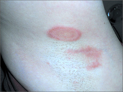
The FP diagnosed tinea corporis by recognizing the pattern of the concentric rings, which have a high specificity for tinea infections. To confirm the diagnosis, the FP performed a scraping of the area and found branching septate hyphae. This is the microscopic appearance of dermatophytes, which come in 3 genera: microsporum, epidermophyton, and trichophyton. There are approximately 40 species in these 3 genera and these fungi cause tinea pedis and manus, tinea capitis, tinea corporis, tinea cruris, tinea faciei, and onychomycosis.
Potassium hydroxide (KOH) test characteristics (without fungal stains) have a sensitivity of 77% to 88% and a specificity of 62% to 95%.1 The sensitivity and specificity is higher with fungal stains and the experience of the person performing the test. Fungal infections of the skin and mucous membranes are ubiquitous and common. There are many types of fungus that grow on humans, but they all share a predilection for warm and moist areas. Consequently, hot and humid climates promote fungal infections. The FP also asked to check the patient’s feet and found a mild case of tinea pedis between the 4th and 5th toes bilaterally.
The FP explained the diagnoses to the patient and recommended that she purchase topical terbinafine over the counter. He explained that there are no better topical antifungals by prescription and that she could find this medicine in the athlete’s foot area of any pharmacy or large grocery store. He told her that she did not have to stop running and that anyone can get a fungal infection. The FP recommended that the patient use the cream for at least one week after clinical clearance and to do her best to keep her feet and axillae dry to avoid new fungal infections.
1. Thomas B. Clear choices in managing epidermal tinea infections. J Fam Pract. 2003;52:850-862.
Photos and text for Photo Rounds Friday courtesy of Richard P. Usatine, MD. This case was adapted from: Usatine R. Fungal overview. In: Usatine R, Smith M, Mayeaux EJ, et al, eds. Color Atlas of Family Medicine. 2nd ed. New York, NY: McGraw-Hill; 2013:771-776.
To learn more about the Color Atlas of Family Medicine, see: www.amazon.com/Color-Family-Medicine-Richard-Usatine/dp/0071769641/
You can now get the second edition of the Color Atlas of Family Medicine as an app by clicking on this link: usatinemedia.com
