User login
Rotavirus vaccine again linked to small increase in risk of intussusception hospitalization
The first dose of rotavirus vaccine was again linked to a small, short-term increase in the risk of hospitalization for intussusception, but the benefits of the vaccine exceed this risk, according to a report.
In absolute numbers, there were an estimated 7 to 26 more intussusception cases per year among U.S. children aged 8-11 weeks during the postvaccine era, compared with the prevaccine era, said Jacqueline E. Tate, PhD, of the National Center for Immunization and Respiratory Diseases, Centers for Disease Control and Prevention, Atlanta, and her associates.

The increased risk did not extend to older children, which “is consistent with other U.S. studies,” the investigators said. “[Given] the magnitude of the declines in rotavirus disease compared with the small increased risk of intussusception, the public health benefits of rotavirus vaccination far exceed the increased risk of intussusception.”
Human rotavirus vaccines have been linked to rare events of intussusception since at least 1999, when Rotashield was withdrawn from the market for this reason. The next two rotavirus vaccines to receive Food and Drug Administration approval, RotaTeq and Rotarix, were not linked to intussusception in large trials or early postmarketing studies, but were estimated to cause 1-5 excess cases of intussusception per 100,000 population in more recent studies. Furthermore, a prior analysis of U.S. hospital discharge data found a small increase in the risk of intussusception hospitalization among 8- to 11-week-olds between 2007 to 2009, compared with baseline data from the prevaccine era, the investigators noted (Pediatrics. 2016 Aug 24. doi: 10.1542/peds.2016-1082).
To build on that analysis, they calculated rates of intussusception between 2000 and 2013 overall and among recommended age windows for rotavirus vaccination, which are 6-14 weeks for the first dose, 15-24 weeks for the second dose, and 25-34 weeks for the third dose. For consistency, they also looked at hospitalization rates among children aged 8-11 weeks.
The investigators identified 15,231 intussusception hospitalizations among children under 1 year of age during the study from 2000-2013. There were no overall trends in intussusception hospitalizations for all children under 1 year of age or subgroups of children aged 15-24 weeks or 25-34 weeks. Among children aged 8-11 weeks, intussusception hospitalization rates did rise significantly during the postvaccine era, compared with the prevaccine era for all years except 2011 and 2013. Excluding those two years, annual rates of intussusception hospitalizations among 8- to 11-week-olds were 46%-101% higher (16.7 to 22.9 hospitalizations per 100,000 population) during the postvaccine era than during the prevaccine era (11.4 hospitalizations per 100,000 population).
These results are “consistent with other U.S. studies that have been able to associate an increased risk of intussusception in the first week after the first dose of vaccine,” the researchers concluded. The advent of rotavirus vaccination prevented more than 176,000 hospitalizations, 242,000 emergency department visits, and 1.1 million outpatient visits from 2007 to 2011 (JAMA. 2015;313(22):2282-4), they noted.
The investigators reported no external funding sources and had no disclosures.
The first dose of rotavirus vaccine was again linked to a small, short-term increase in the risk of hospitalization for intussusception, but the benefits of the vaccine exceed this risk, according to a report.
In absolute numbers, there were an estimated 7 to 26 more intussusception cases per year among U.S. children aged 8-11 weeks during the postvaccine era, compared with the prevaccine era, said Jacqueline E. Tate, PhD, of the National Center for Immunization and Respiratory Diseases, Centers for Disease Control and Prevention, Atlanta, and her associates.
The increased risk did not extend to older children, which “is consistent with other U.S. studies,” the investigators said. “[Given] the magnitude of the declines in rotavirus disease compared with the small increased risk of intussusception, the public health benefits of rotavirus vaccination far exceed the increased risk of intussusception.”
Human rotavirus vaccines have been linked to rare events of intussusception since at least 1999, when Rotashield was withdrawn from the market for this reason. The next two rotavirus vaccines to receive Food and Drug Administration approval, RotaTeq and Rotarix, were not linked to intussusception in large trials or early postmarketing studies, but were estimated to cause 1-5 excess cases of intussusception per 100,000 population in more recent studies. Furthermore, a prior analysis of U.S. hospital discharge data found a small increase in the risk of intussusception hospitalization among 8- to 11-week-olds between 2007 to 2009, compared with baseline data from the prevaccine era, the investigators noted (Pediatrics. 2016 Aug 24. doi: 10.1542/peds.2016-1082).
To build on that analysis, they calculated rates of intussusception between 2000 and 2013 overall and among recommended age windows for rotavirus vaccination, which are 6-14 weeks for the first dose, 15-24 weeks for the second dose, and 25-34 weeks for the third dose. For consistency, they also looked at hospitalization rates among children aged 8-11 weeks.
The investigators identified 15,231 intussusception hospitalizations among children under 1 year of age during the study from 2000-2013. There were no overall trends in intussusception hospitalizations for all children under 1 year of age or subgroups of children aged 15-24 weeks or 25-34 weeks. Among children aged 8-11 weeks, intussusception hospitalization rates did rise significantly during the postvaccine era, compared with the prevaccine era for all years except 2011 and 2013. Excluding those two years, annual rates of intussusception hospitalizations among 8- to 11-week-olds were 46%-101% higher (16.7 to 22.9 hospitalizations per 100,000 population) during the postvaccine era than during the prevaccine era (11.4 hospitalizations per 100,000 population).
These results are “consistent with other U.S. studies that have been able to associate an increased risk of intussusception in the first week after the first dose of vaccine,” the researchers concluded. The advent of rotavirus vaccination prevented more than 176,000 hospitalizations, 242,000 emergency department visits, and 1.1 million outpatient visits from 2007 to 2011 (JAMA. 2015;313(22):2282-4), they noted.
The investigators reported no external funding sources and had no disclosures.
The first dose of rotavirus vaccine was again linked to a small, short-term increase in the risk of hospitalization for intussusception, but the benefits of the vaccine exceed this risk, according to a report.
In absolute numbers, there were an estimated 7 to 26 more intussusception cases per year among U.S. children aged 8-11 weeks during the postvaccine era, compared with the prevaccine era, said Jacqueline E. Tate, PhD, of the National Center for Immunization and Respiratory Diseases, Centers for Disease Control and Prevention, Atlanta, and her associates.
The increased risk did not extend to older children, which “is consistent with other U.S. studies,” the investigators said. “[Given] the magnitude of the declines in rotavirus disease compared with the small increased risk of intussusception, the public health benefits of rotavirus vaccination far exceed the increased risk of intussusception.”
Human rotavirus vaccines have been linked to rare events of intussusception since at least 1999, when Rotashield was withdrawn from the market for this reason. The next two rotavirus vaccines to receive Food and Drug Administration approval, RotaTeq and Rotarix, were not linked to intussusception in large trials or early postmarketing studies, but were estimated to cause 1-5 excess cases of intussusception per 100,000 population in more recent studies. Furthermore, a prior analysis of U.S. hospital discharge data found a small increase in the risk of intussusception hospitalization among 8- to 11-week-olds between 2007 to 2009, compared with baseline data from the prevaccine era, the investigators noted (Pediatrics. 2016 Aug 24. doi: 10.1542/peds.2016-1082).
To build on that analysis, they calculated rates of intussusception between 2000 and 2013 overall and among recommended age windows for rotavirus vaccination, which are 6-14 weeks for the first dose, 15-24 weeks for the second dose, and 25-34 weeks for the third dose. For consistency, they also looked at hospitalization rates among children aged 8-11 weeks.
The investigators identified 15,231 intussusception hospitalizations among children under 1 year of age during the study from 2000-2013. There were no overall trends in intussusception hospitalizations for all children under 1 year of age or subgroups of children aged 15-24 weeks or 25-34 weeks. Among children aged 8-11 weeks, intussusception hospitalization rates did rise significantly during the postvaccine era, compared with the prevaccine era for all years except 2011 and 2013. Excluding those two years, annual rates of intussusception hospitalizations among 8- to 11-week-olds were 46%-101% higher (16.7 to 22.9 hospitalizations per 100,000 population) during the postvaccine era than during the prevaccine era (11.4 hospitalizations per 100,000 population).
These results are “consistent with other U.S. studies that have been able to associate an increased risk of intussusception in the first week after the first dose of vaccine,” the researchers concluded. The advent of rotavirus vaccination prevented more than 176,000 hospitalizations, 242,000 emergency department visits, and 1.1 million outpatient visits from 2007 to 2011 (JAMA. 2015;313(22):2282-4), they noted.
The investigators reported no external funding sources and had no disclosures.
FROM PEDIATRICS
Key clinical point: The first dose of rotavirus vaccination is associated with a slight increase in risk of hospitalization for intussusception.
Major finding: Among 8-week-old infants to 11-week-old infants, rates of intussusception hospitalizations were significantly (46% to 101%) higher than in the prevaccine era for all postvaccine years except 2011 and 2013.
Data source: An analysis of hospitalization data from 2000 through 2013 from the State Inpatient Database, which covers 26 U.S. states.
Disclosures: The investigators had no external funding sources and no disclosures.

Loss of autonomy
At the summer meetings of the American Academy of Dermatology, AAD President Abel Torres screened a video of members responding to the question, “What keeps you up at night?” A recurring refrain in many of their responses was, “loss of autonomy.”
Many physicians feel they are losing autonomy. No doubt they are right. But physicians are not alone in their loss.
A young academic friend of mine had a similar lament. “Some assistant dean sent me an email ordering me to grade my students in a way that made no sense,” he said. “I challenged him to explain why. He answered that my school was following the guidelines of some organization I’d never heard of.”

“Academics used to be autonomous,” he said. “No more.”
Another professor friend decided to retire. “Forty years in the department,” he said, “10 as chair. Now a junior administrator tells me that I have to spend more hours on campus, even though I don’t have anything useful to do when I’m there. She said there are new rules for more academic efficiency.”
New administrators. Guidelines. Efficiency. Experienced hands dropping out or retiring out of frustration. Any of these sound familiar?
Teachers also complain to me about their loss of autonomy. “I used to be able to use judgment,” said one. “I knew what worked for a specific student. Now I just teach to the standardized test.
“For every one of my 23 kindergartners, I spent 1 hour filling out an iPad questionnaire on reading readiness. I’ve had it.”
“What will you do instead?” I asked him.
“Something with dogs,” he said.
And so it goes. Accountants and attorneys complain about heavy reporting regulations, with new ones added each year. Judges in Wisconsin make sentencing decisions using proprietary algorithms that no one outside the company that sells the algorithms has validated. Financial advisers have clients sign boilerplate statements documenting that they accept a certain level of risk. These clients may or may not understand what “level of risk” really implies, but either way they must sign a form, and the form must be filed. If you didn’t document it, you didn’t do it. If you documented it, you did it, even though you may not have really done anything meaningful.
An internist told me how things are in her new dispensation.
“They allow 15 minutes for a physical,” she said, “which is not enough anyway. But I also have to check off boxes for the EMR that add nothing to patient care. Last year we had to start asking about gender status. ‘What was your gender assignment at birth?’ ‘What is it now?’ We have to ask that every year – and click the box that says we did it.
“Several docs in our group retired. Another bunch went concierge. They couldn’t deal with it anymore.”
Metrics. Algorithms. Higher authorities who tell professionals what to do, how to do it, how to record it, business quants with scant understanding of what professionals actually profess. All so familiar and tiresome. It’s everywhere, and it’s bigger than any of us.
Loss of autonomy by professionals across the board reflects a changed understanding by society at large of what quality service is and how it should be judged. Numbers are in. Personal judgment – in our case, clinical judgment – is out. Since judgment can’t be measured, it cannot be trusted.
To a certain extent, autonomy is an illusion. We can do what we want as long as powers larger than we are – natural, social, political – let us do it. Those powers may lie dormant for a while, but they’re always there, and always have been. When they wake up and change the rules of the game, everyone has to adapt. New burdens in the practice of medicine are just one instance of a much broader trend.
Our professional organizations know this well. They are hard at work giving the authorities, government, and insurance administrators what they demand: data showing that what we do is useful, in the quantitative terms the authorities will accept.
To the extent that they succeed, we will be able to do some of what we want to do. Young people entering the medical field will expect nothing more. Some of their older colleagues will be satisfied that they are autonomous enough. The rest will have to find something else to do.
Dr. Rockoff practices dermatology in Brookline, Mass. He is on the clinical faculty at Tufts University School of Medicine, Boston, and has taught senior medical students and other trainees for 30 years.
At the summer meetings of the American Academy of Dermatology, AAD President Abel Torres screened a video of members responding to the question, “What keeps you up at night?” A recurring refrain in many of their responses was, “loss of autonomy.”
Many physicians feel they are losing autonomy. No doubt they are right. But physicians are not alone in their loss.
A young academic friend of mine had a similar lament. “Some assistant dean sent me an email ordering me to grade my students in a way that made no sense,” he said. “I challenged him to explain why. He answered that my school was following the guidelines of some organization I’d never heard of.”
“Academics used to be autonomous,” he said. “No more.”
Another professor friend decided to retire. “Forty years in the department,” he said, “10 as chair. Now a junior administrator tells me that I have to spend more hours on campus, even though I don’t have anything useful to do when I’m there. She said there are new rules for more academic efficiency.”
New administrators. Guidelines. Efficiency. Experienced hands dropping out or retiring out of frustration. Any of these sound familiar?
Teachers also complain to me about their loss of autonomy. “I used to be able to use judgment,” said one. “I knew what worked for a specific student. Now I just teach to the standardized test.
“For every one of my 23 kindergartners, I spent 1 hour filling out an iPad questionnaire on reading readiness. I’ve had it.”
“What will you do instead?” I asked him.
“Something with dogs,” he said.
And so it goes. Accountants and attorneys complain about heavy reporting regulations, with new ones added each year. Judges in Wisconsin make sentencing decisions using proprietary algorithms that no one outside the company that sells the algorithms has validated. Financial advisers have clients sign boilerplate statements documenting that they accept a certain level of risk. These clients may or may not understand what “level of risk” really implies, but either way they must sign a form, and the form must be filed. If you didn’t document it, you didn’t do it. If you documented it, you did it, even though you may not have really done anything meaningful.
An internist told me how things are in her new dispensation.
“They allow 15 minutes for a physical,” she said, “which is not enough anyway. But I also have to check off boxes for the EMR that add nothing to patient care. Last year we had to start asking about gender status. ‘What was your gender assignment at birth?’ ‘What is it now?’ We have to ask that every year – and click the box that says we did it.
“Several docs in our group retired. Another bunch went concierge. They couldn’t deal with it anymore.”
Metrics. Algorithms. Higher authorities who tell professionals what to do, how to do it, how to record it, business quants with scant understanding of what professionals actually profess. All so familiar and tiresome. It’s everywhere, and it’s bigger than any of us.
Loss of autonomy by professionals across the board reflects a changed understanding by society at large of what quality service is and how it should be judged. Numbers are in. Personal judgment – in our case, clinical judgment – is out. Since judgment can’t be measured, it cannot be trusted.
To a certain extent, autonomy is an illusion. We can do what we want as long as powers larger than we are – natural, social, political – let us do it. Those powers may lie dormant for a while, but they’re always there, and always have been. When they wake up and change the rules of the game, everyone has to adapt. New burdens in the practice of medicine are just one instance of a much broader trend.
Our professional organizations know this well. They are hard at work giving the authorities, government, and insurance administrators what they demand: data showing that what we do is useful, in the quantitative terms the authorities will accept.
To the extent that they succeed, we will be able to do some of what we want to do. Young people entering the medical field will expect nothing more. Some of their older colleagues will be satisfied that they are autonomous enough. The rest will have to find something else to do.
Dr. Rockoff practices dermatology in Brookline, Mass. He is on the clinical faculty at Tufts University School of Medicine, Boston, and has taught senior medical students and other trainees for 30 years.
At the summer meetings of the American Academy of Dermatology, AAD President Abel Torres screened a video of members responding to the question, “What keeps you up at night?” A recurring refrain in many of their responses was, “loss of autonomy.”
Many physicians feel they are losing autonomy. No doubt they are right. But physicians are not alone in their loss.
A young academic friend of mine had a similar lament. “Some assistant dean sent me an email ordering me to grade my students in a way that made no sense,” he said. “I challenged him to explain why. He answered that my school was following the guidelines of some organization I’d never heard of.”
“Academics used to be autonomous,” he said. “No more.”
Another professor friend decided to retire. “Forty years in the department,” he said, “10 as chair. Now a junior administrator tells me that I have to spend more hours on campus, even though I don’t have anything useful to do when I’m there. She said there are new rules for more academic efficiency.”
New administrators. Guidelines. Efficiency. Experienced hands dropping out or retiring out of frustration. Any of these sound familiar?
Teachers also complain to me about their loss of autonomy. “I used to be able to use judgment,” said one. “I knew what worked for a specific student. Now I just teach to the standardized test.
“For every one of my 23 kindergartners, I spent 1 hour filling out an iPad questionnaire on reading readiness. I’ve had it.”
“What will you do instead?” I asked him.
“Something with dogs,” he said.
And so it goes. Accountants and attorneys complain about heavy reporting regulations, with new ones added each year. Judges in Wisconsin make sentencing decisions using proprietary algorithms that no one outside the company that sells the algorithms has validated. Financial advisers have clients sign boilerplate statements documenting that they accept a certain level of risk. These clients may or may not understand what “level of risk” really implies, but either way they must sign a form, and the form must be filed. If you didn’t document it, you didn’t do it. If you documented it, you did it, even though you may not have really done anything meaningful.
An internist told me how things are in her new dispensation.
“They allow 15 minutes for a physical,” she said, “which is not enough anyway. But I also have to check off boxes for the EMR that add nothing to patient care. Last year we had to start asking about gender status. ‘What was your gender assignment at birth?’ ‘What is it now?’ We have to ask that every year – and click the box that says we did it.
“Several docs in our group retired. Another bunch went concierge. They couldn’t deal with it anymore.”
Metrics. Algorithms. Higher authorities who tell professionals what to do, how to do it, how to record it, business quants with scant understanding of what professionals actually profess. All so familiar and tiresome. It’s everywhere, and it’s bigger than any of us.
Loss of autonomy by professionals across the board reflects a changed understanding by society at large of what quality service is and how it should be judged. Numbers are in. Personal judgment – in our case, clinical judgment – is out. Since judgment can’t be measured, it cannot be trusted.
To a certain extent, autonomy is an illusion. We can do what we want as long as powers larger than we are – natural, social, political – let us do it. Those powers may lie dormant for a while, but they’re always there, and always have been. When they wake up and change the rules of the game, everyone has to adapt. New burdens in the practice of medicine are just one instance of a much broader trend.
Our professional organizations know this well. They are hard at work giving the authorities, government, and insurance administrators what they demand: data showing that what we do is useful, in the quantitative terms the authorities will accept.
To the extent that they succeed, we will be able to do some of what we want to do. Young people entering the medical field will expect nothing more. Some of their older colleagues will be satisfied that they are autonomous enough. The rest will have to find something else to do.
Dr. Rockoff practices dermatology in Brookline, Mass. He is on the clinical faculty at Tufts University School of Medicine, Boston, and has taught senior medical students and other trainees for 30 years.

Uridine triacetate provides antidote for 5-fluorouracil overdose and toxicity
Fluorouracil or 5-fluorouracil (5-FU) is an effective cytotoxic drug that is incorporated into various chemotherapeutic regimens for the treatment of numerous tumor types, but its clinical utility is limited by its narrow therapeutic index and the risk of overdose and serious toxic effects. Until recently, these outcomes were managed with supportive care, but the approval of uridine triacetate provides an antidote to reverse 5-FU-associated toxicity, to prevent death and potentially allow some patients to resume chemotherapy.
Click on the PDF icon at the top of this introduction to read the full article.
Fluorouracil or 5-fluorouracil (5-FU) is an effective cytotoxic drug that is incorporated into various chemotherapeutic regimens for the treatment of numerous tumor types, but its clinical utility is limited by its narrow therapeutic index and the risk of overdose and serious toxic effects. Until recently, these outcomes were managed with supportive care, but the approval of uridine triacetate provides an antidote to reverse 5-FU-associated toxicity, to prevent death and potentially allow some patients to resume chemotherapy.
Click on the PDF icon at the top of this introduction to read the full article.
Fluorouracil or 5-fluorouracil (5-FU) is an effective cytotoxic drug that is incorporated into various chemotherapeutic regimens for the treatment of numerous tumor types, but its clinical utility is limited by its narrow therapeutic index and the risk of overdose and serious toxic effects. Until recently, these outcomes were managed with supportive care, but the approval of uridine triacetate provides an antidote to reverse 5-FU-associated toxicity, to prevent death and potentially allow some patients to resume chemotherapy.
Click on the PDF icon at the top of this introduction to read the full article.
Sterilization rates decline for women despite coverage
Fewer women aged 20-44 years are using sterilization as their primary method of contraception, despite no-cost coverage of the procedure under the Affordable Care Act.
Sterilization rates in 2011-2013 for women in this age group was 25%, down from 27% in 2002, according to a new report from the Kaiser Family Foundation. Rates declined across income groups, dropping from 41% in 2002 to 37% in 2011-2013 for women whose incomes were up to 149% of the federal poverty level (FPL). For women with incomes within 150%-299% of the FPL, the rate declined from 33% to 28%, and for those at 300% or more of the FPL, the rate declined from 20% to 18%, according to the report.

While expanded coverage under the ACA may, in the long run, lead to greater use of sterilization by women, “it remains to be seen whether the growing availability of [long-acting reversible contraception] will offset this as IUDs and implants serve as semi-permanent substitutes for sterilization,” the Kaiser researchers wrote.
From 2011 to 2013, about 5% of men aged 15-44 years reported undergoing a vasectomy, according to Kaiser. The ACA does not require health plans to cover sterilization for men, but two states – Vermont and Maryland – have passed laws requiring coverage within their borders in the future.
Male sterilization rates increased with age (1% of those aged 15-34 years, compared with 13% of those aged 35-44 years), income (3% for those below 200% of the FPL, 5% for those 200%-399%, and 10% for those above 400%), and education (3% of those with up to a high school diploma/GED, 5% of those with some college, and 13% of those with at least a 4-year college degree). Comparative changes in male sterilization rates were not provided.
Fewer women aged 20-44 years are using sterilization as their primary method of contraception, despite no-cost coverage of the procedure under the Affordable Care Act.
Sterilization rates in 2011-2013 for women in this age group was 25%, down from 27% in 2002, according to a new report from the Kaiser Family Foundation. Rates declined across income groups, dropping from 41% in 2002 to 37% in 2011-2013 for women whose incomes were up to 149% of the federal poverty level (FPL). For women with incomes within 150%-299% of the FPL, the rate declined from 33% to 28%, and for those at 300% or more of the FPL, the rate declined from 20% to 18%, according to the report.
While expanded coverage under the ACA may, in the long run, lead to greater use of sterilization by women, “it remains to be seen whether the growing availability of [long-acting reversible contraception] will offset this as IUDs and implants serve as semi-permanent substitutes for sterilization,” the Kaiser researchers wrote.
From 2011 to 2013, about 5% of men aged 15-44 years reported undergoing a vasectomy, according to Kaiser. The ACA does not require health plans to cover sterilization for men, but two states – Vermont and Maryland – have passed laws requiring coverage within their borders in the future.
Male sterilization rates increased with age (1% of those aged 15-34 years, compared with 13% of those aged 35-44 years), income (3% for those below 200% of the FPL, 5% for those 200%-399%, and 10% for those above 400%), and education (3% of those with up to a high school diploma/GED, 5% of those with some college, and 13% of those with at least a 4-year college degree). Comparative changes in male sterilization rates were not provided.
Fewer women aged 20-44 years are using sterilization as their primary method of contraception, despite no-cost coverage of the procedure under the Affordable Care Act.
Sterilization rates in 2011-2013 for women in this age group was 25%, down from 27% in 2002, according to a new report from the Kaiser Family Foundation. Rates declined across income groups, dropping from 41% in 2002 to 37% in 2011-2013 for women whose incomes were up to 149% of the federal poverty level (FPL). For women with incomes within 150%-299% of the FPL, the rate declined from 33% to 28%, and for those at 300% or more of the FPL, the rate declined from 20% to 18%, according to the report.
While expanded coverage under the ACA may, in the long run, lead to greater use of sterilization by women, “it remains to be seen whether the growing availability of [long-acting reversible contraception] will offset this as IUDs and implants serve as semi-permanent substitutes for sterilization,” the Kaiser researchers wrote.
From 2011 to 2013, about 5% of men aged 15-44 years reported undergoing a vasectomy, according to Kaiser. The ACA does not require health plans to cover sterilization for men, but two states – Vermont and Maryland – have passed laws requiring coverage within their borders in the future.
Male sterilization rates increased with age (1% of those aged 15-34 years, compared with 13% of those aged 35-44 years), income (3% for those below 200% of the FPL, 5% for those 200%-399%, and 10% for those above 400%), and education (3% of those with up to a high school diploma/GED, 5% of those with some college, and 13% of those with at least a 4-year college degree). Comparative changes in male sterilization rates were not provided.

It’s health care … but not as we know it
Click on the PDF icon at the top of this introduction to read the full article.
Click on the PDF icon at the top of this introduction to read the full article.
Click on the PDF icon at the top of this introduction to read the full article.
Expert shares new insights on the pathophysiology of rosacea
NEWPORT BEACH, CALIF. – In the clinical opinion of Richard L. Gallo, MD, PhD, current nomenclature for the diagnosis of rosacea could use a makeover.
“Currently, we’re still operating with an almost 20-year-old set of diagnostic subtypes of rosacea,” Dr. Gallo said at the annual meeting of the Pacific Dermatologic Association. He plans to participate in consensus meeting of experts who will convene this fall in an effort to update and modify these diagnostic criteria.

According to current nomenclature, subtype 1 is erythematotelangiectatic rosacea characterized by facial redness; subtype 2 is papulopustular, marked by bumps and pimples; subtype 3 is phymatous, characterized by enlargement of the nose, and subtype 4 is ocular, marked by eye irritation. Dr. Gallo pointed out that it’s rare to see just one of these subtypes in rosacea patients, with the exception of the erythematotelangiectatic rosacea (ETR). “There is a large population with ETR alone,” he said.” Most patients with the papulopustular subtype have aspects of ETR. There is a mix of subtypes of rosacea and we clearly need to modify our diagnostic criteria.”
Secondary rosacea features may include burning or stinging, plaque, dry appearance and scale, edema, ocular manifestations, peripheral location, and phymatous changes. Work by several researchers in recent years has shed light on the pathophysiology of rosacea. “We’re learning that there are many aspects to this disease that both trigger it and result in progression of the disease,” said Dr. Gallo, professor and chair of the department of dermatology at the University of California, San Diego. “It seems to have biological triggers that exist both in the environment and initiate from internal sources. We’re understanding more about the nature of those, at least specific molecules externally from microbes and so forth. Internally we understand more about the unique inflammatory signals.”
For example, he and other researchers began to look at the innate immune system patients with rosacea and identified LL37, a multifunction peptide that plays a role in a number of skin diseases, as something that can promote inflammation (Nat Med. 2007;13[8]:975-80). “It also promotes the vascular changes [that occur with the disease],” Dr. Gallo said. “We’re now learning how a dysregulation of enzymes in the skin contributes to making too much of these types of peptides. Therefore, treatment approaches that might modify enzymatic activity become useful.” Researchers have also discovered that some of the innate recognition molecules like toll-like receptor 2 (TLR2) are overexpressed in rosacea patients. “Similarly, in terms of the vascular signals, a number of labs are identifying some of the newer vascular transmitters that seem to be uniquely elevated in rosacea, so there’s great reason to be optimistic that given the increased specificity and understanding of what uniquely makes this disease happen, we’ll be able to target it in a safe way.”
A number of published studies have supported these notions, including an analysis of 275 twin pairs (JAMA Dermatol. 2015;151[11]:1213-9). The researchers found that compared with fraternal twins, identical twins had a higher association of National Rosacea Society scores (P = .04), “supporting the concept that there are fundamental genetic factors that are influencing disease,” Dr. Gallo said. Environmental factors found to be associated with rosacea include lifetime UV exposure, smoking, obesity, and alcohol use.
In an assessment of the genetic basis of rosacea by genome-wide association study, researchers identified one confirmed single-nucleotide polymorphism that could be associated with rosacea (J Invest Dermatol. 2015;135[6]:1548-55). It was located in an intergenic region between HLA-DRA and BTNL2, “which is consistent with the overall concept that there is perhaps a genetic abnormality that is leading to increased amino modulation of difficulties,” Dr. Gallo said. For another recent study, researchers analyzed 14 randomized or case control trials involving rosacea patients (Int J Med Sci. 2015;12[5]:387-96). They concluded that vasculature, chronic inflammatory responses, environmental triggers, food and chemicals ingested, and microorganisms either alone or in combination are responsible for rosacea.
Dr. Gallo went on to highlight findings from several studies that link rosacea to an increased risk for certain comorbidities. One, a nationwide case-control study from Taiwan that comprised 35,553 rosacea patients, found that the disease was significantly associated with a risk of certain cardiovascular comorbidities (J Acad Dermatol. 2015;73:249-54). These included dyslipidemia (OR 1.41), coronary artery disease (OR 1.35), and hypertension (OR 1.17). A separate analysis, based 93,314 participants in the Nurse’s Health Study, found that rosacea was significantly associated with a risk of coronary artery disease (OR 2.2). The researchers also observed that comorbidities seemed to increase with duration of the disease (Clin Gastroenterol Hepatol. 2016;14[2]:220-5). Another smaller case-control study of 65 patients and 65 controls found an increased risk with rosacea for coronary artery disease, hyperlipidemia, hypertension, and gastroesophageal reflux disease (J Am Acad Dermatol. 2015;73[4]:604-8).
A recent analysis of 75,088 participants in the Nurses’ Health Study found that women with rosacea faced an increased risk of thyroid cancer (HR 1.59) and basal cell carcinoma (HR 1.50; Br J Cancer 2015;113[3]:520-3). Rosacea may also impact one’s risk for developing certain neurological conditions. One study found an increased risk for dementia (HR 1.42) and Alzheimer’s disease (HR 1.92; Ann Neurol. 2016;79:921-8), while another found an increased risk for Parkinson’s disease (an adjusted incident ratio of 1.71 in patients with rosacea, compared with the referent population; JAMA Neurol. 2016;73[5]:529-34).
As for therapy, a recent Cochrane systematic review found strong evidence supporting benefits of several therapies over placebo, including metronidazole, azelaic acid, brimonidine, tetracycline, doxycycline 40 mg, ivermectin, and isotretinoin (Br J Dermatol. 2015;173[3]:651-2). A separate, 7-year retrospective study of 275 adults with rosacea published online in The Journal of Dermatology on Oct. 28, 2015, found that patients with the PPR subtype had a better overall prognosis, compared with their counterparts with the other subtypes. Overall, the median time to complete remission was 56 months. Complete remission was achieved in 46% of those with PPR subtype, compared with 19% of those with mixed subtype and 11% of those with ETR subtype.
Dr. Gallo disclosed that he has received research grants from the National Institutes of Health, Allergan, L’Oreal, Colgate-Palmolive, Regeneron, GSK, Galderma, and Bayer. He is a consultant for Allergan, Colgate-Palmolive, Sente, Matrisys, Dermata, Alnylam, Abbvie, Roche, and Promius.
NEWPORT BEACH, CALIF. – In the clinical opinion of Richard L. Gallo, MD, PhD, current nomenclature for the diagnosis of rosacea could use a makeover.
“Currently, we’re still operating with an almost 20-year-old set of diagnostic subtypes of rosacea,” Dr. Gallo said at the annual meeting of the Pacific Dermatologic Association. He plans to participate in consensus meeting of experts who will convene this fall in an effort to update and modify these diagnostic criteria.
According to current nomenclature, subtype 1 is erythematotelangiectatic rosacea characterized by facial redness; subtype 2 is papulopustular, marked by bumps and pimples; subtype 3 is phymatous, characterized by enlargement of the nose, and subtype 4 is ocular, marked by eye irritation. Dr. Gallo pointed out that it’s rare to see just one of these subtypes in rosacea patients, with the exception of the erythematotelangiectatic rosacea (ETR). “There is a large population with ETR alone,” he said.” Most patients with the papulopustular subtype have aspects of ETR. There is a mix of subtypes of rosacea and we clearly need to modify our diagnostic criteria.”
Secondary rosacea features may include burning or stinging, plaque, dry appearance and scale, edema, ocular manifestations, peripheral location, and phymatous changes. Work by several researchers in recent years has shed light on the pathophysiology of rosacea. “We’re learning that there are many aspects to this disease that both trigger it and result in progression of the disease,” said Dr. Gallo, professor and chair of the department of dermatology at the University of California, San Diego. “It seems to have biological triggers that exist both in the environment and initiate from internal sources. We’re understanding more about the nature of those, at least specific molecules externally from microbes and so forth. Internally we understand more about the unique inflammatory signals.”
For example, he and other researchers began to look at the innate immune system patients with rosacea and identified LL37, a multifunction peptide that plays a role in a number of skin diseases, as something that can promote inflammation (Nat Med. 2007;13[8]:975-80). “It also promotes the vascular changes [that occur with the disease],” Dr. Gallo said. “We’re now learning how a dysregulation of enzymes in the skin contributes to making too much of these types of peptides. Therefore, treatment approaches that might modify enzymatic activity become useful.” Researchers have also discovered that some of the innate recognition molecules like toll-like receptor 2 (TLR2) are overexpressed in rosacea patients. “Similarly, in terms of the vascular signals, a number of labs are identifying some of the newer vascular transmitters that seem to be uniquely elevated in rosacea, so there’s great reason to be optimistic that given the increased specificity and understanding of what uniquely makes this disease happen, we’ll be able to target it in a safe way.”
A number of published studies have supported these notions, including an analysis of 275 twin pairs (JAMA Dermatol. 2015;151[11]:1213-9). The researchers found that compared with fraternal twins, identical twins had a higher association of National Rosacea Society scores (P = .04), “supporting the concept that there are fundamental genetic factors that are influencing disease,” Dr. Gallo said. Environmental factors found to be associated with rosacea include lifetime UV exposure, smoking, obesity, and alcohol use.
In an assessment of the genetic basis of rosacea by genome-wide association study, researchers identified one confirmed single-nucleotide polymorphism that could be associated with rosacea (J Invest Dermatol. 2015;135[6]:1548-55). It was located in an intergenic region between HLA-DRA and BTNL2, “which is consistent with the overall concept that there is perhaps a genetic abnormality that is leading to increased amino modulation of difficulties,” Dr. Gallo said. For another recent study, researchers analyzed 14 randomized or case control trials involving rosacea patients (Int J Med Sci. 2015;12[5]:387-96). They concluded that vasculature, chronic inflammatory responses, environmental triggers, food and chemicals ingested, and microorganisms either alone or in combination are responsible for rosacea.
Dr. Gallo went on to highlight findings from several studies that link rosacea to an increased risk for certain comorbidities. One, a nationwide case-control study from Taiwan that comprised 35,553 rosacea patients, found that the disease was significantly associated with a risk of certain cardiovascular comorbidities (J Acad Dermatol. 2015;73:249-54). These included dyslipidemia (OR 1.41), coronary artery disease (OR 1.35), and hypertension (OR 1.17). A separate analysis, based 93,314 participants in the Nurse’s Health Study, found that rosacea was significantly associated with a risk of coronary artery disease (OR 2.2). The researchers also observed that comorbidities seemed to increase with duration of the disease (Clin Gastroenterol Hepatol. 2016;14[2]:220-5). Another smaller case-control study of 65 patients and 65 controls found an increased risk with rosacea for coronary artery disease, hyperlipidemia, hypertension, and gastroesophageal reflux disease (J Am Acad Dermatol. 2015;73[4]:604-8).
A recent analysis of 75,088 participants in the Nurses’ Health Study found that women with rosacea faced an increased risk of thyroid cancer (HR 1.59) and basal cell carcinoma (HR 1.50; Br J Cancer 2015;113[3]:520-3). Rosacea may also impact one’s risk for developing certain neurological conditions. One study found an increased risk for dementia (HR 1.42) and Alzheimer’s disease (HR 1.92; Ann Neurol. 2016;79:921-8), while another found an increased risk for Parkinson’s disease (an adjusted incident ratio of 1.71 in patients with rosacea, compared with the referent population; JAMA Neurol. 2016;73[5]:529-34).
As for therapy, a recent Cochrane systematic review found strong evidence supporting benefits of several therapies over placebo, including metronidazole, azelaic acid, brimonidine, tetracycline, doxycycline 40 mg, ivermectin, and isotretinoin (Br J Dermatol. 2015;173[3]:651-2). A separate, 7-year retrospective study of 275 adults with rosacea published online in The Journal of Dermatology on Oct. 28, 2015, found that patients with the PPR subtype had a better overall prognosis, compared with their counterparts with the other subtypes. Overall, the median time to complete remission was 56 months. Complete remission was achieved in 46% of those with PPR subtype, compared with 19% of those with mixed subtype and 11% of those with ETR subtype.
Dr. Gallo disclosed that he has received research grants from the National Institutes of Health, Allergan, L’Oreal, Colgate-Palmolive, Regeneron, GSK, Galderma, and Bayer. He is a consultant for Allergan, Colgate-Palmolive, Sente, Matrisys, Dermata, Alnylam, Abbvie, Roche, and Promius.
NEWPORT BEACH, CALIF. – In the clinical opinion of Richard L. Gallo, MD, PhD, current nomenclature for the diagnosis of rosacea could use a makeover.
“Currently, we’re still operating with an almost 20-year-old set of diagnostic subtypes of rosacea,” Dr. Gallo said at the annual meeting of the Pacific Dermatologic Association. He plans to participate in consensus meeting of experts who will convene this fall in an effort to update and modify these diagnostic criteria.
According to current nomenclature, subtype 1 is erythematotelangiectatic rosacea characterized by facial redness; subtype 2 is papulopustular, marked by bumps and pimples; subtype 3 is phymatous, characterized by enlargement of the nose, and subtype 4 is ocular, marked by eye irritation. Dr. Gallo pointed out that it’s rare to see just one of these subtypes in rosacea patients, with the exception of the erythematotelangiectatic rosacea (ETR). “There is a large population with ETR alone,” he said.” Most patients with the papulopustular subtype have aspects of ETR. There is a mix of subtypes of rosacea and we clearly need to modify our diagnostic criteria.”
Secondary rosacea features may include burning or stinging, plaque, dry appearance and scale, edema, ocular manifestations, peripheral location, and phymatous changes. Work by several researchers in recent years has shed light on the pathophysiology of rosacea. “We’re learning that there are many aspects to this disease that both trigger it and result in progression of the disease,” said Dr. Gallo, professor and chair of the department of dermatology at the University of California, San Diego. “It seems to have biological triggers that exist both in the environment and initiate from internal sources. We’re understanding more about the nature of those, at least specific molecules externally from microbes and so forth. Internally we understand more about the unique inflammatory signals.”
For example, he and other researchers began to look at the innate immune system patients with rosacea and identified LL37, a multifunction peptide that plays a role in a number of skin diseases, as something that can promote inflammation (Nat Med. 2007;13[8]:975-80). “It also promotes the vascular changes [that occur with the disease],” Dr. Gallo said. “We’re now learning how a dysregulation of enzymes in the skin contributes to making too much of these types of peptides. Therefore, treatment approaches that might modify enzymatic activity become useful.” Researchers have also discovered that some of the innate recognition molecules like toll-like receptor 2 (TLR2) are overexpressed in rosacea patients. “Similarly, in terms of the vascular signals, a number of labs are identifying some of the newer vascular transmitters that seem to be uniquely elevated in rosacea, so there’s great reason to be optimistic that given the increased specificity and understanding of what uniquely makes this disease happen, we’ll be able to target it in a safe way.”
A number of published studies have supported these notions, including an analysis of 275 twin pairs (JAMA Dermatol. 2015;151[11]:1213-9). The researchers found that compared with fraternal twins, identical twins had a higher association of National Rosacea Society scores (P = .04), “supporting the concept that there are fundamental genetic factors that are influencing disease,” Dr. Gallo said. Environmental factors found to be associated with rosacea include lifetime UV exposure, smoking, obesity, and alcohol use.
In an assessment of the genetic basis of rosacea by genome-wide association study, researchers identified one confirmed single-nucleotide polymorphism that could be associated with rosacea (J Invest Dermatol. 2015;135[6]:1548-55). It was located in an intergenic region between HLA-DRA and BTNL2, “which is consistent with the overall concept that there is perhaps a genetic abnormality that is leading to increased amino modulation of difficulties,” Dr. Gallo said. For another recent study, researchers analyzed 14 randomized or case control trials involving rosacea patients (Int J Med Sci. 2015;12[5]:387-96). They concluded that vasculature, chronic inflammatory responses, environmental triggers, food and chemicals ingested, and microorganisms either alone or in combination are responsible for rosacea.
Dr. Gallo went on to highlight findings from several studies that link rosacea to an increased risk for certain comorbidities. One, a nationwide case-control study from Taiwan that comprised 35,553 rosacea patients, found that the disease was significantly associated with a risk of certain cardiovascular comorbidities (J Acad Dermatol. 2015;73:249-54). These included dyslipidemia (OR 1.41), coronary artery disease (OR 1.35), and hypertension (OR 1.17). A separate analysis, based 93,314 participants in the Nurse’s Health Study, found that rosacea was significantly associated with a risk of coronary artery disease (OR 2.2). The researchers also observed that comorbidities seemed to increase with duration of the disease (Clin Gastroenterol Hepatol. 2016;14[2]:220-5). Another smaller case-control study of 65 patients and 65 controls found an increased risk with rosacea for coronary artery disease, hyperlipidemia, hypertension, and gastroesophageal reflux disease (J Am Acad Dermatol. 2015;73[4]:604-8).
A recent analysis of 75,088 participants in the Nurses’ Health Study found that women with rosacea faced an increased risk of thyroid cancer (HR 1.59) and basal cell carcinoma (HR 1.50; Br J Cancer 2015;113[3]:520-3). Rosacea may also impact one’s risk for developing certain neurological conditions. One study found an increased risk for dementia (HR 1.42) and Alzheimer’s disease (HR 1.92; Ann Neurol. 2016;79:921-8), while another found an increased risk for Parkinson’s disease (an adjusted incident ratio of 1.71 in patients with rosacea, compared with the referent population; JAMA Neurol. 2016;73[5]:529-34).
As for therapy, a recent Cochrane systematic review found strong evidence supporting benefits of several therapies over placebo, including metronidazole, azelaic acid, brimonidine, tetracycline, doxycycline 40 mg, ivermectin, and isotretinoin (Br J Dermatol. 2015;173[3]:651-2). A separate, 7-year retrospective study of 275 adults with rosacea published online in The Journal of Dermatology on Oct. 28, 2015, found that patients with the PPR subtype had a better overall prognosis, compared with their counterparts with the other subtypes. Overall, the median time to complete remission was 56 months. Complete remission was achieved in 46% of those with PPR subtype, compared with 19% of those with mixed subtype and 11% of those with ETR subtype.
Dr. Gallo disclosed that he has received research grants from the National Institutes of Health, Allergan, L’Oreal, Colgate-Palmolive, Regeneron, GSK, Galderma, and Bayer. He is a consultant for Allergan, Colgate-Palmolive, Sente, Matrisys, Dermata, Alnylam, Abbvie, Roche, and Promius.
EXPERT ANALYSIS AT PDA 2016

Anastrozole-Induced Subacute Cutaneous Lupus Erythematosus
Drug-induced subacute cutaneous lupus erythematosus (DI-SCLE) was first described in 1985 in 5 patients who had been taking hydrochlorothiazide.1 The skin lesions in these patients were identical to those seen in idiopathic subacute cutaneous lupus erythematosus (SCLE) and were accompanied by the same autoantibodies (anti-Ro/Sjögren syndrome antigen A [SS-A] and anti-La/Sjögren syndrome antigen B [SS-B]) and HLA type (HLA-DR2/DR3) that are known to be associated with idiopathic SCLE. The skin lesions of SCLE in these 5 patients resolved spontaneously after discontinuing hydrochlorothiazide; however, anti-Ro/SS-A antibodies persisted in all except 1 patient.1 Over the last decade, an increasing number of drugs from different classes have been implicated to be associated with DI-SCLE. Since the concept of DI-SCLE was introduced, it has been reported to look identical to idiopathic SCLE, both clinically and histopathologically; however, one report suggested that the 2 entities can be distinguished based on clinical variations.2 In general, patients with DI-SCLE develop the same anti-Ro antibodies as seen in idiopathic SCLE. In addition, although the rash in DI-SCLE typically resolves with withdrawal of the offending drug, the antibodies tend to persist. Herein, we report a case of a patient being treated with an aromatase inhibitor who presented with clinical, serologic, and histopathologic evidence of DI-SCLE.
Case Report
A 69-year-old woman diagnosed with breast cancer 4 years prior to her presentation to dermatology initially underwent a lumpectomy and radiation treatment. She was subsequently started on anastrozole 2 years later. After 16 months of treatment with anastrozole, she developed an erythematous scaly rash on sun-exposed areas of the skin. The patient was seen by an outside dermatologist who treated her for a patient-perceived drug rash based on biopsy results that simply demonstrated interface dermatitis. She was treated with both topical and oral steroids with little improvement and therefore presented to our office approximately 6 months after starting treatment seeking a second opinion.
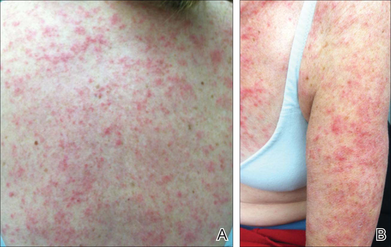
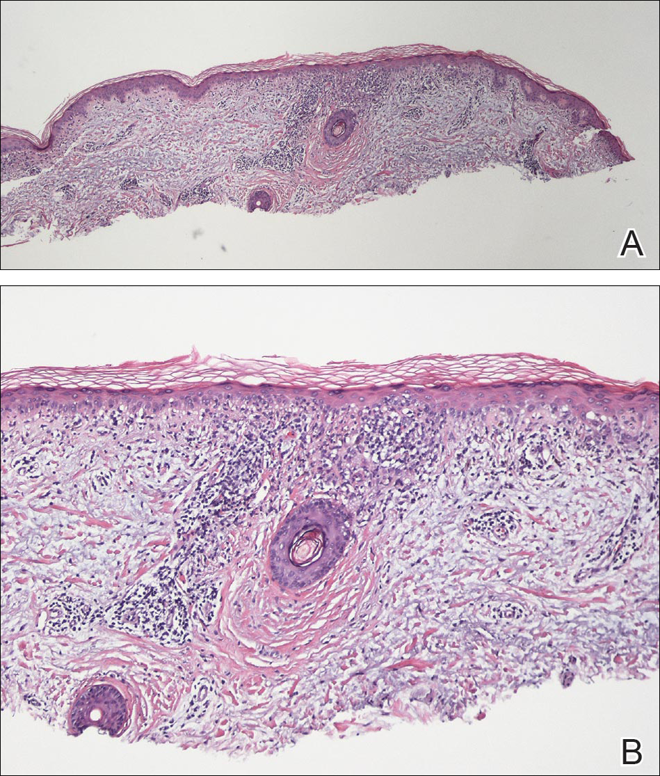
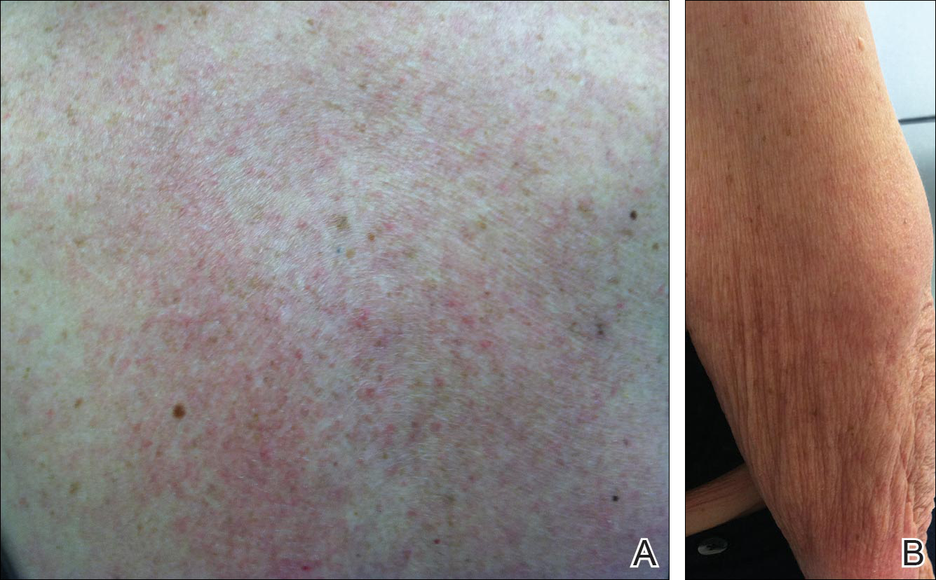
Physical examination revealed numerous erythematous scaly papules and plaques in a photodistributed pattern on the chest, back, legs, and arms (Figure 1). On further questioning, the patient noted that the rash became worse when she was at the beach or playing tennis outside as well as under indoor lights. A repeat biopsy was performed, revealing interface and perivascular dermatitis with an infiltrate composed of lymphocytes, histiocytes, and scattered pigment-laden macrophages (Figure 2). Given the appearance and distribution of the rash as well as the clinical scenario, drug-induced lupus was suspected. Anastrozole was the only medication being taken. Laboratory evaluation was performed and was negative for antinuclear antibodies, antihistone antibodies, and anti-La/SS-B antibodies but was positive for anti-Ro/SS-A antibodies (>8.0 U [reference range, <1.0 U]). Based on these findings, anastrozole-induced SCLE was the most likely explanation for this presentation. The patient was started on a sun-protective regimen (ie, wide-brimmed hat, daily sunscreen) and anastrozole was discontinued by her oncologist; the combination led to moderate improvement in symptoms. One week later, oral hydroxychloroquine 200 mg twice daily was started, which led to notable improvement (Figure 3). The patient was seen for 2 additional follow-up visits, each time with sustained resolution of the rash. The hydroxychloroquine was then stopped at her last visit 3 months after diagnosis. The patient was subsequently lost to follow-up.
Comment
Presentation of SCLE
Subacute cutaneous lupus erythematosus is a form of lupus erythematosus characterized by nonscarring, annular, scaly, erythematous plaques that occur on sun-exposed skin. The lesions are classically distributed on the upper back, chest, dorsal arms, and lateral neck but also can be found in other locations.3,4 Subacute cutaneous lupus erythematosus may be idiopathic; may occur in patients with systemic lupus erythematosus, Sjögren syndrome, or deficiency of the second component of complement (C2d); or may be drug induced.5 On histology SCLE presents as a lichenoid tissue reaction with focal vacuolization of the epidermal basal layer and perivascular lymphocytic infiltrate. On direct immunofluorescence, both idiopathic and drug-induced SCLE present with granular deposition of IgM, IgG, and C3 in a bandlike array at the dermoepidermal junction and circulating anti-Ro/SS-A antibodies. Therefore, histopathologically and immunologically, DI-SCLE is indistinguishable from idiopathic cases.6
Differential Diagnosis
It was previously thought that the clinical presentation of DI-SCLE and idiopathic SCLE were indistinguishable; however, Marzano et al2 described remarkable differences in the cutaneous manifestations of the 2 diseases. Drug-induced SCLE lesions are more widespread, occur more frequently on the legs, and may be bullous or erythema multiforme–like versus the idiopathic lesions, which tend to be more concentrated on the upper body and classically present as scaly erythematous plaques. Additionally, malar rash and vasculitic lesions, such as purpura and necrotic-ulcerative lesions, are seen more often in DI-SCLE.
Drug-induced systemic lupus erythematosus (DI-SLE) is a lupuslike syndrome that can be differentiated from DI-SCLE by virtue of its clinical and serological presentation. It differs from DI-SCLE in that DI-SLE typically does not present with skin symptoms; rather, systemic symptoms such as fever, weight loss, arthralgia, polyarthritis, pericarditis, and pleuritis are more commonly seen. Additionally, it has been associated with antihistone antibodies.4 More than 80 drugs have been reported to cause DI-SLE, including procainamide, hydralazine, and quinidine.7
To be classified as either DI-SCLE or DI-SLE, symptoms need to present after administration of the triggering drug and must resolve after the drug is discontinued.7 The drugs most commonly associated with DI-SCLE are thiazides, calcium channel blockers, tumor necrosis factor α inhibitors, angiotensin-converting enzyme inhibitors, and terbinafine, with few cases citing anastrozole as the inciting agent.4,6,8,9 The incubation period for DI-SCLE varies substantially. Thiazide diuretics and calcium channel blockers typically have the longest incubation period, ranging from 6 months to 5 years for thiazides,1,6,10,11 while calcium channel blockers have an average incubation period of 3 years.12 Drug-induced SCLE associated with antifungals, however, usually is much more rapid in onset; the incubation period on average is 5 weeks for terbinafine and 2 weeks for griseofulvin.13-15
Antiestrogen Drugs and SCLE
Anastrozole, the inciting agent in our case, is a third-generation, selective, nonsteroidal, aromatase inhibitor with no progestogenic, androgenic, or estrogenic activity. Anastrozole, when taken at its recommended dosage of 1 mg daily, will suppress estradiol. It is used as an adjuvant treatment of estrogen-sensitive breast cancer in postmenopausal women. In contrast to a prior case of DI-SCLE secondary to anastrozole in which the incubation period was approximately 1 month,8 our patient had an incubation period of approximately 16 months. Tamoxifen, another antiestrogen drug, also has been associated with DI-SCLE.9 In cases of tamoxifen-induced SCLE, the incubation period was several years, which is more similar to our patient. Although these drugs do not have the same mechanism of action, they both have antiestrogen properties.9 A systemic review of DI-SCLE reported that incubation periods between drug exposure and appearance of DI-SCLE varied greatly and were drug class dependent. It is possible that reactions associated with antiestrogen medications have a delayed presentation; however, given there are limited cases of anastrozole-induced DI-SCLE, we cannot make a clear statement on incubation periods.6
Reports of DI-SCLE caused by antiestrogen drugs are particularly interesting because sex hormones in relation to lupus disease activity have been the subject of debate for decades. Women are considerably more likely to develop autoimmune diseases than men, suggesting that steroid hormones, especially estrogen and progesterone, influence the immune system.16 Estrogen actions are proinflammatory, while the actions of progesterone, androgens, and glucocorticoids are anti-inflammatory.17 Studies in women with lupus revealed an increased rate of mild- to moderate-intensity disease flares associated with estrogen-containing hormone replace-ment therapy.18-20
Over the years, several antiestrogen therapies have been used in murine models, which showed remarkable clinical improvement in the course of SLE. The precise mechanisms involved in disease immunomodulation by these therapies have not been elucidated.21-23 It is thought that estrogen plays a role in the synthesis and expression of Ro antigens on the surface of keratinocytes, increasing the fixation of anti-Ro antibodies in keratinocytes and provoking the appearance of a cutaneous eruption in patients with a susceptible HLA profile.6
Conclusion
We report a rare case of SCLE induced by anastrozole use. Cases such as ours and others that implicate antiestrogen drugs in association with DI-SCLE are particularly noteworthy, considering many studies are looking at the potential usefulness of antiestrogen therapy in the treatment of SLE. Further research on this relationship is warranted.
- Reed B, Huff J, Jones S, et al. Subacute cutaneous lupus erythematosus associated with hydrochlorothiazide therapy. Ann Intern Med. 1985;103:49-51.
- Marzano A, Lazzari R, Polloni I, et al. Drug-induced subacute cutaneous lupus erythematosus: evidence for differences from its idiopathic counterpart. Br J Dermatol. 2011;165:335-341.
- Bonsmann G, Schiller M, Luger T, et al. Terbinafine-induced subacute cutaneous lupus erythematosus. J Am Acad Dermatol. 2001;44:925-931.
- Callen J. Review: drug induced subacute cutaneous lupus erythematosus. Lupus. 2010;19:1107-1111.
- Lin J, Callen JP. Subacute cutaneous lupus erythematosus (SCLE). Medscape website. http://emedicine.medscape.com/article/1065657-overview. Updated March 7, 2016. Accessed April 29, 2016.
- Lowe GC, Henderson CL, Grau RH, et al. A systematic review of drug-induced subacute cutaneous lupus erythematosus. Br J Dermatol. 2011;164:465-472.
- Vedove C, Giglio M, Schena D, et al. Drug-induced lupus erythematosus. Arch Dermatol Res. 2009;301:99-105.
- Trancart M, Cavailhes A, Balme B, et al. Anastrozole-induced subacute cutaneous lupus erythematosus [published online December 6, 2007]. Br J Dermatol. 2008;158:628-629.
- Fumal I, Danchin A, Cosserat F, et al. Subacute cutaneous lupus erythematosus associated with tamoxifen therapy: two cases. Dermatology. 2005;210:251-252.
- Brown C, Deng J. Thiazide diuretics induce cutaneous lupus-like adverse reaction. J Toxicol Clin Toxicol. 1995;33:729-733.
- Sontheimer R. Subacute cutaneous lupus erythematosus: 25-year evolution of a prototypic subset (subphenotype) of lupus erythematosus defined by characteristic cutaneous, pathological, immunological, and genetic findings. Autoimmun Rev. 2005;4:253-263.
- Crowson A, Magro C. Subacute cutaneous lupus erythematosus arising in the setting of calcium channel blocker therapy. Hum Pathol. 1997;28:67-73.
- Lorentz K, Booken N, Goerdt S, et al. Subacute cutaneous lupus erythematosus induced by terbinafine: case report and review of literature. J Dtsch Dermatol Ges. 2008;6:823-837.
- Kasperkiewicz M, Anemüller W, Angelova-Fischer I, et al. Subacute cutaneous lupus erythematosus associated with terbinafine. Clin Exp Dermatol. 2009;34:403-404.
- Miyagawa S, Okuchi T, Shiomi Y, et al. Subacute cutaneous lupus erythematosus lesions precipitated by griseofulvin. J Am Acad Dermatol. 1989;21:343-346.
- Inman RD. Immunologic sex differences and the female predominance in systemic lupus erythematosus. Arthritis Rheum. 1978;21:849-854.
- Cutolo M, Wilder RL. Different roles of androgens and estrogens in the susceptibility to autoimmune rheumatic diseases. Rheum Dis Clin North Am. 2000;26:825-839.
- Petri M. Sex hormones and systemic lupus erythematosus. Lupus. 2008;17:412-415.
- Lateef A, Petri M. Hormone replacement and contraceptive therapy in autoimmune diseases [published online January 18, 2012]. J Autoimmun. 2012;38:J170-J176.
- Buyon JP, Petri M, Kim MY, et al. The effect of combined estrogen and progesterone hormone replacement therapy on disease activity in systemic lupus erythematosus: a randomized trial. Ann Intern Med. 2005;142:954-962.
- Wu W, Suen J, Lin B, et al. Tamoxifen alleviates disease severity and decreases double negative T cells in autoimmune MRL-lpr/lpr mice. Immunology. 2000;100:110-118.
- Dayan M, Zinger H, Kalush F, et al. The beneficial effects of treatment with tamoxifen and anti-oestradiol antibody on experimental systemic lupus erythematosus are associated with cytokine modulations. Immunology. 1997;90:101-108.
- Sthoeger Z, Zinger H, Mozes E. Beneficial effects of the anti-oestrogen tamoxifen on systemic lupus erythematosus of (NZBxNZW)F1 female mice are associated with specific reduction of IgG3 autoantibodies. Ann Rheum Dis. 2003;62:341-346.
Drug-induced subacute cutaneous lupus erythematosus (DI-SCLE) was first described in 1985 in 5 patients who had been taking hydrochlorothiazide.1 The skin lesions in these patients were identical to those seen in idiopathic subacute cutaneous lupus erythematosus (SCLE) and were accompanied by the same autoantibodies (anti-Ro/Sjögren syndrome antigen A [SS-A] and anti-La/Sjögren syndrome antigen B [SS-B]) and HLA type (HLA-DR2/DR3) that are known to be associated with idiopathic SCLE. The skin lesions of SCLE in these 5 patients resolved spontaneously after discontinuing hydrochlorothiazide; however, anti-Ro/SS-A antibodies persisted in all except 1 patient.1 Over the last decade, an increasing number of drugs from different classes have been implicated to be associated with DI-SCLE. Since the concept of DI-SCLE was introduced, it has been reported to look identical to idiopathic SCLE, both clinically and histopathologically; however, one report suggested that the 2 entities can be distinguished based on clinical variations.2 In general, patients with DI-SCLE develop the same anti-Ro antibodies as seen in idiopathic SCLE. In addition, although the rash in DI-SCLE typically resolves with withdrawal of the offending drug, the antibodies tend to persist. Herein, we report a case of a patient being treated with an aromatase inhibitor who presented with clinical, serologic, and histopathologic evidence of DI-SCLE.
Case Report
A 69-year-old woman diagnosed with breast cancer 4 years prior to her presentation to dermatology initially underwent a lumpectomy and radiation treatment. She was subsequently started on anastrozole 2 years later. After 16 months of treatment with anastrozole, she developed an erythematous scaly rash on sun-exposed areas of the skin. The patient was seen by an outside dermatologist who treated her for a patient-perceived drug rash based on biopsy results that simply demonstrated interface dermatitis. She was treated with both topical and oral steroids with little improvement and therefore presented to our office approximately 6 months after starting treatment seeking a second opinion.
Physical examination revealed numerous erythematous scaly papules and plaques in a photodistributed pattern on the chest, back, legs, and arms (Figure 1). On further questioning, the patient noted that the rash became worse when she was at the beach or playing tennis outside as well as under indoor lights. A repeat biopsy was performed, revealing interface and perivascular dermatitis with an infiltrate composed of lymphocytes, histiocytes, and scattered pigment-laden macrophages (Figure 2). Given the appearance and distribution of the rash as well as the clinical scenario, drug-induced lupus was suspected. Anastrozole was the only medication being taken. Laboratory evaluation was performed and was negative for antinuclear antibodies, antihistone antibodies, and anti-La/SS-B antibodies but was positive for anti-Ro/SS-A antibodies (>8.0 U [reference range, <1.0 U]). Based on these findings, anastrozole-induced SCLE was the most likely explanation for this presentation. The patient was started on a sun-protective regimen (ie, wide-brimmed hat, daily sunscreen) and anastrozole was discontinued by her oncologist; the combination led to moderate improvement in symptoms. One week later, oral hydroxychloroquine 200 mg twice daily was started, which led to notable improvement (Figure 3). The patient was seen for 2 additional follow-up visits, each time with sustained resolution of the rash. The hydroxychloroquine was then stopped at her last visit 3 months after diagnosis. The patient was subsequently lost to follow-up.
Comment
Presentation of SCLE
Subacute cutaneous lupus erythematosus is a form of lupus erythematosus characterized by nonscarring, annular, scaly, erythematous plaques that occur on sun-exposed skin. The lesions are classically distributed on the upper back, chest, dorsal arms, and lateral neck but also can be found in other locations.3,4 Subacute cutaneous lupus erythematosus may be idiopathic; may occur in patients with systemic lupus erythematosus, Sjögren syndrome, or deficiency of the second component of complement (C2d); or may be drug induced.5 On histology SCLE presents as a lichenoid tissue reaction with focal vacuolization of the epidermal basal layer and perivascular lymphocytic infiltrate. On direct immunofluorescence, both idiopathic and drug-induced SCLE present with granular deposition of IgM, IgG, and C3 in a bandlike array at the dermoepidermal junction and circulating anti-Ro/SS-A antibodies. Therefore, histopathologically and immunologically, DI-SCLE is indistinguishable from idiopathic cases.6
Differential Diagnosis
It was previously thought that the clinical presentation of DI-SCLE and idiopathic SCLE were indistinguishable; however, Marzano et al2 described remarkable differences in the cutaneous manifestations of the 2 diseases. Drug-induced SCLE lesions are more widespread, occur more frequently on the legs, and may be bullous or erythema multiforme–like versus the idiopathic lesions, which tend to be more concentrated on the upper body and classically present as scaly erythematous plaques. Additionally, malar rash and vasculitic lesions, such as purpura and necrotic-ulcerative lesions, are seen more often in DI-SCLE.
Drug-induced systemic lupus erythematosus (DI-SLE) is a lupuslike syndrome that can be differentiated from DI-SCLE by virtue of its clinical and serological presentation. It differs from DI-SCLE in that DI-SLE typically does not present with skin symptoms; rather, systemic symptoms such as fever, weight loss, arthralgia, polyarthritis, pericarditis, and pleuritis are more commonly seen. Additionally, it has been associated with antihistone antibodies.4 More than 80 drugs have been reported to cause DI-SLE, including procainamide, hydralazine, and quinidine.7
To be classified as either DI-SCLE or DI-SLE, symptoms need to present after administration of the triggering drug and must resolve after the drug is discontinued.7 The drugs most commonly associated with DI-SCLE are thiazides, calcium channel blockers, tumor necrosis factor α inhibitors, angiotensin-converting enzyme inhibitors, and terbinafine, with few cases citing anastrozole as the inciting agent.4,6,8,9 The incubation period for DI-SCLE varies substantially. Thiazide diuretics and calcium channel blockers typically have the longest incubation period, ranging from 6 months to 5 years for thiazides,1,6,10,11 while calcium channel blockers have an average incubation period of 3 years.12 Drug-induced SCLE associated with antifungals, however, usually is much more rapid in onset; the incubation period on average is 5 weeks for terbinafine and 2 weeks for griseofulvin.13-15
Antiestrogen Drugs and SCLE
Anastrozole, the inciting agent in our case, is a third-generation, selective, nonsteroidal, aromatase inhibitor with no progestogenic, androgenic, or estrogenic activity. Anastrozole, when taken at its recommended dosage of 1 mg daily, will suppress estradiol. It is used as an adjuvant treatment of estrogen-sensitive breast cancer in postmenopausal women. In contrast to a prior case of DI-SCLE secondary to anastrozole in which the incubation period was approximately 1 month,8 our patient had an incubation period of approximately 16 months. Tamoxifen, another antiestrogen drug, also has been associated with DI-SCLE.9 In cases of tamoxifen-induced SCLE, the incubation period was several years, which is more similar to our patient. Although these drugs do not have the same mechanism of action, they both have antiestrogen properties.9 A systemic review of DI-SCLE reported that incubation periods between drug exposure and appearance of DI-SCLE varied greatly and were drug class dependent. It is possible that reactions associated with antiestrogen medications have a delayed presentation; however, given there are limited cases of anastrozole-induced DI-SCLE, we cannot make a clear statement on incubation periods.6
Reports of DI-SCLE caused by antiestrogen drugs are particularly interesting because sex hormones in relation to lupus disease activity have been the subject of debate for decades. Women are considerably more likely to develop autoimmune diseases than men, suggesting that steroid hormones, especially estrogen and progesterone, influence the immune system.16 Estrogen actions are proinflammatory, while the actions of progesterone, androgens, and glucocorticoids are anti-inflammatory.17 Studies in women with lupus revealed an increased rate of mild- to moderate-intensity disease flares associated with estrogen-containing hormone replace-ment therapy.18-20
Over the years, several antiestrogen therapies have been used in murine models, which showed remarkable clinical improvement in the course of SLE. The precise mechanisms involved in disease immunomodulation by these therapies have not been elucidated.21-23 It is thought that estrogen plays a role in the synthesis and expression of Ro antigens on the surface of keratinocytes, increasing the fixation of anti-Ro antibodies in keratinocytes and provoking the appearance of a cutaneous eruption in patients with a susceptible HLA profile.6
Conclusion
We report a rare case of SCLE induced by anastrozole use. Cases such as ours and others that implicate antiestrogen drugs in association with DI-SCLE are particularly noteworthy, considering many studies are looking at the potential usefulness of antiestrogen therapy in the treatment of SLE. Further research on this relationship is warranted.
Drug-induced subacute cutaneous lupus erythematosus (DI-SCLE) was first described in 1985 in 5 patients who had been taking hydrochlorothiazide.1 The skin lesions in these patients were identical to those seen in idiopathic subacute cutaneous lupus erythematosus (SCLE) and were accompanied by the same autoantibodies (anti-Ro/Sjögren syndrome antigen A [SS-A] and anti-La/Sjögren syndrome antigen B [SS-B]) and HLA type (HLA-DR2/DR3) that are known to be associated with idiopathic SCLE. The skin lesions of SCLE in these 5 patients resolved spontaneously after discontinuing hydrochlorothiazide; however, anti-Ro/SS-A antibodies persisted in all except 1 patient.1 Over the last decade, an increasing number of drugs from different classes have been implicated to be associated with DI-SCLE. Since the concept of DI-SCLE was introduced, it has been reported to look identical to idiopathic SCLE, both clinically and histopathologically; however, one report suggested that the 2 entities can be distinguished based on clinical variations.2 In general, patients with DI-SCLE develop the same anti-Ro antibodies as seen in idiopathic SCLE. In addition, although the rash in DI-SCLE typically resolves with withdrawal of the offending drug, the antibodies tend to persist. Herein, we report a case of a patient being treated with an aromatase inhibitor who presented with clinical, serologic, and histopathologic evidence of DI-SCLE.
Case Report
A 69-year-old woman diagnosed with breast cancer 4 years prior to her presentation to dermatology initially underwent a lumpectomy and radiation treatment. She was subsequently started on anastrozole 2 years later. After 16 months of treatment with anastrozole, she developed an erythematous scaly rash on sun-exposed areas of the skin. The patient was seen by an outside dermatologist who treated her for a patient-perceived drug rash based on biopsy results that simply demonstrated interface dermatitis. She was treated with both topical and oral steroids with little improvement and therefore presented to our office approximately 6 months after starting treatment seeking a second opinion.
Physical examination revealed numerous erythematous scaly papules and plaques in a photodistributed pattern on the chest, back, legs, and arms (Figure 1). On further questioning, the patient noted that the rash became worse when she was at the beach or playing tennis outside as well as under indoor lights. A repeat biopsy was performed, revealing interface and perivascular dermatitis with an infiltrate composed of lymphocytes, histiocytes, and scattered pigment-laden macrophages (Figure 2). Given the appearance and distribution of the rash as well as the clinical scenario, drug-induced lupus was suspected. Anastrozole was the only medication being taken. Laboratory evaluation was performed and was negative for antinuclear antibodies, antihistone antibodies, and anti-La/SS-B antibodies but was positive for anti-Ro/SS-A antibodies (>8.0 U [reference range, <1.0 U]). Based on these findings, anastrozole-induced SCLE was the most likely explanation for this presentation. The patient was started on a sun-protective regimen (ie, wide-brimmed hat, daily sunscreen) and anastrozole was discontinued by her oncologist; the combination led to moderate improvement in symptoms. One week later, oral hydroxychloroquine 200 mg twice daily was started, which led to notable improvement (Figure 3). The patient was seen for 2 additional follow-up visits, each time with sustained resolution of the rash. The hydroxychloroquine was then stopped at her last visit 3 months after diagnosis. The patient was subsequently lost to follow-up.
Comment
Presentation of SCLE
Subacute cutaneous lupus erythematosus is a form of lupus erythematosus characterized by nonscarring, annular, scaly, erythematous plaques that occur on sun-exposed skin. The lesions are classically distributed on the upper back, chest, dorsal arms, and lateral neck but also can be found in other locations.3,4 Subacute cutaneous lupus erythematosus may be idiopathic; may occur in patients with systemic lupus erythematosus, Sjögren syndrome, or deficiency of the second component of complement (C2d); or may be drug induced.5 On histology SCLE presents as a lichenoid tissue reaction with focal vacuolization of the epidermal basal layer and perivascular lymphocytic infiltrate. On direct immunofluorescence, both idiopathic and drug-induced SCLE present with granular deposition of IgM, IgG, and C3 in a bandlike array at the dermoepidermal junction and circulating anti-Ro/SS-A antibodies. Therefore, histopathologically and immunologically, DI-SCLE is indistinguishable from idiopathic cases.6
Differential Diagnosis
It was previously thought that the clinical presentation of DI-SCLE and idiopathic SCLE were indistinguishable; however, Marzano et al2 described remarkable differences in the cutaneous manifestations of the 2 diseases. Drug-induced SCLE lesions are more widespread, occur more frequently on the legs, and may be bullous or erythema multiforme–like versus the idiopathic lesions, which tend to be more concentrated on the upper body and classically present as scaly erythematous plaques. Additionally, malar rash and vasculitic lesions, such as purpura and necrotic-ulcerative lesions, are seen more often in DI-SCLE.
Drug-induced systemic lupus erythematosus (DI-SLE) is a lupuslike syndrome that can be differentiated from DI-SCLE by virtue of its clinical and serological presentation. It differs from DI-SCLE in that DI-SLE typically does not present with skin symptoms; rather, systemic symptoms such as fever, weight loss, arthralgia, polyarthritis, pericarditis, and pleuritis are more commonly seen. Additionally, it has been associated with antihistone antibodies.4 More than 80 drugs have been reported to cause DI-SLE, including procainamide, hydralazine, and quinidine.7
To be classified as either DI-SCLE or DI-SLE, symptoms need to present after administration of the triggering drug and must resolve after the drug is discontinued.7 The drugs most commonly associated with DI-SCLE are thiazides, calcium channel blockers, tumor necrosis factor α inhibitors, angiotensin-converting enzyme inhibitors, and terbinafine, with few cases citing anastrozole as the inciting agent.4,6,8,9 The incubation period for DI-SCLE varies substantially. Thiazide diuretics and calcium channel blockers typically have the longest incubation period, ranging from 6 months to 5 years for thiazides,1,6,10,11 while calcium channel blockers have an average incubation period of 3 years.12 Drug-induced SCLE associated with antifungals, however, usually is much more rapid in onset; the incubation period on average is 5 weeks for terbinafine and 2 weeks for griseofulvin.13-15
Antiestrogen Drugs and SCLE
Anastrozole, the inciting agent in our case, is a third-generation, selective, nonsteroidal, aromatase inhibitor with no progestogenic, androgenic, or estrogenic activity. Anastrozole, when taken at its recommended dosage of 1 mg daily, will suppress estradiol. It is used as an adjuvant treatment of estrogen-sensitive breast cancer in postmenopausal women. In contrast to a prior case of DI-SCLE secondary to anastrozole in which the incubation period was approximately 1 month,8 our patient had an incubation period of approximately 16 months. Tamoxifen, another antiestrogen drug, also has been associated with DI-SCLE.9 In cases of tamoxifen-induced SCLE, the incubation period was several years, which is more similar to our patient. Although these drugs do not have the same mechanism of action, they both have antiestrogen properties.9 A systemic review of DI-SCLE reported that incubation periods between drug exposure and appearance of DI-SCLE varied greatly and were drug class dependent. It is possible that reactions associated with antiestrogen medications have a delayed presentation; however, given there are limited cases of anastrozole-induced DI-SCLE, we cannot make a clear statement on incubation periods.6
Reports of DI-SCLE caused by antiestrogen drugs are particularly interesting because sex hormones in relation to lupus disease activity have been the subject of debate for decades. Women are considerably more likely to develop autoimmune diseases than men, suggesting that steroid hormones, especially estrogen and progesterone, influence the immune system.16 Estrogen actions are proinflammatory, while the actions of progesterone, androgens, and glucocorticoids are anti-inflammatory.17 Studies in women with lupus revealed an increased rate of mild- to moderate-intensity disease flares associated with estrogen-containing hormone replace-ment therapy.18-20
Over the years, several antiestrogen therapies have been used in murine models, which showed remarkable clinical improvement in the course of SLE. The precise mechanisms involved in disease immunomodulation by these therapies have not been elucidated.21-23 It is thought that estrogen plays a role in the synthesis and expression of Ro antigens on the surface of keratinocytes, increasing the fixation of anti-Ro antibodies in keratinocytes and provoking the appearance of a cutaneous eruption in patients with a susceptible HLA profile.6
Conclusion
We report a rare case of SCLE induced by anastrozole use. Cases such as ours and others that implicate antiestrogen drugs in association with DI-SCLE are particularly noteworthy, considering many studies are looking at the potential usefulness of antiestrogen therapy in the treatment of SLE. Further research on this relationship is warranted.
- Reed B, Huff J, Jones S, et al. Subacute cutaneous lupus erythematosus associated with hydrochlorothiazide therapy. Ann Intern Med. 1985;103:49-51.
- Marzano A, Lazzari R, Polloni I, et al. Drug-induced subacute cutaneous lupus erythematosus: evidence for differences from its idiopathic counterpart. Br J Dermatol. 2011;165:335-341.
- Bonsmann G, Schiller M, Luger T, et al. Terbinafine-induced subacute cutaneous lupus erythematosus. J Am Acad Dermatol. 2001;44:925-931.
- Callen J. Review: drug induced subacute cutaneous lupus erythematosus. Lupus. 2010;19:1107-1111.
- Lin J, Callen JP. Subacute cutaneous lupus erythematosus (SCLE). Medscape website. http://emedicine.medscape.com/article/1065657-overview. Updated March 7, 2016. Accessed April 29, 2016.
- Lowe GC, Henderson CL, Grau RH, et al. A systematic review of drug-induced subacute cutaneous lupus erythematosus. Br J Dermatol. 2011;164:465-472.
- Vedove C, Giglio M, Schena D, et al. Drug-induced lupus erythematosus. Arch Dermatol Res. 2009;301:99-105.
- Trancart M, Cavailhes A, Balme B, et al. Anastrozole-induced subacute cutaneous lupus erythematosus [published online December 6, 2007]. Br J Dermatol. 2008;158:628-629.
- Fumal I, Danchin A, Cosserat F, et al. Subacute cutaneous lupus erythematosus associated with tamoxifen therapy: two cases. Dermatology. 2005;210:251-252.
- Brown C, Deng J. Thiazide diuretics induce cutaneous lupus-like adverse reaction. J Toxicol Clin Toxicol. 1995;33:729-733.
- Sontheimer R. Subacute cutaneous lupus erythematosus: 25-year evolution of a prototypic subset (subphenotype) of lupus erythematosus defined by characteristic cutaneous, pathological, immunological, and genetic findings. Autoimmun Rev. 2005;4:253-263.
- Crowson A, Magro C. Subacute cutaneous lupus erythematosus arising in the setting of calcium channel blocker therapy. Hum Pathol. 1997;28:67-73.
- Lorentz K, Booken N, Goerdt S, et al. Subacute cutaneous lupus erythematosus induced by terbinafine: case report and review of literature. J Dtsch Dermatol Ges. 2008;6:823-837.
- Kasperkiewicz M, Anemüller W, Angelova-Fischer I, et al. Subacute cutaneous lupus erythematosus associated with terbinafine. Clin Exp Dermatol. 2009;34:403-404.
- Miyagawa S, Okuchi T, Shiomi Y, et al. Subacute cutaneous lupus erythematosus lesions precipitated by griseofulvin. J Am Acad Dermatol. 1989;21:343-346.
- Inman RD. Immunologic sex differences and the female predominance in systemic lupus erythematosus. Arthritis Rheum. 1978;21:849-854.
- Cutolo M, Wilder RL. Different roles of androgens and estrogens in the susceptibility to autoimmune rheumatic diseases. Rheum Dis Clin North Am. 2000;26:825-839.
- Petri M. Sex hormones and systemic lupus erythematosus. Lupus. 2008;17:412-415.
- Lateef A, Petri M. Hormone replacement and contraceptive therapy in autoimmune diseases [published online January 18, 2012]. J Autoimmun. 2012;38:J170-J176.
- Buyon JP, Petri M, Kim MY, et al. The effect of combined estrogen and progesterone hormone replacement therapy on disease activity in systemic lupus erythematosus: a randomized trial. Ann Intern Med. 2005;142:954-962.
- Wu W, Suen J, Lin B, et al. Tamoxifen alleviates disease severity and decreases double negative T cells in autoimmune MRL-lpr/lpr mice. Immunology. 2000;100:110-118.
- Dayan M, Zinger H, Kalush F, et al. The beneficial effects of treatment with tamoxifen and anti-oestradiol antibody on experimental systemic lupus erythematosus are associated with cytokine modulations. Immunology. 1997;90:101-108.
- Sthoeger Z, Zinger H, Mozes E. Beneficial effects of the anti-oestrogen tamoxifen on systemic lupus erythematosus of (NZBxNZW)F1 female mice are associated with specific reduction of IgG3 autoantibodies. Ann Rheum Dis. 2003;62:341-346.
- Reed B, Huff J, Jones S, et al. Subacute cutaneous lupus erythematosus associated with hydrochlorothiazide therapy. Ann Intern Med. 1985;103:49-51.
- Marzano A, Lazzari R, Polloni I, et al. Drug-induced subacute cutaneous lupus erythematosus: evidence for differences from its idiopathic counterpart. Br J Dermatol. 2011;165:335-341.
- Bonsmann G, Schiller M, Luger T, et al. Terbinafine-induced subacute cutaneous lupus erythematosus. J Am Acad Dermatol. 2001;44:925-931.
- Callen J. Review: drug induced subacute cutaneous lupus erythematosus. Lupus. 2010;19:1107-1111.
- Lin J, Callen JP. Subacute cutaneous lupus erythematosus (SCLE). Medscape website. http://emedicine.medscape.com/article/1065657-overview. Updated March 7, 2016. Accessed April 29, 2016.
- Lowe GC, Henderson CL, Grau RH, et al. A systematic review of drug-induced subacute cutaneous lupus erythematosus. Br J Dermatol. 2011;164:465-472.
- Vedove C, Giglio M, Schena D, et al. Drug-induced lupus erythematosus. Arch Dermatol Res. 2009;301:99-105.
- Trancart M, Cavailhes A, Balme B, et al. Anastrozole-induced subacute cutaneous lupus erythematosus [published online December 6, 2007]. Br J Dermatol. 2008;158:628-629.
- Fumal I, Danchin A, Cosserat F, et al. Subacute cutaneous lupus erythematosus associated with tamoxifen therapy: two cases. Dermatology. 2005;210:251-252.
- Brown C, Deng J. Thiazide diuretics induce cutaneous lupus-like adverse reaction. J Toxicol Clin Toxicol. 1995;33:729-733.
- Sontheimer R. Subacute cutaneous lupus erythematosus: 25-year evolution of a prototypic subset (subphenotype) of lupus erythematosus defined by characteristic cutaneous, pathological, immunological, and genetic findings. Autoimmun Rev. 2005;4:253-263.
- Crowson A, Magro C. Subacute cutaneous lupus erythematosus arising in the setting of calcium channel blocker therapy. Hum Pathol. 1997;28:67-73.
- Lorentz K, Booken N, Goerdt S, et al. Subacute cutaneous lupus erythematosus induced by terbinafine: case report and review of literature. J Dtsch Dermatol Ges. 2008;6:823-837.
- Kasperkiewicz M, Anemüller W, Angelova-Fischer I, et al. Subacute cutaneous lupus erythematosus associated with terbinafine. Clin Exp Dermatol. 2009;34:403-404.
- Miyagawa S, Okuchi T, Shiomi Y, et al. Subacute cutaneous lupus erythematosus lesions precipitated by griseofulvin. J Am Acad Dermatol. 1989;21:343-346.
- Inman RD. Immunologic sex differences and the female predominance in systemic lupus erythematosus. Arthritis Rheum. 1978;21:849-854.
- Cutolo M, Wilder RL. Different roles of androgens and estrogens in the susceptibility to autoimmune rheumatic diseases. Rheum Dis Clin North Am. 2000;26:825-839.
- Petri M. Sex hormones and systemic lupus erythematosus. Lupus. 2008;17:412-415.
- Lateef A, Petri M. Hormone replacement and contraceptive therapy in autoimmune diseases [published online January 18, 2012]. J Autoimmun. 2012;38:J170-J176.
- Buyon JP, Petri M, Kim MY, et al. The effect of combined estrogen and progesterone hormone replacement therapy on disease activity in systemic lupus erythematosus: a randomized trial. Ann Intern Med. 2005;142:954-962.
- Wu W, Suen J, Lin B, et al. Tamoxifen alleviates disease severity and decreases double negative T cells in autoimmune MRL-lpr/lpr mice. Immunology. 2000;100:110-118.
- Dayan M, Zinger H, Kalush F, et al. The beneficial effects of treatment with tamoxifen and anti-oestradiol antibody on experimental systemic lupus erythematosus are associated with cytokine modulations. Immunology. 1997;90:101-108.
- Sthoeger Z, Zinger H, Mozes E. Beneficial effects of the anti-oestrogen tamoxifen on systemic lupus erythematosus of (NZBxNZW)F1 female mice are associated with specific reduction of IgG3 autoantibodies. Ann Rheum Dis. 2003;62:341-346.
Practice Points
- There are numerous cases of drug-induced subacute cutaneous lupus erythematosus (DI-SCLE) published in the literature; however, there are limited reports with anastrozole implicated as the causative agent.
- Cases of DI-SCLE are clinically and histologically indistinguishable from idiopathic cases. It is important to recognize and withdraw the offending agent.
Studies suggest moving SNAP outside of Alzheimer’s framework
Two studies from JAMA Neurology raise doubts about whether suspected non-Alzheimer’s pathophysiology – a brain biomarker construct suspected by some to be an Alzheimer’s disease precursor – truly belongs on the Alzheimer’s spectrum.
A team from Washington University in St. Louis concluded that neurodegeneration in isolation without beta-amyloid deposition – the definition of suspected non-Alzheimer’s pathophysiology (SNAP) – “often represents comorbid influences rather than emerging” Alzheimer’s disease (JAMA Neurol. 2016 Aug 22. doi:10.1001/jamaneurol.2016.2642).

A team from Harvard Medical School in Boston came to a similar conclusion, finding that patterns of neurodegeneration in brain regions vulnerable to Alzheimer’s disease (AD) “are not specific to AD processes among [clinically normal] individuals. Instead, multiple causes likely contribute to the biomarker construct of SNAP” (JAMA Neurol. 2016 Aug 22. doi:10.1001/jamaneurol.2016.2237).
The relationship between SNAP and AD is, for now, mostly of concern to researchers. SNAP was originally linked to cerebral vascular disease and other age-related problems, but since it was proposed a few years ago as an addition to the National Institute on Aging (NIA)–Alzheimer’s Association’s research criteria for preclinical AD, it’s been unclear how – or if – it really fits. The two research groups tackled the issue.
Patients in stage 1 of the NIA scheme have biomarker evidence of amyloid deposition without neurodegeneration (ND); stage 2 patients have both. Stage 3 adds cognitive decline to the amyloid and ND signs. SNAP captures cognitively normal patients with ND but no evidence of amyloid deposition.
Over a period of 9 years, the St. Louis team split 174 cognitively normal adults – about equal numbers of men and women with an average age of 66 – into the four groups at baseline, plus a fifth: no evidence of ND or amyloid. Amyloid deposition was assessed by positron emission tomography, and ND was assessed by both hippocampal volume loss on MRI and cerebrospinal fluid levels of the AD-linked tau protein.
Amyloid accumulation and hippocampal loss were greater in the later NIA stages than in either SNAP or biomarker-free subjects. Among the 34 SNAP patients (20%), only about 5 went on to deposit amyloid.
“The rates of [amyloid] accumulation and loss of hippocampal volume in individuals with SNAP were indistinguishable from those without any pathologic features at baseline. … SNAP appears most likely to capture inherent individual variability in brain structure or to represent comorbid pathologic features rather than early emerging AD. Low hippocampal volumes or elevated levels of [tau protein] in isolation may not accurately represent ongoing neurodegenerative processes,” said investigators led by Brian Gordon, PhD.
Meanwhile, in the Harvard study, only “a small subset” of the 64 SNAP patients went on to rapid cognitive decline. SNAP seems to be “heterogeneous, and further biomarker refinement will be necessary to characterize this group,” said investigators led by Elizabeth Mormino, PhD.
The Harvard team split 247 clinically normal individuals – a bit more women than men this time, with an average age of 74 years – into NIA categories, SNAP, and patients with no ND or amyloid biomarkers. Hippocampal volume was again assessed by MRI; positron emission tomography scans were used to assess amyloid deposition, but also regional brain tau protein levels and other pathologies, and there was serial cognitive testing. The study started in 2010 and is ongoing, with subjects evaluated yearly.
SNAP patients had less pathology and better ongoing cognitive function than peers in the accepted NIA categories, and had levels of amyloid deposition no greater than biomarker-free subjects. Tau protein “levels in the medial and inferior temporal lobes were indistinguishable between” the two groups.
SNAP patients did slightly worse on serial cognitive tests, but these findings were driven mostly by two SNAP patients who declined faster than the others. (The study by Dr. Gordon and his associates did not report cognitive outcomes.)
Overall, “clinically normal adults with SNAP,” the Harvard group said, “did not exhibit evidence of elevated tau levels, which suggests that this biomarker construct does not represent amyloid-independent tauopathy,” one of the bridges proposed from SNAP to AD.
“SNAP is likely heterogeneous,” with only a subset of patients at risk “for short-term decline. Future refinement of biomarkers will be necessary to subclassify this group and determine the biologic correlates of ND markers.” SNAP does not “appear to be specific to AD,” and is likely influenced by “age-related pathologic processes, the normal aging process, and [individual] differences,” the investigators said.

When asked to comment on the studies, Richard J. Caselli, MD, professor of neurology at the Mayo Clinic, Scottsdale, Ariz., noted that the Mormino study’s findings of a flattened test-retest effect on the serial cognitive tests seemed to be a sensitive cognitive correlate of SNAP, and consistent with the previously reported increased rate of decline in MCI [mild cognitive impairment] patients with SNAP relative to both preclinical stage 0 and preclinical stage 1 (Neurology. 2015 Feb 13;84[5]:508-15).
Overall, the two studies support SNAP as a “generally non-AD heterogeneous diagnosis that does in fact exhibit greater cognitive decline with age, even if subtle in the Mormino study, with an increased risk of MCI and dementia. If we look at the neuropathology of AD cases, we find similar additional pathologies, so maybe SNAP is that minus the AD piece,” said Dr. Caselli, who was not involved in either study.
The heterogeneity explains “our difficulties understanding” the nature of SNAP, Sylvia Villeneuve, PhD, said in an editorial to the Harvard study (JAMA Neurol. 2016 Aug 22. doi:10.1001/jamaneurol.2016.2842).
“Given that the cognitive and brain profile of most SNAP individuals are stable over time, some authors have suggested that neurodegeneration should not be a defining feature of SNAP. … Part of what we capture as SNAP represents a tail of the normal aging distribution,” said Dr. Villeneuve of McGill University in Montreal. “A better comprehension of the biomarkers used to characterize SNAP will be important, with an awareness of the limitations of bimodal markers” – the yes/no answers used for NIA and SNAP classification – “especially those with cutoffs that do not have a biological basis.”
Both studies were funded at least in part by the National Institutes of Health. Dr. Gordon reported research work with Avid Radiopharmaceuticals; Dr. Mormino had no industry disclosures. Other investigators in both projects reported ties to several companies, including Eli Lilly, Janssen, AbbVie, Roche, and Pfizer. Dr. Villeneuve had no disclosures.
The authors have provided a valuable contribution to an area that is receiving increasing attention. While they seem to ascribe small hippocampal volumes to variability in anatomic brain development, it may be that in older individuals, this is more appropriately ascribed to variability in non-Alzheimer’s disease pathologies, resistance to age-related neurodegeneration, or both – all of which should have slow rates of atrophy.
The authors recommended that the field shift from using hippocampal volume as a neurodegenerative measure to using a summary volumetric signature selective for Alzheimer’s disease. This seems justified, although not because hippocampal volume loss with increasing age represents individual variation in brain development. We recommend an Alzheimer’s disease signature (cortical) thickness measure because cortical thickness, unlike volume, is not dependent on head size. Thus, thickness does not need to be adjusted for variation in head size, which imparts a sex effect to all volume adjustments.
Clifford Jack, MD, is a professor of radiology at the Mayo Clinic in Rochester, Minn. He is an originator of the concept of suspected non-Alzheimer’s pathophysiology (SNAP) and also the National Institute on Aging–Alzheimer’s Association’s research criteria for preclinical Alzheimer’s disease. He is a consultant for Eli Lilly and owns stock in Johnson and Johnson. His commentary is adapted from his editorial accompanying the report from Dr. Gordon and his associates (JAMA Neurol. 2016 Aug 22. doi:10.1001/jamaneurol.2016.2843).
The authors have provided a valuable contribution to an area that is receiving increasing attention. While they seem to ascribe small hippocampal volumes to variability in anatomic brain development, it may be that in older individuals, this is more appropriately ascribed to variability in non-Alzheimer’s disease pathologies, resistance to age-related neurodegeneration, or both – all of which should have slow rates of atrophy.
The authors recommended that the field shift from using hippocampal volume as a neurodegenerative measure to using a summary volumetric signature selective for Alzheimer’s disease. This seems justified, although not because hippocampal volume loss with increasing age represents individual variation in brain development. We recommend an Alzheimer’s disease signature (cortical) thickness measure because cortical thickness, unlike volume, is not dependent on head size. Thus, thickness does not need to be adjusted for variation in head size, which imparts a sex effect to all volume adjustments.
Clifford Jack, MD, is a professor of radiology at the Mayo Clinic in Rochester, Minn. He is an originator of the concept of suspected non-Alzheimer’s pathophysiology (SNAP) and also the National Institute on Aging–Alzheimer’s Association’s research criteria for preclinical Alzheimer’s disease. He is a consultant for Eli Lilly and owns stock in Johnson and Johnson. His commentary is adapted from his editorial accompanying the report from Dr. Gordon and his associates (JAMA Neurol. 2016 Aug 22. doi:10.1001/jamaneurol.2016.2843).
The authors have provided a valuable contribution to an area that is receiving increasing attention. While they seem to ascribe small hippocampal volumes to variability in anatomic brain development, it may be that in older individuals, this is more appropriately ascribed to variability in non-Alzheimer’s disease pathologies, resistance to age-related neurodegeneration, or both – all of which should have slow rates of atrophy.
The authors recommended that the field shift from using hippocampal volume as a neurodegenerative measure to using a summary volumetric signature selective for Alzheimer’s disease. This seems justified, although not because hippocampal volume loss with increasing age represents individual variation in brain development. We recommend an Alzheimer’s disease signature (cortical) thickness measure because cortical thickness, unlike volume, is not dependent on head size. Thus, thickness does not need to be adjusted for variation in head size, which imparts a sex effect to all volume adjustments.
Clifford Jack, MD, is a professor of radiology at the Mayo Clinic in Rochester, Minn. He is an originator of the concept of suspected non-Alzheimer’s pathophysiology (SNAP) and also the National Institute on Aging–Alzheimer’s Association’s research criteria for preclinical Alzheimer’s disease. He is a consultant for Eli Lilly and owns stock in Johnson and Johnson. His commentary is adapted from his editorial accompanying the report from Dr. Gordon and his associates (JAMA Neurol. 2016 Aug 22. doi:10.1001/jamaneurol.2016.2843).
Two studies from JAMA Neurology raise doubts about whether suspected non-Alzheimer’s pathophysiology – a brain biomarker construct suspected by some to be an Alzheimer’s disease precursor – truly belongs on the Alzheimer’s spectrum.
A team from Washington University in St. Louis concluded that neurodegeneration in isolation without beta-amyloid deposition – the definition of suspected non-Alzheimer’s pathophysiology (SNAP) – “often represents comorbid influences rather than emerging” Alzheimer’s disease (JAMA Neurol. 2016 Aug 22. doi:10.1001/jamaneurol.2016.2642).
A team from Harvard Medical School in Boston came to a similar conclusion, finding that patterns of neurodegeneration in brain regions vulnerable to Alzheimer’s disease (AD) “are not specific to AD processes among [clinically normal] individuals. Instead, multiple causes likely contribute to the biomarker construct of SNAP” (JAMA Neurol. 2016 Aug 22. doi:10.1001/jamaneurol.2016.2237).
The relationship between SNAP and AD is, for now, mostly of concern to researchers. SNAP was originally linked to cerebral vascular disease and other age-related problems, but since it was proposed a few years ago as an addition to the National Institute on Aging (NIA)–Alzheimer’s Association’s research criteria for preclinical AD, it’s been unclear how – or if – it really fits. The two research groups tackled the issue.
Patients in stage 1 of the NIA scheme have biomarker evidence of amyloid deposition without neurodegeneration (ND); stage 2 patients have both. Stage 3 adds cognitive decline to the amyloid and ND signs. SNAP captures cognitively normal patients with ND but no evidence of amyloid deposition.
Over a period of 9 years, the St. Louis team split 174 cognitively normal adults – about equal numbers of men and women with an average age of 66 – into the four groups at baseline, plus a fifth: no evidence of ND or amyloid. Amyloid deposition was assessed by positron emission tomography, and ND was assessed by both hippocampal volume loss on MRI and cerebrospinal fluid levels of the AD-linked tau protein.
Amyloid accumulation and hippocampal loss were greater in the later NIA stages than in either SNAP or biomarker-free subjects. Among the 34 SNAP patients (20%), only about 5 went on to deposit amyloid.
“The rates of [amyloid] accumulation and loss of hippocampal volume in individuals with SNAP were indistinguishable from those without any pathologic features at baseline. … SNAP appears most likely to capture inherent individual variability in brain structure or to represent comorbid pathologic features rather than early emerging AD. Low hippocampal volumes or elevated levels of [tau protein] in isolation may not accurately represent ongoing neurodegenerative processes,” said investigators led by Brian Gordon, PhD.
Meanwhile, in the Harvard study, only “a small subset” of the 64 SNAP patients went on to rapid cognitive decline. SNAP seems to be “heterogeneous, and further biomarker refinement will be necessary to characterize this group,” said investigators led by Elizabeth Mormino, PhD.
The Harvard team split 247 clinically normal individuals – a bit more women than men this time, with an average age of 74 years – into NIA categories, SNAP, and patients with no ND or amyloid biomarkers. Hippocampal volume was again assessed by MRI; positron emission tomography scans were used to assess amyloid deposition, but also regional brain tau protein levels and other pathologies, and there was serial cognitive testing. The study started in 2010 and is ongoing, with subjects evaluated yearly.
SNAP patients had less pathology and better ongoing cognitive function than peers in the accepted NIA categories, and had levels of amyloid deposition no greater than biomarker-free subjects. Tau protein “levels in the medial and inferior temporal lobes were indistinguishable between” the two groups.
SNAP patients did slightly worse on serial cognitive tests, but these findings were driven mostly by two SNAP patients who declined faster than the others. (The study by Dr. Gordon and his associates did not report cognitive outcomes.)
Overall, “clinically normal adults with SNAP,” the Harvard group said, “did not exhibit evidence of elevated tau levels, which suggests that this biomarker construct does not represent amyloid-independent tauopathy,” one of the bridges proposed from SNAP to AD.
“SNAP is likely heterogeneous,” with only a subset of patients at risk “for short-term decline. Future refinement of biomarkers will be necessary to subclassify this group and determine the biologic correlates of ND markers.” SNAP does not “appear to be specific to AD,” and is likely influenced by “age-related pathologic processes, the normal aging process, and [individual] differences,” the investigators said.
When asked to comment on the studies, Richard J. Caselli, MD, professor of neurology at the Mayo Clinic, Scottsdale, Ariz., noted that the Mormino study’s findings of a flattened test-retest effect on the serial cognitive tests seemed to be a sensitive cognitive correlate of SNAP, and consistent with the previously reported increased rate of decline in MCI [mild cognitive impairment] patients with SNAP relative to both preclinical stage 0 and preclinical stage 1 (Neurology. 2015 Feb 13;84[5]:508-15).
Overall, the two studies support SNAP as a “generally non-AD heterogeneous diagnosis that does in fact exhibit greater cognitive decline with age, even if subtle in the Mormino study, with an increased risk of MCI and dementia. If we look at the neuropathology of AD cases, we find similar additional pathologies, so maybe SNAP is that minus the AD piece,” said Dr. Caselli, who was not involved in either study.
The heterogeneity explains “our difficulties understanding” the nature of SNAP, Sylvia Villeneuve, PhD, said in an editorial to the Harvard study (JAMA Neurol. 2016 Aug 22. doi:10.1001/jamaneurol.2016.2842).
“Given that the cognitive and brain profile of most SNAP individuals are stable over time, some authors have suggested that neurodegeneration should not be a defining feature of SNAP. … Part of what we capture as SNAP represents a tail of the normal aging distribution,” said Dr. Villeneuve of McGill University in Montreal. “A better comprehension of the biomarkers used to characterize SNAP will be important, with an awareness of the limitations of bimodal markers” – the yes/no answers used for NIA and SNAP classification – “especially those with cutoffs that do not have a biological basis.”
Both studies were funded at least in part by the National Institutes of Health. Dr. Gordon reported research work with Avid Radiopharmaceuticals; Dr. Mormino had no industry disclosures. Other investigators in both projects reported ties to several companies, including Eli Lilly, Janssen, AbbVie, Roche, and Pfizer. Dr. Villeneuve had no disclosures.
Two studies from JAMA Neurology raise doubts about whether suspected non-Alzheimer’s pathophysiology – a brain biomarker construct suspected by some to be an Alzheimer’s disease precursor – truly belongs on the Alzheimer’s spectrum.
A team from Washington University in St. Louis concluded that neurodegeneration in isolation without beta-amyloid deposition – the definition of suspected non-Alzheimer’s pathophysiology (SNAP) – “often represents comorbid influences rather than emerging” Alzheimer’s disease (JAMA Neurol. 2016 Aug 22. doi:10.1001/jamaneurol.2016.2642).
A team from Harvard Medical School in Boston came to a similar conclusion, finding that patterns of neurodegeneration in brain regions vulnerable to Alzheimer’s disease (AD) “are not specific to AD processes among [clinically normal] individuals. Instead, multiple causes likely contribute to the biomarker construct of SNAP” (JAMA Neurol. 2016 Aug 22. doi:10.1001/jamaneurol.2016.2237).
The relationship between SNAP and AD is, for now, mostly of concern to researchers. SNAP was originally linked to cerebral vascular disease and other age-related problems, but since it was proposed a few years ago as an addition to the National Institute on Aging (NIA)–Alzheimer’s Association’s research criteria for preclinical AD, it’s been unclear how – or if – it really fits. The two research groups tackled the issue.
Patients in stage 1 of the NIA scheme have biomarker evidence of amyloid deposition without neurodegeneration (ND); stage 2 patients have both. Stage 3 adds cognitive decline to the amyloid and ND signs. SNAP captures cognitively normal patients with ND but no evidence of amyloid deposition.
Over a period of 9 years, the St. Louis team split 174 cognitively normal adults – about equal numbers of men and women with an average age of 66 – into the four groups at baseline, plus a fifth: no evidence of ND or amyloid. Amyloid deposition was assessed by positron emission tomography, and ND was assessed by both hippocampal volume loss on MRI and cerebrospinal fluid levels of the AD-linked tau protein.
Amyloid accumulation and hippocampal loss were greater in the later NIA stages than in either SNAP or biomarker-free subjects. Among the 34 SNAP patients (20%), only about 5 went on to deposit amyloid.
“The rates of [amyloid] accumulation and loss of hippocampal volume in individuals with SNAP were indistinguishable from those without any pathologic features at baseline. … SNAP appears most likely to capture inherent individual variability in brain structure or to represent comorbid pathologic features rather than early emerging AD. Low hippocampal volumes or elevated levels of [tau protein] in isolation may not accurately represent ongoing neurodegenerative processes,” said investigators led by Brian Gordon, PhD.
Meanwhile, in the Harvard study, only “a small subset” of the 64 SNAP patients went on to rapid cognitive decline. SNAP seems to be “heterogeneous, and further biomarker refinement will be necessary to characterize this group,” said investigators led by Elizabeth Mormino, PhD.
The Harvard team split 247 clinically normal individuals – a bit more women than men this time, with an average age of 74 years – into NIA categories, SNAP, and patients with no ND or amyloid biomarkers. Hippocampal volume was again assessed by MRI; positron emission tomography scans were used to assess amyloid deposition, but also regional brain tau protein levels and other pathologies, and there was serial cognitive testing. The study started in 2010 and is ongoing, with subjects evaluated yearly.
SNAP patients had less pathology and better ongoing cognitive function than peers in the accepted NIA categories, and had levels of amyloid deposition no greater than biomarker-free subjects. Tau protein “levels in the medial and inferior temporal lobes were indistinguishable between” the two groups.
SNAP patients did slightly worse on serial cognitive tests, but these findings were driven mostly by two SNAP patients who declined faster than the others. (The study by Dr. Gordon and his associates did not report cognitive outcomes.)
Overall, “clinically normal adults with SNAP,” the Harvard group said, “did not exhibit evidence of elevated tau levels, which suggests that this biomarker construct does not represent amyloid-independent tauopathy,” one of the bridges proposed from SNAP to AD.
“SNAP is likely heterogeneous,” with only a subset of patients at risk “for short-term decline. Future refinement of biomarkers will be necessary to subclassify this group and determine the biologic correlates of ND markers.” SNAP does not “appear to be specific to AD,” and is likely influenced by “age-related pathologic processes, the normal aging process, and [individual] differences,” the investigators said.
When asked to comment on the studies, Richard J. Caselli, MD, professor of neurology at the Mayo Clinic, Scottsdale, Ariz., noted that the Mormino study’s findings of a flattened test-retest effect on the serial cognitive tests seemed to be a sensitive cognitive correlate of SNAP, and consistent with the previously reported increased rate of decline in MCI [mild cognitive impairment] patients with SNAP relative to both preclinical stage 0 and preclinical stage 1 (Neurology. 2015 Feb 13;84[5]:508-15).
Overall, the two studies support SNAP as a “generally non-AD heterogeneous diagnosis that does in fact exhibit greater cognitive decline with age, even if subtle in the Mormino study, with an increased risk of MCI and dementia. If we look at the neuropathology of AD cases, we find similar additional pathologies, so maybe SNAP is that minus the AD piece,” said Dr. Caselli, who was not involved in either study.
The heterogeneity explains “our difficulties understanding” the nature of SNAP, Sylvia Villeneuve, PhD, said in an editorial to the Harvard study (JAMA Neurol. 2016 Aug 22. doi:10.1001/jamaneurol.2016.2842).
“Given that the cognitive and brain profile of most SNAP individuals are stable over time, some authors have suggested that neurodegeneration should not be a defining feature of SNAP. … Part of what we capture as SNAP represents a tail of the normal aging distribution,” said Dr. Villeneuve of McGill University in Montreal. “A better comprehension of the biomarkers used to characterize SNAP will be important, with an awareness of the limitations of bimodal markers” – the yes/no answers used for NIA and SNAP classification – “especially those with cutoffs that do not have a biological basis.”
Both studies were funded at least in part by the National Institutes of Health. Dr. Gordon reported research work with Avid Radiopharmaceuticals; Dr. Mormino had no industry disclosures. Other investigators in both projects reported ties to several companies, including Eli Lilly, Janssen, AbbVie, Roche, and Pfizer. Dr. Villeneuve had no disclosures.
FROM JAMA NEUROLOGY
Key clinical point: Two studies from JAMA Neurology raise doubts about whether suspected non-Alzheimer’s pathophysiology truly belongs on the Alzheimer’s spectrum.
Major finding: SNAP patients had less pathology and better ongoing cognitive function than peers in the accepted NIA categories and had levels of amyloid deposition no greater than biomarker-free subjects.
Data source: One longitudinal study from Harvard Medical School, Boston, and one from Washington University, St. Louis.
Disclosures: Both studies were funded at least in part by the National Institutes of Health. One lead investigator reported research work with Avid Radiopharmaceuticals; the other had no industry disclosures. Other investigators in both projects reported ties to several companies, including Eli Lilly, Janssen, AbbVie, Roche, and Pfizer.

Prednisone and Vardenafil Hydrochloride for Refractory Levamisole-Induced Vasculitis
Levamisole is an immunomodulatory drug that had been used to treat various medical conditions, including parasitic infections, nephrotic syndrome, and colorectal cancer,1 before being withdrawn from the US market in 2000. The most common reasons for levamisole discontinuation were leukopenia and rashes (1%–2%),1 many of which included leg ulcers and necrotizing purpura of the ears.1,2 The drug is currently available only as a deworming agent in veterinary medicine.
Since 2007, increasing amounts of levamisole have been used as an adulterant in cocaine. In 2007, less than 10% of cocaine was contaminated with levamisole, with an increase to 77% by 2010.3 In addition, 78% of 249 urine toxicology screens that were positive for cocaine in an inner city hospital also tested positive for levamisole.4 Levamisole-cut cocaine has become a concern because it is associated with a life-threatening syndrome involving a necrotizing purpuric rash, autoantibody production, and leukopenia.5
Levamisole-induced vasculitis is an independent entity from cocaine-induced vasculitis, which is associated with skin findings ranging from palpable purpura and chronic ulcers to digital infarction secondary to its vasospastic activity.6-8 Cocaine-induced vasculopathy has been related to cytoplasmic antineutrophil cytoplasmic antibody positivity and often resembles Wegener granulomatosis.6 Although both cocaine and levamisole have reportedly caused acrally distributed purpura and vasculopathy, levamisole is specifically associated with retiform purpura, ear involvement, and leukopenia.6,9 In addition, levamisole-induced skin reactions have been linked to specific antibodies, including antinuclear, antiphospholipid, and perinuclear antineutrophil cytoplasmic antibody (p-ANCA).2,5-7,9-14
We present a case of refractory levamisole-induced vasculitis and review its clinical presentation, diagnostic approach, laboratory findings, histology, and management. Furthermore, we discuss the possibility of a new treatment option for levamisole-induced vasculitis for patients with refractory disease or for patients who continue to use levamisole.
Case Report
A 49-year-old man with a history of polysubstance abuse presented with intermittent fevers and painful swollen ears as well as joint pain of 3 weeks’ duration. One week after the lesions developed on the ears, similar lesions were seen on the legs, arms, and trunk. He admitted to cocaine use 3 weeks prior to presentation when the symptoms began.
On physical examination, violaceous patches with necrotic bleeding edges and overlying black eschars were noted on the helices, antihelices, and ear lobules bilaterally (Figure 1). Retiform, purpuric to dark brown patches, some with signs of epidermal necrosis, were scattered on the arms, legs, and chest (Figure 2).
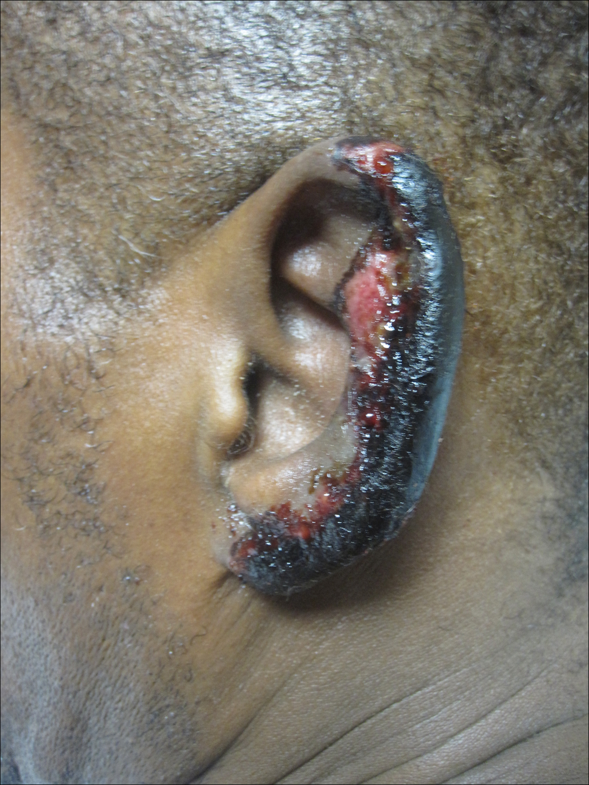
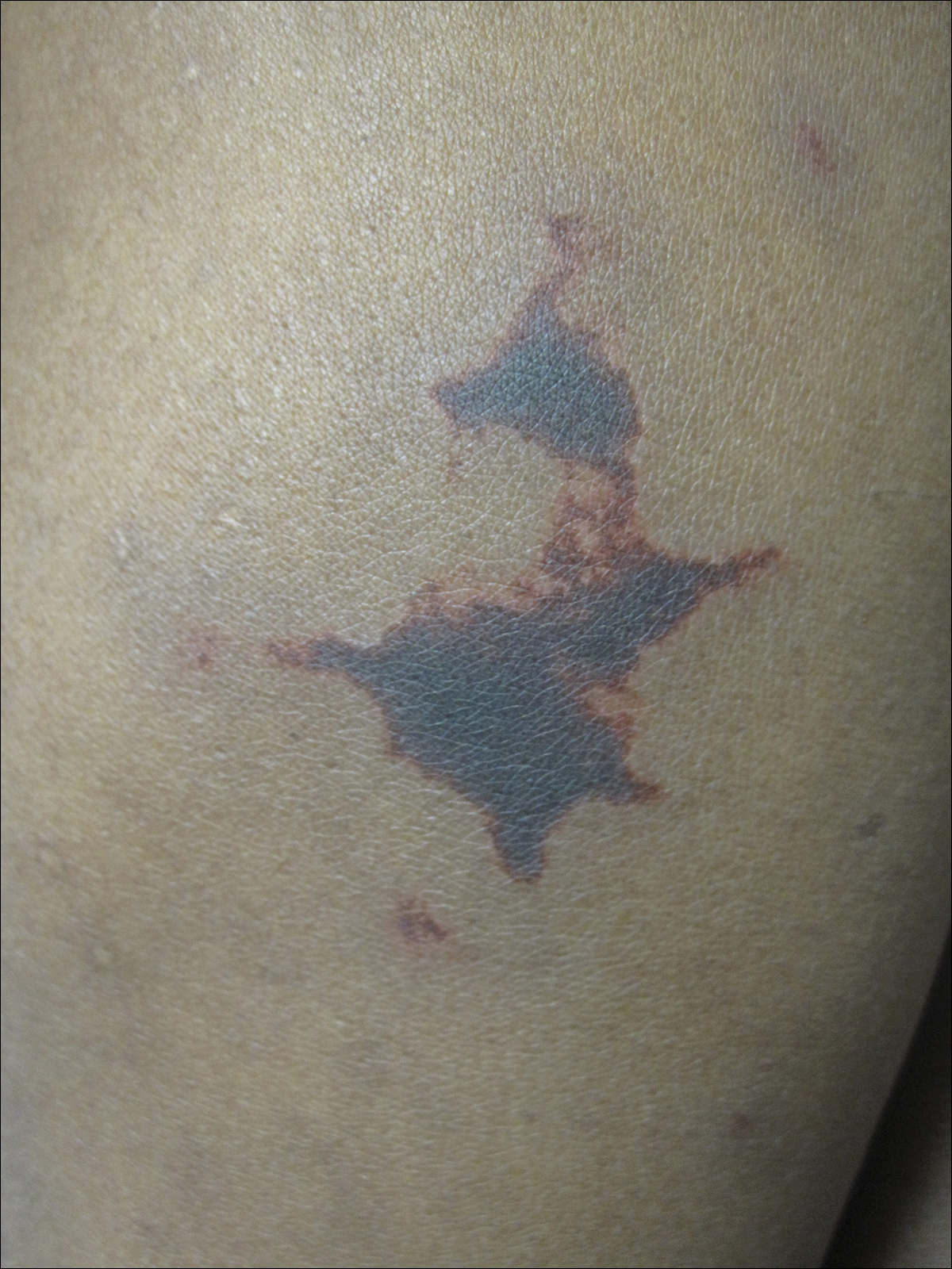
Laboratory examination revealed renal failure, anemia of chronic disease, and thrombocytosis (Table). The patient also screened positive for lupus anticoagulant and antinuclear antibodies and had elevated p-ANCA and anti–double-stranded DNA (Table). He also had an elevated sedimentation rate (109 mm/h [reference range, 0–20 mm/h]) and C-reactive protein level (11.3 mg/dL [reference range, 0–1.0 mg/dL])(Table). Urine toxicology was positive for cocaine.
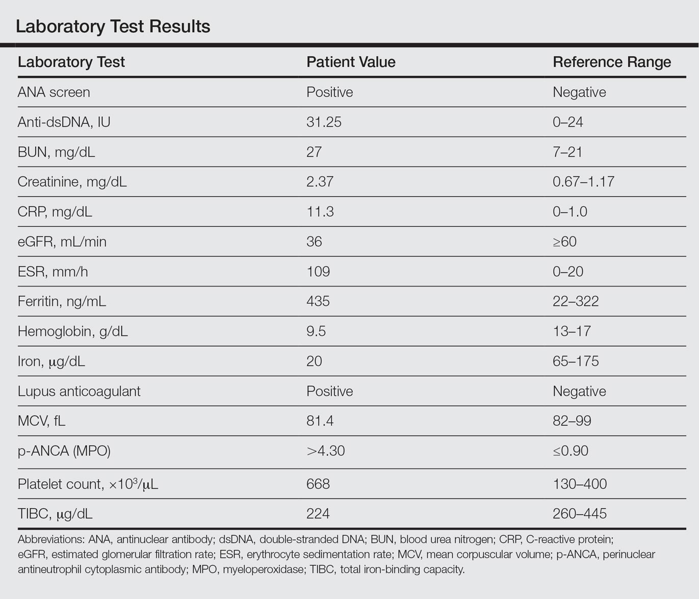
A punch biopsy of the left thigh was performed on the edge of a retiform purpuric patch. Histopathologic examination revealed epidermal necrosis with subjacent intraluminal vascular thrombi along with extravasated red blood cells and neutrophilic debris (leukocytoclasis) and fibrin in and around vessel walls, consistent with vasculitis (Figure 3).
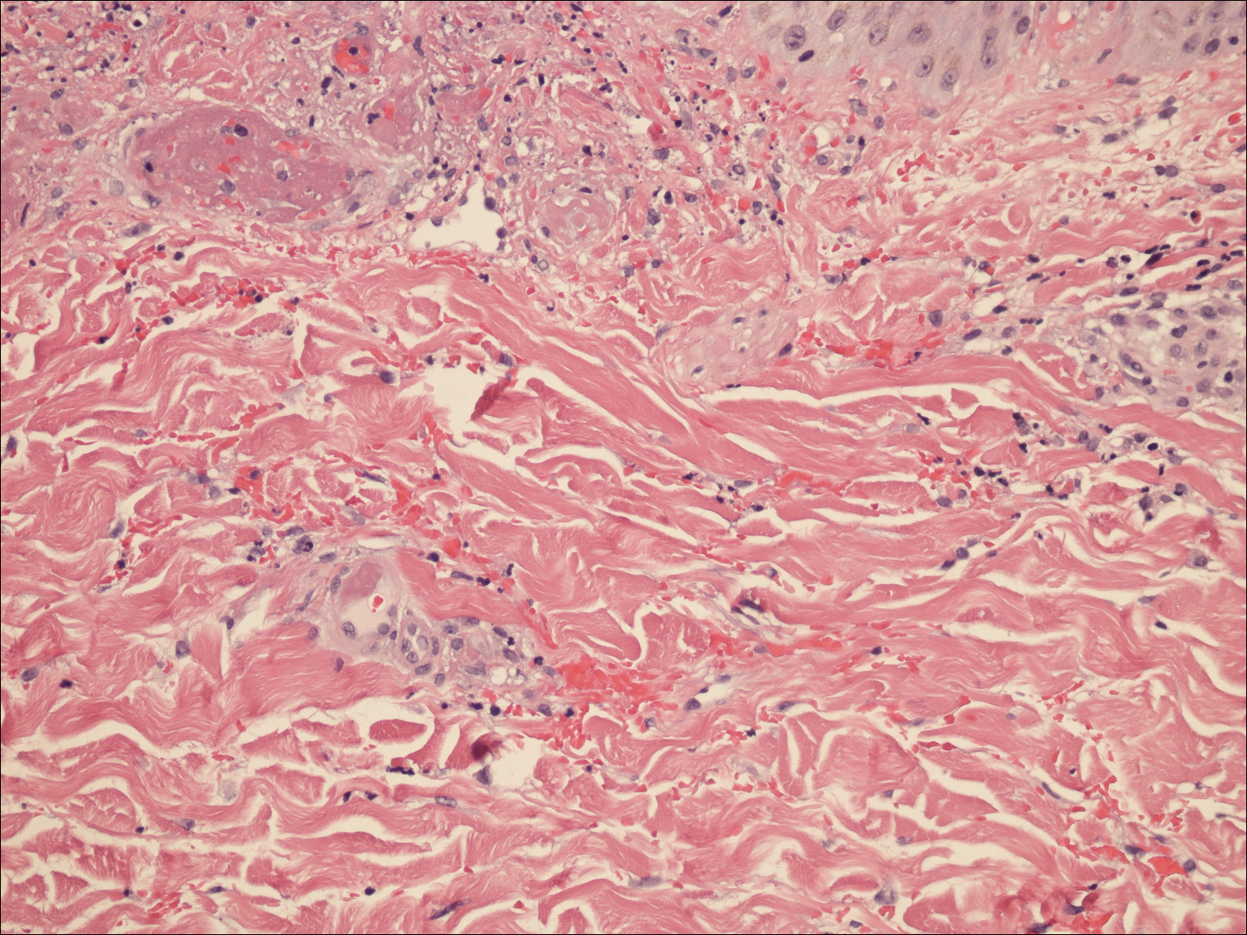
The patient was admitted to the hospital for pain management and wound care. Despite cocaine cessation and oral prednisone taper, the lesions on the legs worsened over the next several weeks. His condition was further complicated by wound infections, nonhealing ulcers, and subjective fevers and chills requiring frequent hospitalization. The patient was managed by the dermatology department as an outpatient and in clinic between hospital visits. He was treated with antibiotics, ulcer debridement, compression wraps, and aspirin (81 mg once daily) with moderate improvement.
Ten weeks after the first visit, the patient returned with worsening and recurrent leg and ear lesions. He denied any cocaine use since the initial hospital admission; however, a toxicology screen was never obtained. It was decided that the patient would need additional treatment along with traditional trigger (cocaine) avoidance and wound care. Combined treatment with aspirin (81 mg once daily), oral prednisone (40 mg once daily), and vardenafil hydrochloride (20 mg twice weekly) was initiated. At the end of week 1, the patient began to exhibit signs of improvement, which continued over the next 4 weeks. He was then lost to follow-up.
Comment
Our patient presented with severe necrotizing cutaneous vasculitis, likely secondary to levamisole exposure. Some of our patient’s cutaneous findings may be explained exclusively on the basis of cocaine exposure, but the characteristic lesion distribution and histopathologic findings along with the evidence of autoantibody positivity and concurrent arthralgias make the combination of levamisole and cocaine a more likely cause. Similar skin lesions were first described in children treated with levamisole for nephrotic syndrome.2 The most common site of clinical involvement in these children was the ears, as seen in our patient. Our patient tested positive for p-ANCA, which is the most commonly reported autoantibody associated with this patient population. Sixty-one percent (20/33) of patients with levamisole-induced vasculitis from 2 separate reviews showed p-ANCA positivity.7,10
On histopathology, our patient’s skin biopsy findings were consistent with those of prior reports of levamisole-induced vasculitis, which describe patterns of thrombotic vasculitis, leukocytoclasis, and fibrin deposition or occlusive disease.2,6,7,9-14 Mixed histologic findings of vasculitis and thrombosis, usually with varying ages of thrombi, are characteristic of levamisole-induced purpura. In addition, the disease can present nonspecifically with pure microvascular thrombosis without vasculitis, especially later in the course.9
The recommended management of levamisole-induced vasculitis currently involves the withdrawal of the culprit adulterated cocaine along with supportive treatment. Spontaneous and complete clinical resolution of lesions has been reported within 2 to 3 weeks and serology normalization within 2 to 14 months of levamisole cessation.2,6 A 2011 review of patients with levamisole-induced vasculitis reported 66% (19/29) of cases with either full cutaneous resolution after levamisole withdrawal or recurrence with resumed use, supporting a causal relationship.7 Walsh et al9 described 2 patients with recurrent and exacerbated retiform purpura following cocaine binges. Both of these patients had urine samples that tested positive for levamisole.9 In more severe cases, medications shown to be effective include colchicine, polidocanol, antibiotics, methotrexate, anticoagulants, and most commonly systemic corticosteroids.7,10,11,15 Nonsteroidal anti-inflammatory drugs were successful in treating lesions in 2 patients with concurrent arthralgia.7 Rarely, patients have required surgical debridement or skin grafting due to advanced disease at initial presentation.9,12-14 One of the most severe cases of levamisole-induced vasculitis reported in the literature involved 52% of the patient’s total body surface area with skin, soft tissue, and bony necrosis requiring nasal amputation, upper lip excision, skin grafting, and extremity amputation.14 Another severe case with widespread skin involvement was recently reported.16
For unclear reasons, our patient continued to develop cutaneous lesions despite self-reported cocaine cessation. Complete resolution required the combination of vardenafil, prednisone, and aspirin, along with debridement and wound care. Vardenafil, a selective phosphodiesterase 5 inhibitor, enhances the effect of nitrous oxide by increasing levels of cyclic guanosine monophosphate,17 which results in smooth muscle relaxation and vasodilatation. The primary indication for vardenafil is the treatment of erectile dysfunction, but it often is used off label in diseases that may benefit from vasodilatation. Because of its mechanism of action, it is understandable that a vasodilator such as vardenafil could be therapeutic in a condition associated with thrombosis. Moreover, the autoinflammatory nature of levamisole-induced vasculitis makes corticosteroid treatment effective. Given the 10-week delay in improvement, we suspect that it was the combination of treatment or an individual agent that led to our patient’s eventual recovery.
There are few reports in the literature focusing on optimal treatment of levamisole-induced vasculitis and none that mention alternative management for patients who continue to develop new lesions despite cocaine avoidance. Although the discontinuation of levamisole seems to be imperative for resolution of cutaneous lesions, it may not always be enough. It is possible that there is a subpopulation of patients that may not respond to the simple withdrawal of cocaine. It also should be mentioned that there was no urine toxicology screen obtained to support our patient’s reported cocaine cessation. Therefore, it is possible that his worsening condition was secondary to continued cocaine use. However, the patient successfully responded to the combination of vardenafil and prednisone, regardless of whether his condition persisted due to continued use of cocaine or not. This case suggests the possibility of a new treatment option for levamisole-induced vasculitis for patients who continue to use levamisole despite instruction for cessation or for patients with refractory disease.
Conclusion
A trial of prednisone and vardenafil should be considered for patients with refractory levamisole-induced vasculitis. Further studies and discussions of disease course are needed to identify the best treatment of this skin condition, especially for patients with refractory lesions.
- Scheinfeld N, Rosenberg JD, Weinberg JM. Levamisole in dermatology: a review. Am J Clin Dermatol. 2004;5:97-104.
- Rongioletti F, Ghio L, Ginevri F, et al. Purpura of the ears: a distinctive vasculopathy with circulating autoantibodies complicating long-term treatment with levamisole in children. Br J Dermatol. 1999;140:948-951.
- National Drug Threat Assessment 2011. US Department of Justice National Drug Intelligence Center website. https://www.justice.gov/archive/ndic/pubs44/44849/44849p.pdf. Published August 2011. Accessed August 7, 2016.
- Buchanan JA, Heard K, Burbach C, et al. Prevalence of levamisole in urine toxicology screens positive for cocaine in an inner-city hospital. JAMA. 2011;305:1657-1658.
- Gross RL, Brucker J, Bahce-Altuntas A, et al. A novel cutaneous vasculitis syndrome induced by levamisole-contaminated cocaine. Clin Rheumatol. 2011;30:1385-1392.
- Waller JM, Feramisco JD, Alberta-Wszolek L, et al. Cocaine-associated retiform purpura and neutropenia: is levamisole the culprit? J Am Acad Dermatol. 2010;63:530-535.
- Poon SH, Baliog CR, Sams RN, et al. Syndrome of cocaine-levamisole-induced cutaneous vasculitis and immune-mediated leukopenia. Semin Arthritis Rheum. 2011;41:434-444.
- Brewer JD, Meves A, Bostwick JM, et al. Cocaine abuse: dermatologic manifestations and therapeutic approaches. J Am Acad Dermatol. 2008;59:483-487.
- Walsh NMG, Green PJ, Burlingame RW, et al. Cocaine-related retiform purpura: evidence to incriminate the adulterant, levamisole. J Cutan Pathol. 2010;37:1212-1219.
- Chung C, Tumeh PC, Birnbaum R, et al. Characteristic purpura of the ears, vasculitis, and neutropenia—a potential public health epidemic associated with levamisole adultered cocaine. J Am Acad Dermatol. 2011;65:722-725.
- Kahn TA, Cuchacovich R, Espinoza LR, et al. Vasculopathy, hematological, and immune abnormalities associated with levamisole-contaminated cocaine use. Semin Arthritis Rheum. 2011;41:445-454.
- Graf J, Lynch K, Yeh CL, et al. Purpura, cutaneous necrosis, and antineutrophil cytoplasmic antibodies associated with levamisole-adulterated cocaine. Arthritis Rheum. 2011;63:3998-4001.
- Farmer RW, Malhotra PS, Mays MP, et al. Necrotizing peripheral vasculitis/vasculopathy following the use of cocaine laced with levamisole. J Burn Care Res. 2012;33:e6-e11.
- Ching JA, Smith DJ Jr. Levamisole-induced skin necrosis of skin, soft tissue, and bone: case report and review of literature. J Burn Care Res. 2012;33:e1-e5.
- Buchanan JA, Vogel JA, Eberhardt AM. Levamisole-induced occlusive necrotizing vasculitis of the ears after use of cocaine contaminated with levamisole. J Med Toxicol. 2011;7:83-84.
- Graff N, Whitworth K, Trigger C. Purpuric skin eruption in an illicit drug user: levamisole-induced vasculitis. Am J Emer Med. 2016;34:1321.
- Schwartz BG, Kloner RA. Drug interactions with phosphodiesterase-5 inhibitors used for the treatment of erectile dysfunction or pulmonary hypertension. Circulation. 2010;122:88-95.
Levamisole is an immunomodulatory drug that had been used to treat various medical conditions, including parasitic infections, nephrotic syndrome, and colorectal cancer,1 before being withdrawn from the US market in 2000. The most common reasons for levamisole discontinuation were leukopenia and rashes (1%–2%),1 many of which included leg ulcers and necrotizing purpura of the ears.1,2 The drug is currently available only as a deworming agent in veterinary medicine.
Since 2007, increasing amounts of levamisole have been used as an adulterant in cocaine. In 2007, less than 10% of cocaine was contaminated with levamisole, with an increase to 77% by 2010.3 In addition, 78% of 249 urine toxicology screens that were positive for cocaine in an inner city hospital also tested positive for levamisole.4 Levamisole-cut cocaine has become a concern because it is associated with a life-threatening syndrome involving a necrotizing purpuric rash, autoantibody production, and leukopenia.5
Levamisole-induced vasculitis is an independent entity from cocaine-induced vasculitis, which is associated with skin findings ranging from palpable purpura and chronic ulcers to digital infarction secondary to its vasospastic activity.6-8 Cocaine-induced vasculopathy has been related to cytoplasmic antineutrophil cytoplasmic antibody positivity and often resembles Wegener granulomatosis.6 Although both cocaine and levamisole have reportedly caused acrally distributed purpura and vasculopathy, levamisole is specifically associated with retiform purpura, ear involvement, and leukopenia.6,9 In addition, levamisole-induced skin reactions have been linked to specific antibodies, including antinuclear, antiphospholipid, and perinuclear antineutrophil cytoplasmic antibody (p-ANCA).2,5-7,9-14
We present a case of refractory levamisole-induced vasculitis and review its clinical presentation, diagnostic approach, laboratory findings, histology, and management. Furthermore, we discuss the possibility of a new treatment option for levamisole-induced vasculitis for patients with refractory disease or for patients who continue to use levamisole.
Case Report
A 49-year-old man with a history of polysubstance abuse presented with intermittent fevers and painful swollen ears as well as joint pain of 3 weeks’ duration. One week after the lesions developed on the ears, similar lesions were seen on the legs, arms, and trunk. He admitted to cocaine use 3 weeks prior to presentation when the symptoms began.
On physical examination, violaceous patches with necrotic bleeding edges and overlying black eschars were noted on the helices, antihelices, and ear lobules bilaterally (Figure 1). Retiform, purpuric to dark brown patches, some with signs of epidermal necrosis, were scattered on the arms, legs, and chest (Figure 2).
Laboratory examination revealed renal failure, anemia of chronic disease, and thrombocytosis (Table). The patient also screened positive for lupus anticoagulant and antinuclear antibodies and had elevated p-ANCA and anti–double-stranded DNA (Table). He also had an elevated sedimentation rate (109 mm/h [reference range, 0–20 mm/h]) and C-reactive protein level (11.3 mg/dL [reference range, 0–1.0 mg/dL])(Table). Urine toxicology was positive for cocaine.
A punch biopsy of the left thigh was performed on the edge of a retiform purpuric patch. Histopathologic examination revealed epidermal necrosis with subjacent intraluminal vascular thrombi along with extravasated red blood cells and neutrophilic debris (leukocytoclasis) and fibrin in and around vessel walls, consistent with vasculitis (Figure 3).
The patient was admitted to the hospital for pain management and wound care. Despite cocaine cessation and oral prednisone taper, the lesions on the legs worsened over the next several weeks. His condition was further complicated by wound infections, nonhealing ulcers, and subjective fevers and chills requiring frequent hospitalization. The patient was managed by the dermatology department as an outpatient and in clinic between hospital visits. He was treated with antibiotics, ulcer debridement, compression wraps, and aspirin (81 mg once daily) with moderate improvement.
Ten weeks after the first visit, the patient returned with worsening and recurrent leg and ear lesions. He denied any cocaine use since the initial hospital admission; however, a toxicology screen was never obtained. It was decided that the patient would need additional treatment along with traditional trigger (cocaine) avoidance and wound care. Combined treatment with aspirin (81 mg once daily), oral prednisone (40 mg once daily), and vardenafil hydrochloride (20 mg twice weekly) was initiated. At the end of week 1, the patient began to exhibit signs of improvement, which continued over the next 4 weeks. He was then lost to follow-up.
Comment
Our patient presented with severe necrotizing cutaneous vasculitis, likely secondary to levamisole exposure. Some of our patient’s cutaneous findings may be explained exclusively on the basis of cocaine exposure, but the characteristic lesion distribution and histopathologic findings along with the evidence of autoantibody positivity and concurrent arthralgias make the combination of levamisole and cocaine a more likely cause. Similar skin lesions were first described in children treated with levamisole for nephrotic syndrome.2 The most common site of clinical involvement in these children was the ears, as seen in our patient. Our patient tested positive for p-ANCA, which is the most commonly reported autoantibody associated with this patient population. Sixty-one percent (20/33) of patients with levamisole-induced vasculitis from 2 separate reviews showed p-ANCA positivity.7,10
On histopathology, our patient’s skin biopsy findings were consistent with those of prior reports of levamisole-induced vasculitis, which describe patterns of thrombotic vasculitis, leukocytoclasis, and fibrin deposition or occlusive disease.2,6,7,9-14 Mixed histologic findings of vasculitis and thrombosis, usually with varying ages of thrombi, are characteristic of levamisole-induced purpura. In addition, the disease can present nonspecifically with pure microvascular thrombosis without vasculitis, especially later in the course.9
The recommended management of levamisole-induced vasculitis currently involves the withdrawal of the culprit adulterated cocaine along with supportive treatment. Spontaneous and complete clinical resolution of lesions has been reported within 2 to 3 weeks and serology normalization within 2 to 14 months of levamisole cessation.2,6 A 2011 review of patients with levamisole-induced vasculitis reported 66% (19/29) of cases with either full cutaneous resolution after levamisole withdrawal or recurrence with resumed use, supporting a causal relationship.7 Walsh et al9 described 2 patients with recurrent and exacerbated retiform purpura following cocaine binges. Both of these patients had urine samples that tested positive for levamisole.9 In more severe cases, medications shown to be effective include colchicine, polidocanol, antibiotics, methotrexate, anticoagulants, and most commonly systemic corticosteroids.7,10,11,15 Nonsteroidal anti-inflammatory drugs were successful in treating lesions in 2 patients with concurrent arthralgia.7 Rarely, patients have required surgical debridement or skin grafting due to advanced disease at initial presentation.9,12-14 One of the most severe cases of levamisole-induced vasculitis reported in the literature involved 52% of the patient’s total body surface area with skin, soft tissue, and bony necrosis requiring nasal amputation, upper lip excision, skin grafting, and extremity amputation.14 Another severe case with widespread skin involvement was recently reported.16
For unclear reasons, our patient continued to develop cutaneous lesions despite self-reported cocaine cessation. Complete resolution required the combination of vardenafil, prednisone, and aspirin, along with debridement and wound care. Vardenafil, a selective phosphodiesterase 5 inhibitor, enhances the effect of nitrous oxide by increasing levels of cyclic guanosine monophosphate,17 which results in smooth muscle relaxation and vasodilatation. The primary indication for vardenafil is the treatment of erectile dysfunction, but it often is used off label in diseases that may benefit from vasodilatation. Because of its mechanism of action, it is understandable that a vasodilator such as vardenafil could be therapeutic in a condition associated with thrombosis. Moreover, the autoinflammatory nature of levamisole-induced vasculitis makes corticosteroid treatment effective. Given the 10-week delay in improvement, we suspect that it was the combination of treatment or an individual agent that led to our patient’s eventual recovery.
There are few reports in the literature focusing on optimal treatment of levamisole-induced vasculitis and none that mention alternative management for patients who continue to develop new lesions despite cocaine avoidance. Although the discontinuation of levamisole seems to be imperative for resolution of cutaneous lesions, it may not always be enough. It is possible that there is a subpopulation of patients that may not respond to the simple withdrawal of cocaine. It also should be mentioned that there was no urine toxicology screen obtained to support our patient’s reported cocaine cessation. Therefore, it is possible that his worsening condition was secondary to continued cocaine use. However, the patient successfully responded to the combination of vardenafil and prednisone, regardless of whether his condition persisted due to continued use of cocaine or not. This case suggests the possibility of a new treatment option for levamisole-induced vasculitis for patients who continue to use levamisole despite instruction for cessation or for patients with refractory disease.
Conclusion
A trial of prednisone and vardenafil should be considered for patients with refractory levamisole-induced vasculitis. Further studies and discussions of disease course are needed to identify the best treatment of this skin condition, especially for patients with refractory lesions.
Levamisole is an immunomodulatory drug that had been used to treat various medical conditions, including parasitic infections, nephrotic syndrome, and colorectal cancer,1 before being withdrawn from the US market in 2000. The most common reasons for levamisole discontinuation were leukopenia and rashes (1%–2%),1 many of which included leg ulcers and necrotizing purpura of the ears.1,2 The drug is currently available only as a deworming agent in veterinary medicine.
Since 2007, increasing amounts of levamisole have been used as an adulterant in cocaine. In 2007, less than 10% of cocaine was contaminated with levamisole, with an increase to 77% by 2010.3 In addition, 78% of 249 urine toxicology screens that were positive for cocaine in an inner city hospital also tested positive for levamisole.4 Levamisole-cut cocaine has become a concern because it is associated with a life-threatening syndrome involving a necrotizing purpuric rash, autoantibody production, and leukopenia.5
Levamisole-induced vasculitis is an independent entity from cocaine-induced vasculitis, which is associated with skin findings ranging from palpable purpura and chronic ulcers to digital infarction secondary to its vasospastic activity.6-8 Cocaine-induced vasculopathy has been related to cytoplasmic antineutrophil cytoplasmic antibody positivity and often resembles Wegener granulomatosis.6 Although both cocaine and levamisole have reportedly caused acrally distributed purpura and vasculopathy, levamisole is specifically associated with retiform purpura, ear involvement, and leukopenia.6,9 In addition, levamisole-induced skin reactions have been linked to specific antibodies, including antinuclear, antiphospholipid, and perinuclear antineutrophil cytoplasmic antibody (p-ANCA).2,5-7,9-14
We present a case of refractory levamisole-induced vasculitis and review its clinical presentation, diagnostic approach, laboratory findings, histology, and management. Furthermore, we discuss the possibility of a new treatment option for levamisole-induced vasculitis for patients with refractory disease or for patients who continue to use levamisole.
Case Report
A 49-year-old man with a history of polysubstance abuse presented with intermittent fevers and painful swollen ears as well as joint pain of 3 weeks’ duration. One week after the lesions developed on the ears, similar lesions were seen on the legs, arms, and trunk. He admitted to cocaine use 3 weeks prior to presentation when the symptoms began.
On physical examination, violaceous patches with necrotic bleeding edges and overlying black eschars were noted on the helices, antihelices, and ear lobules bilaterally (Figure 1). Retiform, purpuric to dark brown patches, some with signs of epidermal necrosis, were scattered on the arms, legs, and chest (Figure 2).
Laboratory examination revealed renal failure, anemia of chronic disease, and thrombocytosis (Table). The patient also screened positive for lupus anticoagulant and antinuclear antibodies and had elevated p-ANCA and anti–double-stranded DNA (Table). He also had an elevated sedimentation rate (109 mm/h [reference range, 0–20 mm/h]) and C-reactive protein level (11.3 mg/dL [reference range, 0–1.0 mg/dL])(Table). Urine toxicology was positive for cocaine.
A punch biopsy of the left thigh was performed on the edge of a retiform purpuric patch. Histopathologic examination revealed epidermal necrosis with subjacent intraluminal vascular thrombi along with extravasated red blood cells and neutrophilic debris (leukocytoclasis) and fibrin in and around vessel walls, consistent with vasculitis (Figure 3).
The patient was admitted to the hospital for pain management and wound care. Despite cocaine cessation and oral prednisone taper, the lesions on the legs worsened over the next several weeks. His condition was further complicated by wound infections, nonhealing ulcers, and subjective fevers and chills requiring frequent hospitalization. The patient was managed by the dermatology department as an outpatient and in clinic between hospital visits. He was treated with antibiotics, ulcer debridement, compression wraps, and aspirin (81 mg once daily) with moderate improvement.
Ten weeks after the first visit, the patient returned with worsening and recurrent leg and ear lesions. He denied any cocaine use since the initial hospital admission; however, a toxicology screen was never obtained. It was decided that the patient would need additional treatment along with traditional trigger (cocaine) avoidance and wound care. Combined treatment with aspirin (81 mg once daily), oral prednisone (40 mg once daily), and vardenafil hydrochloride (20 mg twice weekly) was initiated. At the end of week 1, the patient began to exhibit signs of improvement, which continued over the next 4 weeks. He was then lost to follow-up.
Comment
Our patient presented with severe necrotizing cutaneous vasculitis, likely secondary to levamisole exposure. Some of our patient’s cutaneous findings may be explained exclusively on the basis of cocaine exposure, but the characteristic lesion distribution and histopathologic findings along with the evidence of autoantibody positivity and concurrent arthralgias make the combination of levamisole and cocaine a more likely cause. Similar skin lesions were first described in children treated with levamisole for nephrotic syndrome.2 The most common site of clinical involvement in these children was the ears, as seen in our patient. Our patient tested positive for p-ANCA, which is the most commonly reported autoantibody associated with this patient population. Sixty-one percent (20/33) of patients with levamisole-induced vasculitis from 2 separate reviews showed p-ANCA positivity.7,10
On histopathology, our patient’s skin biopsy findings were consistent with those of prior reports of levamisole-induced vasculitis, which describe patterns of thrombotic vasculitis, leukocytoclasis, and fibrin deposition or occlusive disease.2,6,7,9-14 Mixed histologic findings of vasculitis and thrombosis, usually with varying ages of thrombi, are characteristic of levamisole-induced purpura. In addition, the disease can present nonspecifically with pure microvascular thrombosis without vasculitis, especially later in the course.9
The recommended management of levamisole-induced vasculitis currently involves the withdrawal of the culprit adulterated cocaine along with supportive treatment. Spontaneous and complete clinical resolution of lesions has been reported within 2 to 3 weeks and serology normalization within 2 to 14 months of levamisole cessation.2,6 A 2011 review of patients with levamisole-induced vasculitis reported 66% (19/29) of cases with either full cutaneous resolution after levamisole withdrawal or recurrence with resumed use, supporting a causal relationship.7 Walsh et al9 described 2 patients with recurrent and exacerbated retiform purpura following cocaine binges. Both of these patients had urine samples that tested positive for levamisole.9 In more severe cases, medications shown to be effective include colchicine, polidocanol, antibiotics, methotrexate, anticoagulants, and most commonly systemic corticosteroids.7,10,11,15 Nonsteroidal anti-inflammatory drugs were successful in treating lesions in 2 patients with concurrent arthralgia.7 Rarely, patients have required surgical debridement or skin grafting due to advanced disease at initial presentation.9,12-14 One of the most severe cases of levamisole-induced vasculitis reported in the literature involved 52% of the patient’s total body surface area with skin, soft tissue, and bony necrosis requiring nasal amputation, upper lip excision, skin grafting, and extremity amputation.14 Another severe case with widespread skin involvement was recently reported.16
For unclear reasons, our patient continued to develop cutaneous lesions despite self-reported cocaine cessation. Complete resolution required the combination of vardenafil, prednisone, and aspirin, along with debridement and wound care. Vardenafil, a selective phosphodiesterase 5 inhibitor, enhances the effect of nitrous oxide by increasing levels of cyclic guanosine monophosphate,17 which results in smooth muscle relaxation and vasodilatation. The primary indication for vardenafil is the treatment of erectile dysfunction, but it often is used off label in diseases that may benefit from vasodilatation. Because of its mechanism of action, it is understandable that a vasodilator such as vardenafil could be therapeutic in a condition associated with thrombosis. Moreover, the autoinflammatory nature of levamisole-induced vasculitis makes corticosteroid treatment effective. Given the 10-week delay in improvement, we suspect that it was the combination of treatment or an individual agent that led to our patient’s eventual recovery.
There are few reports in the literature focusing on optimal treatment of levamisole-induced vasculitis and none that mention alternative management for patients who continue to develop new lesions despite cocaine avoidance. Although the discontinuation of levamisole seems to be imperative for resolution of cutaneous lesions, it may not always be enough. It is possible that there is a subpopulation of patients that may not respond to the simple withdrawal of cocaine. It also should be mentioned that there was no urine toxicology screen obtained to support our patient’s reported cocaine cessation. Therefore, it is possible that his worsening condition was secondary to continued cocaine use. However, the patient successfully responded to the combination of vardenafil and prednisone, regardless of whether his condition persisted due to continued use of cocaine or not. This case suggests the possibility of a new treatment option for levamisole-induced vasculitis for patients who continue to use levamisole despite instruction for cessation or for patients with refractory disease.
Conclusion
A trial of prednisone and vardenafil should be considered for patients with refractory levamisole-induced vasculitis. Further studies and discussions of disease course are needed to identify the best treatment of this skin condition, especially for patients with refractory lesions.
- Scheinfeld N, Rosenberg JD, Weinberg JM. Levamisole in dermatology: a review. Am J Clin Dermatol. 2004;5:97-104.
- Rongioletti F, Ghio L, Ginevri F, et al. Purpura of the ears: a distinctive vasculopathy with circulating autoantibodies complicating long-term treatment with levamisole in children. Br J Dermatol. 1999;140:948-951.
- National Drug Threat Assessment 2011. US Department of Justice National Drug Intelligence Center website. https://www.justice.gov/archive/ndic/pubs44/44849/44849p.pdf. Published August 2011. Accessed August 7, 2016.
- Buchanan JA, Heard K, Burbach C, et al. Prevalence of levamisole in urine toxicology screens positive for cocaine in an inner-city hospital. JAMA. 2011;305:1657-1658.
- Gross RL, Brucker J, Bahce-Altuntas A, et al. A novel cutaneous vasculitis syndrome induced by levamisole-contaminated cocaine. Clin Rheumatol. 2011;30:1385-1392.
- Waller JM, Feramisco JD, Alberta-Wszolek L, et al. Cocaine-associated retiform purpura and neutropenia: is levamisole the culprit? J Am Acad Dermatol. 2010;63:530-535.
- Poon SH, Baliog CR, Sams RN, et al. Syndrome of cocaine-levamisole-induced cutaneous vasculitis and immune-mediated leukopenia. Semin Arthritis Rheum. 2011;41:434-444.
- Brewer JD, Meves A, Bostwick JM, et al. Cocaine abuse: dermatologic manifestations and therapeutic approaches. J Am Acad Dermatol. 2008;59:483-487.
- Walsh NMG, Green PJ, Burlingame RW, et al. Cocaine-related retiform purpura: evidence to incriminate the adulterant, levamisole. J Cutan Pathol. 2010;37:1212-1219.
- Chung C, Tumeh PC, Birnbaum R, et al. Characteristic purpura of the ears, vasculitis, and neutropenia—a potential public health epidemic associated with levamisole adultered cocaine. J Am Acad Dermatol. 2011;65:722-725.
- Kahn TA, Cuchacovich R, Espinoza LR, et al. Vasculopathy, hematological, and immune abnormalities associated with levamisole-contaminated cocaine use. Semin Arthritis Rheum. 2011;41:445-454.
- Graf J, Lynch K, Yeh CL, et al. Purpura, cutaneous necrosis, and antineutrophil cytoplasmic antibodies associated with levamisole-adulterated cocaine. Arthritis Rheum. 2011;63:3998-4001.
- Farmer RW, Malhotra PS, Mays MP, et al. Necrotizing peripheral vasculitis/vasculopathy following the use of cocaine laced with levamisole. J Burn Care Res. 2012;33:e6-e11.
- Ching JA, Smith DJ Jr. Levamisole-induced skin necrosis of skin, soft tissue, and bone: case report and review of literature. J Burn Care Res. 2012;33:e1-e5.
- Buchanan JA, Vogel JA, Eberhardt AM. Levamisole-induced occlusive necrotizing vasculitis of the ears after use of cocaine contaminated with levamisole. J Med Toxicol. 2011;7:83-84.
- Graff N, Whitworth K, Trigger C. Purpuric skin eruption in an illicit drug user: levamisole-induced vasculitis. Am J Emer Med. 2016;34:1321.
- Schwartz BG, Kloner RA. Drug interactions with phosphodiesterase-5 inhibitors used for the treatment of erectile dysfunction or pulmonary hypertension. Circulation. 2010;122:88-95.
- Scheinfeld N, Rosenberg JD, Weinberg JM. Levamisole in dermatology: a review. Am J Clin Dermatol. 2004;5:97-104.
- Rongioletti F, Ghio L, Ginevri F, et al. Purpura of the ears: a distinctive vasculopathy with circulating autoantibodies complicating long-term treatment with levamisole in children. Br J Dermatol. 1999;140:948-951.
- National Drug Threat Assessment 2011. US Department of Justice National Drug Intelligence Center website. https://www.justice.gov/archive/ndic/pubs44/44849/44849p.pdf. Published August 2011. Accessed August 7, 2016.
- Buchanan JA, Heard K, Burbach C, et al. Prevalence of levamisole in urine toxicology screens positive for cocaine in an inner-city hospital. JAMA. 2011;305:1657-1658.
- Gross RL, Brucker J, Bahce-Altuntas A, et al. A novel cutaneous vasculitis syndrome induced by levamisole-contaminated cocaine. Clin Rheumatol. 2011;30:1385-1392.
- Waller JM, Feramisco JD, Alberta-Wszolek L, et al. Cocaine-associated retiform purpura and neutropenia: is levamisole the culprit? J Am Acad Dermatol. 2010;63:530-535.
- Poon SH, Baliog CR, Sams RN, et al. Syndrome of cocaine-levamisole-induced cutaneous vasculitis and immune-mediated leukopenia. Semin Arthritis Rheum. 2011;41:434-444.
- Brewer JD, Meves A, Bostwick JM, et al. Cocaine abuse: dermatologic manifestations and therapeutic approaches. J Am Acad Dermatol. 2008;59:483-487.
- Walsh NMG, Green PJ, Burlingame RW, et al. Cocaine-related retiform purpura: evidence to incriminate the adulterant, levamisole. J Cutan Pathol. 2010;37:1212-1219.
- Chung C, Tumeh PC, Birnbaum R, et al. Characteristic purpura of the ears, vasculitis, and neutropenia—a potential public health epidemic associated with levamisole adultered cocaine. J Am Acad Dermatol. 2011;65:722-725.
- Kahn TA, Cuchacovich R, Espinoza LR, et al. Vasculopathy, hematological, and immune abnormalities associated with levamisole-contaminated cocaine use. Semin Arthritis Rheum. 2011;41:445-454.
- Graf J, Lynch K, Yeh CL, et al. Purpura, cutaneous necrosis, and antineutrophil cytoplasmic antibodies associated with levamisole-adulterated cocaine. Arthritis Rheum. 2011;63:3998-4001.
- Farmer RW, Malhotra PS, Mays MP, et al. Necrotizing peripheral vasculitis/vasculopathy following the use of cocaine laced with levamisole. J Burn Care Res. 2012;33:e6-e11.
- Ching JA, Smith DJ Jr. Levamisole-induced skin necrosis of skin, soft tissue, and bone: case report and review of literature. J Burn Care Res. 2012;33:e1-e5.
- Buchanan JA, Vogel JA, Eberhardt AM. Levamisole-induced occlusive necrotizing vasculitis of the ears after use of cocaine contaminated with levamisole. J Med Toxicol. 2011;7:83-84.
- Graff N, Whitworth K, Trigger C. Purpuric skin eruption in an illicit drug user: levamisole-induced vasculitis. Am J Emer Med. 2016;34:1321.
- Schwartz BG, Kloner RA. Drug interactions with phosphodiesterase-5 inhibitors used for the treatment of erectile dysfunction or pulmonary hypertension. Circulation. 2010;122:88-95.
Practice Points
- Levamisole is an immunomodulatory drug that, before being withdrawn from the US market in 2000, was previously used to treat various medical conditions.
- A majority of the cocaine in the United States is contaminated with levamisole, which is added as an adulterant or bulking agent.
- Levamisole-cut cocaine is a concern because it is associated with a life-threatening syndrome involving a necrotizing purpuric rash, autoantibody production, and leukopenia.
- Although treatment of levamisole toxicity is primarily supportive and includes cessation of levamisole-cut cocaine, a trial of prednisone and vardenafil hydrochloride can be considered for refractory levamisole-induced vasculopathy or for patients who continue to use the drug.

Food allergy testing only rarely needed for AD patients
BOSTON – Between 15% and 30% of children with moderate to severe atopic dermatitis also have food allergies, but the allergies are a trigger for AD in only a small subset of patients, according to Mercedes E. Gonzalez, MD.
In most cases, allergy testing is not indicated, she said at the American Academy of Dermatology summer meeting.

She described a scenario involving a parent who is concerned that a food allergy is causing her child’s AD. The child has had no hives, no lip swelling, and no other signs of immediate hypersensitivity. In such a case, the best approach is to treat with topical therapies and follow the patient clinically.
“Allergy testing independent of history is not recommended,” she said.
However, in cases involving a significant concern about food allergy, such as the presence of hives or urticaria, or when the child has severe dermatitis that is not improving with optimized topical therapies, an assessment can be undertaken, said Dr. Gonzalez of the University of Miami.
She recommended limited food allergy testing – for common culprits such as cow’s milk, eggs, wheat, soy, and peanuts – in children younger than age 5 years with moderate to severe AD, if the AD persists despite optimized topical treatment and/or a history of immediate and reproducible reaction after ingestion of a specific food.
Food elimination diets based solely on the findings of food allergy test results are not recommended for managing AD, she noted.
If a patient has true immunoglobulin E–mediated allergy they should practice avoidance to prevent potential serious health sequelae, Dr. Gonzalez said.
When testing is done, keep in mind that skin prick tests and serum-specific IgE levels have high negative predictive values above 95%, but low specificity and positive predictive values of 40%-60%, she pointed out. Positive tests should be verified with a food elimination diet or oral food challenge.
Also, most children develop tolerance to the foods over time and should be retested, Dr. Gonzalez said.
Early peanut introduction advised in infants with AD
There is no need to delay the introduction of peanuts into the diet of an infant at high risk for atopic dermatitis, Dr. Gonzalez said.
A 2015 consensus communication from the American Academy of Pediatrics and numerous other organizations, including the American Academy of Allergy, Asthma & Immunology and the Society of Pediatric Dermatology, offering interim guidance on the topic calls for introduction of peanut products into the diets of high-risk infants in countries where peanut allergy is present, she said.
High-risk infants were defined in the study as those with egg allergy and/or severe eczema.
The guidance, which the AAP “endorses and accepts as its policy” pending more formal guidelines currently in development, was based largely on findings from the LEAP (Learn Early About Peanut Allergy) trial – a 5-year randomized, controlled trial of 640 high-risk infants aged 4-11 months. The trial showed that 17.2% of infants who avoided peanuts had peanut allergy at 5 years, compared with 3.2% of those with peanut consumption three times weekly, a relative risk reduction of 81% (N Engl J Med. 2015; 372:803-13).
In infants with egg allergy or severe eczema, an evaluation by an allergist or dermatologist familiar with the guidance may be warranted to assist in implementing the suggestions, Dr. Gonzalez said.
Dr. Gonzalez reported receiving honoraria for serving as a speaker and/or advisory board member for Pierre Fabre Dermatologie, Anacor Pharmaceuticals, Encore Dermatology, and PuraCap Pharmaceutical.
BOSTON – Between 15% and 30% of children with moderate to severe atopic dermatitis also have food allergies, but the allergies are a trigger for AD in only a small subset of patients, according to Mercedes E. Gonzalez, MD.
In most cases, allergy testing is not indicated, she said at the American Academy of Dermatology summer meeting.
She described a scenario involving a parent who is concerned that a food allergy is causing her child’s AD. The child has had no hives, no lip swelling, and no other signs of immediate hypersensitivity. In such a case, the best approach is to treat with topical therapies and follow the patient clinically.
“Allergy testing independent of history is not recommended,” she said.
However, in cases involving a significant concern about food allergy, such as the presence of hives or urticaria, or when the child has severe dermatitis that is not improving with optimized topical therapies, an assessment can be undertaken, said Dr. Gonzalez of the University of Miami.
She recommended limited food allergy testing – for common culprits such as cow’s milk, eggs, wheat, soy, and peanuts – in children younger than age 5 years with moderate to severe AD, if the AD persists despite optimized topical treatment and/or a history of immediate and reproducible reaction after ingestion of a specific food.
Food elimination diets based solely on the findings of food allergy test results are not recommended for managing AD, she noted.
If a patient has true immunoglobulin E–mediated allergy they should practice avoidance to prevent potential serious health sequelae, Dr. Gonzalez said.
When testing is done, keep in mind that skin prick tests and serum-specific IgE levels have high negative predictive values above 95%, but low specificity and positive predictive values of 40%-60%, she pointed out. Positive tests should be verified with a food elimination diet or oral food challenge.
Also, most children develop tolerance to the foods over time and should be retested, Dr. Gonzalez said.
Early peanut introduction advised in infants with AD
There is no need to delay the introduction of peanuts into the diet of an infant at high risk for atopic dermatitis, Dr. Gonzalez said.
A 2015 consensus communication from the American Academy of Pediatrics and numerous other organizations, including the American Academy of Allergy, Asthma & Immunology and the Society of Pediatric Dermatology, offering interim guidance on the topic calls for introduction of peanut products into the diets of high-risk infants in countries where peanut allergy is present, she said.
High-risk infants were defined in the study as those with egg allergy and/or severe eczema.
The guidance, which the AAP “endorses and accepts as its policy” pending more formal guidelines currently in development, was based largely on findings from the LEAP (Learn Early About Peanut Allergy) trial – a 5-year randomized, controlled trial of 640 high-risk infants aged 4-11 months. The trial showed that 17.2% of infants who avoided peanuts had peanut allergy at 5 years, compared with 3.2% of those with peanut consumption three times weekly, a relative risk reduction of 81% (N Engl J Med. 2015; 372:803-13).
In infants with egg allergy or severe eczema, an evaluation by an allergist or dermatologist familiar with the guidance may be warranted to assist in implementing the suggestions, Dr. Gonzalez said.
Dr. Gonzalez reported receiving honoraria for serving as a speaker and/or advisory board member for Pierre Fabre Dermatologie, Anacor Pharmaceuticals, Encore Dermatology, and PuraCap Pharmaceutical.
BOSTON – Between 15% and 30% of children with moderate to severe atopic dermatitis also have food allergies, but the allergies are a trigger for AD in only a small subset of patients, according to Mercedes E. Gonzalez, MD.
In most cases, allergy testing is not indicated, she said at the American Academy of Dermatology summer meeting.
She described a scenario involving a parent who is concerned that a food allergy is causing her child’s AD. The child has had no hives, no lip swelling, and no other signs of immediate hypersensitivity. In such a case, the best approach is to treat with topical therapies and follow the patient clinically.
“Allergy testing independent of history is not recommended,” she said.
However, in cases involving a significant concern about food allergy, such as the presence of hives or urticaria, or when the child has severe dermatitis that is not improving with optimized topical therapies, an assessment can be undertaken, said Dr. Gonzalez of the University of Miami.
She recommended limited food allergy testing – for common culprits such as cow’s milk, eggs, wheat, soy, and peanuts – in children younger than age 5 years with moderate to severe AD, if the AD persists despite optimized topical treatment and/or a history of immediate and reproducible reaction after ingestion of a specific food.
Food elimination diets based solely on the findings of food allergy test results are not recommended for managing AD, she noted.
If a patient has true immunoglobulin E–mediated allergy they should practice avoidance to prevent potential serious health sequelae, Dr. Gonzalez said.
When testing is done, keep in mind that skin prick tests and serum-specific IgE levels have high negative predictive values above 95%, but low specificity and positive predictive values of 40%-60%, she pointed out. Positive tests should be verified with a food elimination diet or oral food challenge.
Also, most children develop tolerance to the foods over time and should be retested, Dr. Gonzalez said.
Early peanut introduction advised in infants with AD
There is no need to delay the introduction of peanuts into the diet of an infant at high risk for atopic dermatitis, Dr. Gonzalez said.
A 2015 consensus communication from the American Academy of Pediatrics and numerous other organizations, including the American Academy of Allergy, Asthma & Immunology and the Society of Pediatric Dermatology, offering interim guidance on the topic calls for introduction of peanut products into the diets of high-risk infants in countries where peanut allergy is present, she said.
High-risk infants were defined in the study as those with egg allergy and/or severe eczema.
The guidance, which the AAP “endorses and accepts as its policy” pending more formal guidelines currently in development, was based largely on findings from the LEAP (Learn Early About Peanut Allergy) trial – a 5-year randomized, controlled trial of 640 high-risk infants aged 4-11 months. The trial showed that 17.2% of infants who avoided peanuts had peanut allergy at 5 years, compared with 3.2% of those with peanut consumption three times weekly, a relative risk reduction of 81% (N Engl J Med. 2015; 372:803-13).
In infants with egg allergy or severe eczema, an evaluation by an allergist or dermatologist familiar with the guidance may be warranted to assist in implementing the suggestions, Dr. Gonzalez said.
Dr. Gonzalez reported receiving honoraria for serving as a speaker and/or advisory board member for Pierre Fabre Dermatologie, Anacor Pharmaceuticals, Encore Dermatology, and PuraCap Pharmaceutical.
EXPERT ANALYSIS FROM AAD SUMMER ACADEMY 2016