User login
ICD same-day discharge safe, but not a money saver
San Francisco – Same day discharge is generally safe after cardioverter defibrillator implantation for primary prevention, but it doesn’t save money.
Furthermore, guidelines are needed to standardize the practice as it becomes increasingly common in the United States, according to a 25-site investigation.

After implantable cardioverter defibrillator (ICD) procedures, patients were monitored for 3-4 hours, and their devices were checked for proper functioning; 129 patients who were stable at that point were randomized to early discharge and 136 to next day discharge (NDD).
The overall 30-day procedural complication rate was 3.1% in the same day discharge (SDD) group and 1.6% in the NDD group, a nonsignificant difference (P = .37). Three patients in the SDD group developed hematomas that resolved on their own, and one had a cardiac perforation. One NDD patient dislodged a lead and another developed an infection. There were no differences in quality of life measures between the two groups at 30 days.
However, there were also no differences in procedural and perioperative direct costs, which was surprising because saving money is a major driver of SDD, and the most expensive part of ICD implantation is the first 24 hours. Direct per-patient medical costs in the study – estimated by applying hospital cost-to-charge ratios to the Medicare-reported charge – were $31,771 for SDD and $30,437 for NDD, but NDD was more expensive than SDD at several sites. The investigators suspect a flaw in their analysis related to the opaque nature of hospital accounting, and plan to look into the matter further with modeling to identify savings opportunities with SDD.
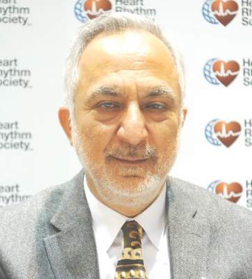
“We can insert ICDs on an outpatient basis, but this study will be difficult to replicate because clinical practice is moving towards SDD. In view of this, we think professional societies should be thinking of standardizing criteria for SDD; guidelines would help with the adoption of this approach. There are clinicians who are astute and have great clinical judgment, but there are others who need a scoring system. We believe that by using the 270,000 patients in the [American College of Cardiology’s ICD Registry], there is the ability to identify patients who have low periprocedural risk,” said lead investigator Dr. Ranjit Suri, a cardiologist at Mt. Sinai Hospital in New York.
The study excluded patients receiving an ICD for secondary prevention, as well as those on periprocedural heparin and patients who were pacemaker dependent. SDD seemed safe otherwise, but it’s unknown “if our concept of low risk is acceptable to all implanting physicians,” Dr. Suri said at the annual scientific sessions of the Heart Rhythm Society.
The study groups were well matched. About 75% in each arm were men, and ischemic cardiomyopathy was the leading ICD indication. Patients were amenable to the idea of SDD; the advent of remote monitoring “adds a certain sense of safety” for both patients and physicians, he said.
Dr. Suri is a speaker for Boehringer Ingelheim and St. Jude Medical. He is also a consultant for Biosense Webster and Zoll, and receives research funding from St. Jude.

|
Dr. Thomas Deering |
The vast majority of primary prevention patients who are clinically stable enough to come in as outpatients can go home as outpatients if you watch them for a short period of time and make sure they are clinically stable. Most patients don’t want to be in the hospital, and many hospitals are crunched for available beds. It would be great to have guidelines on how to handle this, but we have to allow for clinical judgment.
Dr. Thomas Deering is chief of the Arrhythmia Center at the Piedmont Heart Institute in Atlanta, where he is also chairman of the Executive Council and the Clinical Centers for Excellence. He moderated Dr. Suri’s presentation and was not involved in the work.
|
|
Dr. Thomas Deering |
The vast majority of primary prevention patients who are clinically stable enough to come in as outpatients can go home as outpatients if you watch them for a short period of time and make sure they are clinically stable. Most patients don’t want to be in the hospital, and many hospitals are crunched for available beds. It would be great to have guidelines on how to handle this, but we have to allow for clinical judgment.
Dr. Thomas Deering is chief of the Arrhythmia Center at the Piedmont Heart Institute in Atlanta, where he is also chairman of the Executive Council and the Clinical Centers for Excellence. He moderated Dr. Suri’s presentation and was not involved in the work.
San Francisco – Same day discharge is generally safe after cardioverter defibrillator implantation for primary prevention, but it doesn’t save money.
Furthermore, guidelines are needed to standardize the practice as it becomes increasingly common in the United States, according to a 25-site investigation.
After implantable cardioverter defibrillator (ICD) procedures, patients were monitored for 3-4 hours, and their devices were checked for proper functioning; 129 patients who were stable at that point were randomized to early discharge and 136 to next day discharge (NDD).
The overall 30-day procedural complication rate was 3.1% in the same day discharge (SDD) group and 1.6% in the NDD group, a nonsignificant difference (P = .37). Three patients in the SDD group developed hematomas that resolved on their own, and one had a cardiac perforation. One NDD patient dislodged a lead and another developed an infection. There were no differences in quality of life measures between the two groups at 30 days.
However, there were also no differences in procedural and perioperative direct costs, which was surprising because saving money is a major driver of SDD, and the most expensive part of ICD implantation is the first 24 hours. Direct per-patient medical costs in the study – estimated by applying hospital cost-to-charge ratios to the Medicare-reported charge – were $31,771 for SDD and $30,437 for NDD, but NDD was more expensive than SDD at several sites. The investigators suspect a flaw in their analysis related to the opaque nature of hospital accounting, and plan to look into the matter further with modeling to identify savings opportunities with SDD.
“We can insert ICDs on an outpatient basis, but this study will be difficult to replicate because clinical practice is moving towards SDD. In view of this, we think professional societies should be thinking of standardizing criteria for SDD; guidelines would help with the adoption of this approach. There are clinicians who are astute and have great clinical judgment, but there are others who need a scoring system. We believe that by using the 270,000 patients in the [American College of Cardiology’s ICD Registry], there is the ability to identify patients who have low periprocedural risk,” said lead investigator Dr. Ranjit Suri, a cardiologist at Mt. Sinai Hospital in New York.
The study excluded patients receiving an ICD for secondary prevention, as well as those on periprocedural heparin and patients who were pacemaker dependent. SDD seemed safe otherwise, but it’s unknown “if our concept of low risk is acceptable to all implanting physicians,” Dr. Suri said at the annual scientific sessions of the Heart Rhythm Society.
The study groups were well matched. About 75% in each arm were men, and ischemic cardiomyopathy was the leading ICD indication. Patients were amenable to the idea of SDD; the advent of remote monitoring “adds a certain sense of safety” for both patients and physicians, he said.
Dr. Suri is a speaker for Boehringer Ingelheim and St. Jude Medical. He is also a consultant for Biosense Webster and Zoll, and receives research funding from St. Jude.
AT HEART RHYTHM 2016
Key clinical point: Same-day discharge is generally safe after cardioverter defibrillator implantation for primary prevention, but it doesn’t save money and guidelines are needed to standardize the practice as it becomes increasingly common in the United States.
Major finding: The overall 30-day procedural complication rate was 3.1% in the same day discharge (SDD) group and 1.5% in the next-day discharge group, a nonsignificant difference (P = .37).
Data source: Randomized trial of 265 ICD patients.
Disclosures: The lead investigator is a speaker for Boehringer Ingelheim and St. Jude Medical. He is also a consultant for Biosense Webster and Zoll, and receives research funding from St. Jude.
Lifetime Achievement Award to Dr. Cronenwett
This year’s Lifetime Achievement Award winner is called many things by his many nominators: quadruple hitter, visionary, mentor, revolutionary, unsurpassed, profoundly effective. But one thing is mentioned by all.
“Everybody,” they say, “calls him Jack.” That may be because Dr. Jack Cronenwett is not just a legend among SVS members, but he is also unassuming.
“Jack is one of the kindest and most humble person that you could meet,” said Dr. Marc L. Schermerhorn. “I was immediately surprised when I arrived at Dartmouth as a vascular fellow and heard him say, ‘Call me Jack.’ ”
Of Dr. Cronenwett’s – excuse us – Jack’s major achievements, the first was his work at Dartmouth-Hitchcock Medical Center.
In the 1980s, Dartmouth had a relatively small, little-known division of vascular surgery, and Jack was its first vascular surgeon. “He has trained and recruited the entire faculty and turned it into a powerhouse, a division of vascular surgery that has national and international recognition,” wrote Dr. Jens Eldrup-Jorgensen in his nomination.
In addition, Jack was largely responsible for the development at Dartmouth of a new vascular surgery training paradigm – the 0+5 residency. “He had the first program and was instrumental in making this type of program a possibility at the board level,” Dr. Schermerhorn wrote.
Getting a 0+5 program approved by the American Board of Surgery was an almost insurmountable hurdle.
“From my perspective,” said Jack, “it was all about training. We were advocating for a concept. You didn’t need to have full general surgery training in order to become a vascular surgeon. It was inefficient. We needed to be more focused. We couldn’t expand training every time a new procedure came along and still think that these residents needed to know how to do breast surgery.”
“Many of us work in institutions or divisions where we simply had to carry on what had been started by others,” noted Dr. Richard Cambria. “That was not the case at Dartmouth, where the present status of that outstanding vascular surgery division can be traced to Jack Cronenwett.”
The experience of building a program and expanding the processes of a vascular residency program seems to have whetted Jack’s appetite for leadership and educational excellence.
After the millennium, Jack’s legacy mushroomed with these outstanding achievements:
• 2002 – Spearheaded the Vascular Study Group of New England, a regional quality outcomes registry, which became the model for the future Vascular Quality Initiative
• 2003 – Took the reins of SVS as president and, with Dr. Tom Riles, then president of the American Association for Vascular Surgery (AAVS), convinced these organizations to merge and create an independent administrative office in Chicago, creating today’s more inclusive SVS.
• 2003-2008 – Was senior co-editor of the Journal of Vascular Surgery with Dr. James Seeger.
• 2010, 2014 – Was co-editor of the 7th and 8th editions of Rutherford’s Vascular Surgery textbook with Dr. K. Wayne Johnston.
• 2011 – Helped launch the SVS-PSO as an official new entity and has served as medical director since.
The Vascular Study Group of New England: This now well-known quality improvement registry was partially modeled after the Northern New England Cardiovascular Disease Study Group. “It was not just a copycat of NNECVDSG,” noted Dr. Eldrup-Jorgensen. “Biannual regional meetings were developed to promote quality improvement, which is somewhat uncommon for a registry.”
The organization grew and earned the attention of other centers around the country and the world. Under the umbrella of the SVS, the study group opened its membership and renamed itself the Vascular Quality Initiative, which today has 375 centers and a multidisciplinary membership that includes cardiologists and radiologists.
Vascular Quality Initiative: The huge success of the VQI, Dr. Jorgensen added, “required not only wisdom and vision, but also great judgment and the ability to work collaboratively and collegially.”
“Some of us may now take it for granted,” said Dr. Anton Sidawy, “but those of us who were involved in the discussions about VQI remember the endless night calls that we spent listening to Jack making various arguments about why the Society should invest in such a program. He actually had the plan completely conceived, describing the details of the regional concept used currently by VQI. I also remember the many meetings during VAM to discuss the program. Jack had the vision, the persistence and the details to be able to convince the SVS leadership to adopt it. We did, and as they say, the rest is history.”
President of SVS: While serving as SVS president, Jack worked with Dr. Tom Riles to push for the merger of two vascular societies, the SVS and the AAVS, into today’s Society for Vascular Surgery.
Senior co-editor, Journal of Vascular Surgery: As co-editor from 2003-2008, Jack has been recognized for leading significant changes that improved the content, quality and impact of the publication.
“Jack’s tenure on the Journal is when the number of submissions began to accelerate, exceeding a thousand per year,” recalled SVS President Dr. Bruce A. Perler. “He also started a program at the annual meeting to teach reviewers.”
He also served as a senior editor for recent editions of Rutherford’s Vascular Surgery textbook. The modern understanding of the pathophysiology and surgical decision-making surrounding the management of aortoiliac occlusive and infrarenal aortic aneurysm disease is profoundly influenced by his early work. His early studies of actuarial survival after aortic aneurysm surgery and cost effectiveness of asymptomatic carotid endarterectomy have inspired a generation of researchers.
A prolific writer and publisher himself, Jack has published more than 200 peer-reviewed journal articles, contributed chapters to more than 50 books, co-edited 14 books, delivered more than 300 invited presentations, and made nearly 200 peer-reviewed presentations at regional and national meetings.
In response to receiving this award, Jack expressed profound gratitude to his colleagues in the SVS for this recognition. “To me, this should really be called the SVS Lifetime ‘Opportunity’ Award. I have been afforded so many opportunities by the SVS during my life, which are only exceeded by the great number of close friendships that I have developed through the SVS. This has been a privilege and source of great gratification for me throughout my career.”
Among all his nominators and their excellent, thorough and effusive recommendations, Dr. Larry Kraiss summed it up as well as anyone: “Jack’s contributions outside of the VQI would make him a very strong candidate for the Lifetime Achievement Award. When you credit him with the VQI and its long-term positive impact on the SVS, I think he is a slam dunk.”
Congratulations, Dr. Slam Dunk Cronenwett.

Smoking causes worse outcome in MS
NATIONAL HARBOR, MD. – Follow-up of a cohort of patients in the United Kingdom has demonstrated associations between smoking and a higher risk of development of multiple sclerosis (MS), progression of MS-related disability, higher risk of premature death, and shortened life expectancy.
The findings highlight the need for clinical trials of the effects of quitting smoking and provide data that will be useful in the development of effective intervention strategies.

Dr. Cris S. Constantinescu of the University of Nottingham (England) discussed findings from the Nottingham University Hospitals MS Clinics database at the annual meeting of the Consortium of Multiple Sclerosis Centers.
“Although smokers had higher levels of comorbid conditions, it appeared that the influence of smoking is independent of the presence of comorbid conditions. Those who gave up smoking could do as well as nonsmokers,” said Dr. Constantinescu.
While ample epidemiologic evidence indicates that smoking is a driver of the development and progression of MS, there are few hard data. To gain insight, the researchers analyzed data on over 1,200 MS patients throughout England who were followed up beginning in the mid-1990s. About 60% of the patients had relapsing-remitting MS. The duration of MS and smoking were both about 20 years. About 60% of men and 50% of women were current smokers.
Regular smokers were 64% more likely to develop MS than were nonsmokers. Having ever smoked carried a 44% increased risk of MS (both P less than .001). MS patients who grew up in a household where one parent smoked were 50% more likely to become regular smokers. The risk climbed to 85% if both parents were smokers.
No association was evident between smoking and the development of primary progressive MS, but current smokers were almost 2.5 times more likely to develop secondary progressive MS. Smoking correlated with more severe MS disability, compared with nonsmokers. Ex-smokers had risks similar to those of nonsmokers of developing secondary MS and in the level of disease severity.
Every year a person refrained from smoking decreased the risk of severe disability by 5%. Current and ex-smokers displayed increased psychological and physical detriments of MS.
In a subgroup of 923 patients, of whom 80 died, current smokers were almost 3 times and 1.5 times more likely to die, compared with never smokers and ex-smokers, respectively, and were twice as likely to die as were people without MS in the UK general population.
The findings have prompted studies into major aspects of smoking, such as the age when smoking begins, the success of various smoking cessation programs, and development of interventions. Some of the data discussed at the meeting were published in 2013 (Brain;136:2298-2304) and some are part of a manuscript in preparation.
NATIONAL HARBOR, MD. – Follow-up of a cohort of patients in the United Kingdom has demonstrated associations between smoking and a higher risk of development of multiple sclerosis (MS), progression of MS-related disability, higher risk of premature death, and shortened life expectancy.
The findings highlight the need for clinical trials of the effects of quitting smoking and provide data that will be useful in the development of effective intervention strategies.
Dr. Cris S. Constantinescu of the University of Nottingham (England) discussed findings from the Nottingham University Hospitals MS Clinics database at the annual meeting of the Consortium of Multiple Sclerosis Centers.
“Although smokers had higher levels of comorbid conditions, it appeared that the influence of smoking is independent of the presence of comorbid conditions. Those who gave up smoking could do as well as nonsmokers,” said Dr. Constantinescu.
While ample epidemiologic evidence indicates that smoking is a driver of the development and progression of MS, there are few hard data. To gain insight, the researchers analyzed data on over 1,200 MS patients throughout England who were followed up beginning in the mid-1990s. About 60% of the patients had relapsing-remitting MS. The duration of MS and smoking were both about 20 years. About 60% of men and 50% of women were current smokers.
Regular smokers were 64% more likely to develop MS than were nonsmokers. Having ever smoked carried a 44% increased risk of MS (both P less than .001). MS patients who grew up in a household where one parent smoked were 50% more likely to become regular smokers. The risk climbed to 85% if both parents were smokers.
No association was evident between smoking and the development of primary progressive MS, but current smokers were almost 2.5 times more likely to develop secondary progressive MS. Smoking correlated with more severe MS disability, compared with nonsmokers. Ex-smokers had risks similar to those of nonsmokers of developing secondary MS and in the level of disease severity.
Every year a person refrained from smoking decreased the risk of severe disability by 5%. Current and ex-smokers displayed increased psychological and physical detriments of MS.
In a subgroup of 923 patients, of whom 80 died, current smokers were almost 3 times and 1.5 times more likely to die, compared with never smokers and ex-smokers, respectively, and were twice as likely to die as were people without MS in the UK general population.
The findings have prompted studies into major aspects of smoking, such as the age when smoking begins, the success of various smoking cessation programs, and development of interventions. Some of the data discussed at the meeting were published in 2013 (Brain;136:2298-2304) and some are part of a manuscript in preparation.
AT THE CMSC ANNUAL MEETING
Key clinical point: Smoking increases the risk of developing multiple sclerosis and worsens the severity of the disease.
Major finding: Regular smokers are 64% more likely to develop MS than are nonsmokers.
Data source: Nottingham MS Clinic Study.
Disclosures: Dr. Constantinescu reported having no financial disclosures.

Tandem beats single ASCT for childhood neuroblastoma
CHICAGO – More children treated for high-risk neuroblastoma who received a second autologous stem cell transplant in consolidation after induction chemotherapy were alive after 3 years compared with children getting a single transplant, Dr. Julie R. Park reported at the annual meeting of the American Society of Clinical Oncology.
Neuroblastoma (NB) is the most common extracranial tumor of childhood and arises in the sympathetic nervous system of very young children. Fewer than 50% of children with high-risk NB survive 5 years following today’s multiagent, aggressive therapy. Single autologous hematopoietic stem cell transplant (ASCT) has improved outcomes, and in pilot studies, tandem ASCT appeared tolerable with better efficacy as consolidation therapy for high-risk NB.

The present trial enrolled 665 patients (mean age 3.1 years), who received an induction regimen of six cycles of chemotherapy, with harvest of peripheral blood stem cells after the first two cycles and surgery after five cycles. Patients with adequate stem cell collection, adequate organ function, and no evidence of disease progression were randomized to either standard therapy with a single ASCT with carboplatin, etoposide, melphalan and local radiotherapy (n = 179); or to a double (tandem) ASCT with cyclophosphamide and thiotepa prior to the first ASCT followed 6 weeks later by a dose-modified regimen of carboplatin, etoposide, melphalan and radiotherapy prior to a second ASCT (n = 176). The two transplants were separated by 6-8 weeks.
About 70% of patients in each arm received dinutuximab plus cytokine immunotherapy after their transplants. Dinutuximab is an antibody directed against GD2, an antigen present on neuroblastoma cells. About 38% of patients had high-risk tumors based on the presence of MYCN gene amplification.
The children who were randomized to receive a tandem transplant had a statistically significant, improved event-free survival, with a 3-year event-free survival of 61%, compared to those children receiving a single transplant, with a 3-year event-free survival of 48% (P = .0081), reported Dr. Park, professor of pediatrics at the University of Washington, Seattle.
Three-year overall survival did not differ between the two groups, at 74% for the tandem transplant group and 69% for the single transplant group (P = .185). The study was powered to see a difference in event-free survival, and the study was probably not long enough to detect a difference in overall survival, Dr. Park said.
Anti-GD2 immunotherapy improved both event-free and overall survival for both the tandem and single ASCT groups. At 3 years from the time of the patients’ receiving immunotherapy, event-free survival was 73.7% and 56%, respectively (P = .0033), and overall survival was 83.7% and 74.4% (P = .0322), respectively.
The benefit of tandem transplant occurred without an increase in toxicity or regimen-related mortality. The rates of severe toxicities were similar in the two arms. Two patients receiving a tandem ASCT died, compared with eight receiving a single ASCT.
“This finding will change the way we treat children with high-risk neuroblastoma in North America, which still claims many young lives and is in urgent need of better treatments,” Dr. Park said in a press release.
Dr. Park noted that most NB recurrences happen within 2-3 years from diagnosis and that patients who have not had a recurrence by 3 years have a better chance of long-term survival. Patients in this study will continue to be followed for 10 years.
Dr. Park disclosed ties with Roche. Dr. Hunger reported ties with Merck, Sigma Tau, Jazz Pharmaceuticals, and Spectrum Pharmaceuticals.
CHICAGO – More children treated for high-risk neuroblastoma who received a second autologous stem cell transplant in consolidation after induction chemotherapy were alive after 3 years compared with children getting a single transplant, Dr. Julie R. Park reported at the annual meeting of the American Society of Clinical Oncology.
Neuroblastoma (NB) is the most common extracranial tumor of childhood and arises in the sympathetic nervous system of very young children. Fewer than 50% of children with high-risk NB survive 5 years following today’s multiagent, aggressive therapy. Single autologous hematopoietic stem cell transplant (ASCT) has improved outcomes, and in pilot studies, tandem ASCT appeared tolerable with better efficacy as consolidation therapy for high-risk NB.
The present trial enrolled 665 patients (mean age 3.1 years), who received an induction regimen of six cycles of chemotherapy, with harvest of peripheral blood stem cells after the first two cycles and surgery after five cycles. Patients with adequate stem cell collection, adequate organ function, and no evidence of disease progression were randomized to either standard therapy with a single ASCT with carboplatin, etoposide, melphalan and local radiotherapy (n = 179); or to a double (tandem) ASCT with cyclophosphamide and thiotepa prior to the first ASCT followed 6 weeks later by a dose-modified regimen of carboplatin, etoposide, melphalan and radiotherapy prior to a second ASCT (n = 176). The two transplants were separated by 6-8 weeks.
About 70% of patients in each arm received dinutuximab plus cytokine immunotherapy after their transplants. Dinutuximab is an antibody directed against GD2, an antigen present on neuroblastoma cells. About 38% of patients had high-risk tumors based on the presence of MYCN gene amplification.
The children who were randomized to receive a tandem transplant had a statistically significant, improved event-free survival, with a 3-year event-free survival of 61%, compared to those children receiving a single transplant, with a 3-year event-free survival of 48% (P = .0081), reported Dr. Park, professor of pediatrics at the University of Washington, Seattle.
Three-year overall survival did not differ between the two groups, at 74% for the tandem transplant group and 69% for the single transplant group (P = .185). The study was powered to see a difference in event-free survival, and the study was probably not long enough to detect a difference in overall survival, Dr. Park said.
Anti-GD2 immunotherapy improved both event-free and overall survival for both the tandem and single ASCT groups. At 3 years from the time of the patients’ receiving immunotherapy, event-free survival was 73.7% and 56%, respectively (P = .0033), and overall survival was 83.7% and 74.4% (P = .0322), respectively.
The benefit of tandem transplant occurred without an increase in toxicity or regimen-related mortality. The rates of severe toxicities were similar in the two arms. Two patients receiving a tandem ASCT died, compared with eight receiving a single ASCT.
“This finding will change the way we treat children with high-risk neuroblastoma in North America, which still claims many young lives and is in urgent need of better treatments,” Dr. Park said in a press release.
Dr. Park noted that most NB recurrences happen within 2-3 years from diagnosis and that patients who have not had a recurrence by 3 years have a better chance of long-term survival. Patients in this study will continue to be followed for 10 years.
Dr. Park disclosed ties with Roche. Dr. Hunger reported ties with Merck, Sigma Tau, Jazz Pharmaceuticals, and Spectrum Pharmaceuticals.
AT THE 2016 ASCO ANNUAL MEETING
Key clinical point: Neuroblastoma event-free survival was better with tandem than with single ASCT.
Major finding: Event-free survival was 61% with tandem vs. 48% with single ASCT.
Data source: Randomized phase III trial of 355 patients assigned equally to single vs. tandem autologous stem cell transplant.
Disclosures: Dr. Park disclosed ties with Roche. Dr. Hunger reported ties with Merck, Sigma Tau, Jazz Pharmaceuticals, and Spectrum Pharmaceuticals.
Lessons learned working in the clinical trial industry
As a resident in psychiatry, I am being trained in the art of diagnosis, treatment, and prevention of mental illness and emotional problems. As part of my training, research and scholarly activities are encouraged—reminding us that clinical medicine is always evolving and that it is every physician’s duty to be at the forefront of advancements in medical science.
Last year, I worked in the clinical trial industry under a seasoned principal investigator. I learned several lessons from my time with him and in the industry. Here, I present these lessons as a starting point for residents who are looking to gain experience or contemplating a career as an expert trialist or principal investigator.
Lesson 1: Know the lingo
To make the transition from physician to principal investigator go more smoothly, I recommend taking the time to learn the language of the industry. The good news? Clinical trials involve patients who have a medical history and take medications, which you are well acquainted with. In addition to medical jargon, the industry has developed its own distinctive terminology and abbreviations: adverse drug reaction (ADR), good clinical practice (GCP), contract research organization (CRO), and more.
Don’t stop there, however. I recommend that you read FDA research guidelines and guidelines of the International Conference on Harmonisation of Good Clinical Practice (ICH-GCP) to be familiar with the ethics and standard regulations of the industry.
Lesson 2: When in doubt, refer to the Protocol
Every clinical trial has a manual, so to speak, known as the Study Protocol, which outlines approved methods of performing diagnostic tests and procedures; provides information on the study timeline; and specifies patient inclusion and exclusion criteria. This document ensures conformity across all study sites, helps prevent errors, limits bias, and answers questions that might come up during the study. It’s worth noting that, in my experience, many of the questions about exclusionary medications arise when psychiatric drugs are involved.
Lesson 3: Document. Document. And document.
The golden rule in clinical practice and research is: “If it isn’t documented, it didn’t happen.” (Recall what I said about reading FDA and ICH-GCP guidelines to learn about regulations.) Documentation of all study-related activities must be meticulous. At any time, your documents might be subject to external or internal audit, conducted to preserve conformity to the protocol and maintain patient safety. Improper documentation can delay, even invalidate, your research.
Lesson 4: Remember that advertising is an art
The real work begins when your site is ready to accept patients. To fill the study, patients need to be aware that you are recruiting participants. A good starting point is to inform likely candidates from your existing patient population about any studies from which they might benefit.
Most times, however, recruiting among your patients is not enough to meet necessary enrollment numbers. You will have to advertise the study to the general public. Advertisements must target the specific patient population, informing them of the study but, at the same time, not be coercive or make false promises. The advertisements must be approved by the study’s institutional review board, which is responsible for protecting the rights and welfare of study participants.
Advertising can be tricky. If an advertisement is too vague, you will get a huge response, causing time and resources to be spent screening patients—most of whom might not be suitable for the study. If an advertisement is too specific, on the other hand, the response might be poor or none at all.
Advertising is its own industry. It might be best to hire an advertising expert who can help you decide on the selection of media (radio, television, print, digital) and can design a campaign that best suits your needs. If you decide to hire a professional, I recommend close collaboration with him (her), to help him understand the medical nature of the study.
Related Resources
• ClinicalTrials.gov. About clinical studies. https://clinicaltrials.gov/ct2/about-studies.
• U.S. Food and Drug Administration. Clinical trials and human subject protection. http://www.fda.gov/ScienceResearch/SpecialTopics/RunningClinicalTrials/default.htm.
• ICH GCP. International Conference on Harmonisation of technical requirements for registration of pharmaceuticals for human use. http://ichgcp.net/.

Daratumumab yields “unprecedented” PFS benefit in refractory myeloma
CHICAGO – Adding daratumumab to the current two-drug standard of care for relapsed or refractory multiple myeloma dramatically improves outcomes, according to results of an interim analysis of the phase III CASTOR trial.
When added to bortezomib and dexamethasone, the anti-CD38 antibody, which has both direct and indirect antimyeloma activity, reduced the risk of progression or death by 61%, with little increase in toxicity, investigators reported in a plenary session and press briefing at the annual meeting of the American Society of Clinical Oncology.

This magnitude of benefit is “unprecedented in randomized studies that compare novel treatments for relapsed, refractory multiple myeloma,” contended lead investigator Dr. Antonio Palumbo, chief of the multiple myeloma unit at the University of Torino, Italy.
“We hope this [daratumumab combination for myeloma] will be the translation of R-CHOP for lymphoma,” he added, referring to the addition of rituximab to an established backbone regimen. “Daratumumab-DVd [bortezomib-dexamethasone] might be considered today a new standard of care for relapsed and refractory multiple myeloma.”
At present, daratumumab is approved by the Food and Drug Administration for use only after patients have received at least three other therapies, Dr. Palumbo said.
“This study will open the opportunity to have a combination approach after diagnosis, therefore, at first relapse,” he commented, while noting that sufficient evidence should be obtained before using a drug for a new indication. “It’s very important to move as fast as we can this combination into the early phases of disease, where there is the major impact, even from a cost-efficacy point of view.”
The CASTOR findings add to evidence suggesting that daratumumab is synergistic with other myeloma therapies, according to invited discussant Dr. Paul G. Richardson, the R.J. Corman Professor of Medicine, Harvard Medical School, Boston, and the clinical program leader and director of clinical research at the Jerome Lipper Multiple Myeloma Center, Dana-Farber Cancer Institute, Boston.
“What I think is particularly impressive is the effect on the one-prior-line-of-treatment stratum,” he said, where the reduction in risk of progression or death was 69% with the addition of daratumumab.
Findings from the POLLUX trial, soon to be presented at another meeting, show a similar benefit when daratumumab is added to lenalidomide and dexamethasone in this setting, yielding a 63% reduction in the risk of events, according to Dr. Richardson. Additionally, other anti-CD38 antibodies and immune checkpoint inhibitors are being studied in this disease.
“I would first and foremost emphasize that this is not a zero-sum game. We need all of these drugs and all of these combinations to fight this disease,” he maintained, reviewing the many options now available. “But truly, the advent of the monoclonal antibody platform in this setting has resulted in hazard ratios that are unprecedented,” he agreed.
In CASTOR, 498 patients were randomized evenly to eight cycles of bortezomib (Velcade) plus dexamethasone, either alone or with the addition of daratumumab (Darzalex) given concurrently and then as monotherapy after completion of those cycles.
In the interim analysis, conducted at a median follow-up of about 7 months, the trial met its primary endpoint of progression-free survival: the median time to an event had not been reached with the daratumumab three-drug therapy but was 7.2 months with the standard two-drug therapy (hazard ratio, 0.39; P less than .0001). The difference translated to a more than doubling of the 1-year rate of progression-free survival (60.7% vs. 26.9%).
Benefit was generally consistent across patient subgroups stratified by disease characteristics and previous treatments, although greater impact was seen among the subgroups having earlier-stage or less heavily pretreated disease.
On the basis of these interim findings, patients in the standard therapy arm were allowed to cross over to get daratumumab, according to Dr. Palumbo.
The triple regimen was also associated with a higher overall response rate (83% vs. 63%; P less than .0001) as well as deeper responses, with a doubling in rates of both very good partial response (VGPR) or better and complete response (CR) or better.
“More patients do achieve a profound cytoreduction,” Dr. Palumbo said. “The remission duration is almost [tripled] in patients with a CR or VGPR versus patients with a minimal response or partial response.”
Data for overall survival are not yet mature, but at the time of the analysis, there was also a trend toward fewer deaths with daratumumab than without it (hazard ratio, 0.77).
“Daratumumab did not significantly increase any toxicity that was already present with the combination of bortezomib and dexamethasone,” Dr. Palumbo reported.
The three-drug regimen yielded somewhat higher rates of treatment-emergent thrombocytopenia (59% vs. 44%) and sensory peripheral neuropathy (47% vs. 38%). However, “this was mainly due to the fact that the experimental arm was longer exposed to bortezomib in comparison to the control arm that had a higher proportion of early progressions,” he explained. Additionally, the increase in thrombocytopenia did not translate into increased bleeding.
Forty-five percent of patients in the daratumumab arm had an infusion-related reaction, but nearly all reactions were limited to the first infusion.
The rate of discontinuation because of treatment-emergent adverse events was 7% with daratumumab and 9% without it.
Dr. Palumbo disclosed that he has a consulting or advisory role with and receives honoraria and research funding (institutional) from Genmab, Janssen-Cilag, and Takeda. The trial was sponsored by Janssen Research & Development.
CHICAGO – Adding daratumumab to the current two-drug standard of care for relapsed or refractory multiple myeloma dramatically improves outcomes, according to results of an interim analysis of the phase III CASTOR trial.
When added to bortezomib and dexamethasone, the anti-CD38 antibody, which has both direct and indirect antimyeloma activity, reduced the risk of progression or death by 61%, with little increase in toxicity, investigators reported in a plenary session and press briefing at the annual meeting of the American Society of Clinical Oncology.
This magnitude of benefit is “unprecedented in randomized studies that compare novel treatments for relapsed, refractory multiple myeloma,” contended lead investigator Dr. Antonio Palumbo, chief of the multiple myeloma unit at the University of Torino, Italy.
“We hope this [daratumumab combination for myeloma] will be the translation of R-CHOP for lymphoma,” he added, referring to the addition of rituximab to an established backbone regimen. “Daratumumab-DVd [bortezomib-dexamethasone] might be considered today a new standard of care for relapsed and refractory multiple myeloma.”
At present, daratumumab is approved by the Food and Drug Administration for use only after patients have received at least three other therapies, Dr. Palumbo said.
“This study will open the opportunity to have a combination approach after diagnosis, therefore, at first relapse,” he commented, while noting that sufficient evidence should be obtained before using a drug for a new indication. “It’s very important to move as fast as we can this combination into the early phases of disease, where there is the major impact, even from a cost-efficacy point of view.”
The CASTOR findings add to evidence suggesting that daratumumab is synergistic with other myeloma therapies, according to invited discussant Dr. Paul G. Richardson, the R.J. Corman Professor of Medicine, Harvard Medical School, Boston, and the clinical program leader and director of clinical research at the Jerome Lipper Multiple Myeloma Center, Dana-Farber Cancer Institute, Boston.
“What I think is particularly impressive is the effect on the one-prior-line-of-treatment stratum,” he said, where the reduction in risk of progression or death was 69% with the addition of daratumumab.
Findings from the POLLUX trial, soon to be presented at another meeting, show a similar benefit when daratumumab is added to lenalidomide and dexamethasone in this setting, yielding a 63% reduction in the risk of events, according to Dr. Richardson. Additionally, other anti-CD38 antibodies and immune checkpoint inhibitors are being studied in this disease.
“I would first and foremost emphasize that this is not a zero-sum game. We need all of these drugs and all of these combinations to fight this disease,” he maintained, reviewing the many options now available. “But truly, the advent of the monoclonal antibody platform in this setting has resulted in hazard ratios that are unprecedented,” he agreed.
In CASTOR, 498 patients were randomized evenly to eight cycles of bortezomib (Velcade) plus dexamethasone, either alone or with the addition of daratumumab (Darzalex) given concurrently and then as monotherapy after completion of those cycles.
In the interim analysis, conducted at a median follow-up of about 7 months, the trial met its primary endpoint of progression-free survival: the median time to an event had not been reached with the daratumumab three-drug therapy but was 7.2 months with the standard two-drug therapy (hazard ratio, 0.39; P less than .0001). The difference translated to a more than doubling of the 1-year rate of progression-free survival (60.7% vs. 26.9%).
Benefit was generally consistent across patient subgroups stratified by disease characteristics and previous treatments, although greater impact was seen among the subgroups having earlier-stage or less heavily pretreated disease.
On the basis of these interim findings, patients in the standard therapy arm were allowed to cross over to get daratumumab, according to Dr. Palumbo.
The triple regimen was also associated with a higher overall response rate (83% vs. 63%; P less than .0001) as well as deeper responses, with a doubling in rates of both very good partial response (VGPR) or better and complete response (CR) or better.
“More patients do achieve a profound cytoreduction,” Dr. Palumbo said. “The remission duration is almost [tripled] in patients with a CR or VGPR versus patients with a minimal response or partial response.”
Data for overall survival are not yet mature, but at the time of the analysis, there was also a trend toward fewer deaths with daratumumab than without it (hazard ratio, 0.77).
“Daratumumab did not significantly increase any toxicity that was already present with the combination of bortezomib and dexamethasone,” Dr. Palumbo reported.
The three-drug regimen yielded somewhat higher rates of treatment-emergent thrombocytopenia (59% vs. 44%) and sensory peripheral neuropathy (47% vs. 38%). However, “this was mainly due to the fact that the experimental arm was longer exposed to bortezomib in comparison to the control arm that had a higher proportion of early progressions,” he explained. Additionally, the increase in thrombocytopenia did not translate into increased bleeding.
Forty-five percent of patients in the daratumumab arm had an infusion-related reaction, but nearly all reactions were limited to the first infusion.
The rate of discontinuation because of treatment-emergent adverse events was 7% with daratumumab and 9% without it.
Dr. Palumbo disclosed that he has a consulting or advisory role with and receives honoraria and research funding (institutional) from Genmab, Janssen-Cilag, and Takeda. The trial was sponsored by Janssen Research & Development.
AT THE 2016 ASCO ANNUAL MEETING
Key clinical point: Daratumumab improves outcomes when added to the current standard of care in relapsed or refractory myeloma.
Major finding: Adding daratumumab to bortezomib and dexamethasone reduced the risk of progression or death by 61%.
Data source: An open-label randomized phase III trial among 498 patients with relapsed or refractory multiple myeloma.
Disclosures: Dr. Palumbo disclosed that he has a consulting or advisory role with and receives honoraria and research funding (institutional) from Genmab, Janssen-Cilag, and Takeda. The trial was sponsored by Janssen Research & Development.
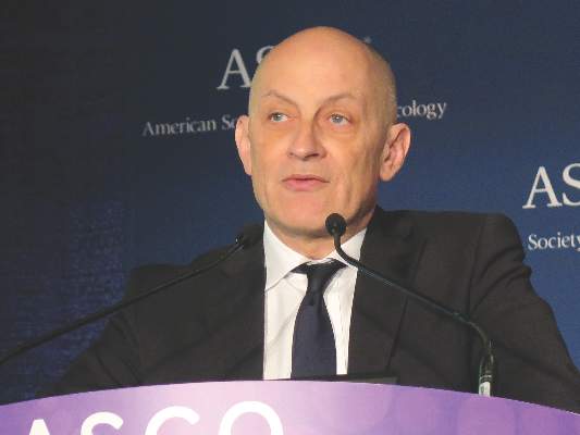
Acne: Maybe She's Born With It?
A recent treatment regimen worsened this 13-year-old Native American girl’s “acne.” Within hours of beginning the treatment (application of benzoyl peroxide–containing gel and twice-daily use of a benzoyl peroxide–based cleanser), she reported intense burning and redness in the treated areas. She stopped this regimen after a week, and her primary care provider referred her for evaluation.
The lesions have been on her face since age 2 and are not usually problematic.
The patient has a notable history of atopy, with seasonal allergies and occasional bouts of asthma. She claims to have no other health problems but does admit to being overweight for most of her life; this is an issue for most of her family members, as well. Several of her relatives developed diabetes as adults.
EXAMINATION
The patient is 5 ft tall and weighs 250 lb. She has type IV skin, consistent with her Native American heritage. The posterior aspects of both cheeks are quite red and covered with hyperkeratotic, rough, pinpoint papules. There are no comedones or pustules there or elsewhere on her body. 
No other blemishes or lesions are seen on her face, but there are hundreds of hyperkeratotic papules on her bilateral triceps. The skin in all her intertriginous (skin-on-skin) areas is quite dark and has a faintly papular, velvety appearance.
What is the diagnosis?
DISCUSSION
This girl’s facial and arm condition is keratosis pilaris (KP), an extremely common and harmless problem that affects up to 70% of newborns, though it may not fully express until age 1 or 2. Caused by an overproduction of perifollicular keratin, KP is inherited in an autosomal dominant pattern and is often seen in conjunction with atopic dermatitis and related conditions (eg, eczema, xerosis, asthma, icthyosis). KP can affect skin anywhere on the body except glabrous skin (palms and soles).
Variants of KP are also common; the one affecting this patient is keratosis pilaris rubra facei. This condition is often confused with acne, but treating it as such worsens irritation—especially in the wintertime, when humidity levels are low.
The presence of the patient’s condition from such an early age, the lack of comedones or pustules, and the discovery of more typical KP lesions on her arms all served to rule out acne and rosacea (the other item in the differential). The intertriginous hyperpigmentation represents acanthosis nigricans, reflecting her obese and (probable) prediabetic state, and is unrelated to her KP.
She will be treated with a one-week course of 2.5% hydrocortisone cream (bid application) and cessation of benzoyl peroxide–containing products. The condition should improve rapidly and become less prominent over time. Given the nature of KP, I have also found it helpful to encourage parents of affected children to research the condition online, to reinforce the information I’ve provided in the clinic.
TAKE-HOME LEARNING POINTS
• Keratosis pilaris (KP) is extremely common, affecting up to 70% of all newborns.
• Keratosis pilaris rubra facei, a common variant, affects the posterior two-thirds of the cheeks. Virtually all patients with this form will also exhibit KP on more typical areas (eg, the triceps).
• While there is no cure, moisturization helps to ease symptoms, as do other products (eg, salicylic acid–containing creams).
• KP is often mistaken for acne; unfortunately, treatment as such is counterproductive.
A recent treatment regimen worsened this 13-year-old Native American girl’s “acne.” Within hours of beginning the treatment (application of benzoyl peroxide–containing gel and twice-daily use of a benzoyl peroxide–based cleanser), she reported intense burning and redness in the treated areas. She stopped this regimen after a week, and her primary care provider referred her for evaluation.
The lesions have been on her face since age 2 and are not usually problematic.
The patient has a notable history of atopy, with seasonal allergies and occasional bouts of asthma. She claims to have no other health problems but does admit to being overweight for most of her life; this is an issue for most of her family members, as well. Several of her relatives developed diabetes as adults.
EXAMINATION
The patient is 5 ft tall and weighs 250 lb. She has type IV skin, consistent with her Native American heritage. The posterior aspects of both cheeks are quite red and covered with hyperkeratotic, rough, pinpoint papules. There are no comedones or pustules there or elsewhere on her body.
No other blemishes or lesions are seen on her face, but there are hundreds of hyperkeratotic papules on her bilateral triceps. The skin in all her intertriginous (skin-on-skin) areas is quite dark and has a faintly papular, velvety appearance.
What is the diagnosis?
DISCUSSION
This girl’s facial and arm condition is keratosis pilaris (KP), an extremely common and harmless problem that affects up to 70% of newborns, though it may not fully express until age 1 or 2. Caused by an overproduction of perifollicular keratin, KP is inherited in an autosomal dominant pattern and is often seen in conjunction with atopic dermatitis and related conditions (eg, eczema, xerosis, asthma, icthyosis). KP can affect skin anywhere on the body except glabrous skin (palms and soles).
Variants of KP are also common; the one affecting this patient is keratosis pilaris rubra facei. This condition is often confused with acne, but treating it as such worsens irritation—especially in the wintertime, when humidity levels are low.
The presence of the patient’s condition from such an early age, the lack of comedones or pustules, and the discovery of more typical KP lesions on her arms all served to rule out acne and rosacea (the other item in the differential). The intertriginous hyperpigmentation represents acanthosis nigricans, reflecting her obese and (probable) prediabetic state, and is unrelated to her KP.
She will be treated with a one-week course of 2.5% hydrocortisone cream (bid application) and cessation of benzoyl peroxide–containing products. The condition should improve rapidly and become less prominent over time. Given the nature of KP, I have also found it helpful to encourage parents of affected children to research the condition online, to reinforce the information I’ve provided in the clinic.
TAKE-HOME LEARNING POINTS
• Keratosis pilaris (KP) is extremely common, affecting up to 70% of all newborns.
• Keratosis pilaris rubra facei, a common variant, affects the posterior two-thirds of the cheeks. Virtually all patients with this form will also exhibit KP on more typical areas (eg, the triceps).
• While there is no cure, moisturization helps to ease symptoms, as do other products (eg, salicylic acid–containing creams).
• KP is often mistaken for acne; unfortunately, treatment as such is counterproductive.

VIDEO: Lobectomy quality requires linking outcomes to process change
BALTIMORE – An “introspective” analysis connecting patient outcomes with process changes may lead to significant surgical quality improvement, according to a study presented at the 2016 annual meeting of the American Association for Thoracic Surgery.
The case study detailed the University of Alabama at Birmingham School of Medicine’s attempt to identify the metrics used for the Society of Thoracic Surgeons lobectomy ranking, and show how the institution used root cause analysis with “lean” and process improvements to improve outcomes from Jan. 2006 until July 2014 in order to achieve a three star STS ranking.
UAB researchers found that their most common root cause analysis was failure to escalate care. The institution implemented process improvements such as increasing pulmonary rehabilitation prior to surgery, adding a respiratory therapist, eliminating (lean) non-valued steps, favoring stereotactic radiotherapy and segmentectomy instead of lobectomy for marginal patients, and using minimally invasive lobectomy. They ultimately achieved a three-star STS ranking.
The video associated with this article is no longer available on this site. Please view all of our videos on the MDedge YouTube channel
Dr. Stephen D. Cassivi, professor of surgery at the Mayo Clinic in Rochester, Minn., and a discussant on the paper at AATS 2016, said in an interview that the research was important because it encourages surgeons to discuss and reevaluate quality improvement measures. He noted that early phases of surgical quality improvement was based on process measures, specifically around the idea that if surgeons were attentive to process measures, their outcome measures would improve. But over time, the emphasis on process measures has dissipated in favor of outcomes-focused analysis.
“Now that we have more robust [outcomes] data... we can examine our practices in a more thoughtful, data-driven, evidence-based way,” Dr. Cassivi said. He added that the shift from process measures to outcome measures is important in that surgeons can easily interpret and compare outcomes data across facilities. But he noted that there is a downside: If an institution’s outcome measures are not up to standard, it is sometimes difficult to determine why.
“The current way that the [outcomes] data are reported and processed is not easily interpretable into which processes we need to adapt,” Dr. Cassivi said. “There is still work that needs to be done, but [this paper] is a first step.”
Dr. Cassivi reported no relevant financial disclosures.
On Twitter @richpizzi
AT THE AATS ANNUAL MEETING
Precapillary pulmonary hypertension redefined
The hemodynamic characteristics of precapillary pulmonary hypertension differ from those typically thought of as defining the disease, suggests a study published in Chest.
A pulmonary hypertension patient’s diagnostic category is used to select which patients will or won’t receive pulmonary arterial hypertension (PAH)-targeted therapies, such as treprostinil. Currently, patients with a mean pulmonary arterial wedge pressure (mPAWP) less than or equal to 15 mm Hg are classified as having precapillary pulmonary hypertension, which includes PAH; and patients with a mPAWP above 15 mm Hg are classified as having postcapillary pulmonary hypertension. Current recommendations advise against treating patients with postcapillary hypertension with PAH-targeted therapies.
First author Dr. Christian Gerges of the division of cardiology, Vienna General Hospital, Medical University of Vienna and colleagues hypothesized that a precapillary hemodynamic profile is characterized by a mPAWP that is lower than the currently accepted 15 mm Hg, an elevated diastolic pulmonary vascular pressure gradient (DPG), “and a beneficial response to PAH-targeted therapies.”
The study identified an mPAWP less than 12 mm Hg and a DPG greater than or equal to 7 mm Hg as the best hemodynamic discriminators between idiopathic PAH and postcapillary pulmonary hypertension.
This study also found that patients with a mPAWP less than 12 mm Hg combined with a diastolic pulmonary vascular pressure gradient (DPG) greater than 20 mm Hg are likely to have a significant response to PAH-targeted-therapy.
The researchers analyzed hemodynamic data from a retrospective cohort of 4,363 stable patients who underwent a first diagnostic right heart catheterization at the Medical University of Vienna between May 1996 and June 2006. Of these patients, 3,524 (81%) also received a left heart catheterization. Additionally, the researchers analyzed deidentified individual data for 541 patients from four randomized placebo-controlled trials and 437 patients from one open-label trial of treprostinil in PAH. These trials all had similar inclusion criteria and data collection processes (Chest. 2016 Apr;149[4]:1061-73. doi: 10.1378/chest.15-0928).
Funding was provided by educational grants from Bayer and United Therapeutics Corporation. All but two coauthors had no disclosures; the remaining authors disclosed numerous ties to industry sources.*
*Correction, 6/14/16: An earlier version of this article misstated the investigators' conflicts of interest.
FROM CHEST
Key clinical point: The hemodynamic characteristics of precapillary pulmonary hypertension differ from those typically thought of as defining the disease, suggests a study published in Chest.
Major finding: Patients with a mean pulmonary arterial wedge pressure less than 12 mm Hg combined with a diastolic pulmonary vascular pressure gradient greater than 20 mm Hg are likely to display a significant response to pulmonary arterial hypertension (PAH)-targeted therapy.
Data sources: Data for 978 patients with PAH from four randomized placebo-controlled trials and one open-open label trial, and a retrospective cohort of 4,363 stable patients who underwent a first diagnostic right heart catheterization.
Disclosures: Funding was provided by educational grants from Bayer and United Therapeutics Corporation. All but two coauthors had no disclosures; the remaining authors disclosed numerous ties to industry sources.*
Veteran Cancer Research Highlighted in ASCO Posters and Abstracts
At the 2016 American Society of Clinical Oncology in Chicago, Illinois, researchers from the VA submitted new abstracts on more than 20 different research projects focused on veteran’s oncology care. The researchers conducted studies with the scope of veteran diagnosis, treatment, and quality of care. The abstracts were presented at the annual ASCO meeting during poster sessions and online.
General Oncology
The Impact of a Veterans Administration NURSE Disease Management Program: the Oncology Aspect
Author(s): George Anthony Dawson, et al.
Background: In an effort to decrease time to diagnosis and/or treatment intervention in newly diagnosed veteran cancer patients, Hudson Valley administrators chose the disease case management team model as a potential useful solution to enhance and / or expedite health care delivery. This disease management program began in 2008. The team consists of 3 registered nurses who work under the auspices of the quality management division rather than a specific oncology clinic. This report will show the clinical impact of Hudson Valley VA’s disease management team.
To read the full abstract click here: http://meetinglibrary.asco.org/content/161764-176
Implementation of a Nurse Communication Strategy to Improve Perception of Nurse-Patient Communication at the Bedside on an Inpatient Oncology Unit
Author(s): Lyn Cain Zehner, et al.
Background: Patients spend more time with nurses when in the hospital than with any other health care professional. Patient experience scores have been used as a measure of quality patient outcomes. The Hospital Consumer Assessment of Healthcare Providers and Systems Survey (HCAHPS) score communication with nurses, correlates with improvements in 4 other dimensions: pain, communication about medications, responsiveness of staff, and overall rating. As patient experience scores are tied to Medicare reimbursement, nurses have an impact on reimbursement. The nurse communication scores on the inpatient 29 bed oncology unit reflected an opportunity for improvement. The purpose of this project was to develop a communication strategy that staff would implement at the bedside. The goal was to achieve an improvement in the HCAHPS Communication with Nurses question.
To read the full abstract click here: http://meetinglibrary.asco.org/content/166250-176
Modified Ketogenic Diet in Advanced Malignancies–Final Results of a Safety and Feasibility Trial Within the Veterans Affairs Healthcare System
Author(s): Jocelyn Tan-Shalaby, et al.
Final report of a safety and feasibility trial within the Pittsburgh Veteran Affairs HealthCare System- Modified Ketogenic Diet in Advanced Cancer.
Background: Malignant cells have dysfunctional mitochondria, which limit their ability to use energy from fatty acids and ketones. Preclinical animal studies using a ketogenic diet in cancer show encouraging results. The authors tested safety and feasibility of the modified ketogenic diet in a veteran population of advanced cancer patients across a variety of solid tumors.
To read the full abstract click here: http://meetinglibrary.asco.org/content/170081-176
[Click through for : Prostate Cancer, Liver, Radiation Oncology, Lung Cancer, Colorectal, HIV and Cancer, Palliative Care, Esophageal]
Prostate Cancer
Improving Risk Stratification Among Veterans With Newly Diagnosed, Clinically Low-Risk Prostate Cancer Using the 17-Gene Genomic Prostate Score Assay.
Author(s): Julie Ann Lynch, et al.
Background: Active surveillance (AS) is a recommended treatment option for low risk prostate cancer (PCa). Studies have shown high rates of AS in the VA yet treatment variation exists between VAMCs, likely due to concern about missing aggressive disease. The 17-gene Genomic Prostate Score (GPS) has been validated to predict likelihood of favorable pathology in men with low risk PCa. This study compares treatment patterns before and after introduction of the GPS to determine if the assay assists in risk-stratification. Men with PCa who met National Comprehensive Cancer Network criteria for very low, low, or intermediate risk PCa were eligible. Chart review of men across 6 VAMCs established baseline treatment in untested patients in 2013-2014. In 2015, Veterans at the same VAMCs were offered the assay in a prospective study. Treatment recommendations and treatment implemented were captured.
To read the full abstract click here: http://meetinglibrary.asco.org/content/170703-176
Trends in Utilization of Novel Oral Therapeutic Agents for Men With Metastatic Castrate-Resistant Prostate Cancer Within the United States Veteran’s Affairs Health System.
Author(s): Elizabeth Henry, et al.
Background: Therapeutic options for men with metastatic castrate resistant prostate cancer (mCRPC) have expanded significantly over the past 5 years with several new agents demonstrating improved survival, including two oral agents. Abiraterone acetate, a CYP-17 androgen synthesis inhibitor, obtained initial FDA approval in 2011 for post-docetaxel (D) use, and gained expanded approval in 2012 for pre-D use. Enzalutamide, an androgen receptor signaling inhibitor, also gained FDA approval in 2012 (post-D) and indication was expanded in 2014 (pre-D). Due to the relatively recent approvals of these agents, there is limited data on rates of uptake and prescribing patterns for patients with mCRPC.
To read the full abstract click here: http://meetinglibrary.asco.org/content/157970-172
Liver
A Comparison of Liver Cancer Patients Receiving Palliative Care at Two Veteran Affairs Medical Centers.
Author(s): Sarah Lee, Zhen Wang, et al.
Background: Liver cancer is a leading cause of death. Lack of data exists on palliative care in this group, and care varies by location. The authors aim to determine if there are differences in palliative care for liver cancer patients by VA site.
To read the full abstract click here: http://meetinglibrary.asco.org/content/171726-176
[Click through for : Radiation Oncology, Lung Cancer, Colorectal, HIV and Cancer, Palliative Care, Esophageal]
Radiation Oncology
A Cross-Sectional View of Radiation Dose Fractionation Schemes Used for Treating Painful Bone Metastases (PBM) Within Veterans Health Administration’s Radiation Oncology Centers.
Author(s): George Anthony Dawson, et al.
Background: The use of single fraction radiotherapy in the treatment of PBM in the United States is now gaining clinical acceptance, especially at large institutional levels. In this cross-sectional report, the authors report the varying dose fractionation schemes used to treat PBM by VHA radiation oncology centers.
To read the full abstract click here: http://meetinglibrary.asco.org/content/161496-176
Will Academic and Community Physicians Engage and Share Knowledge in an Online Physician Social Network? Lessons from the Radiation Oncology Community.
Author(s): Nadine Housri, et al.
Background: The exponential growth of medical knowledge has made it increasingly difficult for clinicians to make sense of new information and recognize how to incorporate new research results into clinical practice. The authors sought to determine whether a social question and answer website designed to connect academic and community physicians would consistently engage radiation oncologists to share evidence based information and clinical insights with each other.
To read the full abstract click here: http://meetinglibrary.asco.org/content/171497-176
Lung Cancer
Non–Small Cell Lung Cancer in Veterans: Disparities in Prevalence and Survival Among Different Histologic Subtypes.
Author(s): Hussein Assi, Efstratios Koutroumpakis, et al.
Background: Since the 1980s, adenocarcinoma (AC) of the lung has become more common than squamous cell carcinoma (SCC). Given the differences in smoking patterns, it is unclear if the same holds true among veterans. In this study, the authors compare the distribution of lung AC and SCC in the veterans’ population to that in the general population. They also looked at the survival of patients with different histologies in the VA population.
To read the full abstract click here: http://meetinglibrary.asco.org/content/167505-176
VA Oncologists’ Attitudes and Behaviors Regarding Genomic-Based Targeted Therapy for the Management of Advanced Lung Cancer.
Author(s): Jennifer Arney, et al.
Background: Genomic-based targeted therapy (GBTT) has emerged as a treatment option for patients with advanced lung cancer. Clinical practice guidelines of the National Comprehensive Cancer Network and the American Society of Clinical Oncology recommend testing all metastatic adenocarcinomas for EGFR mutation, and use of EGFR-TKIs (erlotinib) as first-line therapy for advanced adenocarcinoma lung cancer patients with EGFR mutation positive. Little is known about how oncologists utilize genomic testing and GBTT in a clinical setting. Drawing from the Cabana and colleagues theoretical framework on providers’ adherence to guidelines, this study aims to elicit provider and facility-level barriers and facilitators to using GBTT in VA healthcare delivery system.
To read the full abstract click here: http://meetinglibrary.asco.org/content/166517-176
[Click through for: Colorectal, HIV and Cancer, Palliative Care, Esophageal]
Colorectal
Survival Advantage for Hepatectomy With or Without Hemicolectomy in Colorectal Liver Metastases.
Author(s): Dalia A. Mobarek, et al.
Background: Surgical resection of colorectal liver metastases offers the greatest chance of long-term survival. The authors aimed to study survival in patients presenting with hepatic metastasis at the outset and rates of surgical intervention in the VHA.
To read the full abstract click here: http://meetinglibrary.asco.org/content/171640-176
Colorectal Liver Metastases Management in the Veterans Health Administration: Geographic Disparity.
Author(s): Dalia A. Mobarek, et al.
Background: Multidisciplinary management including surgical resection of colorectal liver metastases offers the greatest chance of long-term survival. The authors aimed to study surgical intervention types, rates and factors affecting the decision making in the VHA.
To read the full abstract click here: http://meetinglibrary.asco.org/content/160233-173
HIV and Cancer
Gleason Grade in HIV+ Versus Uninfected Prostate Cancer Patients in the Veterans Aging Cohort Study.
Author(s): Roxanne Jimmy Wadia, et al.
Background: Little is known about how the biology of prostate cancer in HIV+ men compares to that of uninfected men. The purpose of this study was to compare the Gleason grade (GL) of prostate adenocarcinoma (PrCA) in HIV+ vs uninfected patients in the combination antiretroviral therapy (ART) era (1997-present).
To read the full abstract click here: http://meetinglibrary.asco.org/content/162980-176
Disparities in AIDS-Related Kaposi Sarcoma Incidence and Survival.
Author(s): Firas El Chaer, et al.
Background: With advances in combined antiretroviral therapy (ART), Kaposi’s sarcoma (KS) incidence has decreased over the past decade. Geographical and racial disparities may contribute to variation in the incidence and outcomes of multiple cancers in the U.S., including KS. Using the Surveillance, Epidemiology, and End Results (SEER) database, the authors analyzed KS incidence and survival by race and geographical region in the ART era.
To read the full abstract click here: http://meetinglibrary.asco.org/content/168953-176
[Click through for : Palliative Care, Esophageal]
Palliative Care
Palliative Care Interventions and EOL Care Outcomes for Hepatocellular Patients (pts) at 2 VA Medical Centers.
Author(s): Zhen Wang, et al.
Background: Palliative care interventions and its effect on EOL outcomes for liver cancer pts have not been described. The authors investigated the association between palliative care intervention and EOL care outcomes.
To read the full abstract click here: http://meetinglibrary.asco.org/content/167081-176
Esophageal
Characteristics of Esophageal Carcinoma in a Veteran Population.
Author(s): Theresa Ratajczak, et al.
Background: Esophageal cancer is projected to increase by approximately 35% through 2025 in the U.S. The authors investigated the characteristics of esophageal carcinoma in a veteran population.
To read the full abstract click here: http://meetinglibrary.asco.org/content/162712-176