User login
Restrictive Transfusion Strategy No Benefit for Cardiac Surgery Patients
Clinical question: Does a restrictive transfusion strategy improve outcomes following nonemergent cardiac surgery?
Bottom line: For patients undergoing cardiac surgery, using a restrictive transfusion strategy with a hemoglobin threshold of 7.5 g/dL does not decrease serious infections or ischemic events and may lead to increased all-cause mortality at 90 days. (LOE = 1b)
Study design: Randomized controlled trial (single-blinded)
Funding source: Government
Allocation: Concealed
Setting: Inpatient (any location)
Synopsis
This is yet another study that compares restrictive and liberal transfusion strategies, this time in a cardiac surgery population. These investigators enrolled patients undergoing nonemergency cardiac surgery (mostly coronary artery bypass grafts or valvular procedures) who had a drop of hemoglobin level to below 9 g/dL following surgery.
Patients were randomized, using concealed allocation, to the restrictive transfusion threshold group (threshold hemoglobin 7.5 g/dL) or liberal transfusion threshold group (threshold hemoglobin 9 g/dL). Patients were masked but physicians and nurses were aware of the group assignments.
In the liberal group, patients received one unit of red cell transfusion immediately after randomization followed by an additional unit if the hemoglobin level remained below or dropped below 9 g/dL again during the hospitalization. In the restrictive group, patients received one unit of red cells only if the hemoglobin level dropped below 7.5 g/dL. An additional unit was then given if hemoglobin remained below or dropped below 7.5 g/dL again during the hospitalization. The 2 groups were similar at baseline and analysis was by intention to treat. Not surprisingly, more patients in the liberal strategy group received transfusions than did those in the restrictive strategy group (95% vs 64%).
For the primary outcome—a composite of sepsis, wound infection, stroke, myocardial infarction, gut infarction, or acute kidney injury within 3 months of randomization—there was no significant difference detected between the 2 groups. However, the restrictive group had a higher mortality rate than the liberal group (4.2% vs 2.6%; P = .045). Although this was a secondary outcome, it is possible that a restrictive strategy may be harmful in this cohort, given that they may have less cardiovascular reserve than the general patient population.
Dr. Kulkarni is an assistant professor of hospital medicine at Northwestern University in Chicago.
Endovascular Treatment for Acute Ischemic Stroke Decreases Mortality
Clinical question: Does endovascular treatment improve outcomes for patients with acute ischemic stroke?
Bottom line: For patients with acute ischemic stroke and imaging that suggests a proximal artery occlusion with evidence of good collateral circulation, the use of rapid endovascular treatment improves functional outcomes and reduces mortality. (LOE = 1b)
Study design: Randomized controlled trial (nonblinded)
Funding source: Industry
Allocation: Concealed
Setting: Inpatient (any location) with outpatient follow-up
Synopsis
The recent MR CLEAN study showed improved functional outcomes with the use of endovascular therapy for the treatment of acute ischemic stroke (N Engl J Med 2015;372:11-20).
In this study, investigators enrolled patients with acute disabling ischemic strokes and computed tomographic evidence of a small infarct core, an occluded proximal artery in the anterior circulation, and moderate-to-good collateral circulation. Patients were randomized, using concealed allocation, to receive either usual care or usual care plus rapid endovascular treatment with the use of mechanical thrombectomy and retrievable stents.
The 2 groups had similar baseline characteristics with a mean age of 70 years and a median National Institutes of Health Stroke Scale score of 16 to 17. The median time from stroke onset to reperfusion was 4 hours in the intervention group. The trial was stopped early because of the efficacy of the endovascular therapy. The primary outcome was a common odds ratio, indicating the odds of improvement by 1 point on the modified Rankin scale of 0 to 6 (0 = no symptoms, 1–2 = slight disability, 6 = death). This ratio favored the intervention (common odds ratio 2.6, 95% CI 1.7-3.8; P < .001).
Overall, at 90-day follow-up, the intervention group had a greater proportion of patients with a modified Rankin score of 0–2 (53% vs. 29%; P < .001), as well as decreased mortality (10% vs 19% in control group, P = .04). There was no difference between the 2 groups in the rate of symptomatic intracerebral bleeds.
Dr. Kulkarni is an assistant professor of hospital medicine at Northwestern University in Chicago.
NASPAG: Migraines don’t always preclude combined OCs
ORLANDO– Most adolescent girls who report having headaches – including some of those who report migraines with aura – can safely use combined oral contraceptive pills, according to Dr. Sari Kives.
The available literature suggests that there is some resistance to prescribing such contraception for adolescents with headaches, but most adolescents don’t have the types of headaches that are of concern, Dr. Kives of the University of Toronto said at the annual meeting of the North American Society for Pediatric and Adolescent Gynecology.

owever, it is important to get a good characterization of the headaches, keeping in mind that teens may have difficulty relating their symptoms, she said.
“It’s important to understand what the headache actually is. Is it a tension headache, which is by far the most common headache you will see in adolescents?” she said, noting that menstrual migraines and classical migraines are less common in adolescents.
Menstrual migraines account for about 7%-8% of migraines, occur 2-3 days before menses, can last throughout the period, don’t occur any other time of the month, and can be quite debilitating. They usually are secondary to estrogen withdrawal, Dr. Kives said, adding that a decade ago, add-back estrogen was commonly given during the week off of oral contraceptives in those with menstrual headaches.

Now it is common practice to use continuous pills or extended-cycle pills, she said, explaining that eliminating the estrogen fluctuation improves the headaches.
Classical migraines also occur commonly in adolescent girls. Some may include focal neurological symptoms that may be triggered by hormonal changes, stress, certain foods and beverages, certain scents or fumes, fatigue, hunger, or trauma.
It is important to ask about such symptoms, Dr. Kives said.
“And that’s probably the most important question you can ask. For me, a focal neurological symptom is, ‘I go blind in my left eye. I lose sensation in my right arm,’ ” she said, providing examples.
Some symptoms are characteristic of “atypical aura,” and some are associated with “typical aura” – an important distinction when determining whether combined OCs are safe for a given patient.
Atypical aura usually has sudden unilateral onset and lasts more than 30-60 minutes. Headache may or may not be present, and visual symptoms may include loss of vision, amaurosis fugax (painless transient monocular visual loss), and visual field anomaly. Sensory and motor symptoms can include lower limb anesthesia or hypoesthesia (decrease in sensation).
Typical aura has more progressive onset, lasts less than an hour, and precedes migraine. Patients may experience bilateral scintillating scotoma, fortification spectra, and blurred vision. These are usually limited to visual symptoms, Dr. Kives said, but sensory and motor symptoms can occur. They tend to occur in relation to the visual symptoms, and may affect the upper limbs, mouth, and tongue – causing tingling or pinching sensations.
Individuals who have migraine with aura account for only about 20% of those with migraine headaches, and the vast majority are going to have visual aura.
“They can have sensory and motor symptoms, but the visual ones are the ones where you have to be very specific,” she said, noting that in her experience, 99% of cases are visual.
“If it’s a short visual aura, less than an hour, and it’s not repetitive, I will consider an oral contraceptive pill in this group of patients, but you have to balance it against what their history sounds like,” she said.
Typically, combined OCs are contraindicated in patients with migraine with aura because of an increased risk of cerebrovascular accident, but in Canada, guidelines provide allowances for this “unique group of individuals with migraines with aura that are limited to visual symptoms and that last less than 1 hour,” she said.
Remember that photophobia, phonophobia, nausea and vomiting, visual blurring, and generalized visual spots/flashing lights do not constitute aura, she said.
This is important, because using too stringent a definition of “migraine with aura” will leave a substantial number of individuals with limited contraceptive options, particularly options that are effective and promote cycle control and compliance, she said.
Although there is a definite risk associated with combined OCs in those with migraine with aura – with an added risk in those who smoke, the risks are low in adolescents, compared with older patients.
“The adult women who walks in with hypertension, or who is a smoker who gets oral contraception – that is a very different patient than the 14-year-old who says, ‘I may get flashing lights before my headache, but not on a regular basis.’ Those are completely different entity patients, in my opinion,” Dr. Kives said.
If she does prescribe combined OCs for an adolescent with migraines, she advises the patient to stop the pills if the headaches get worse, she said. In many cases, however, headaches improve, because they were menstrual migraines and not classical migraines, she said, adding that “improvement in headaches is a reassuring sign.” The bottom line, Dr. Kives said, is that migraine without aura doesn’t preclude prescribing of any contraceptive options in adolescents, and that migraine with aura is a relative contraindication; low-dose combined oral contraceptive pills are safe for those with migraine with aura that primarily includes visual symptoms lasting less than an hour.
However, the risk of cerebrovascular accidents is increased in those with migraines, so other risk factors, such as family history, obesity, hypertension, and smoking, should be considered.
“If they have no other risk factors, their risk probably is quite low,” Dr. Kives said.
She reported having no relevant financial disclosures.
ORLANDO– Most adolescent girls who report having headaches – including some of those who report migraines with aura – can safely use combined oral contraceptive pills, according to Dr. Sari Kives.
The available literature suggests that there is some resistance to prescribing such contraception for adolescents with headaches, but most adolescents don’t have the types of headaches that are of concern, Dr. Kives of the University of Toronto said at the annual meeting of the North American Society for Pediatric and Adolescent Gynecology.
owever, it is important to get a good characterization of the headaches, keeping in mind that teens may have difficulty relating their symptoms, she said.
“It’s important to understand what the headache actually is. Is it a tension headache, which is by far the most common headache you will see in adolescents?” she said, noting that menstrual migraines and classical migraines are less common in adolescents.
Menstrual migraines account for about 7%-8% of migraines, occur 2-3 days before menses, can last throughout the period, don’t occur any other time of the month, and can be quite debilitating. They usually are secondary to estrogen withdrawal, Dr. Kives said, adding that a decade ago, add-back estrogen was commonly given during the week off of oral contraceptives in those with menstrual headaches.
Now it is common practice to use continuous pills or extended-cycle pills, she said, explaining that eliminating the estrogen fluctuation improves the headaches.
Classical migraines also occur commonly in adolescent girls. Some may include focal neurological symptoms that may be triggered by hormonal changes, stress, certain foods and beverages, certain scents or fumes, fatigue, hunger, or trauma.
It is important to ask about such symptoms, Dr. Kives said.
“And that’s probably the most important question you can ask. For me, a focal neurological symptom is, ‘I go blind in my left eye. I lose sensation in my right arm,’ ” she said, providing examples.
Some symptoms are characteristic of “atypical aura,” and some are associated with “typical aura” – an important distinction when determining whether combined OCs are safe for a given patient.
Atypical aura usually has sudden unilateral onset and lasts more than 30-60 minutes. Headache may or may not be present, and visual symptoms may include loss of vision, amaurosis fugax (painless transient monocular visual loss), and visual field anomaly. Sensory and motor symptoms can include lower limb anesthesia or hypoesthesia (decrease in sensation).
Typical aura has more progressive onset, lasts less than an hour, and precedes migraine. Patients may experience bilateral scintillating scotoma, fortification spectra, and blurred vision. These are usually limited to visual symptoms, Dr. Kives said, but sensory and motor symptoms can occur. They tend to occur in relation to the visual symptoms, and may affect the upper limbs, mouth, and tongue – causing tingling or pinching sensations.
Individuals who have migraine with aura account for only about 20% of those with migraine headaches, and the vast majority are going to have visual aura.
“They can have sensory and motor symptoms, but the visual ones are the ones where you have to be very specific,” she said, noting that in her experience, 99% of cases are visual.
“If it’s a short visual aura, less than an hour, and it’s not repetitive, I will consider an oral contraceptive pill in this group of patients, but you have to balance it against what their history sounds like,” she said.
Typically, combined OCs are contraindicated in patients with migraine with aura because of an increased risk of cerebrovascular accident, but in Canada, guidelines provide allowances for this “unique group of individuals with migraines with aura that are limited to visual symptoms and that last less than 1 hour,” she said.
Remember that photophobia, phonophobia, nausea and vomiting, visual blurring, and generalized visual spots/flashing lights do not constitute aura, she said.
This is important, because using too stringent a definition of “migraine with aura” will leave a substantial number of individuals with limited contraceptive options, particularly options that are effective and promote cycle control and compliance, she said.
Although there is a definite risk associated with combined OCs in those with migraine with aura – with an added risk in those who smoke, the risks are low in adolescents, compared with older patients.
“The adult women who walks in with hypertension, or who is a smoker who gets oral contraception – that is a very different patient than the 14-year-old who says, ‘I may get flashing lights before my headache, but not on a regular basis.’ Those are completely different entity patients, in my opinion,” Dr. Kives said.
If she does prescribe combined OCs for an adolescent with migraines, she advises the patient to stop the pills if the headaches get worse, she said. In many cases, however, headaches improve, because they were menstrual migraines and not classical migraines, she said, adding that “improvement in headaches is a reassuring sign.” The bottom line, Dr. Kives said, is that migraine without aura doesn’t preclude prescribing of any contraceptive options in adolescents, and that migraine with aura is a relative contraindication; low-dose combined oral contraceptive pills are safe for those with migraine with aura that primarily includes visual symptoms lasting less than an hour.
However, the risk of cerebrovascular accidents is increased in those with migraines, so other risk factors, such as family history, obesity, hypertension, and smoking, should be considered.
“If they have no other risk factors, their risk probably is quite low,” Dr. Kives said.
She reported having no relevant financial disclosures.
EXPERT ANALYSIS FROM THE NASPAG ANNUAL MEETING

No causal effect found between high uric acid concentrations and diabetes
Despite observed associations between uric acid concentrations and diabetes risk, the relationship has not been found to be causal, according to results published April 27 in the journal Diabetes.
Dr. Ivonne Sluijs of University Medical Center Utrecht (the Netherlands) and her associates performed a Mendelian randomization study in a cohort of 24,265 European patients using 24 uric acid loci.
Higher uric acid concentrations were associated with greater diabetes risk (hazard ratio, 1.20 per 59.48 μmol/L; 95% CI, 1.11-1.30), though this did not have a causal effect on diabetes, the authors reported (HR, 1.01; 95% CI, 0.87-1.16).
The findings suggest that “increased uric acid concentrations are a consequence of an adverse metabolic profile, rather than a cause of diabetes,” Dr. Sluijs and her associates wrote (Diabetes 2015 April 27 [doi:10.2337/db14-0742]). Thus, therapies that lower uric acid may not be effective in lowering diabetes risk, they said. “Uric acid has limited value as therapeutic target in preventing diabetes,” the investigators concluded.
Dr. Slujis and her associates did not disclose any conflicts of interest.
FROM DIABETES
Medicare at 50: Or, the end of fee-for-service
Few of my readers were around when Medicare was signed into law in 1965 by President Lyndon Johnson. Mr. Johnson was into his first elected term as president, having first taken the oath of office with the assassination of John F. Kennedy in November 1963.
Hardly a darling of the liberal left, he was maligned as the perpetrator, if not for the expansion, of the Vietnam War. He was elected with the largest presidential plurality in history, receiving 61% of the votes, and he carried with him a solid Democratic Congress bolstered by a large block of solid Southern Democrats, a firmament from which he had emerged. He can be credited for the most far-reaching social legislation of the 20th century, second only to Franklin Delano Roosevelt of the 1930s. Johnson was able to move the Civil Rights Act of 1964 and the Voting Rights Act and Medicare Act through Congress in 1965, the latter of which we celebrate this year.

At the time of passage of Medicare, 35% of Americans over 65 had no health insurance, either because they found insurance unaffordable or because of preexisting illness. Health insurance premiums for the elderly cost roughly three times more than did those for younger individuals. With many of the elderly living on Social Security alone, health insurance was an impossibility. If they needed care, they might find it at the emergency department of their local hospitals.
The battle for the passage of Medicare was formidable. In contrast to the passive role of the American Medical Association in the passage of the Affordable Care Act, Medicare was, to put it mildly, vigorously opposed by most doctors and by the AMA, which viewed it as the harbinger of long-anticipated socialized medicine. It ended up being far from it.
Medicare provided a pathway to an economic bonanza for the practicing physician and to hospitals that they could never have imagined. It immediately expanded the patient population and allowed enterprising and not-so-enterprising doctors and hospitals to run up the costs of health care by strengthening the fee-for-service style of medical care. Doctors, hospitals, and patients had no concern for cost: Medicare paid for everything.
Hospitals found Medicare a ready source of income. Doctor and hospital bills were based on the usual cost to each in their respective communities. Medical educators as well as house staff also saw their income lifted as a result of the federal government’s paying for a large part if not all of the cost of house staff education. Even the small number of physicians who opted out of Medicare, because of the red tape or to preserve their independence from the federal government, benefited. Their fees increased as the payment schedules of Medicare increased in their communities.
One of the by-products of Medicare was the end of hospital segregation. For those of you who were not around then, you can imagine what an impact that had on hospitals and doctors in both the North and the South.
Over time, Medicare expanded its footprint to include treatment of chronic kidney disease at any age, as well as drug coverage. I would guess that most Americans over 65 (this writer included) love Medicare. And doctors? Well, they still grumble about it.
With the recent readjustment of physicians’ Medicare reimbursement rates, the door has been opened to new initiatives based on quality of care. No matter what your political persuasion, it is clear that Americans cannot afford Medicare as we know it. Physicians undoubtedly will find their fee-for-service style of reimbursement curtailed as we learn the definition of “quality care standards.” Quality standards will not only define clinical performance, but will also include cost of rendering that care. So long to the world of fee-for-service reimbursement.
Dr. Goldstein, medical editor of Cardiology News, is professor of medicine at Wayne State University and division head emeritus of cardiovascular medicine at Henry Ford Hospital, both in Detroit. He is on data safety monitoring committees for the National Institutes of Health and several pharmaceutical companies.
Few of my readers were around when Medicare was signed into law in 1965 by President Lyndon Johnson. Mr. Johnson was into his first elected term as president, having first taken the oath of office with the assassination of John F. Kennedy in November 1963.
Hardly a darling of the liberal left, he was maligned as the perpetrator, if not for the expansion, of the Vietnam War. He was elected with the largest presidential plurality in history, receiving 61% of the votes, and he carried with him a solid Democratic Congress bolstered by a large block of solid Southern Democrats, a firmament from which he had emerged. He can be credited for the most far-reaching social legislation of the 20th century, second only to Franklin Delano Roosevelt of the 1930s. Johnson was able to move the Civil Rights Act of 1964 and the Voting Rights Act and Medicare Act through Congress in 1965, the latter of which we celebrate this year.
At the time of passage of Medicare, 35% of Americans over 65 had no health insurance, either because they found insurance unaffordable or because of preexisting illness. Health insurance premiums for the elderly cost roughly three times more than did those for younger individuals. With many of the elderly living on Social Security alone, health insurance was an impossibility. If they needed care, they might find it at the emergency department of their local hospitals.
The battle for the passage of Medicare was formidable. In contrast to the passive role of the American Medical Association in the passage of the Affordable Care Act, Medicare was, to put it mildly, vigorously opposed by most doctors and by the AMA, which viewed it as the harbinger of long-anticipated socialized medicine. It ended up being far from it.
Medicare provided a pathway to an economic bonanza for the practicing physician and to hospitals that they could never have imagined. It immediately expanded the patient population and allowed enterprising and not-so-enterprising doctors and hospitals to run up the costs of health care by strengthening the fee-for-service style of medical care. Doctors, hospitals, and patients had no concern for cost: Medicare paid for everything.
Hospitals found Medicare a ready source of income. Doctor and hospital bills were based on the usual cost to each in their respective communities. Medical educators as well as house staff also saw their income lifted as a result of the federal government’s paying for a large part if not all of the cost of house staff education. Even the small number of physicians who opted out of Medicare, because of the red tape or to preserve their independence from the federal government, benefited. Their fees increased as the payment schedules of Medicare increased in their communities.
One of the by-products of Medicare was the end of hospital segregation. For those of you who were not around then, you can imagine what an impact that had on hospitals and doctors in both the North and the South.
Over time, Medicare expanded its footprint to include treatment of chronic kidney disease at any age, as well as drug coverage. I would guess that most Americans over 65 (this writer included) love Medicare. And doctors? Well, they still grumble about it.
With the recent readjustment of physicians’ Medicare reimbursement rates, the door has been opened to new initiatives based on quality of care. No matter what your political persuasion, it is clear that Americans cannot afford Medicare as we know it. Physicians undoubtedly will find their fee-for-service style of reimbursement curtailed as we learn the definition of “quality care standards.” Quality standards will not only define clinical performance, but will also include cost of rendering that care. So long to the world of fee-for-service reimbursement.
Dr. Goldstein, medical editor of Cardiology News, is professor of medicine at Wayne State University and division head emeritus of cardiovascular medicine at Henry Ford Hospital, both in Detroit. He is on data safety monitoring committees for the National Institutes of Health and several pharmaceutical companies.

Sports Purpura From Floorball, Indoor Climbing, and Archery
To the Editor:
Sports purpura can be broken down into different types including traumatic purpura,1 exercise-induced cutaneous vasculitis,2 occurrence of coincidental systemic purpura,3 and other conditions.4-6 Traumatic purpura results from brutal contact with an opponent, the court, the equipment, or the ball. Three cases of sports purpura related to equipment and balls are reported.
An otherwise healthy 27-year-old woman presented with multiple ecchymotic round patches on her legs. The largest patch was 70 mm and displayed a heterogeneous Swiss cheese–like pattern with discrete whiter round areas within the patch (Figure 1). She reported that she played as a defender in a second division floorball team weekly, acknowledging frequent body contacts and being hit on the legs with the sticks and balls. Purpura was diagnosed due to hits from the floorball.
A 32-year-old healthy man presented with purpuric petechiae of the left palm after indoor climbing. He had been regularly climbing indoors for 3 years and denied a history of similar eruptions. The lesions were painless, noninfiltrated, and did not disappear after pressure (Figure 2). Lesions presumably were due to repeated friction on the climbing hold. Petechiae took a transiently golden hue before resolving within a week.

| 
|
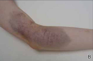
A 26-year-old right-handed woman injured the left forearm while practicing target archery. She was not wearing an arm guard at the time of the injury. Once released, the bowstring scraped the volar aspect of the forearm, causing a painful warm ecchymotic and swollen plaque. She denied neurologic or vascular symptoms. The hematoma rapidly evolved from red to blue (Figure 3) and spontaneously resolved within weeks.
|
|

Purpura related to the high-velocity impact of sport balls has been previously reported with ping-pong,7 paintball,8,9 racquetball, squash,10 and baseball. Floorball, one of the most popular team sports in Finland, is played indoors and resembles ice hockey. The players use graphite compound sticks and a light hollow plastic ball. Except for the goalkeeper, players do not wear specific protective gear. Accidental body contact, including a direct hit from the floorball stick or ball, are frequent.11 The ball weighs 23 g, measures 72 mm in diameter, and has 26 holes that are 11 mm in diameter. The fastest shot was recorded at 127 miles per hour.12 The cutaneous imprint from the ball impact on bare skin, as shown with patient 1, initially is annular,8-10 but the bruise later takes an unusual design due to the peculiar shape of the ball. This complication is no stranger to floorball players but has been rarely reported. The diagnosis is easy, the condition is benign and asymptomatic, and it resolves when the season is over; therefore, players commonly will not seek medical attention. Of note, lower limb injuries, including joint sprains, muscle strains, and soft-tissue contusions, are frequent in female athletes.11 Additional causes of purpura include collision with another player or with boards and stick hits.
Palmar petechiae from indoor climbing is similar to black palm from weight lifting.13 Although the typical black discoloration is absent, the mechanisms of friction and brutal trauma, clinical presentation, and evolution are similar.
Lastly, archery-induced hematomas are caused by the absence of an arm guard, which protects the wrist and forearm when the string snaps back.14 This complication is not often reported but is known by archers. Because archers usually wear protective gear, these injuries are expected to occur in novices or when safety measures are not respected.
1. Aguayo-Leiva I, Vano-Galvan S, Arrazola JM. A purpuric rash. Aust Fam Physician. 2009;38:889-890.
2. Ramelet AA. Exercise-induced vasculitis. J Eur Acad Dermatol Venereol. 2006;20:423-427.
3. Leonard JC, Rieger M. Idiopathic thrombocytopenic purpura presenting in a high school football player: a case report. J Athl Train. 1998;33:269-270.
4. Nordlind K, Bondesson L, Johansson SG, et al. Purpura provoked by cold exposure in a skier. Dermatologica. 1983;167:101-103.
5. Latenser BA, Hempstead RW. Exercise-associated solar purpura in an atypical location. Cutis. 1985;35:365-366.
6. Allan SJ, Humphreys F, Buxton PK. Annular purpura and step aerobics. Clin Exp Dermatol. 1994;19:418.
7. Scott MJ Jr, Scott MJ 3rd. Ping pong patches. Cutis. 1989;43:363-364.
8. Aboutalebi S, Stetson CL. Paintball purpura. J Am Acad Dermatol. 2005;53:901-902.
9. Levsky ME, Crowe M. What is your diagnosis? paintball purpura. Cutis. 2005;75:148, 157-158.
10. Barazi H, Adams BB. Sports purpura. Int J Dermatol. 2006;45:1443.
11. Pasanen K, Parkkari J, Kannus P, et al. Injury risk in female floorball: a prospective one-season follow-up [published online ahead of print May 9, 2007]. Scand J Med Sci Sports. 2008;18:49-54.
12. New world record. Floorball Central Web site. http://www.floorballcentral.com/2010/11/new-world -record.html. Published November 5, 2010. Accessed April 8, 2015.
13. Izumi AK. Letter: pigmented palmar petechiae (black palm). Arch Dermatol. 1974;109:261.
14. Rayan GM. Archery-related injuries of the hand, forearm, and elbow. South Med J. 1992;85:961-964.
To the Editor:
Sports purpura can be broken down into different types including traumatic purpura,1 exercise-induced cutaneous vasculitis,2 occurrence of coincidental systemic purpura,3 and other conditions.4-6 Traumatic purpura results from brutal contact with an opponent, the court, the equipment, or the ball. Three cases of sports purpura related to equipment and balls are reported.
An otherwise healthy 27-year-old woman presented with multiple ecchymotic round patches on her legs. The largest patch was 70 mm and displayed a heterogeneous Swiss cheese–like pattern with discrete whiter round areas within the patch (Figure 1). She reported that she played as a defender in a second division floorball team weekly, acknowledging frequent body contacts and being hit on the legs with the sticks and balls. Purpura was diagnosed due to hits from the floorball.
A 32-year-old healthy man presented with purpuric petechiae of the left palm after indoor climbing. He had been regularly climbing indoors for 3 years and denied a history of similar eruptions. The lesions were painless, noninfiltrated, and did not disappear after pressure (Figure 2). Lesions presumably were due to repeated friction on the climbing hold. Petechiae took a transiently golden hue before resolving within a week.
|
|
|
A 26-year-old right-handed woman injured the left forearm while practicing target archery. She was not wearing an arm guard at the time of the injury. Once released, the bowstring scraped the volar aspect of the forearm, causing a painful warm ecchymotic and swollen plaque. She denied neurologic or vascular symptoms. The hematoma rapidly evolved from red to blue (Figure 3) and spontaneously resolved within weeks.
|
|
Purpura related to the high-velocity impact of sport balls has been previously reported with ping-pong,7 paintball,8,9 racquetball, squash,10 and baseball. Floorball, one of the most popular team sports in Finland, is played indoors and resembles ice hockey. The players use graphite compound sticks and a light hollow plastic ball. Except for the goalkeeper, players do not wear specific protective gear. Accidental body contact, including a direct hit from the floorball stick or ball, are frequent.11 The ball weighs 23 g, measures 72 mm in diameter, and has 26 holes that are 11 mm in diameter. The fastest shot was recorded at 127 miles per hour.12 The cutaneous imprint from the ball impact on bare skin, as shown with patient 1, initially is annular,8-10 but the bruise later takes an unusual design due to the peculiar shape of the ball. This complication is no stranger to floorball players but has been rarely reported. The diagnosis is easy, the condition is benign and asymptomatic, and it resolves when the season is over; therefore, players commonly will not seek medical attention. Of note, lower limb injuries, including joint sprains, muscle strains, and soft-tissue contusions, are frequent in female athletes.11 Additional causes of purpura include collision with another player or with boards and stick hits.
Palmar petechiae from indoor climbing is similar to black palm from weight lifting.13 Although the typical black discoloration is absent, the mechanisms of friction and brutal trauma, clinical presentation, and evolution are similar.
Lastly, archery-induced hematomas are caused by the absence of an arm guard, which protects the wrist and forearm when the string snaps back.14 This complication is not often reported but is known by archers. Because archers usually wear protective gear, these injuries are expected to occur in novices or when safety measures are not respected.
1. Aguayo-Leiva I, Vano-Galvan S, Arrazola JM. A purpuric rash. Aust Fam Physician. 2009;38:889-890.
2. Ramelet AA. Exercise-induced vasculitis. J Eur Acad Dermatol Venereol. 2006;20:423-427.
3. Leonard JC, Rieger M. Idiopathic thrombocytopenic purpura presenting in a high school football player: a case report. J Athl Train. 1998;33:269-270.
4. Nordlind K, Bondesson L, Johansson SG, et al. Purpura provoked by cold exposure in a skier. Dermatologica. 1983;167:101-103.
5. Latenser BA, Hempstead RW. Exercise-associated solar purpura in an atypical location. Cutis. 1985;35:365-366.
6. Allan SJ, Humphreys F, Buxton PK. Annular purpura and step aerobics. Clin Exp Dermatol. 1994;19:418.
7. Scott MJ Jr, Scott MJ 3rd. Ping pong patches. Cutis. 1989;43:363-364.
8. Aboutalebi S, Stetson CL. Paintball purpura. J Am Acad Dermatol. 2005;53:901-902.
9. Levsky ME, Crowe M. What is your diagnosis? paintball purpura. Cutis. 2005;75:148, 157-158.
10. Barazi H, Adams BB. Sports purpura. Int J Dermatol. 2006;45:1443.
11. Pasanen K, Parkkari J, Kannus P, et al. Injury risk in female floorball: a prospective one-season follow-up [published online ahead of print May 9, 2007]. Scand J Med Sci Sports. 2008;18:49-54.
12. New world record. Floorball Central Web site. http://www.floorballcentral.com/2010/11/new-world -record.html. Published November 5, 2010. Accessed April 8, 2015.
13. Izumi AK. Letter: pigmented palmar petechiae (black palm). Arch Dermatol. 1974;109:261.
14. Rayan GM. Archery-related injuries of the hand, forearm, and elbow. South Med J. 1992;85:961-964.

NASPAG: Parity, postpartum status predict adolescent LARC use
ORLANDO – The decision to use long-acting reversible contraception appears largely reactionary among adolescent girls, as the only factors significantly associated with the decision in a recent cross-sectional study were increased parity and postpartum status.
The findings could help with future efforts to identify and remove barriers to long-acting reversible contraceptive (LARC) use among adolescents, according to Dr. Lisa Moon, a third-year resident at the University of Oklahoma, Oklahoma City.

Of 209 adolescents included in the study, 66 used oral contraceptive (OC) pills, and 143 used LARC methods. Levonorgestrel intrauterine devices were used most often (77 subjects), followed by etonogestrel implants (61 subjects). Five of the adolescents used a copper intrauterine device (IUD), Dr. Moon reported at the annual meeting of the North American Society for Pediatric and Adolescent Gynecology.
A breakdown of the findings by age showed that with the exception of those aged 15 years, LARC use increased with increasing age; 1 subject was aged 14 years, and she used OCs; 5 were aged 15 years, and all used a LARC; 15 were aged 16 years, and 9 (60%) used a LARC; 44 were aged 17 years, and 28 (64%) used a LARC; 62 were aged 18 years and 44 (71%) used a LARC; and 82 were aged 19 years, and 57 (70%) used a LARC.
Multivariate analysis showed that having previously given birth and postpartum status were significant predictors of LARC vs. OC use (odds ratios, 3.5 and 3.9, respectively). Age, race, marital status, and documented citizenship were not associated with choice of contraception.
The vast majority of adolescent pregnancies – about 82% – are unplanned, and 50% of teens with unplanned pregnancies report having used some form of contraception at the time of pregnancy. LARC methods have the potential to improve teen pregnancy rates because non-LARC methods have been reported to have a more than 20-fold greater risk of failure; that risk was almost doubled in adolescents, but while 8.5% of U.S. women use such methods, 4.5% of those aged 15-19 years do so, Dr. Moon said (N. Engl. J. Med. 2012;366:1998-2007).
Lack of familiarity with LARCs, misperceptions, cost, lack of access, health care provider concerns, and confidentiality concerns are possible barriers to increased LARC use, she noted.
Confusion about recommendations for LARC use also may play a role, she said, noting that as recently as 2004, a World Health Organization report stated that “While there are no restrictions based on age or parity for IUDs, many adolescents still will not qualify as candidates, because of the risk of exposure to STIs [sexually transmitted infections]. Ideal candidates for IUDs are in long-term mutually monogamous relationships, are parous, and do not have unexplained vaginal bleeding,”
That view has changed. In 2012, both the Centers for Disease Control and Prevention and the American College of Obstetricians and Gynecologists released recommendations promoting LARC use among adolescents, and a 2013 WHO report stated that “LARC methods are appropriate for most women, including adolescent and nulliparous women.”
The ACOG recommendation specifically notes that LARC methods should be first line for all women (Obstet. Gynecol. 2012;120:983-8).
“It takes a little bit of time for that information to percolate out to our community clinics, which is where we get a little bit behind sometimes in our recommendations,” Dr. Moon noted.
That is concerning, given that a 2010 survey of physicians showed that 30.7% agreed that IUDs were appropriate for teenagers, 49.6% said they would offer an IUD to an unmarried teenager with one child, and 19% said they would offer an IUD to a nulliparous unmarried teenager (Contraception 2010;81:112-6).
“There’s kind of this disconnect between what we know is effective and reliable for preventing pregnancy in our teen population, and what we recommend to them,” Dr. Moon said.
The findings have prompted a deeper look into barriers to adolescent LARC use in Oklahoma, which ranks 48th in the nation for teen pregnancy rates among 15- to 17-year olds (22.8 births per 1,000 vs. 14.1 nationally), 50th for unplanned pregnancies among 18- and 19-year olds (83.1 per 1,000 vs. 51.4 nationally), and 49th overall (MMWR 2013;62:249-55).
“What’s most staggering to me is that 20% of those are to teens who are already parents, which highlights this unmet need that we have in our state,” she said.
The current findings demonstrate that parity and postpartum status predict LARC choice, but they don’t explain why that is, Dr. Moon said.
To characterize barriers to LARC use, as well as biases on the part of both patients and physicians, researchers are currently meeting with focus groups of primary care practitioners to identify provider biases, and focus groups of adolescent are planned, she said.
“Our hope is that with education and identifying some of those barriers, we can catch these people – before they get pregnant – and get them the contraception that they need,” she said.
Dr. Moon reported having no relevant financial disclosures.
ORLANDO – The decision to use long-acting reversible contraception appears largely reactionary among adolescent girls, as the only factors significantly associated with the decision in a recent cross-sectional study were increased parity and postpartum status.
The findings could help with future efforts to identify and remove barriers to long-acting reversible contraceptive (LARC) use among adolescents, according to Dr. Lisa Moon, a third-year resident at the University of Oklahoma, Oklahoma City.
Of 209 adolescents included in the study, 66 used oral contraceptive (OC) pills, and 143 used LARC methods. Levonorgestrel intrauterine devices were used most often (77 subjects), followed by etonogestrel implants (61 subjects). Five of the adolescents used a copper intrauterine device (IUD), Dr. Moon reported at the annual meeting of the North American Society for Pediatric and Adolescent Gynecology.
A breakdown of the findings by age showed that with the exception of those aged 15 years, LARC use increased with increasing age; 1 subject was aged 14 years, and she used OCs; 5 were aged 15 years, and all used a LARC; 15 were aged 16 years, and 9 (60%) used a LARC; 44 were aged 17 years, and 28 (64%) used a LARC; 62 were aged 18 years and 44 (71%) used a LARC; and 82 were aged 19 years, and 57 (70%) used a LARC.
Multivariate analysis showed that having previously given birth and postpartum status were significant predictors of LARC vs. OC use (odds ratios, 3.5 and 3.9, respectively). Age, race, marital status, and documented citizenship were not associated with choice of contraception.
The vast majority of adolescent pregnancies – about 82% – are unplanned, and 50% of teens with unplanned pregnancies report having used some form of contraception at the time of pregnancy. LARC methods have the potential to improve teen pregnancy rates because non-LARC methods have been reported to have a more than 20-fold greater risk of failure; that risk was almost doubled in adolescents, but while 8.5% of U.S. women use such methods, 4.5% of those aged 15-19 years do so, Dr. Moon said (N. Engl. J. Med. 2012;366:1998-2007).
Lack of familiarity with LARCs, misperceptions, cost, lack of access, health care provider concerns, and confidentiality concerns are possible barriers to increased LARC use, she noted.
Confusion about recommendations for LARC use also may play a role, she said, noting that as recently as 2004, a World Health Organization report stated that “While there are no restrictions based on age or parity for IUDs, many adolescents still will not qualify as candidates, because of the risk of exposure to STIs [sexually transmitted infections]. Ideal candidates for IUDs are in long-term mutually monogamous relationships, are parous, and do not have unexplained vaginal bleeding,”
That view has changed. In 2012, both the Centers for Disease Control and Prevention and the American College of Obstetricians and Gynecologists released recommendations promoting LARC use among adolescents, and a 2013 WHO report stated that “LARC methods are appropriate for most women, including adolescent and nulliparous women.”
The ACOG recommendation specifically notes that LARC methods should be first line for all women (Obstet. Gynecol. 2012;120:983-8).
“It takes a little bit of time for that information to percolate out to our community clinics, which is where we get a little bit behind sometimes in our recommendations,” Dr. Moon noted.
That is concerning, given that a 2010 survey of physicians showed that 30.7% agreed that IUDs were appropriate for teenagers, 49.6% said they would offer an IUD to an unmarried teenager with one child, and 19% said they would offer an IUD to a nulliparous unmarried teenager (Contraception 2010;81:112-6).
“There’s kind of this disconnect between what we know is effective and reliable for preventing pregnancy in our teen population, and what we recommend to them,” Dr. Moon said.
The findings have prompted a deeper look into barriers to adolescent LARC use in Oklahoma, which ranks 48th in the nation for teen pregnancy rates among 15- to 17-year olds (22.8 births per 1,000 vs. 14.1 nationally), 50th for unplanned pregnancies among 18- and 19-year olds (83.1 per 1,000 vs. 51.4 nationally), and 49th overall (MMWR 2013;62:249-55).
“What’s most staggering to me is that 20% of those are to teens who are already parents, which highlights this unmet need that we have in our state,” she said.
The current findings demonstrate that parity and postpartum status predict LARC choice, but they don’t explain why that is, Dr. Moon said.
To characterize barriers to LARC use, as well as biases on the part of both patients and physicians, researchers are currently meeting with focus groups of primary care practitioners to identify provider biases, and focus groups of adolescent are planned, she said.
“Our hope is that with education and identifying some of those barriers, we can catch these people – before they get pregnant – and get them the contraception that they need,” she said.
Dr. Moon reported having no relevant financial disclosures.
AT THE NASPAG ANNUAL MEETING
Key clinical point: LARC methods are underutilized in adolescents.
Major finding: Significant predictors of LARC vs. OC use were previous childbirth and postpartum status (odds ratios, 3.5 and 3.9, respectively).
Data source: A cross-sectional study of 209 adolescents.
Disclosures: Dr. Moon reported having no relevant financial disclosures.
ACOG, SMFM, and others address safety concerns in labor and delivery
At least half of all cases of maternal morbidity and mortality could be prevented, or so studies suggest.1,2
The main stumbling block?
Faulty communication.
That’s the word from the American College of Obstetricians and Gynecologists, the Society for Maternal-Fetal Medicine, the American College of Nurse-Midwives, and the Association of Women’s Health, Obstetric and Neonatal Nurses.3
In a joint “blueprint” to transform communication and enhance the safety culture in intrapartum care, these organizations, led by Audrey Lyndon, PhD, RN, FAAN, from the University of California, San Francisco, School of Nursing, describe the extent of the problem, steps that various team members can take to improve safety, notable success stories, and communication strategies.3 In this article, the joint blueprint is summarized, with a focus on steps obstetricians can take to improve the intrapartum safety culture.
Scope of the problem
A study of more than 3,282 physicians, midwives, and registered nurses produced a troubling statistic: More than 90% of respondents said that they had “witnessed shortcuts, missing competencies, disrespect, or performance problems” during the preceding year of practice.4 Few of these clinicians reported that they had discussed their concerns with the parties involved.
A second study of 1,932 clinicians found that 34% of physicians, 40% of midwives, and 56% of registered nurses had witnessed patients being put at risk within the preceding 2 years by other team members’ inattentiveness or lack of responsiveness.5
These findings suggest that health care providers often witness weak links in intrapartum safety but do not always address or report them. Among the reasons team members may be hesitant to speak up when they perceive a potential problem:
- feelings of resignation or inability to change the situation
- fear of retribution or ridicule
- fear of interpersonal or intrateam conflict.
Although Lyndon and colleagues acknowledge that it is impossible to eliminate adverse outcomes entirely or completely eradicate human error, they argue that significant improvements can be made by adopting a number of manageable strategies.
Recommended strategies
Lyndon and colleagues describe some of the challenges of effective communication in a health care setting:
Lyndon and colleagues go on to mention a number of strategies to improve communication, boost safety, and reduce medical errors.
1. Remember that the patient is part of the team
The patient and her family play a key role in identifying the potential for harm during labor and delivery, Lyndon and colleagues assert. They should be considered members of the intrapartum team, care should be patient-focused, and any communications from the patient should not only be heard but fully considered. In fact, explicit elicitation of her experience and concerns is recommended.
2. Consider that you might be part of the problem
It is human nature to attribute a communication problem to the other people involved, rather than take responsibility for it oneself. One potential solution to this mindset is team training, where all members are encouraged to communicate clearly and listen attentively. Organizations that have been successful at improving their culture of safety have implemented such training, as well as the use of checklists, training in fetal heart-rate monitoring, formation of a patient safety committee, external review of safety practices, and designation of a key clinician to lead the safety program and oversee team training.
3. Structure handoffs
The team should standardize handoffs so that they occur smoothly and all channels of communication remain open and clear.
“Having structured formats for debriefing and handoffs are steps in the right direction, but solving the problem of communication breakdowns is more complicated than standardizing the flow and format of information transfer,” Lyndon and colleagues assert. “Indeed, solving communication breakdowns is a matter of individual, group, organizational, and professional responsibility for creating and sustaining an environment of mutual respect, curiosity, and accountability for behavior and performance.”3
4. Learn to communicate responsibly
“Differences of opinion about clinical assessments, goals of care, and the pathway to optimal outcomes are bound to occur with some regularity in the dynamic environment of labor and delivery,” note Lyndon and colleagues. “Every person has the responsibility to contribute to improving how we relate to and communicate with each other. Collectively, we must create environments in which every team member (woman, family member, physician, midwife, nurse, unit clerk, patient care assistant, or scrub tech) is comfortable expressing and discussing concerns about safety or performance, is encouraged to do so, and has the support of the team to articulate the rationale for and urgency of the concern without fear of put-downs, retribution, or receiving poor-quality care.”3
5. Be persistent and proactive
When team members have differing expectations and communication styles, useful approaches include structured communication tools such as situation, background, assessment, recommendation (SBAR); structured handoffs; board rounds; huddles; attentive listening; and explicit elicitation of the patient’s concerns and desires.3
If someone fails to pay attention to a concern you raise, be persistent about restating that concern until you elicit a response.
If someone exhibits disruptive behavior, point to or establish a code of conduct that clearly describes professional behavior.
If there is a difference of opinion on patient management, such as fetal monitoring and interpretation, conduct regular case reviews and standardize a plan for notification of complications.
6. If you’re a team leader, set clear goals
Then ask team members what will be needed to achieve the outcomes desired.
“Team leaders need to develop outstanding skills for listening and eliciting feedback and cross-monitoring (being aware of each other’s actions and performance) from other team members,” note Lyndon and colleagues.
7. Increase public awareness of safety concepts
When these concepts and best practices are made known to the public, women and families become “empowered” to speak up when they have concerns about care.
And when they do speak up, it pays to listen.
Share your thoughts on this article! Send your Letter to the Editor to [email protected]. Please include your name and the city and state in which you practice.
1. Geller SE, Rosenberg D, Cox SM, et al. The continuum of maternal morbidity and mortality: factors associated with severity. Am J Obstet Gynecol. 2004;191(3):939–944.
2. Mitchell C, Lawton E, Morton C, McCain C, Holtby S, Main E. California Pregnancy-Associated Mortality Review: mixed methods approach for improved case identification, cause of death analyses and translation of findings. Matern Child Health J. 2014;18(3):518–526.
3. Lyndon A, Johnson MC, Bingham D, et al. Transforming communication and safety culture in intrapartum care: a multi-organization blueprint. Obstet Gynecol. 2015;125(5):1049–1055.
4. Maxfield DG, Lyndon A, Kennedy HP, O’Keeffe DF, Ziatnik MG. Confronting safety gaps across labor and delivery teams. Am J Obstet Gynecol. 2013;209(5):402–408.e3.
5. Lyndon A, Zlatnik MG, Maxfield DG, Lewis A, McMillan C, Kennedy HP. Contributions of clinical disconnections and unresolved conflict to failures in intrapartum safety. J Obstet Gynecol Neonatal Nurs. 2014;43(1):2–12.
At least half of all cases of maternal morbidity and mortality could be prevented, or so studies suggest.1,2
The main stumbling block?
Faulty communication.
That’s the word from the American College of Obstetricians and Gynecologists, the Society for Maternal-Fetal Medicine, the American College of Nurse-Midwives, and the Association of Women’s Health, Obstetric and Neonatal Nurses.3
In a joint “blueprint” to transform communication and enhance the safety culture in intrapartum care, these organizations, led by Audrey Lyndon, PhD, RN, FAAN, from the University of California, San Francisco, School of Nursing, describe the extent of the problem, steps that various team members can take to improve safety, notable success stories, and communication strategies.3 In this article, the joint blueprint is summarized, with a focus on steps obstetricians can take to improve the intrapartum safety culture.
Scope of the problem
A study of more than 3,282 physicians, midwives, and registered nurses produced a troubling statistic: More than 90% of respondents said that they had “witnessed shortcuts, missing competencies, disrespect, or performance problems” during the preceding year of practice.4 Few of these clinicians reported that they had discussed their concerns with the parties involved.
A second study of 1,932 clinicians found that 34% of physicians, 40% of midwives, and 56% of registered nurses had witnessed patients being put at risk within the preceding 2 years by other team members’ inattentiveness or lack of responsiveness.5
These findings suggest that health care providers often witness weak links in intrapartum safety but do not always address or report them. Among the reasons team members may be hesitant to speak up when they perceive a potential problem:
- feelings of resignation or inability to change the situation
- fear of retribution or ridicule
- fear of interpersonal or intrateam conflict.
Although Lyndon and colleagues acknowledge that it is impossible to eliminate adverse outcomes entirely or completely eradicate human error, they argue that significant improvements can be made by adopting a number of manageable strategies.
Recommended strategies
Lyndon and colleagues describe some of the challenges of effective communication in a health care setting:
Lyndon and colleagues go on to mention a number of strategies to improve communication, boost safety, and reduce medical errors.
1. Remember that the patient is part of the team
The patient and her family play a key role in identifying the potential for harm during labor and delivery, Lyndon and colleagues assert. They should be considered members of the intrapartum team, care should be patient-focused, and any communications from the patient should not only be heard but fully considered. In fact, explicit elicitation of her experience and concerns is recommended.
2. Consider that you might be part of the problem
It is human nature to attribute a communication problem to the other people involved, rather than take responsibility for it oneself. One potential solution to this mindset is team training, where all members are encouraged to communicate clearly and listen attentively. Organizations that have been successful at improving their culture of safety have implemented such training, as well as the use of checklists, training in fetal heart-rate monitoring, formation of a patient safety committee, external review of safety practices, and designation of a key clinician to lead the safety program and oversee team training.
3. Structure handoffs
The team should standardize handoffs so that they occur smoothly and all channels of communication remain open and clear.
“Having structured formats for debriefing and handoffs are steps in the right direction, but solving the problem of communication breakdowns is more complicated than standardizing the flow and format of information transfer,” Lyndon and colleagues assert. “Indeed, solving communication breakdowns is a matter of individual, group, organizational, and professional responsibility for creating and sustaining an environment of mutual respect, curiosity, and accountability for behavior and performance.”3
4. Learn to communicate responsibly
“Differences of opinion about clinical assessments, goals of care, and the pathway to optimal outcomes are bound to occur with some regularity in the dynamic environment of labor and delivery,” note Lyndon and colleagues. “Every person has the responsibility to contribute to improving how we relate to and communicate with each other. Collectively, we must create environments in which every team member (woman, family member, physician, midwife, nurse, unit clerk, patient care assistant, or scrub tech) is comfortable expressing and discussing concerns about safety or performance, is encouraged to do so, and has the support of the team to articulate the rationale for and urgency of the concern without fear of put-downs, retribution, or receiving poor-quality care.”3
5. Be persistent and proactive
When team members have differing expectations and communication styles, useful approaches include structured communication tools such as situation, background, assessment, recommendation (SBAR); structured handoffs; board rounds; huddles; attentive listening; and explicit elicitation of the patient’s concerns and desires.3
If someone fails to pay attention to a concern you raise, be persistent about restating that concern until you elicit a response.
If someone exhibits disruptive behavior, point to or establish a code of conduct that clearly describes professional behavior.
If there is a difference of opinion on patient management, such as fetal monitoring and interpretation, conduct regular case reviews and standardize a plan for notification of complications.
6. If you’re a team leader, set clear goals
Then ask team members what will be needed to achieve the outcomes desired.
“Team leaders need to develop outstanding skills for listening and eliciting feedback and cross-monitoring (being aware of each other’s actions and performance) from other team members,” note Lyndon and colleagues.
7. Increase public awareness of safety concepts
When these concepts and best practices are made known to the public, women and families become “empowered” to speak up when they have concerns about care.
And when they do speak up, it pays to listen.
Share your thoughts on this article! Send your Letter to the Editor to [email protected]. Please include your name and the city and state in which you practice.
1. Geller SE, Rosenberg D, Cox SM, et al. The continuum of maternal morbidity and mortality: factors associated with severity. Am J Obstet Gynecol. 2004;191(3):939–944.
2. Mitchell C, Lawton E, Morton C, McCain C, Holtby S, Main E. California Pregnancy-Associated Mortality Review: mixed methods approach for improved case identification, cause of death analyses and translation of findings. Matern Child Health J. 2014;18(3):518–526.
3. Lyndon A, Johnson MC, Bingham D, et al. Transforming communication and safety culture in intrapartum care: a multi-organization blueprint. Obstet Gynecol. 2015;125(5):1049–1055.
4. Maxfield DG, Lyndon A, Kennedy HP, O’Keeffe DF, Ziatnik MG. Confronting safety gaps across labor and delivery teams. Am J Obstet Gynecol. 2013;209(5):402–408.e3.
5. Lyndon A, Zlatnik MG, Maxfield DG, Lewis A, McMillan C, Kennedy HP. Contributions of clinical disconnections and unresolved conflict to failures in intrapartum safety. J Obstet Gynecol Neonatal Nurs. 2014;43(1):2–12.
Clinicians are adept at estimating uterine size prior to benign hysterectomy
In a poster presented at the 2015 ACOG Annual Clinical Meeting in San Francisco, Neal Marc Lonky, MD, and colleagues from the Southern California Permanente Group assessed the clinical acumen of physicians in estimating uterine size prior to elective hysterectomy for benign indications. They found that the correlation between estimates and actual uterine weight was 0.79 (P<.001), with a very low conversion rate for the surgery.1
Lonky and colleagues collected preoperative uterine estimates and actual specimen weights prospectively for 1,079 cases of benign hysterectomy. The surgeries were performed by 186 primary surgeons and assistant surgeons at 10 Kaiser Permanente Southern California medical centers. Surgeons based the route of hysterectomy on estimates of uterine size, which were calculated using bimanual examination, ultrasonography, or both. Linear regression was used to measure and compare the relationship between estimated uterine size and the pelvic specimen weight.
Uterine size estimates ranged from 4 cm to 40 cm, and specimen weights ranged from 2 g to 4,607 g. The mean (SD) estimate of uterine size was 11.7 (4.43) cm, and the mean actual specimen weight was 334.6 (401.42) g.
The mean age of women in the sample was 47.2 (8.35) years. Overall, 379 women (35.1%) were Hispanic, 325 (30.1%) were non-Hispanic white, 281 (26.0%) were non-Hispanic black, and 81 (7.5%) were Asian/Pacific Islander. The mean body mass index (BMI) was 30.0 (6.37) kg/m2, with a range of 16.8 to 67.9 kg/m2.
“This is real world research,” said Dr. Lonky. “It’s called comparative effectiveness research. Basically, all patients who are undergoing the procedure are entered in the registry, and the clinical acumen of the physician—either using or not using ultrasound—is assessed.”
“We looked at whether or not we had a bias toward one patient age group, race/ethnicity, BMI, or estimated uterine size. But there were no clusters, so this was truly a random distribution,” said Dr. Lonky.
“These findings may be population-specific to my group of doctors,” he added. “They should be replicated in other settings. It may be that residents are not going to be as linear.”
Reference
- Lonky NM, Chiu V, Mohan Y. Clinical utility of the estimation of uterine size in planning hysterectomy approach. Obstet Gynecol. 2015;125(5 suppl):19S.
In a poster presented at the 2015 ACOG Annual Clinical Meeting in San Francisco, Neal Marc Lonky, MD, and colleagues from the Southern California Permanente Group assessed the clinical acumen of physicians in estimating uterine size prior to elective hysterectomy for benign indications. They found that the correlation between estimates and actual uterine weight was 0.79 (P<.001), with a very low conversion rate for the surgery.1
Lonky and colleagues collected preoperative uterine estimates and actual specimen weights prospectively for 1,079 cases of benign hysterectomy. The surgeries were performed by 186 primary surgeons and assistant surgeons at 10 Kaiser Permanente Southern California medical centers. Surgeons based the route of hysterectomy on estimates of uterine size, which were calculated using bimanual examination, ultrasonography, or both. Linear regression was used to measure and compare the relationship between estimated uterine size and the pelvic specimen weight.
Uterine size estimates ranged from 4 cm to 40 cm, and specimen weights ranged from 2 g to 4,607 g. The mean (SD) estimate of uterine size was 11.7 (4.43) cm, and the mean actual specimen weight was 334.6 (401.42) g.
The mean age of women in the sample was 47.2 (8.35) years. Overall, 379 women (35.1%) were Hispanic, 325 (30.1%) were non-Hispanic white, 281 (26.0%) were non-Hispanic black, and 81 (7.5%) were Asian/Pacific Islander. The mean body mass index (BMI) was 30.0 (6.37) kg/m2, with a range of 16.8 to 67.9 kg/m2.
“This is real world research,” said Dr. Lonky. “It’s called comparative effectiveness research. Basically, all patients who are undergoing the procedure are entered in the registry, and the clinical acumen of the physician—either using or not using ultrasound—is assessed.”
“We looked at whether or not we had a bias toward one patient age group, race/ethnicity, BMI, or estimated uterine size. But there were no clusters, so this was truly a random distribution,” said Dr. Lonky.
“These findings may be population-specific to my group of doctors,” he added. “They should be replicated in other settings. It may be that residents are not going to be as linear.”
Reference
- Lonky NM, Chiu V, Mohan Y. Clinical utility of the estimation of uterine size in planning hysterectomy approach. Obstet Gynecol. 2015;125(5 suppl):19S.
Reference
- Lonky NM, Chiu V, Mohan Y. Clinical utility of the estimation of uterine size in planning hysterectomy approach. Obstet Gynecol. 2015;125(5 suppl):19S.
Hospital Medicine 2015 Photo Gallery - Day One
Photographs from the first day of Hospital Medicine 2015, which took place March 29-April 1 at the Gaylord National Hotel and Conference Center in National Harbor, Md.
Photos by Manuel Noguera
[gallery ids="9553,9555,9556,9557,9558,9559,9560,9561,9562,9563,9564,9565,9566,9567,9568,9569,9570,9571,9572,9573,9574,9575,9576,9577,9578,9579,9580,9581,9582,9583,9584"]