User login
SURVIVORSHIP Embracing the ‘new normal’
Since 1971, when President Richard M. Nixon announced the “war on cancer” with the signing of the National Cancer Act, we have seen an increase of 300% in the number of survivors, which is now reaching more than 12 million in the United States, according to the Centers for Disease Control. By 2020, that number will likely approach 20 million. Investment in research, early detection, and prevention has contributed to making these numbers a reality, and community-based oncology centers have played a critical role in delivering quality care and improved survival numbers based on the findings of that research. Therefore, it is logical that these same networks of community-based providers that have helped create survivors now help take the next step in addressing the needs of cancer patients on their journey to a life beyond cancer.
*For a PDF of the full article, click in the link to the left of this introduction.
Retrospective Study of Patients with Metastatic Triple-Negative Breast Cancer: Survival, Health Care Utilization, and Cost
Background: Triple-negative breast cancer (TNBC) is a subset of breast cancer. Health care cost and utilization data for TNBC are lacking.
Objective: We examined differences between metastatic TNBC and metastatic non-TNBC in survival and health care costs and utilization.
Methods: This retrospective analysis of metastatic TNBC (134 patients) and metastatic non-TNBC (445 patients) used a proprietary oncology registry, the Impact Intelligence Oncology Management registry database, linked with health insurance claims and social security mortality data.
Results: We found metastatic patients whose breast cancer is triple negative to be younger (56.49 vs 59.24 years), to be more likely to have recurrent disease (64.93 vs 45.39%), and to have greater mortality vs metastatic non-TNBC patients (67.16 vs 50.79%) (all P less than .05). Recurrent patients with metastatic TNBC have the highest risk of death (HR, 1.9; P less than .001), whereas survival was greatest for de novo metastatic non-TNBC. Patients with metastatic TNBC had more all-cause annual hospitalizations, more hospitalized days, and higher total costs vs metastatic non-TNBC. Annual payer’s total costs, annual payer’s inpatient costs, cancer-related hospitalizations, and cancer-related inpatient costs also were greater among patients with metastatic TNBC.
Limitations: While the study spans slightly more than 2 years, 5-10 years would have been preferable to achieve a full clinical profile of indexed patients. The database also omitted factors that potentially confound the results, such as race and socioeconomic status.
Conclusions: Metastatic TNBC is associated with significant burden of disease and higher health care utilization vs metastatic non-TNBC, which may be due in part to the aggressive clinical course of the disease...
* For a PDF of the full article, click in the link to the left of this introduction.
Put Medical School Debt in Perspective As You Enter Job Market
According to the Association of American Medical Colleges, the average debt for a young physician graduating from medical school in 2010 was $158,000. How do you keep that debt in perspective as you plot your career path?
Look at the Bigger Picture
Making plans for repaying medical school debt should start before your job search, says Danielle Salovich, president of the American Medical Student Association in Sterling, Va. “Part of your medical school exit interview should include campus financial advisors who give you options for loan repayment,” she says.
—Danielle Salovich, president, American Medical Student Association, Sterling, Va.
Consider what payment schedule will work best with your projected budget. Many medical students do not have financial or business backgrounds, so she advises enlisting the services of a trusted financial advisor.
Variety of Options
Recruitment packages for early-career hospitalists vary from region to region, based on an area’s appeal and marketplace pressures, says Kent McMackin, senior vice president of Physician Services for Cogent HMG in Brentwood, Tenn. Hospitals are looking to control costs in the wake of regulatory and reimbursement pressures. One alternative is to investigate loan repayment and scholarship programs.
Bonuses and Benefits
Many young physicians are especially interested in the size of signing bonuses, which can be a plus, Salovich says, if they’re planning on making a down payment on a house or taking a chunk out of their school loan principal. Joel Greenwald, MD, CFP, partner at Sterling Retirement Resources in St. Louis Park, Minn., cautions against the “lure” of a big signing bonus. More important, he says, is the total compensation and benefits package. Comprehensive disability insurance should be at the top of the list, he says, since your ability to work is your most important asset.
Another factor to consider, McMackin says, is the type of mentoring available when you start that first job.
“Physicians work so hard in medical school, where they are not taught about how to develop and understand the inter-relationships between executives in the C-suite, X-ray, dietary, and other services in the hospital,” he says. “We need to make sure that physicians have access to national networks of mentors and to best practices and information-sharing.”
Those principles—taught at Cogent HMG Academy, SHM’s Leadership Academy, and others—help build career sustainability, he says.
Above all, keep your individual goals in mind and make a decision that works best with your particular situation. “I still believe,” Salovich says, “that a medical education is a wise investment.”
Gretchen Henkel is a freelance writer based in California.
Know What You Owe
Debt management “starts with getting a handle on your loan obligations,” Dr. Greenwald says. Start with a spreadsheet listing all your lenders, principal balances, and interest rates. Then arrange those obligations in descending order, with the highest-interest balances at the top. It’s more important to chisel down the high-interest loans first (e.g. credit-card debt) and to keep paying the minimum amounts on the lower ones. In addition, some interest is tax-deductible, so it is not as toxic as a high-interest credit card balance.
Here are some resources to help:
- Loan Repayment and Scholarship Program Resources.
- The National Health Service Corps offers student loan assistance for providers serving in communities with limited access to healthcare.
- AMSA offers a variety of information and tools to help organize a student-loan payoff plan.
Relationship Building From the Lab to an Island
"In the comfort of the Gottmans’ own island home, learn research-based tools for building and maintaining relationship intimacy. Have you ever planned a wonderful romantic vacation, complete with a lovely room, candle-lit dinners, and white sand beaches, only to have your dream become a nightmare when tense quarrels and silence spoil the sunset? We have been listening to painful stories like these for nearly 35 years.
"Now you can restore your relationship while having a world-class romantic holiday. In an exclusive, small group two-day couples retreat (there will be no more than six couples in each retreat), we offer you research-based skills for deepening intimacy, managing conflicts, and enhancing your sense of shared meaning in a private, comfortable, and beautiful setting."
That is a description from the brochure and website of the Gottman Relationship Institute, a marital therapy retreat conducted on Orcas Island off the coast of Washington, and run by an academic who has crossed over to the public sector: John M. Gottman, Ph.D.

Dr. Gottman is a psychologist who has received National Institute of Mental Health grants and numerous awards, and has authored or coauthored 119 academic articles and 37 books. In addition, Dr. Gottman is cofounder with his wife, Julie Gottman, also a Ph.D psychologist, of the Gottman Relationship Institute in Seattle. (Another personalized marriage counseling retreat is called Marriage Quest.* This one is held in Cabot, Vt., and is run by Israel Helfand, Ph.D., and his wife, Cathie Helfand, M.S. The Helfands are marriage counselors and sex therapists who have been working together with couples since 1983.)
We are all familiar with Dr. Gottman’s relationship interaction ratios. In happy couples, there are 20 positive interactions to 1 negative interaction, in conflicted couples the ratio is 5 to 1, and in soon-to-divorce couples the ratio is 0.8 to 1. Unhappy couples have a filter that screens out positive events and makes even neutral events negative. Happy couples have a filter that screens out negative events and makes neutral events positive.
Dr. Gottman’s research involves a quantitative mathematical approach to understanding the changes in the marital dyad and the relative influence that each has on the other. This model predicts changes in the relationship of a couple in his research laboratory.
His relational ratios and equations are now being applied to the dynamics of psychotherapy (Cogn. Neurodyn. 2011;5:265-75). The influence that the client has on the therapist and the influence that the therapist has on the client are mathematically mapped. Without going into the mathematical computations, I can say that the findings are that the therapist or client who is most responsive to the other ends up being the most positive, often through a series of emotional ups and downs. In other words, being positive, either on the part of the therapist or the client, yields positive outcomes. However, being too responsive to clients produces mediocre outcomes, and being negative to patients produces negative outcomes.
The authors do not claim any major theoretical advances. But they do believe that understanding the dynamic emotional states that exist between the therapist and the patient holds clues to the good therapeutic relationship.
For their part, Dr. Gottman and his wife certainly have balanced out the positives with the negatives when it comes to work and lifestyle balance. They’ve got an island home, the gratification of a successful practice, the avoidance of the health care system, and a continued ability to influence the field of relational psychology. In fact, he has developed a Relationship Vision Quest based on extensive research laboratory findings. One critical question has always been: Does this research translate into clinical practice?
Dr. Gottman now has his utopian island where he can put his research to the test – and he is looking for volunteers.
This column, Families in Psychiatry, appears in Clinical Psychiatry News. Dr. Heru is an associate professor of psychiatry at the University of Colorado at Denver, Aurora. She has been a member of the Association of Family Psychiatrists since 2002 and currently serves as the organization’s treasurer. In addition, she is the coauthor of two books on working with families and author of numerous articles on this topic.
*Correction, 1/9/2012: An earlier version of this column inadvertently referred to the Gottmans' retreat as Marriage Quest.
"In the comfort of the Gottmans’ own island home, learn research-based tools for building and maintaining relationship intimacy. Have you ever planned a wonderful romantic vacation, complete with a lovely room, candle-lit dinners, and white sand beaches, only to have your dream become a nightmare when tense quarrels and silence spoil the sunset? We have been listening to painful stories like these for nearly 35 years.
"Now you can restore your relationship while having a world-class romantic holiday. In an exclusive, small group two-day couples retreat (there will be no more than six couples in each retreat), we offer you research-based skills for deepening intimacy, managing conflicts, and enhancing your sense of shared meaning in a private, comfortable, and beautiful setting."
That is a description from the brochure and website of the Gottman Relationship Institute, a marital therapy retreat conducted on Orcas Island off the coast of Washington, and run by an academic who has crossed over to the public sector: John M. Gottman, Ph.D.
Dr. Gottman is a psychologist who has received National Institute of Mental Health grants and numerous awards, and has authored or coauthored 119 academic articles and 37 books. In addition, Dr. Gottman is cofounder with his wife, Julie Gottman, also a Ph.D psychologist, of the Gottman Relationship Institute in Seattle. (Another personalized marriage counseling retreat is called Marriage Quest.* This one is held in Cabot, Vt., and is run by Israel Helfand, Ph.D., and his wife, Cathie Helfand, M.S. The Helfands are marriage counselors and sex therapists who have been working together with couples since 1983.)
We are all familiar with Dr. Gottman’s relationship interaction ratios. In happy couples, there are 20 positive interactions to 1 negative interaction, in conflicted couples the ratio is 5 to 1, and in soon-to-divorce couples the ratio is 0.8 to 1. Unhappy couples have a filter that screens out positive events and makes even neutral events negative. Happy couples have a filter that screens out negative events and makes neutral events positive.
Dr. Gottman’s research involves a quantitative mathematical approach to understanding the changes in the marital dyad and the relative influence that each has on the other. This model predicts changes in the relationship of a couple in his research laboratory.
His relational ratios and equations are now being applied to the dynamics of psychotherapy (Cogn. Neurodyn. 2011;5:265-75). The influence that the client has on the therapist and the influence that the therapist has on the client are mathematically mapped. Without going into the mathematical computations, I can say that the findings are that the therapist or client who is most responsive to the other ends up being the most positive, often through a series of emotional ups and downs. In other words, being positive, either on the part of the therapist or the client, yields positive outcomes. However, being too responsive to clients produces mediocre outcomes, and being negative to patients produces negative outcomes.
The authors do not claim any major theoretical advances. But they do believe that understanding the dynamic emotional states that exist between the therapist and the patient holds clues to the good therapeutic relationship.
For their part, Dr. Gottman and his wife certainly have balanced out the positives with the negatives when it comes to work and lifestyle balance. They’ve got an island home, the gratification of a successful practice, the avoidance of the health care system, and a continued ability to influence the field of relational psychology. In fact, he has developed a Relationship Vision Quest based on extensive research laboratory findings. One critical question has always been: Does this research translate into clinical practice?
Dr. Gottman now has his utopian island where he can put his research to the test – and he is looking for volunteers.
This column, Families in Psychiatry, appears in Clinical Psychiatry News. Dr. Heru is an associate professor of psychiatry at the University of Colorado at Denver, Aurora. She has been a member of the Association of Family Psychiatrists since 2002 and currently serves as the organization’s treasurer. In addition, she is the coauthor of two books on working with families and author of numerous articles on this topic.
*Correction, 1/9/2012: An earlier version of this column inadvertently referred to the Gottmans' retreat as Marriage Quest.
Hematologists, Hospitalists Differ in Care of Sickle Cell Patients
SAN DIEGO – When caring for patients with sickle cell disease who were hospitalized for vaso-occlusive crisis, hospitalists more often used demand-only patient-controlled analgesia, compared with hematologists.
In addition, symptoms of acute chest syndrome were reported more often among patients cared for by hospitalists than by those cared for by hematologists.
The findings, reported during a poster session at the annual meeting of the American Society of Hematology, underscore the need for improved collaboration between hematologists and hospitalists in the care of patients with sickle cell disease.

These patients have been "increasingly managed by hospitalists rather than adult hematology specialists," the researchers, led by Dr. Nirmish Shah, wrote in their abstract. "Hospitalists provide efficient inpatient management; however, many possess knowledge gaps and carry biases into their relationship with patients with sickle cell disease."
In an effort to better understand the differences between hematologists and hospitalists in the care practices of this patient population, Dr. Shah and his associates reviewed the records of 298 patients with sickle cell disease who were hospitalized for vaso-occlusive crisis at Duke University Medical Center, Durham, N.C., between Sept. 1, 2008, and Jan. 31, 2011. In November of 2009, all inpatient care for patients with sickle cell disease was moved from the hematologists to the hospitalist service, said Dr. Shah, who directs the medical center’s sickle cell transition program.
Of the 298 patients, 120 were cared for by hematologists over 13 months while 178 were cared for by hospitalists over 13 months. The mean age of patients was 32 years.
There were no significant differences between hematologists and hospitalists in standard ordering practices including incentive spirometry, a bowel regimen, an anti-itching medication, and DVT prophylaxis. However, patients cared for by a hematologist had a higher total number of hours on a patient-controlled analgesia device (212 vs. 171 hours).
Dr. Shah also reported that, compared with hospitalists, hematologists were significantly less likely to use demand-only patient-controlled analgesia (23% vs. 42%) and significantly more likely to use both continuous and demand patient-controlled analgesia (67% vs. 54%).
Transfusions were similarly used by clinicians in both specialties. However, the number of units transfused was higher for hematologists, compared with hospitalists (a mean of 2.6 units vs. 1.9 units).
Erythrocytapheresis was used more often by hematologists than by hospitalists, but this difference did not reach statistical significance (6.7% vs. 2.2%).
No patients cared for by hematologists were diagnosed with acute chest syndrome, but 5.6% of those cared for by hospitalists were. In addition, patients cared for by hematologists had a significantly longer hospitalization, compared with those cared for by hospitalists (a mean of 10 days vs. 8.4 days, respectively) and a nonsignificantly lower 7-day readmission rate (6.7% vs. 7.2%) and 30-day readmission rate (35% vs. 40%).
"There often is a knowledge gap in hospitalist care of sickle cell patients," Dr. Shah said in an interview. "Because they may have a gap, it is our job as hematologists to be available as consultants. In addition, we should decide on the right method for controlling pain in this population of patients. I’m not trying to say that [hospitalists are] doing something in a wrong fashion. I think it brings up the idea that we need to collaborate more. At our hospital we’re assigning a nurse practitioner to round independently and help assist hospitalists now, in an attempt to improve our care for patients with sickle cell disease."
Dr. Shah said that he had no relevant financial disclosures.
(**Corrected: An earlier version of this story referred to "anesthesia" rather than "analgesia.")
SAN DIEGO – When caring for patients with sickle cell disease who were hospitalized for vaso-occlusive crisis, hospitalists more often used demand-only patient-controlled analgesia, compared with hematologists.
In addition, symptoms of acute chest syndrome were reported more often among patients cared for by hospitalists than by those cared for by hematologists.
The findings, reported during a poster session at the annual meeting of the American Society of Hematology, underscore the need for improved collaboration between hematologists and hospitalists in the care of patients with sickle cell disease.
These patients have been "increasingly managed by hospitalists rather than adult hematology specialists," the researchers, led by Dr. Nirmish Shah, wrote in their abstract. "Hospitalists provide efficient inpatient management; however, many possess knowledge gaps and carry biases into their relationship with patients with sickle cell disease."
In an effort to better understand the differences between hematologists and hospitalists in the care practices of this patient population, Dr. Shah and his associates reviewed the records of 298 patients with sickle cell disease who were hospitalized for vaso-occlusive crisis at Duke University Medical Center, Durham, N.C., between Sept. 1, 2008, and Jan. 31, 2011. In November of 2009, all inpatient care for patients with sickle cell disease was moved from the hematologists to the hospitalist service, said Dr. Shah, who directs the medical center’s sickle cell transition program.
Of the 298 patients, 120 were cared for by hematologists over 13 months while 178 were cared for by hospitalists over 13 months. The mean age of patients was 32 years.
There were no significant differences between hematologists and hospitalists in standard ordering practices including incentive spirometry, a bowel regimen, an anti-itching medication, and DVT prophylaxis. However, patients cared for by a hematologist had a higher total number of hours on a patient-controlled analgesia device (212 vs. 171 hours).
Dr. Shah also reported that, compared with hospitalists, hematologists were significantly less likely to use demand-only patient-controlled analgesia (23% vs. 42%) and significantly more likely to use both continuous and demand patient-controlled analgesia (67% vs. 54%).
Transfusions were similarly used by clinicians in both specialties. However, the number of units transfused was higher for hematologists, compared with hospitalists (a mean of 2.6 units vs. 1.9 units).
Erythrocytapheresis was used more often by hematologists than by hospitalists, but this difference did not reach statistical significance (6.7% vs. 2.2%).
No patients cared for by hematologists were diagnosed with acute chest syndrome, but 5.6% of those cared for by hospitalists were. In addition, patients cared for by hematologists had a significantly longer hospitalization, compared with those cared for by hospitalists (a mean of 10 days vs. 8.4 days, respectively) and a nonsignificantly lower 7-day readmission rate (6.7% vs. 7.2%) and 30-day readmission rate (35% vs. 40%).
"There often is a knowledge gap in hospitalist care of sickle cell patients," Dr. Shah said in an interview. "Because they may have a gap, it is our job as hematologists to be available as consultants. In addition, we should decide on the right method for controlling pain in this population of patients. I’m not trying to say that [hospitalists are] doing something in a wrong fashion. I think it brings up the idea that we need to collaborate more. At our hospital we’re assigning a nurse practitioner to round independently and help assist hospitalists now, in an attempt to improve our care for patients with sickle cell disease."
Dr. Shah said that he had no relevant financial disclosures.
(**Corrected: An earlier version of this story referred to "anesthesia" rather than "analgesia.")
FROM THE ANNUAL MEETING OF THE AMERICAN SOCIETY OF HEMATOLOGY
Major Finding: Compared with sickle cell patients cared for by a hospitalist, those cared for by a hematologist had more hours on a patient-controlled analgesia device (212 vs. 171 hours). In addition, hematologists were significantly less likely than were hospitalists to use demand-only patient-controlled analgesia (23% vs. 42%) and more likely to use both continuous and demand patient-controlled analgesia (67% vs. 54%).
Data Source: A study of 298 patients with sickle cell disease who were hospitalized for vaso-occlusive crisis at Duke University Medical Center between Sept. 1, 2008, and Jan. 31, 2011.
Disclosures: Dr. Shah said that he had no relevant financial disclosures.
Pomalidomide and loDex achieve impressive results in MM

Convention Center, site of
the 53rd ASH Annual Meeting
Photo courtesy of ASH
SAN DIEGO—Impressive results have been achieved with pomalidomide and low-dose dexamethasone (loDex) in relapsed and refractory patients with multiple myeloma (MM), according to study investigators.
The combination achieved a 34% response rate, including a complete response of 1% and very good partial response of 10% in this population of MM patients refractory to lenalidomide, bortezomib, or both.
Paul Richardson, MD, of Dana-Farber Cancer Institute, reported the results of the phase 1/2 study, known as MM-002, at the 53rd ASH Annual Meeting (abstract 634).
The phase 1 portion of the trial had been reported previously. It established a maximum-tolerated dose of 4 mg daily of pomalidomide.
Dr Richardson called pomalidomide “arguably the most potent of all the IMiDs.” It has significant antimyeloma activity in vitro, promising activity as a single agent in patients with relapsed/refractory MM and efficacy when combined with loDex in patients previously treated with lenalidomide and/or bortezomib.
Dr Richardson pointed out that the investigators defined relapsed and refractory disease rigorously.
“Patients had to have progressed during or 60 days from last treatment, and this had to be confirmed with source documentation prior to study entry,” he said.
In addition, patients had to have measurable levels of M-paraprotein in serum or urine. They must have had 2 or more prior therapies that included 2 or more cycles of lenalidomide and 2 or more cycles of bortezomib, either separately or in combination.
Two hundred twenty-one patients were randomized, 108 to pomalidomide alone and 113 to pomalidomide plus loDex. The pomalidomide regimen was 4 mg daily for 3 weeks with 1 week off. The loDex consisted of 40 mg per week.
Patients were a median age of 63 years. Seventy-eight percent were refractory to lenalidomide, 71% to bortezomib, and 60% to both agents. They had a median of 5 prior therapies, range 2-13.
One hundred ninety-one patients were evaluable for response. They had a median number of 5 cycles of therapy (range, 1-17) and a median duration of 5 months of treatment.
The combination arm had a 34% overall response rate, including partial response or better, compared to 13% in the pomalidomide-alone arm.
Forty-five percent in the combination arm achieved a minor response, compared to 29% in the pomalidomide-alone arm.
And 81% of the patients overall achieved stable disease or better. Responses were rapid and appeared durable, Dr Richardson said.
“What was particularly reassuring and encouraging as well,” he said, “is to see a very similar rate of response for those patients who were refractory to lenalidomide and bortezomib together.” The overall response rate in those patients was 30%.
The median progression-free survival for patients on the combination was 4.7 months. For those on pomalidomide alone, it was 2.7 months.
And the median overall survival on the combination arm was 16.9 months. For those on pomalidomide alone, it was 14 months.
Patients who were refractory to both lenalidomide and bortezomib achieved progression-free survival approaching 4 months with the drug combination and 2 months for pomalidomide alone.
Neutropenia was the dominant grade 3-4 hematologic side effect, followed by thrombocytopenia and anemia. Dose reduction was required in a minority of patients.
The nonhematologic side effects were quite uncommon and included pneumonia and fatigue. No grade 3-4 peripheral neuropathy was reported.
The rate of thromboembolism was less than 5% in either arm. Anticoagulation with low-dose aspirin was required, however.
The investigators concluded that pomalidomide and loDex are synergistic in terms of response rate, and they were impressed that the consistent and durable responses occurred regardless of prior therapy or refractoriness.
On this basis, pomalidomide plus loDex is being investigated in phase 3 trials and as part of combination treatments, including bortezomib. ![]()
The Age of Statins
It has been 17 years since the first publication of the Scandinavian Simvastatin Survival Study (Lancet 1994;344;1383-9) describing the benefit of coenzyme A reductase inhibitors (statins) on cardiovascular events in patients with coronary heart disease.
During this period, statins have become a major part of our strategy for the treatment and prevention of acute and chronic coronary heart disease. A number of statins have been introduced in a variety of clinical settings, and all have shown a consistent benefit. Although associated with some side effects, they have been relatively well tolerated by patients requiring either dose adjustment or switching to other drugs within the class.
Along the way, questions have been raised about the long-term safety of statins, and their potential oncogenic potential. The impact of statins directly on the cardiovascular intima has been elucidated using angiographic and intravascular imaging techniques. These studies have shown that statins mitigate to some degree the progression of localized intravascular abnormalities. Two recent studies provide important information both about the safety and long-term benefit of statins, and their remarkable effect on remodeling of the coronary arteries and regression of vascular lesions.
A recent intracoronary ultrasound study that compared high-dose rosuvastatin with atorvastatin in 1,039 patients with coronary disease provides striking evidence for the benefit of both of these drugs on regression of coronary plaque. In patients with residual lesions of 20%-50% by angiography, treatment with these drugs demonstrated a decrease in percent of atherosclerotic volume by 1.22% with rosuvastatin and by 0.99% with atorvastatin, and a decrease in total atheroma volume of 6.39 mm3 and 4.42 mm3, respectively. With doses of 40 mg atorvastatin and 80 mg rosuvastatin, low-density lipoprotein (LDL) cholesterol was decreased to 62.6 mg/dL and 70.2 mg/dL, respectively, and HDL cholesterol increased to 50.4 mg/dL and 48.6 mg/dL in the rosuvastatin and atorvastatin patients, respectively (N. Engl. J. Med. 2011;365;2078-87).
Regression of coronary lesions was observed in approximately two-thirds of the patients. Because of the short duration of the study, there were very few ischemic events and there was no difference between the two drug strategies. Since remodeling of the coronary artery is a surrogate indicator for clinical disease prevention, we cannot be certain that these ultrasound changes in plaques are directly related to the prevention of coronary events.
The second study of interest is the extended follow-up of the Heart Protection Study, which included more than 20,536 patients treated with 40 mg simvastatin or placebo for 5.3 years in-trial and a post-trial follow-up for a total 11 years (Lancet 2011:378;2013-20). There was an overall 23% decrease in major vascular events in the simvastatin-treated patients in the first year of the trial that continued to decrease annually during the trial phase. After study conclusion, when all patients were placed on therapy, there was a legacy benefit that persisted during the remaining 6 years of the study for the patients who were initially in the simvastatin arm of the study. The study confirms the long-term benefit of simvastatin and its safety without any evidence for an increase in cancer or other adverse events during the 11 years of follow-up.
The demonstration that statins can remodel the coronary artery provides pathologic support for their clinical benefit. Questions still remain in regard to mechanisms of action. The changes in the plaque volume theoretically could explain the therapeutic benefit of statins, but they must still be considered as surrogates for clinical benefit. Newer intravascular imaging technology using near-infrared spectroscopy is now able to characterize and measure the lipid core of the coronary plaque (JACC Cardiovasc. Imaging 2008;1:638-48), and may provide a better understanding of the mechanism by which statins modify plaque architecture.
Challenges still remain in regard to how the one-third of patients who did not experience any plaque regression should be approached. A treatment strategy for these presumably high-risk patients remains elusive.
Much has been learned from statin therapy, but many questions still remain in regard to the pathologic process by which the plaque progresses to cause an acute event, and how to prevent it.
This column, "Heart of the Matter," appears regularly in Cardiology News, a publication of Elsevier. Dr. Goldstein, medical editor of Cardiology News, is a professor of medicine at Wayne State University and division head emeritus of cardiovascular medicine at Henry Ford Hospital, both in Detroit. He is on data safety monitoring committees for the National Institutes of Health and several pharmaceutical companies.
Pediatric Hodgkin's Regimens Suit Adolescents and Young Adults
SAN DIEGO – Adolescents and young adults with Hodgkin’s lymphoma can have high response rates and durable remissions under protocols developed for children, with potentially lower long-term toxicities than those commonly seen with adult-oriented regimens, said investigators at the annual meeting of the American Society of Hematology.
The 15- to 20-year-olds with Hodgkin’s lymphoma who were treated under two similar Children’s Oncology Group protocols had a 5-year event-free survival rate of 85.9%, compared with 87.0% for patients younger than 15 years. The 10-year rate was 77.3% in the older patients and 83.8% in the younger group (P = .515), reported Dr. Karen S. Fernandez of the pediatrics department at the University of Illinois, Peoria.
These 5-year event-free survival results are comparable to outcomes of other studies in which adolescents and young adults were treated with adult cooperative-group protocols. Moreover, the cumulative doses of alkylators, anthracyclines, and etoposide used are below thresholds usually associated with significant long-term toxicities, Dr. Fernandez noted.
"Based on this data, we favor the use of pediatric-focused therapy with dose-limited regimens for adolescents and young adults with Hodgkin’s lymphoma in whom decreasing long-term effects is important to improve quality of life, particularly for adolescents with advanced stages," she said.
There is no widely accepted standard of treatment for adolescents and young adults with Hodgkin’s lymphoma, in part because different centers variously treat teens with adult or pediatric protocols, and published data specifically regarding the treatment of adolescents and young adults with Hodgkin’s lymphoma are scarce, Dr. Fernandez said.
She and her colleagues in the Children’s Oncology Group tried to find a balance between maximum possible cure rates and reduced long-term effects by conducting a retrospective analysis comparing children younger than 15 years with adolescents and young adults aged 15-20 years, in the Children’s Oncology Group protocols P9425 and P9426.
The P9425 study looked at the ABVE-PC regimen (doxorubicin [Adriamycin], bleomycin, vincristine, etoposide, prednisone, and cyclophosphamide in dose-dense application) in patients with advanced-stage Hodgkin’s lymphoma. P9426 evaluated the ABVE regimen (the same combination, also dose-dense but without prednisone or cyclophosphamide) in patients with low-stage Hodgkin’s lymphoma.
Patients with an early response in P9425 received 21 Gy of radiation after three cycles of ABVE-PC spaced 21 days apart; patients with a slow response received the same radiation dose after five cycles.
In the P9426 protocol, patients with early responses received 25 Gy after two cycles, while all others received the same radiation dose after four cycles of ABVE. Patients in both protocols received dexrazoxane at the investigators’ discretion.
The cumulative chemotherapy doses delivered in the ABVE and ABVE-PC regimens in these trials were significantly lower than those delivered in other trials of patients with standard or high- to intermediate-risk Hodgkin’s disease, including the BEACOPP regimen (bleomycin, etoposide, doxorubicin, cyclophosphamide, vincristine [Oncovin], prednisone, and procarbazine), COPP-ABVD (the same drugs as BEACOPP plus vinblastine and dacarbazine), and ABVD (doxorubicin, bleomycin, vinblastine, and dacarbazine).
When Dr. Fernandez and her colleagues conducted a Cox regression analysis of patients in the combined studies, they found that neither sex, histologic subtypes, nor tumor staging was predictive of outcomes.
She noted that the study was limited by the retrospective design and the small number of adolescents in each group (104 in P9425 and 99 in P9426). Another limitation was the fact that neither study was designed to distinguish between children and adolescents/young adults, she said.
The investigators plan to collaborate with adult oncology groups to standardize treatment of adolescents and young adults, Dr. Fernandez concluded.
In the question and response session following the presentation, Dr. Jonathan Friedberg, chief of hematology/oncology at the University of Rochester (N.Y.), noted that the toxicities associated with therapy are not all drug related.
"Your pediatric group seems to be very focused on decreasing the doses of chemotherapy in an effort to mediate late toxicity, but I think most people would accept that radiation is probably the big driver of late toxicity, and it appears that in these regimens you’re giving radiation to patients with advanced-stage Hodgkin’s," commented Dr. Friedberg, who moderated the session but was not involved in the study.
Dr. Fernandez acknowledged that all patients in the P9425 and P9426 protocols received radiation, but added that the group is currently conducting trials in which radiation will be given based on patient responses determined by PET scans following the first cycle of chemotherapy.
The study was supported by the Children’s Oncology Group. Dr. Fernandez reported that she had no relevant conflicts of interest. Dr. Friedberg reported being a consultant to or receiving honoraria from Genentech, Astellas, Lilly, Trubion, Seattle Genetics, and Cephalon.
Adolescents
and young adults with Hodgkin’s lymphoma were treated on two Children’s
Oncology Group clinical trials that were designed to mitigate the late effects
of successful therapy by reducing the cumulative doses of alkylating agents,
anthracyclines, and etoposide. They experienced 5- and 10-year event-free
outcomes which were not statistically different than those experienced by
younger patients.
In most other cancers
in the pediatric age group, age at diagnosis has nearly always exerted
prognostic significance. Hodgkin’s has a peak incidence in adolescence and
young adulthood, and favorable histologic subtypes of the disease are known to
predominate in younger children. The importance of this report is that
excellent outcomes can be obtained in the older patients with a combination
chemotherapy regimen that uses lower cumulative doses of classes of drugs known
to be associated with significant potential for deleterious late effects. This
is, indeed, good news that overall drug doses can be safely reduced without
jeopardizing disease control; however, any long-term quality of life
differences between younger children and the adolescent and young adult
population as a result of reducing cumulative doses is not yet established.
Although dose
reduction of chemotherapy is one strategy to reduce the long-term and delayed
side effects of therapy for a highly curable disease, the reduction in exposure
to therapeutic irradiation or its elimination entirely remains an equally
important consideration for the optimal management of younger children as well
as adolescents and young adults with Hodgkin’s disease.
Dr. Gregory H. Reaman, an associate editor of The Oncology Report, is professor of
pediatrics at the George Washington University
School of Medicine and Health Sciences
and Children’s National Medical Center
in Washington.
SAN DIEGO – Adolescents and young adults with Hodgkin’s lymphoma can have high response rates and durable remissions under protocols developed for children, with potentially lower long-term toxicities than those commonly seen with adult-oriented regimens, said investigators at the annual meeting of the American Society of Hematology.
The 15- to 20-year-olds with Hodgkin’s lymphoma who were treated under two similar Children’s Oncology Group protocols had a 5-year event-free survival rate of 85.9%, compared with 87.0% for patients younger than 15 years. The 10-year rate was 77.3% in the older patients and 83.8% in the younger group (P = .515), reported Dr. Karen S. Fernandez of the pediatrics department at the University of Illinois, Peoria.
These 5-year event-free survival results are comparable to outcomes of other studies in which adolescents and young adults were treated with adult cooperative-group protocols. Moreover, the cumulative doses of alkylators, anthracyclines, and etoposide used are below thresholds usually associated with significant long-term toxicities, Dr. Fernandez noted.
"Based on this data, we favor the use of pediatric-focused therapy with dose-limited regimens for adolescents and young adults with Hodgkin’s lymphoma in whom decreasing long-term effects is important to improve quality of life, particularly for adolescents with advanced stages," she said.
There is no widely accepted standard of treatment for adolescents and young adults with Hodgkin’s lymphoma, in part because different centers variously treat teens with adult or pediatric protocols, and published data specifically regarding the treatment of adolescents and young adults with Hodgkin’s lymphoma are scarce, Dr. Fernandez said.
She and her colleagues in the Children’s Oncology Group tried to find a balance between maximum possible cure rates and reduced long-term effects by conducting a retrospective analysis comparing children younger than 15 years with adolescents and young adults aged 15-20 years, in the Children’s Oncology Group protocols P9425 and P9426.
The P9425 study looked at the ABVE-PC regimen (doxorubicin [Adriamycin], bleomycin, vincristine, etoposide, prednisone, and cyclophosphamide in dose-dense application) in patients with advanced-stage Hodgkin’s lymphoma. P9426 evaluated the ABVE regimen (the same combination, also dose-dense but without prednisone or cyclophosphamide) in patients with low-stage Hodgkin’s lymphoma.
Patients with an early response in P9425 received 21 Gy of radiation after three cycles of ABVE-PC spaced 21 days apart; patients with a slow response received the same radiation dose after five cycles.
In the P9426 protocol, patients with early responses received 25 Gy after two cycles, while all others received the same radiation dose after four cycles of ABVE. Patients in both protocols received dexrazoxane at the investigators’ discretion.
The cumulative chemotherapy doses delivered in the ABVE and ABVE-PC regimens in these trials were significantly lower than those delivered in other trials of patients with standard or high- to intermediate-risk Hodgkin’s disease, including the BEACOPP regimen (bleomycin, etoposide, doxorubicin, cyclophosphamide, vincristine [Oncovin], prednisone, and procarbazine), COPP-ABVD (the same drugs as BEACOPP plus vinblastine and dacarbazine), and ABVD (doxorubicin, bleomycin, vinblastine, and dacarbazine).
When Dr. Fernandez and her colleagues conducted a Cox regression analysis of patients in the combined studies, they found that neither sex, histologic subtypes, nor tumor staging was predictive of outcomes.
She noted that the study was limited by the retrospective design and the small number of adolescents in each group (104 in P9425 and 99 in P9426). Another limitation was the fact that neither study was designed to distinguish between children and adolescents/young adults, she said.
The investigators plan to collaborate with adult oncology groups to standardize treatment of adolescents and young adults, Dr. Fernandez concluded.
In the question and response session following the presentation, Dr. Jonathan Friedberg, chief of hematology/oncology at the University of Rochester (N.Y.), noted that the toxicities associated with therapy are not all drug related.
"Your pediatric group seems to be very focused on decreasing the doses of chemotherapy in an effort to mediate late toxicity, but I think most people would accept that radiation is probably the big driver of late toxicity, and it appears that in these regimens you’re giving radiation to patients with advanced-stage Hodgkin’s," commented Dr. Friedberg, who moderated the session but was not involved in the study.
Dr. Fernandez acknowledged that all patients in the P9425 and P9426 protocols received radiation, but added that the group is currently conducting trials in which radiation will be given based on patient responses determined by PET scans following the first cycle of chemotherapy.
The study was supported by the Children’s Oncology Group. Dr. Fernandez reported that she had no relevant conflicts of interest. Dr. Friedberg reported being a consultant to or receiving honoraria from Genentech, Astellas, Lilly, Trubion, Seattle Genetics, and Cephalon.
FROM THE ANNUAL MEETING OF THE AMERICAN SOCIETY OF HEMATOLOGY
Major Finding: Adolescents and young adults with Hodgkin’s lymphoma who were treated under two similar Children’s Oncology Group protocols had a 5-year event-free survival (EFS) rate of 85.9%, compared with 87.0% for patients younger than 15 years, and a 10-year EFS rate of 77.3% compared with 83.8% (P = .515).
Data Source: A retrospective analysis of two published trials.
Disclosures: The study was supported by the Children’s Oncology Group. Dr. Fernandez reported that she had no relevant conflicts of interest. Dr. Friedberg reported being a consultant to or receiving honoraria from Genentech, Astellas, Lilly, Trubion, Seattle Genetics, and Cephalon.
A Winnable Battle
Redness, pain, and drainage from the skin around a catheter. Bloody, cloudy, or pungent urine and mental confusion. Severe diarrhea and a dilated colon: The potential symptoms of some of the most common hospital-acquired infections in the United States aren’t particularly pleasant.
The numbers don’t paint a pretty picture, either.
One widely cited study estimates that the nation’s hospitals report 1.7 million healthcare-associated infections (HAIs) every year, along with nearly 100,000 associated deaths.1 All told, the additional healthcare costs could tally $30 billion or more annually, according to a 2009 Centers for Disease Control and Prevention (CDC) analysis. Though some bright spots have emerged, healthcare advocates, including Kevin Kavanagh, MD, MS, FACS, director of Kentucky-based patient advocacy group Health Watch USA, contend that the overall size of the problem is likely underestimated. And unless brought under control, he says, emerging pathogens could make a bad situation worse.
The incredible variety of healthcare settings precludes any across-the-board solutions for hospitalists and other care providers. A recent survey of more than 1,200 healthcare professionals from 33 hospitals, however, revealed some common themes. According to the study authors, the data show that “hand hygiene is consistently identified as the greatest barrier to reducing HAIs, and that sustaining the necessary behavioral change to overcome this barrier is difficult.”2 In fact, the study highlighted a widespread belief that medical staff, and doctors in particular, are not fully engaged or in compliance with HAI reduction efforts. “The data suggest that physicians often do not adhere to protocols and guidelines; moreover, some respondents questioned whether many physicians are open to change,” the authors wrote.
So what can hospitalists do to help their colleagues embrace a culture of positive change and turn back a worsening healthcare epidemic? If simple answers are in short supply, studies aimed at central-line-associated bloodstream infections, catheter-associated urinary tract infections, and Clostridium difficile infections have at least highlighted some keys to a successful intervention.
In Focus: Central Lines
Central-line-associated bloodstream infections (CLABSIs) arguably have received the most attention of any HAI. Not coincidentally, most researchers point to the anti-CLABSI effort as the high point in the struggle to reduce infection rates.
In a landmark 2006 study led by hospitalist Peter Pronovost, MD, more than 100 ICUs in Michigan nearly eliminated CLABSIs over an 18-month period.3 The “remarkably successful” intervention, according to its authors, focused on changing provider behavior through education and collaboration with infection-control specialists. Nationwide, a recent CDC study estimated that absolute CLABSI numbers in ICUs dropped to 18,000 in 2009 from 43,000 in 2001, a 58% decline.4
Sheri Chernetsky Tejedor, MD, SFHM, assistant professor in the division of hospital medicine at Emory University School of Medicine in Atlanta, points out a major caveat in CLABSI prevention efforts, however: To date, she says, most have targeted insertion practices and ICU patients, which do little for the more than half of CLABSIs that occur on hospital wards. These wards thus represent “a ripe opportunity for intervention, especially the low-hanging, ‘low tech’ interventions of removing unnecessary devices,” she says.
Among a small random sample of patients with temporary central venous lines (CVCs), including peripherally inserted central catheters (PICCs), a study led by Dr. Chernetsky Tejedor found that 25% of all CVC days were “idle.”5 In other words, in 1 out of every 4 CVC days, the catheter was retained despite failing to meet standard justification criteria. In particular, the study suggested that PICCs were associated with longer catheter use and more idle days, fueling her group’s suspicion that increased PICC availability has changed CVC use patterns.
Among her group’s interventions, a patient safety checklist has been built, with questions about catheter use entered into a daily electronic progress note used by all services on the inpatient wards. One particularly successful strategy empowered nurses to determine whether continued catheter use seems justified and to ask physicians whether an “idle” CVC could be removed.
With a decline in CLABSI rates in ICUs and a shift in focus to the medical wards, Dr. Chernetsky Tejedor says, hospitalists are in a prime position to help reduce the inappropriate use of catheters and other invasive devices. More broadly, hospitalists may be well-suited to help change the culture of medicine toward a wider acceptance of proven interventions, such as central-line checklists.
Dr. Kavanagh argues that the slow and arduous process to move past what he calls “a huge resistance to checklists” and win universal adoption of such protocols “is not a good chapter in medicine.” He isn’t laying the blame at the feet of doctors alone, however. “In some institutions,” he says, “there needs to be a change from a profit-driven to a patient-centered culture.”
Greg Maynard, MD, MSc, SFHM, director of the University of California at San Diego Center for Innovation and Improvement Science and senior vice president of SHM’s Center for Hospital Innovation and Improvement, acknowledges the integral role of checklists in improvement efforts. He cautions, however, that they should be viewed as only one part of a multipronged approach.
As with hand hygiene, Dr. Maynard concedes that some facilities are still facing resistance from medical staff in integrating checklists into their routines, though he argues that an ingrained anti-checklist medical culture may not be solely at fault. “If you insert a checklist in such a way that it’s very inconvenient, that stops the flow of work,” he says, “you’re not going to have as much success as if you’ve thoughtfully designed it so that the checklist is called into play when it makes sense to bring it into play and made it easy to do, as opposed to difficult and awkward to do.”
In Focus: Catheter-Associated UTIs
Catheter-associated urinary tract infections (CAUTIs) account for roughly 1 in 3 healthcare-associated infections, according to the CDC. And yet many researchers say the “Rodney Dangerfield of HAIs” has long been overlooked.
A frequently cited study led by Sanjay Saint, MD, MPH, FHM, professor of internal medicine at the University of Michigan and the Ann Arbor VA Medical Center, found that nearly 40% of attending physicians were unaware that their patients even had an indwelling urinary catheter.6
Dr. Saint has dubbed the phenomenon “immaculate catheterization” to highlight the glaring discrepancy. “Because we also found, in a significant number of patients, there was no documentation anywhere in the medical record that the catheter existed,” he says. “It’s not in the physician’s notes, it’s not in the nursing notes, but we knew it was there because we could see it coming out of the patient.” Catheters that were inappropriately placed, he found, were more often “forgotten” than appropriate ones.
Perhaps more than anything else, the startling observation underscores the intense need for basic awareness of the two biggest UTI risk factors: whether a catheter is used, and how long it’s left in place. “You can get marked reductions by just taking care of those two factors,” Health Watch USA’s Dr. Kavanagh says.
At the Ann Arbor VA Hospital, one unit’s presence of a “Patient Safety Professional” to help ensure better oversight virtually brought the inappropriate use of indwelling catheters to a standstill. Dr. Saint and his colleagues are now gathering data to determine whether that decrease has translated to a drop in infections.

—Greg Maynard, MD, MSc, SFHM, director, University of California at San Diego Center for Innovation and Improvement Science, senior vice president, SHM Center for Hospital Innovation and Improvement
One fundamental key, he says, is paying close attention to whether a catheter is really in the patient’s best interests. “If we ask that question—‘If this was my family member, what would I want?’—we usually do the right thing,” Dr. Saint explains. Another key is leveraging the hospitalist’s core skill in communicating often and well with nurses to ensure that they are in sync during the “team sport” of CAUTI prevention.
With pockets of success in reducing inappropriate catheterization, the larger question now is how to scale up the individual interventions to achieve nationwide reductions. “How do you take what will work in one hospital, given its culture and microculture, and then apply it to hospitals across the state, or even across the country?” Dr. Saint asks.
Karen Clarke, MD, MS, MPH, a hospitalist and assistant professor of medicine at Emory University Hospital in Atlanta, is in the midst of tackling such issues. Using a bundled approach that included four interventions, she and colleagues reduced CAUTI rates by 70% at 276-bed West Georgia Medical Center in La Grange, Ga.7
The interventions were straightforward and inexpensive, Dr. Clarke says, meaning that they could be widely applied. “The only thing is that there has to be a champion overseeing the interventions to make sure that the steps are followed through on,” she says. Even at cash-strapped facilities, then, a similar approach could prove effective as long as someone assumes responsibility—and hospitalists would be a natural choice.
Based on her study’s promising results, Dr. Clarke hopes to begin implementing the intervention in at least one other hospital starting Jan. 1. If the success can be replicated, she says, the CAUTI-reduction protocol will branch out to include more regional hospitals.
In Focus: C. Diff-Associated Disease
Even as many hospitals are improving their CLABSI and CAUTI rates, hospital-acquired Clostridium difficile infections appear to be getting worse, particularly among older patients. In some facilities, the potentially fatal, diarrhea-causing microbe is now the top pathogen (see “Gut Reaction,” December 2011).
With a timely intervention, however, Kaiser Permanente Medical Center in Santa Clara, Calif., cut its own infection rates by one-third.8 In brainstorming how to improve the medical center’s rates, Susanne Mierendorf, MD, MS, FHM, a hospitalist and associate residency program director for internal medicine, joined colleagues in thinking through the barriers for healthcare providers. “It wasn’t ‘Why don’t they follow the infection-control guidelines?’ It was, ‘Why can’t they?’” Dr. Mierendorf says.
The thought exercise led to some eye-opening observations, including the realization that disposable gowns, gloves, and other personal protective equipment weren’t in the room and were hard to find. To help establish habits, Dr. Mierendorf’s team picked a consistent drawer in each patient’s room to stow the equipment and instructed that a wall-mounted holder be filled with gloves at all times.
The researchers also realized that the rooms of patients with suspected or confirmed C. diff infections had warning signs that were too simplistic at first, then overly wordy. Both were being ignored. The solution was simple signage with yellow color-coding and easily recognizable symbols that readily conveyed the infection-control message to staff: sterile gowns, gloves, hands with soap and water, bleach. Those messages were reinforced through a brief, simple, and mandatory educational module for all hospital workers who might come into contact with the patients.
National Implications
On a national scale, hospitalists have helped compile other educational packets and toolkits to address the spectrum of HAIs, from ventilator-associated pneumonia to methicillin-resistant Staphylococcus aureus (MRSA).
More help may be forthcoming through the American Hospital Association’s Health Research and Education Trust. The broad-based effort, funded by the federal Agency for Healthcare Research and Quality (AHRQ), is partnering with SHM and other professional societies to implement small HAI-reduction successes on ever-wider scales. The University of Michigan’s Dr. Saint, for example, is a key partner in the trust’s national initiative to reduce CAUTI rates and improve patient safety.

—Sanjay Saint, MD, MPH, FHM, professor of internal medicine, University of Michigan, Ann Arbor
Medicare has rolled out its own collection of carrots and sticks to address the problem. The largest carrot, the public-private Partnership for Patients, has joined with SHM, other professional societies, and roughly 3,000 hospitals, so far, and set the ambitious goal of reducing hospital-acquired conditions by 40% by the end of 2013, compared with 2010 tallies. Although applauding the program’s overall goals, physicians, including Dr. Maynard, are taking a wait-and-see attitude, pointing out that many of the details have yet to be ironed out.
Among the sticks, Medicare has begun withholding reimbursement payments for such HAIs as CLABSIs and CAUTIs. Due to intricacies in how hospitals bill under the current DRG system, Dr. Kavanagh says Medicare’s nonpayment policy has recouped relatively little money from its first full year: about $18.8 million in all.
“The policies that cover public reporting, however, do have much more of an impact,” he says. “It’s more of a perceptual sting. Believe me, it is more concerning to have data of bad results that are available to the public than to be penalized by the current financial incentives.”
Other financial policies could carry considerably more weight. The threat of nonpayment for hospital readmissions, Dr. Maynard says, “totally changed the game” by intensifying efforts to reduce those rates, addressing contributing factors such as HAIs in the process. In combination, he says, public accountability through mandated reporting plus financial penalties could prove more powerful than either tactic alone.
Regardless of how federal policy plays out, experts say a new era of accountability and transparency is on its way. As the champions of positive change, hospitalists have a distinct opportunity to help lead the way and bring about a culture that consistently embraces effective interventions—before things get really ugly.
Bryn Nelson is a freelance medical writer based in Seattle.
References
- Klevens RM, Edwards JR, Richards CL, Horan TC, et al. Estimating health care-associated infections and deaths in U.S. hospitals, 2002. Public Health Rep. 2007;122:160-166.
- Flanagan ME, Welsh CA, Kiess C, Hoke S, et al. A national collaborative for reducing health care-associated infections: current initiatives, challenges, and opportunities. Am J Infect Control. 2011;39:685-689.
- Pronovost P, Needham D, Berenholtz S, Sinopoli D, et al. An intervention to decrease catheter-related bloodstream infections in the ICU. N Engl J Med. 2006;355:2725-2732.
- Srinivasan A, Wise M, Bell M, Cardo D, et al. Vital signs: central line-associated blood stream infections—United States, 2001, 2008, and 2009. MMWR. 2011;60(8):243-248.
- Chernetsky Tejedor S, Tong D, Stein J, Payne C, et al. Temporary central venous catheter utilization patterns in a large tertiary care center: tracking the “idle central venous catheter.” Infect Control Hosp Epidemiol. 2012;33(1): in press.
- Saint S, Wiese J, Amory JK, Bernstein ML, et al. Are physicians aware of which of their patients have indwelling urinary catheters? Am J Med. 2000;109(6):476-480.
- Clarke K, Norrick B, Easley K, Pan Y, et al. Reduction of catheter-associated urinary tract infections through a bundled intervention in a community hospital. J Hosp Med. 2011;6(4):S22.
- Mierendorf S, Rushton M. Decreasing barriers in prevention of hospital-acquired Clostridium difficile colitis. J Hosp Med. 2011;6(4):S50-S51.
Redness, pain, and drainage from the skin around a catheter. Bloody, cloudy, or pungent urine and mental confusion. Severe diarrhea and a dilated colon: The potential symptoms of some of the most common hospital-acquired infections in the United States aren’t particularly pleasant.
The numbers don’t paint a pretty picture, either.
One widely cited study estimates that the nation’s hospitals report 1.7 million healthcare-associated infections (HAIs) every year, along with nearly 100,000 associated deaths.1 All told, the additional healthcare costs could tally $30 billion or more annually, according to a 2009 Centers for Disease Control and Prevention (CDC) analysis. Though some bright spots have emerged, healthcare advocates, including Kevin Kavanagh, MD, MS, FACS, director of Kentucky-based patient advocacy group Health Watch USA, contend that the overall size of the problem is likely underestimated. And unless brought under control, he says, emerging pathogens could make a bad situation worse.
The incredible variety of healthcare settings precludes any across-the-board solutions for hospitalists and other care providers. A recent survey of more than 1,200 healthcare professionals from 33 hospitals, however, revealed some common themes. According to the study authors, the data show that “hand hygiene is consistently identified as the greatest barrier to reducing HAIs, and that sustaining the necessary behavioral change to overcome this barrier is difficult.”2 In fact, the study highlighted a widespread belief that medical staff, and doctors in particular, are not fully engaged or in compliance with HAI reduction efforts. “The data suggest that physicians often do not adhere to protocols and guidelines; moreover, some respondents questioned whether many physicians are open to change,” the authors wrote.
So what can hospitalists do to help their colleagues embrace a culture of positive change and turn back a worsening healthcare epidemic? If simple answers are in short supply, studies aimed at central-line-associated bloodstream infections, catheter-associated urinary tract infections, and Clostridium difficile infections have at least highlighted some keys to a successful intervention.
In Focus: Central Lines
Central-line-associated bloodstream infections (CLABSIs) arguably have received the most attention of any HAI. Not coincidentally, most researchers point to the anti-CLABSI effort as the high point in the struggle to reduce infection rates.
In a landmark 2006 study led by hospitalist Peter Pronovost, MD, more than 100 ICUs in Michigan nearly eliminated CLABSIs over an 18-month period.3 The “remarkably successful” intervention, according to its authors, focused on changing provider behavior through education and collaboration with infection-control specialists. Nationwide, a recent CDC study estimated that absolute CLABSI numbers in ICUs dropped to 18,000 in 2009 from 43,000 in 2001, a 58% decline.4
Sheri Chernetsky Tejedor, MD, SFHM, assistant professor in the division of hospital medicine at Emory University School of Medicine in Atlanta, points out a major caveat in CLABSI prevention efforts, however: To date, she says, most have targeted insertion practices and ICU patients, which do little for the more than half of CLABSIs that occur on hospital wards. These wards thus represent “a ripe opportunity for intervention, especially the low-hanging, ‘low tech’ interventions of removing unnecessary devices,” she says.
Among a small random sample of patients with temporary central venous lines (CVCs), including peripherally inserted central catheters (PICCs), a study led by Dr. Chernetsky Tejedor found that 25% of all CVC days were “idle.”5 In other words, in 1 out of every 4 CVC days, the catheter was retained despite failing to meet standard justification criteria. In particular, the study suggested that PICCs were associated with longer catheter use and more idle days, fueling her group’s suspicion that increased PICC availability has changed CVC use patterns.
Among her group’s interventions, a patient safety checklist has been built, with questions about catheter use entered into a daily electronic progress note used by all services on the inpatient wards. One particularly successful strategy empowered nurses to determine whether continued catheter use seems justified and to ask physicians whether an “idle” CVC could be removed.
With a decline in CLABSI rates in ICUs and a shift in focus to the medical wards, Dr. Chernetsky Tejedor says, hospitalists are in a prime position to help reduce the inappropriate use of catheters and other invasive devices. More broadly, hospitalists may be well-suited to help change the culture of medicine toward a wider acceptance of proven interventions, such as central-line checklists.
Dr. Kavanagh argues that the slow and arduous process to move past what he calls “a huge resistance to checklists” and win universal adoption of such protocols “is not a good chapter in medicine.” He isn’t laying the blame at the feet of doctors alone, however. “In some institutions,” he says, “there needs to be a change from a profit-driven to a patient-centered culture.”
Greg Maynard, MD, MSc, SFHM, director of the University of California at San Diego Center for Innovation and Improvement Science and senior vice president of SHM’s Center for Hospital Innovation and Improvement, acknowledges the integral role of checklists in improvement efforts. He cautions, however, that they should be viewed as only one part of a multipronged approach.
As with hand hygiene, Dr. Maynard concedes that some facilities are still facing resistance from medical staff in integrating checklists into their routines, though he argues that an ingrained anti-checklist medical culture may not be solely at fault. “If you insert a checklist in such a way that it’s very inconvenient, that stops the flow of work,” he says, “you’re not going to have as much success as if you’ve thoughtfully designed it so that the checklist is called into play when it makes sense to bring it into play and made it easy to do, as opposed to difficult and awkward to do.”
In Focus: Catheter-Associated UTIs
Catheter-associated urinary tract infections (CAUTIs) account for roughly 1 in 3 healthcare-associated infections, according to the CDC. And yet many researchers say the “Rodney Dangerfield of HAIs” has long been overlooked.
A frequently cited study led by Sanjay Saint, MD, MPH, FHM, professor of internal medicine at the University of Michigan and the Ann Arbor VA Medical Center, found that nearly 40% of attending physicians were unaware that their patients even had an indwelling urinary catheter.6
Dr. Saint has dubbed the phenomenon “immaculate catheterization” to highlight the glaring discrepancy. “Because we also found, in a significant number of patients, there was no documentation anywhere in the medical record that the catheter existed,” he says. “It’s not in the physician’s notes, it’s not in the nursing notes, but we knew it was there because we could see it coming out of the patient.” Catheters that were inappropriately placed, he found, were more often “forgotten” than appropriate ones.
Perhaps more than anything else, the startling observation underscores the intense need for basic awareness of the two biggest UTI risk factors: whether a catheter is used, and how long it’s left in place. “You can get marked reductions by just taking care of those two factors,” Health Watch USA’s Dr. Kavanagh says.
At the Ann Arbor VA Hospital, one unit’s presence of a “Patient Safety Professional” to help ensure better oversight virtually brought the inappropriate use of indwelling catheters to a standstill. Dr. Saint and his colleagues are now gathering data to determine whether that decrease has translated to a drop in infections.
—Greg Maynard, MD, MSc, SFHM, director, University of California at San Diego Center for Innovation and Improvement Science, senior vice president, SHM Center for Hospital Innovation and Improvement
One fundamental key, he says, is paying close attention to whether a catheter is really in the patient’s best interests. “If we ask that question—‘If this was my family member, what would I want?’—we usually do the right thing,” Dr. Saint explains. Another key is leveraging the hospitalist’s core skill in communicating often and well with nurses to ensure that they are in sync during the “team sport” of CAUTI prevention.
With pockets of success in reducing inappropriate catheterization, the larger question now is how to scale up the individual interventions to achieve nationwide reductions. “How do you take what will work in one hospital, given its culture and microculture, and then apply it to hospitals across the state, or even across the country?” Dr. Saint asks.
Karen Clarke, MD, MS, MPH, a hospitalist and assistant professor of medicine at Emory University Hospital in Atlanta, is in the midst of tackling such issues. Using a bundled approach that included four interventions, she and colleagues reduced CAUTI rates by 70% at 276-bed West Georgia Medical Center in La Grange, Ga.7
The interventions were straightforward and inexpensive, Dr. Clarke says, meaning that they could be widely applied. “The only thing is that there has to be a champion overseeing the interventions to make sure that the steps are followed through on,” she says. Even at cash-strapped facilities, then, a similar approach could prove effective as long as someone assumes responsibility—and hospitalists would be a natural choice.
Based on her study’s promising results, Dr. Clarke hopes to begin implementing the intervention in at least one other hospital starting Jan. 1. If the success can be replicated, she says, the CAUTI-reduction protocol will branch out to include more regional hospitals.
In Focus: C. Diff-Associated Disease
Even as many hospitals are improving their CLABSI and CAUTI rates, hospital-acquired Clostridium difficile infections appear to be getting worse, particularly among older patients. In some facilities, the potentially fatal, diarrhea-causing microbe is now the top pathogen (see “Gut Reaction,” December 2011).
With a timely intervention, however, Kaiser Permanente Medical Center in Santa Clara, Calif., cut its own infection rates by one-third.8 In brainstorming how to improve the medical center’s rates, Susanne Mierendorf, MD, MS, FHM, a hospitalist and associate residency program director for internal medicine, joined colleagues in thinking through the barriers for healthcare providers. “It wasn’t ‘Why don’t they follow the infection-control guidelines?’ It was, ‘Why can’t they?’” Dr. Mierendorf says.
The thought exercise led to some eye-opening observations, including the realization that disposable gowns, gloves, and other personal protective equipment weren’t in the room and were hard to find. To help establish habits, Dr. Mierendorf’s team picked a consistent drawer in each patient’s room to stow the equipment and instructed that a wall-mounted holder be filled with gloves at all times.
The researchers also realized that the rooms of patients with suspected or confirmed C. diff infections had warning signs that were too simplistic at first, then overly wordy. Both were being ignored. The solution was simple signage with yellow color-coding and easily recognizable symbols that readily conveyed the infection-control message to staff: sterile gowns, gloves, hands with soap and water, bleach. Those messages were reinforced through a brief, simple, and mandatory educational module for all hospital workers who might come into contact with the patients.
National Implications
On a national scale, hospitalists have helped compile other educational packets and toolkits to address the spectrum of HAIs, from ventilator-associated pneumonia to methicillin-resistant Staphylococcus aureus (MRSA).
More help may be forthcoming through the American Hospital Association’s Health Research and Education Trust. The broad-based effort, funded by the federal Agency for Healthcare Research and Quality (AHRQ), is partnering with SHM and other professional societies to implement small HAI-reduction successes on ever-wider scales. The University of Michigan’s Dr. Saint, for example, is a key partner in the trust’s national initiative to reduce CAUTI rates and improve patient safety.
—Sanjay Saint, MD, MPH, FHM, professor of internal medicine, University of Michigan, Ann Arbor
Medicare has rolled out its own collection of carrots and sticks to address the problem. The largest carrot, the public-private Partnership for Patients, has joined with SHM, other professional societies, and roughly 3,000 hospitals, so far, and set the ambitious goal of reducing hospital-acquired conditions by 40% by the end of 2013, compared with 2010 tallies. Although applauding the program’s overall goals, physicians, including Dr. Maynard, are taking a wait-and-see attitude, pointing out that many of the details have yet to be ironed out.
Among the sticks, Medicare has begun withholding reimbursement payments for such HAIs as CLABSIs and CAUTIs. Due to intricacies in how hospitals bill under the current DRG system, Dr. Kavanagh says Medicare’s nonpayment policy has recouped relatively little money from its first full year: about $18.8 million in all.
“The policies that cover public reporting, however, do have much more of an impact,” he says. “It’s more of a perceptual sting. Believe me, it is more concerning to have data of bad results that are available to the public than to be penalized by the current financial incentives.”
Other financial policies could carry considerably more weight. The threat of nonpayment for hospital readmissions, Dr. Maynard says, “totally changed the game” by intensifying efforts to reduce those rates, addressing contributing factors such as HAIs in the process. In combination, he says, public accountability through mandated reporting plus financial penalties could prove more powerful than either tactic alone.
Regardless of how federal policy plays out, experts say a new era of accountability and transparency is on its way. As the champions of positive change, hospitalists have a distinct opportunity to help lead the way and bring about a culture that consistently embraces effective interventions—before things get really ugly.
Bryn Nelson is a freelance medical writer based in Seattle.
References
- Klevens RM, Edwards JR, Richards CL, Horan TC, et al. Estimating health care-associated infections and deaths in U.S. hospitals, 2002. Public Health Rep. 2007;122:160-166.
- Flanagan ME, Welsh CA, Kiess C, Hoke S, et al. A national collaborative for reducing health care-associated infections: current initiatives, challenges, and opportunities. Am J Infect Control. 2011;39:685-689.
- Pronovost P, Needham D, Berenholtz S, Sinopoli D, et al. An intervention to decrease catheter-related bloodstream infections in the ICU. N Engl J Med. 2006;355:2725-2732.
- Srinivasan A, Wise M, Bell M, Cardo D, et al. Vital signs: central line-associated blood stream infections—United States, 2001, 2008, and 2009. MMWR. 2011;60(8):243-248.
- Chernetsky Tejedor S, Tong D, Stein J, Payne C, et al. Temporary central venous catheter utilization patterns in a large tertiary care center: tracking the “idle central venous catheter.” Infect Control Hosp Epidemiol. 2012;33(1): in press.
- Saint S, Wiese J, Amory JK, Bernstein ML, et al. Are physicians aware of which of their patients have indwelling urinary catheters? Am J Med. 2000;109(6):476-480.
- Clarke K, Norrick B, Easley K, Pan Y, et al. Reduction of catheter-associated urinary tract infections through a bundled intervention in a community hospital. J Hosp Med. 2011;6(4):S22.
- Mierendorf S, Rushton M. Decreasing barriers in prevention of hospital-acquired Clostridium difficile colitis. J Hosp Med. 2011;6(4):S50-S51.
Reconciliation Act

Pharmacist Kristine M. Gleason, RPh, got the chance to personally test her ability to help ED providers with medication reconciliation—known by most in healthcare as “med rec”—when she broke her leg a couple of years ago. No problem, she thought: “I’ve been involved in med-rec efforts for eight-plus years.”
But when asked to provide her current medications, Gleason, who is the clinical quality leader in the department of clinical quality and analytics at Northwestern Memorial Hospital in Chicago, says she was in pain and overwhelmed. “I couldn’t even remember my children’s names, let alone the names and dosages of my aspirin and my thyroid medication,” she says. Moreover, she didn’t carry a list in her wallet because “I’m a pharmacist and I do med rec,” she says.
Gleason’s experience highlights why, six years after The Joint Commission introduced medication reconciliation as National Patient Safety Goal (NPSG) No. 8, hospitals and providers still struggle with the process.1 As a younger patient, Gleason took few medications. But for the majority of elderly inpatients with comorbid conditions, just establishing the patient’s medication list can bring the whole process to a halt; without that foundational list, reconciling other medications becomes problematic.
Although the commission has taken the goals under review and has, since July 1, required compliance with the revised NPSG 03.06.01 (see “Additional Resources,”), hospitalization-associated adverse drug events continue to mount. A recent Canadian study caused a ripple this summer with its findings that patients discharged from acute-care hospitals were at higher risk for unintentional discontinuation of their medications prescribed for chronic diseases than control groups, and those who had an ICU stay are at even higher risk.2
There’s been no shortage of med-rec initiatives in recent years. Medication reconciliation was at the top of the list for ways to prevent errors when the Institute for Healthcare Improvement launched its “5 Million Lives Campaign” in December 2006. SHM weighed in on the issue in 2010 with a consensus statement on key principles and necessary first steps in med rec.3
“This isn’t a new problem,” Gleason says. “Med rec has become more heightened because we have many more medications and complex therapies, more care providers, more specialists—more players, if you will.”
The March launch of the Hospital Readmissions Reduction Program, part of the Centers for Medicaid & Medicare Services’ (CMS) Inpatient Prospective Payment System, will again shine the spotlight on med rec’s role in the prevention of 30-day readmissions. The Hospitalist talked with researchers, pharmacists, and hospitalists about the reasons behind medication discrepancies, and their strategies for addressing mismatches.
Why So Difficult?
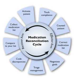
The goal of medication reconciliation is to generate and maintain an accurate and coherent record of patients’ medications across all transitions of care, which sounds straightforward enough. But the process involves much more than just checking items off a list, says Jeffrey Schnipper, MD, MPH, FHM, currently the principal investigator for the $1.5 million study funded by the Agency for Healthcare Research and Quality (AHRQ) to research and implement best practices in med rec, dubbed MARQUIS (Multicenter Medication Reconciliation Quality Improvement Study). Those immersed in med rec know that it’s nonlinear, multilayered, and surprisingly complex, requiring partnerships among diverse providers across many domains of care.
“Medication reconciliation gets right at all the weaknesses of our healthcare system,” says Dr. Schnipper, a hospitalist and director of clinical research for the HM service at Brigham and Women’s Hospital (BWH) and assistant professor of medicine at Harvard Medical School, both in Boston. “We have an excellent healthcare system in so many ways, but what we do not do such a good job of is coordination of care across settings, easy transfer of information, and having one person who is responsible for the accuracy of a patient’s health information.”
Dr. Schnipper’s studies attest to the common occurrence of unintentional medical discrepancies, pointing to the need for accurate medication histories, identifying high-risk patients for intensive interventions, and careful med rec at time of discharge.4
Other factors might come into play, says Ted Tsomides, MD, PhD, an attending physician on the HM service at WakeMed Hospital and assistant professor of medicine at the University of North Carolina’s School of Medicine in Raleigh, N.C. For example, he surmises that a “fatigue factor” sets in for some providers. “After five years of working on any initiative, people get worn out and push it to the back burner, unless they are really incentivized to stay on it,” he says.
List Capture
Medication reconciliation is a multifaceted process, and the first step is to gather the history of medications the patient has been taking. Hospitalist Blake J. Lesselroth, MD, MBI, assistant professor of medicine and medical informatics and director of the Portland Patient Safety Center of Inquiry at the Portland VA Medical Center in Oregon, points out that “the initial exposure to the patient is like a pencil sketch. You start to realize that med rec involves iterative loops of communication between you, the patient, and other knowledge resources (see Figure 1). As you start to pull in more information, you begin to complete your narrative. At the end of hospitalization, you’ve got a vibrant portrait with much more nuance to it. So it can’t be a linear process.”
—Kristine M. Gleason, RPh, clinical quality leader, department of clinical quality and analytics, Northwestern Memorial Hospital, Chicago
The list is dynamic, especially in the ICU setting, says Gleason, where it represents only one point in time.
In a closed system, such as the Veterans Administration or Kaiser Permanente, it’s often easier to establish a patient’s ongoing medications. With an integrated electronic health record (EHR), providers can call up the patient’s list of medications during admittance to the hospital. Verifying those medications remains critical: The health record lists patients’ prescriptions, but that doesn’t always mean they have actually filled or are taking those medications.
At the Kaiser Permanente Southern California site in Santa Clarita, Calif., where hospitalist David W. Wong, MD, works, pharmacists review their medications with patients when they are admitted, provide any needed consultation, then repeat the process at discharge. “So far,” Dr. Wong says, “this has resulted in the best medication reconciliation that we’ve seen.”
Pharmacy Is Key
In 2006, Kenneth Boockvar, MD, of the James J. Peters VA Medical Center in Bronx, N.Y., found in a pre- and post-intervention study that using pharmacists to ferret out and communicate prescribing discrepancies to physicians resulted in lower risk of adverse drug events (ADEs) for patients transferred between the hospital and the nursing home.5 Likewise, Dr. Schnipper and his colleagues found that using pharmacists to conduct medication reviews, counsel patients at discharge, and make follow-up telephone calls to patients was associated with a lower rate of preventable ADEs 30 days after hospital discharge.6
At United Hospital System’s (UHS) Kenosha Medical Center campus in Kenosha, Wis., pharmacists play a key role in generating medication lists for incoming patients. Hospitalist Corey Black, MD, regional medical director for Cogent HMG, says many patients do not recall their medications or the dosages, so UHS utilizes a team approach: If patients come in during evenings or weekends, pharmacists start calling local pharmacies to track down patients’ medication lists. “We also try to have family members bring in any medication containers they can find,” he adds. Due to a Wisconsin state law mandating nursing homes to send medication lists along with patients, generating a list is much easier.
Dr. Tsomides is a physician sponsor of a new med-rec initiative at WakeMed. With a steering committee that includes representatives from stakeholder services (medicine, nursing, pharmacy, administration, etc.), the group plans to hire and train pharmacy techs who will take home medication lists in the ED, lifting that responsibility from physicians’ task lists.
Is IT the Answer?
Would many of the barriers to med rec go away with universal EHR? So far, the literature has not borne out the superiority of using EHR to facilitate better med rec.
Peter Kaboli and colleagues found that the computerized medication record reflected what patients were actually taking for only 5.3% of the 493 VA patients enrolled in a study at the Iowa City VA.7 Kenneth Boockvar and colleagues at the Bronx VA found no difference in the overall incidence of ADEs caused by medication discrepancies between VA patients with an EHR and non-VA patients without an EHR.8 A group of researchers with Partners HealthCare in Boston evaluated a secure, Web-based patient portal to produce more accurate medication lists. The patients using this system had just as many discrepancies between medication lists and self-reporting as those who did not.9
Dr. Lesselroth, who has devised a patient kiosk touch-screen tool for reconciling patients’ medication lists and has faced barriers when implementing said technology, says med rec is much more “organic” than strictly mechanical. “It invokes theories of learning from the cognitive sciences,” he says. “We haven’t actually built tools that help people with their problem representation, with understanding not just how medications reconcile with the prior setting of care, but whether they make clinical sense within the new context of care. That requires a quantum leap in thinking.”
Re-Brand the Message
Drs. Schnipper and Tsomides believe that when The Joint Committee first coined the term “medication reconciliation” and advanced it as a mandate, most providers associated it with a regulatory requirement, and understandably so. Dr. Schnipper says med rec could be improved if providers think about it in the context of accurate orders that translate to greater patient safety. “After all,” he says, “hospitalists are ultimately responsible for the medication orders written for their patients.
“This is not about regulatory requirements,” he continues. “This is about medication safety and transitions of care. You can spend an hour on deciding what dose of Lasix you want to send this patient home on, but if the patient then takes the wrong dose of Lasix because they don’t know what they were supposed to be taking, then all that good medical care is undone.”
The med rec conversation has come full circle, then, as being truly an issue of delivering patient-centered care. (For more on this topic, visit the-hospitalist.org to read “Patient Engagement Critical.”) Rather than focusing on the sometimes-befuddling term of medication reconciliation, providers should see med rec as part of an integrated medication management process that aims to take better care of patients through prevention and treatment, Gleason says.
The med rec issue is about effective communication at every transition of care. And that’s why, says Dr. Schnipper, “Hospitalists should own this process. We don’t have to do the process entirely by ourselves—and shouldn’t. But we are responsible for errors that happen during transitions in care and we should own these initiatives.”
He notes that all six hospitals enrolled in the MARQUIS study have hospitalists at the forefront of their quality-improvement (QI) efforts.
“Medication reconciliation is potentially a high-risk process, and there are no silver bullets” for globally addressing the process, says Dorothea Wild, MD, chief hospitalist at Griffin Hospital, a 160-bed acute care hospital in Derby, Conn.
—Jeffrey Schnipper, MD, MPH, FHM, hospitalist and director of clinical research, Brigham and Women’s Hospital Hospitalist Service, assistant professor of medicine, Harvard Medical School, Boston
Dr. Wild draws a parallel between med rec and blood transfusions. Just as with correct transfusing procedures, “we envision a process where at least two people independently verify what patients’ medications are,” she says. The meds list is started in the ED by nursing staff, is verified by the ED attending, verified again by the admitting team, and triple-checked by the admitting attending. Thus, says Dr. Wild, med rec becomes a shared responsibility.
Dr. Lesselroth wholeheartedly agrees with the approach.
“This is everybody’s job,” he says. “In a larger world view, med rec is all about trying to find a medication regimen that harmonizes with what the patient can do, that improves their probability of adherence, and that also helps us gather information when the patient returns and we re-embrace them in the care model. Theoretically, then, everybody [interfacing with a patient] becomes a clutch player.”
Gretchen Henkel is a freelance writer in California.
Target High-Risk Patients

Pharmacist Kristine M. Gleason, RPh, was the lead author of Northwestern Memorial Hospital’s MATCH (Medications at Transitions and Clinical Handoffs) study, which formed the basis for the recommendations contained in a toolkit generated by the research team (see “Additional Resources,” p. 20).10 Comparing pharmacist and hospitalist medication histories with medication orders, the authors uncovered medication errors for one-third of the study’s 651 patients at admission. The majority of errors (85%) originated in patients’ medication histories. As a result of the study, Gleason explains, the authors recommend that admitting physicians focus on older patients with polypharmacy to reduce medication errors.
Roberta Barber, PharmD, MPH, assistant vice president of pharmacy at Virtua Health in Mount Holly, N.J., oversees pharmacy operations for Virtua’s four hospital campuses. She is involved in med rec/readmission initiatives that focus on such disease states as congestive heart failure that are likely to result in discrepancies. “We can’t have dedicated pharmacists for every unit and every patient, so we have to strategically pick where we can have the most impact.”
References
- Joint Commission on Accreditation of Healthcare Organizations. 2005 Hospital Accreditation Standards. JCO website. Available at: http://www.jointcommissioninternational.org/ JCI-Accredited-Organizations/. Accessed Dec. 7, 2011.
- Bell CM, Brener SS, Gunraj N, et al. Association of ICU or hospital admission with unintentional discontinuation of medications for chronic diseases. JAMA. 2011;306:840-847.
- Greenwald JL, Halasyamani L, Green J, et al. Making inpatient medication patient centered, clinically relevant and implementable: a consensus statement on key principles and necessary first steps. J Hosp Med. 2010;5:477-485.
- Pippins JR, Gandhi TK, Hamann C, et al. Classifying and predicting errors of inpatient medication reconciliation. J Gen Intern Med. 2008;23:1414-1422.
- Boockvar KS, Carlson HL, Giambanco V, et al. Medication reconciliation for reducing drug-discrepancy adverse events. Am J Geriatr Pharmacother. 2006;4:236-243.
- Schnipper JL, Kirwin JL, Cotugno MC, et al. Role of pharmacist counseling in preventing adverse drug events after hospitalization. Arch Intern Med. 2006;166:565-571.
- Kaboli PJ, McClimon JB, Hoth AB, et al. Assessing the accuracy of computerized medication histories. Am J Manag Care. 2004;10(11 Pt 2):872-877.
- Boockvar KS, Livote EE, Goldstein N, et al. Electronic health records and adverse drug events after patient transfer. Qual Saf Health Care. 2010;5:Epub(Aug 19).
- Staroselsky M, Volk LA, Tsurikova R, et al. An effort to improve electronic health record medication list accuracy between visits: patients’ and physicians’ responses. Int J Med Inform. 2008;77:153-160.
- Gleason KM, McDaniel MR, Feinglass J, et al. Results of the Medications at Transitions and Clinical Handoffs (MATCH) study: an analysis of medication reconciliation errors and risk factors at hospital admission. J Gen Intern Med. 2010;25:441-447.
Pharmacist Kristine M. Gleason, RPh, got the chance to personally test her ability to help ED providers with medication reconciliation—known by most in healthcare as “med rec”—when she broke her leg a couple of years ago. No problem, she thought: “I’ve been involved in med-rec efforts for eight-plus years.”
But when asked to provide her current medications, Gleason, who is the clinical quality leader in the department of clinical quality and analytics at Northwestern Memorial Hospital in Chicago, says she was in pain and overwhelmed. “I couldn’t even remember my children’s names, let alone the names and dosages of my aspirin and my thyroid medication,” she says. Moreover, she didn’t carry a list in her wallet because “I’m a pharmacist and I do med rec,” she says.
Gleason’s experience highlights why, six years after The Joint Commission introduced medication reconciliation as National Patient Safety Goal (NPSG) No. 8, hospitals and providers still struggle with the process.1 As a younger patient, Gleason took few medications. But for the majority of elderly inpatients with comorbid conditions, just establishing the patient’s medication list can bring the whole process to a halt; without that foundational list, reconciling other medications becomes problematic.
Although the commission has taken the goals under review and has, since July 1, required compliance with the revised NPSG 03.06.01 (see “Additional Resources,”), hospitalization-associated adverse drug events continue to mount. A recent Canadian study caused a ripple this summer with its findings that patients discharged from acute-care hospitals were at higher risk for unintentional discontinuation of their medications prescribed for chronic diseases than control groups, and those who had an ICU stay are at even higher risk.2
There’s been no shortage of med-rec initiatives in recent years. Medication reconciliation was at the top of the list for ways to prevent errors when the Institute for Healthcare Improvement launched its “5 Million Lives Campaign” in December 2006. SHM weighed in on the issue in 2010 with a consensus statement on key principles and necessary first steps in med rec.3
“This isn’t a new problem,” Gleason says. “Med rec has become more heightened because we have many more medications and complex therapies, more care providers, more specialists—more players, if you will.”
The March launch of the Hospital Readmissions Reduction Program, part of the Centers for Medicaid & Medicare Services’ (CMS) Inpatient Prospective Payment System, will again shine the spotlight on med rec’s role in the prevention of 30-day readmissions. The Hospitalist talked with researchers, pharmacists, and hospitalists about the reasons behind medication discrepancies, and their strategies for addressing mismatches.
Why So Difficult?
The goal of medication reconciliation is to generate and maintain an accurate and coherent record of patients’ medications across all transitions of care, which sounds straightforward enough. But the process involves much more than just checking items off a list, says Jeffrey Schnipper, MD, MPH, FHM, currently the principal investigator for the $1.5 million study funded by the Agency for Healthcare Research and Quality (AHRQ) to research and implement best practices in med rec, dubbed MARQUIS (Multicenter Medication Reconciliation Quality Improvement Study). Those immersed in med rec know that it’s nonlinear, multilayered, and surprisingly complex, requiring partnerships among diverse providers across many domains of care.
“Medication reconciliation gets right at all the weaknesses of our healthcare system,” says Dr. Schnipper, a hospitalist and director of clinical research for the HM service at Brigham and Women’s Hospital (BWH) and assistant professor of medicine at Harvard Medical School, both in Boston. “We have an excellent healthcare system in so many ways, but what we do not do such a good job of is coordination of care across settings, easy transfer of information, and having one person who is responsible for the accuracy of a patient’s health information.”
Dr. Schnipper’s studies attest to the common occurrence of unintentional medical discrepancies, pointing to the need for accurate medication histories, identifying high-risk patients for intensive interventions, and careful med rec at time of discharge.4
Other factors might come into play, says Ted Tsomides, MD, PhD, an attending physician on the HM service at WakeMed Hospital and assistant professor of medicine at the University of North Carolina’s School of Medicine in Raleigh, N.C. For example, he surmises that a “fatigue factor” sets in for some providers. “After five years of working on any initiative, people get worn out and push it to the back burner, unless they are really incentivized to stay on it,” he says.
List Capture
Medication reconciliation is a multifaceted process, and the first step is to gather the history of medications the patient has been taking. Hospitalist Blake J. Lesselroth, MD, MBI, assistant professor of medicine and medical informatics and director of the Portland Patient Safety Center of Inquiry at the Portland VA Medical Center in Oregon, points out that “the initial exposure to the patient is like a pencil sketch. You start to realize that med rec involves iterative loops of communication between you, the patient, and other knowledge resources (see Figure 1). As you start to pull in more information, you begin to complete your narrative. At the end of hospitalization, you’ve got a vibrant portrait with much more nuance to it. So it can’t be a linear process.”
—Kristine M. Gleason, RPh, clinical quality leader, department of clinical quality and analytics, Northwestern Memorial Hospital, Chicago
The list is dynamic, especially in the ICU setting, says Gleason, where it represents only one point in time.
In a closed system, such as the Veterans Administration or Kaiser Permanente, it’s often easier to establish a patient’s ongoing medications. With an integrated electronic health record (EHR), providers can call up the patient’s list of medications during admittance to the hospital. Verifying those medications remains critical: The health record lists patients’ prescriptions, but that doesn’t always mean they have actually filled or are taking those medications.
At the Kaiser Permanente Southern California site in Santa Clarita, Calif., where hospitalist David W. Wong, MD, works, pharmacists review their medications with patients when they are admitted, provide any needed consultation, then repeat the process at discharge. “So far,” Dr. Wong says, “this has resulted in the best medication reconciliation that we’ve seen.”
Pharmacy Is Key
In 2006, Kenneth Boockvar, MD, of the James J. Peters VA Medical Center in Bronx, N.Y., found in a pre- and post-intervention study that using pharmacists to ferret out and communicate prescribing discrepancies to physicians resulted in lower risk of adverse drug events (ADEs) for patients transferred between the hospital and the nursing home.5 Likewise, Dr. Schnipper and his colleagues found that using pharmacists to conduct medication reviews, counsel patients at discharge, and make follow-up telephone calls to patients was associated with a lower rate of preventable ADEs 30 days after hospital discharge.6
At United Hospital System’s (UHS) Kenosha Medical Center campus in Kenosha, Wis., pharmacists play a key role in generating medication lists for incoming patients. Hospitalist Corey Black, MD, regional medical director for Cogent HMG, says many patients do not recall their medications or the dosages, so UHS utilizes a team approach: If patients come in during evenings or weekends, pharmacists start calling local pharmacies to track down patients’ medication lists. “We also try to have family members bring in any medication containers they can find,” he adds. Due to a Wisconsin state law mandating nursing homes to send medication lists along with patients, generating a list is much easier.
Dr. Tsomides is a physician sponsor of a new med-rec initiative at WakeMed. With a steering committee that includes representatives from stakeholder services (medicine, nursing, pharmacy, administration, etc.), the group plans to hire and train pharmacy techs who will take home medication lists in the ED, lifting that responsibility from physicians’ task lists.
Is IT the Answer?
Would many of the barriers to med rec go away with universal EHR? So far, the literature has not borne out the superiority of using EHR to facilitate better med rec.
Peter Kaboli and colleagues found that the computerized medication record reflected what patients were actually taking for only 5.3% of the 493 VA patients enrolled in a study at the Iowa City VA.7 Kenneth Boockvar and colleagues at the Bronx VA found no difference in the overall incidence of ADEs caused by medication discrepancies between VA patients with an EHR and non-VA patients without an EHR.8 A group of researchers with Partners HealthCare in Boston evaluated a secure, Web-based patient portal to produce more accurate medication lists. The patients using this system had just as many discrepancies between medication lists and self-reporting as those who did not.9
Dr. Lesselroth, who has devised a patient kiosk touch-screen tool for reconciling patients’ medication lists and has faced barriers when implementing said technology, says med rec is much more “organic” than strictly mechanical. “It invokes theories of learning from the cognitive sciences,” he says. “We haven’t actually built tools that help people with their problem representation, with understanding not just how medications reconcile with the prior setting of care, but whether they make clinical sense within the new context of care. That requires a quantum leap in thinking.”
Re-Brand the Message
Drs. Schnipper and Tsomides believe that when The Joint Committee first coined the term “medication reconciliation” and advanced it as a mandate, most providers associated it with a regulatory requirement, and understandably so. Dr. Schnipper says med rec could be improved if providers think about it in the context of accurate orders that translate to greater patient safety. “After all,” he says, “hospitalists are ultimately responsible for the medication orders written for their patients.
“This is not about regulatory requirements,” he continues. “This is about medication safety and transitions of care. You can spend an hour on deciding what dose of Lasix you want to send this patient home on, but if the patient then takes the wrong dose of Lasix because they don’t know what they were supposed to be taking, then all that good medical care is undone.”
The med rec conversation has come full circle, then, as being truly an issue of delivering patient-centered care. (For more on this topic, visit the-hospitalist.org to read “Patient Engagement Critical.”) Rather than focusing on the sometimes-befuddling term of medication reconciliation, providers should see med rec as part of an integrated medication management process that aims to take better care of patients through prevention and treatment, Gleason says.
The med rec issue is about effective communication at every transition of care. And that’s why, says Dr. Schnipper, “Hospitalists should own this process. We don’t have to do the process entirely by ourselves—and shouldn’t. But we are responsible for errors that happen during transitions in care and we should own these initiatives.”
He notes that all six hospitals enrolled in the MARQUIS study have hospitalists at the forefront of their quality-improvement (QI) efforts.
“Medication reconciliation is potentially a high-risk process, and there are no silver bullets” for globally addressing the process, says Dorothea Wild, MD, chief hospitalist at Griffin Hospital, a 160-bed acute care hospital in Derby, Conn.
—Jeffrey Schnipper, MD, MPH, FHM, hospitalist and director of clinical research, Brigham and Women’s Hospital Hospitalist Service, assistant professor of medicine, Harvard Medical School, Boston
Dr. Wild draws a parallel between med rec and blood transfusions. Just as with correct transfusing procedures, “we envision a process where at least two people independently verify what patients’ medications are,” she says. The meds list is started in the ED by nursing staff, is verified by the ED attending, verified again by the admitting team, and triple-checked by the admitting attending. Thus, says Dr. Wild, med rec becomes a shared responsibility.
Dr. Lesselroth wholeheartedly agrees with the approach.
“This is everybody’s job,” he says. “In a larger world view, med rec is all about trying to find a medication regimen that harmonizes with what the patient can do, that improves their probability of adherence, and that also helps us gather information when the patient returns and we re-embrace them in the care model. Theoretically, then, everybody [interfacing with a patient] becomes a clutch player.”
Gretchen Henkel is a freelance writer in California.
Target High-Risk Patients
Pharmacist Kristine M. Gleason, RPh, was the lead author of Northwestern Memorial Hospital’s MATCH (Medications at Transitions and Clinical Handoffs) study, which formed the basis for the recommendations contained in a toolkit generated by the research team (see “Additional Resources,” p. 20).10 Comparing pharmacist and hospitalist medication histories with medication orders, the authors uncovered medication errors for one-third of the study’s 651 patients at admission. The majority of errors (85%) originated in patients’ medication histories. As a result of the study, Gleason explains, the authors recommend that admitting physicians focus on older patients with polypharmacy to reduce medication errors.
Roberta Barber, PharmD, MPH, assistant vice president of pharmacy at Virtua Health in Mount Holly, N.J., oversees pharmacy operations for Virtua’s four hospital campuses. She is involved in med rec/readmission initiatives that focus on such disease states as congestive heart failure that are likely to result in discrepancies. “We can’t have dedicated pharmacists for every unit and every patient, so we have to strategically pick where we can have the most impact.”
References
- Joint Commission on Accreditation of Healthcare Organizations. 2005 Hospital Accreditation Standards. JCO website. Available at: http://www.jointcommissioninternational.org/ JCI-Accredited-Organizations/. Accessed Dec. 7, 2011.
- Bell CM, Brener SS, Gunraj N, et al. Association of ICU or hospital admission with unintentional discontinuation of medications for chronic diseases. JAMA. 2011;306:840-847.
- Greenwald JL, Halasyamani L, Green J, et al. Making inpatient medication patient centered, clinically relevant and implementable: a consensus statement on key principles and necessary first steps. J Hosp Med. 2010;5:477-485.
- Pippins JR, Gandhi TK, Hamann C, et al. Classifying and predicting errors of inpatient medication reconciliation. J Gen Intern Med. 2008;23:1414-1422.
- Boockvar KS, Carlson HL, Giambanco V, et al. Medication reconciliation for reducing drug-discrepancy adverse events. Am J Geriatr Pharmacother. 2006;4:236-243.
- Schnipper JL, Kirwin JL, Cotugno MC, et al. Role of pharmacist counseling in preventing adverse drug events after hospitalization. Arch Intern Med. 2006;166:565-571.
- Kaboli PJ, McClimon JB, Hoth AB, et al. Assessing the accuracy of computerized medication histories. Am J Manag Care. 2004;10(11 Pt 2):872-877.
- Boockvar KS, Livote EE, Goldstein N, et al. Electronic health records and adverse drug events after patient transfer. Qual Saf Health Care. 2010;5:Epub(Aug 19).
- Staroselsky M, Volk LA, Tsurikova R, et al. An effort to improve electronic health record medication list accuracy between visits: patients’ and physicians’ responses. Int J Med Inform. 2008;77:153-160.
- Gleason KM, McDaniel MR, Feinglass J, et al. Results of the Medications at Transitions and Clinical Handoffs (MATCH) study: an analysis of medication reconciliation errors and risk factors at hospital admission. J Gen Intern Med. 2010;25:441-447.