User login
Impact of Fellowship Programs on Residents' Case Logs Examined
BOCA RATON, FLA. – Coexisting subspecialty fellowship programs have at most only minimal adverse impact on general surgery residency training operative volumes, according to a national study sponsored by the American Board of Surgery.
The analysis also demonstrated that fellowship-bound general surgery residents tend to select additional cases in their chosen future subspecialty, thereby in effect creating a self-directed early tracking program, Dr. John B. Hanks said at the annual meeting of the American Surgical Association.
The American Board of Surgery (ABS) conducted the study in response to concerns that because 80% of general surgery residents pursue fellowship training, an insufficient number of cases could be available for resident training. But the national data indicate that this is not a problem, according to Dr. Hanks, professor and chief of general surgery at the University of Virginia, Charlottesville.
The study entailed detailed analysis of the operative logs of 976 applicants to the 2009 ABS qualifying exam. The applicants came from 246 general surgery residency programs. In all, 97 of these residency programs coexisted with a vascular surgery fellowship program, 35 with a colorectal surgery fellowship program, 80 with a minimally invasive surgery training program, and 12 with an endocrine surgery fellowship program. The investigators scrutinized case volumes for predefined key operations in the areas of vascular, colorectal, endocrine, and minimally invasive surgery (MIS).
Residents bound for fellowships in vascular, colorectal, and endocrine surgery performed significantly more total cases in each of those areas than did general surgery residents who were not bound for fellowships. For example, operative logs for the 90 vascular surgery fellowship–bound residents showed a mean experience of 165 vascular cases, compared with 123 cases for the other general surgery residents. And residents headed for colorectal surgery fellowships had a mean of 204 colorectal surgery cases, compared with 163 for all other general surgery residents.
In contrast, residents who were headed for an MIS fellowship and those who were not averaged a similar number of minimally invasive operations.
With regard to the effect of coexisting fellowship programs on general surgery residents’ operative experience in those specific areas, there was a negative impact only for MIS. Residents in general surgery training programs with a coexisting MIS fellowship averaged about 10% fewer MIS cases than did residents in institutions without a fellowship.
"That difference reaches statistical significance, although the practical significance of this effect may be open for debate," Dr. Hanks observed.
The situation was different for residents at institutions with coexisting colorectal or vascular surgery fellowships; those fellowships had no impact on general surgery residents’ case volumes in those specialty-specific areas. And the presence of a coexisting endocrine surgery fellowship was actually associated with a significant increase in endocrine surgery case volumes for all residents, he continued.
Discussant Dr. Layton F. Rikkers cautioned that the ABS study describes national trends using broad strokes, and the data don’t necessarily apply to any individual residency program.
"As an example, the only fellowship we developed at the University of Wisconsin, Madison, during the time I was chairman there was a vascular surgery fellowship," said Dr. Rikkers, professor of surgery at the university and past president of the ABS. "The year prior to developing the fellowship, our residents were in the 93rd percentile nationally with respect to vascular surgery cases done. Over the many years since that fellowship was established, our residents are in the 10th to 20th percentile for vascular surgery cases."
Dr. Hanks declared having no financial conflict of interest.
The significance of this is hard to determine, precisely because of the reason stated by Dr. Rikkers – impact on individual programs cannot be assessed, and likely varies greatly from program to program. For example, in some instances the additional specialty cases may be uncovered by residents, and in that situation no impact would be seen. In others, the cases might be directly "poached" from the general surgery residencies. A final scenario might be one in which an initial downturn is seen in general surgery cases, which is overcome over a period of years as the presence of a fellowship often drives up case volumes at an institution.
Another confounding issue generated by the lack of individual institutional information is that the case volumes in certain specialties may be higher in institutions that attract more residents to a specific field – prominence of a specialty in a given institution may impact resident choice of specialty. Indeed, rather than residents choosing to do more cases in their chosen field, they may in fact have chosen their field based on greater exposure to the specialty. This cannot be parsed out from the data at hand.
No mention is made of the involvement of the residents in the pre-operative and post-operative care of the patients when participation is outside of usual service assignments. If this is not robust, benefits to patients and trainees may be lessened.
Dr. Cynthia K. Shortell is a professor of surgery and chief of vascular surgery and program director, vascular residency at Duke University Medical Center, Durham, N.C., and an associate medical editor for Vascular Specialist.
BOCA RATON, FLA. – Coexisting subspecialty fellowship programs have at most only minimal adverse impact on general surgery residency training operative volumes, according to a national study sponsored by the American Board of Surgery.
The analysis also demonstrated that fellowship-bound general surgery residents tend to select additional cases in their chosen future subspecialty, thereby in effect creating a self-directed early tracking program, Dr. John B. Hanks said at the annual meeting of the American Surgical Association.
The American Board of Surgery (ABS) conducted the study in response to concerns that because 80% of general surgery residents pursue fellowship training, an insufficient number of cases could be available for resident training. But the national data indicate that this is not a problem, according to Dr. Hanks, professor and chief of general surgery at the University of Virginia, Charlottesville.
The study entailed detailed analysis of the operative logs of 976 applicants to the 2009 ABS qualifying exam. The applicants came from 246 general surgery residency programs. In all, 97 of these residency programs coexisted with a vascular surgery fellowship program, 35 with a colorectal surgery fellowship program, 80 with a minimally invasive surgery training program, and 12 with an endocrine surgery fellowship program. The investigators scrutinized case volumes for predefined key operations in the areas of vascular, colorectal, endocrine, and minimally invasive surgery (MIS).
Residents bound for fellowships in vascular, colorectal, and endocrine surgery performed significantly more total cases in each of those areas than did general surgery residents who were not bound for fellowships. For example, operative logs for the 90 vascular surgery fellowship–bound residents showed a mean experience of 165 vascular cases, compared with 123 cases for the other general surgery residents. And residents headed for colorectal surgery fellowships had a mean of 204 colorectal surgery cases, compared with 163 for all other general surgery residents.
In contrast, residents who were headed for an MIS fellowship and those who were not averaged a similar number of minimally invasive operations.
With regard to the effect of coexisting fellowship programs on general surgery residents’ operative experience in those specific areas, there was a negative impact only for MIS. Residents in general surgery training programs with a coexisting MIS fellowship averaged about 10% fewer MIS cases than did residents in institutions without a fellowship.
"That difference reaches statistical significance, although the practical significance of this effect may be open for debate," Dr. Hanks observed.
The situation was different for residents at institutions with coexisting colorectal or vascular surgery fellowships; those fellowships had no impact on general surgery residents’ case volumes in those specialty-specific areas. And the presence of a coexisting endocrine surgery fellowship was actually associated with a significant increase in endocrine surgery case volumes for all residents, he continued.
Discussant Dr. Layton F. Rikkers cautioned that the ABS study describes national trends using broad strokes, and the data don’t necessarily apply to any individual residency program.
"As an example, the only fellowship we developed at the University of Wisconsin, Madison, during the time I was chairman there was a vascular surgery fellowship," said Dr. Rikkers, professor of surgery at the university and past president of the ABS. "The year prior to developing the fellowship, our residents were in the 93rd percentile nationally with respect to vascular surgery cases done. Over the many years since that fellowship was established, our residents are in the 10th to 20th percentile for vascular surgery cases."
Dr. Hanks declared having no financial conflict of interest.
New Options to Treat Hepatitis C
Enter text here
Enter text here
Enter text here
New Options to Treat Hepatitis C
Two new drug therapies to treat chronic hepatitis C (HCV) are the first new options for hospitalists in 20 years. But freedom of choice doesn't come cheap.
Telaprevir, from Vertex Pharmaceuticals, will cost $49,200 for a 12-week regimen, while boceprevir, from Merck & Co., will cost from $26,400 to $48,000 for regimens of 24 and 48 weeks, respectively, Reuters reports. Telaprevir will go by the brand name Incivek, while boceprevir is known as Victrelis.
"The price is expensive," but financial support for some patients is expected early on, says Satya Chelamkuri, MD, a hospitalist with Cogent Healthcare at Allegiance Health in Jackson, Mich. "When the generic comes out, it will be a great help to patients with hepatitis C. Cost is a worry, but with help from pharmaceutical companies, the majority of the patients who need it will hopefully get the treatment."
Still, Dr. Chelamkuri expects the drugs to appear relatively quickly on hospital formularies, in large part because of the efficacy they showed in trials. The standard treatment for years, a cocktail of peginterferon-alpha and ribavirin, has a roughly 50% response rate. Both of the new treatments, which have been formally approved by the FDA in the past six weeks, boost the cure rates to a range of 66% to 80% by working in tandem with the current therapy.
"A less than 50% [response rate] for the previous regime and around 70% for the new regime. That’s a considerable difference," Dr. Chelamkuri says.
In the Literature: Research You Need to Know
Clinical question: Does the mortality rate differ between patients treated perioperatively with atenolol versus metoprolol?
Background: Perioperative beta-blockers have been shown to reduce mortality in patients with significant cardiac risk factors. Different beta-blockers have been used in the studies demonstrating benefit, and it is uncertain which beta-blocker is the preferred agent. The purpose of this study was to compare the perioperative benefits of metoprolol versus atenolol.
Study design: Retrospective cohort study.
Setting: San Francisco Veterans Affairs Medical Center.
Synopsis: Computerized records of patients who underwent surgery from 1996 to 2008 were extracted into a database, and patients who received inpatient beta-blockers after surgery were included. Of these patients, 3,787 received beta-blockade exclusively with either atenolol (n=1,011) or metoprolol (n=2,776) during hospitalization. Perioperative risk reduction was better with atenolol versus metoprolol. Mortality rates were 1% versus 3% at 30 days (P=0.0008), and 7% versus 13% at one year (P=<0.0001) with atenolol and metoprolol, respectively. Similar results were found in the analysis of 1,871 patients who were on their respective beta-blocker as an outpatient before surgery.
Because the metoprolol group had higher prevalence of risk factors, such as coronary artery disease, peripheral vascular disease, and congestive heart failure, a propensity-matched analysis was performed to remove differences in risk factors between the groups. After propensity matching, the metoprolol group still had statistically significant higher mortality at 30 days and one year, even though causality cannot be established in this retrospective study.
Bottom line: Atenolol was associated with fewer cases of perioperative mortality compared with metoprolol in patients with cardiac risk factors or established cardiovascular disease.
Citation: Wallace AW, Au S, Cason BA. Perioperative ß-blockade: atenolol is associated with reduced mortality when compared to metoprolol. Anesthesiology. 2011;114:824-836.
For more physician reviews of HM-related research, visit our website.
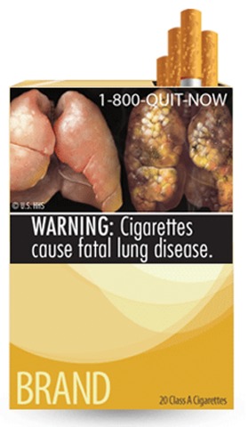
FDA Unveils Graphic Cigarette Packaging Intended to Deter Smoking
The Food and Drug Administration unveiled on June 21 the final nine warning images that will appear on every package of cigarettes by 2012 – graphic photos and drawings intended to educate and even deter consumers from buying cigarettes.
The images, set to debut in stores this September, are required by the 2009 Tobacco Control Act, according to FDA spokesman Jeffrey Ventura, who added that these are the first changes to cigarette pack warnings in 25 years. By Oct. 22, 2012, cigarette manufacturers will no longer be able to distribute cigarettes for sale in the United States unless they display these warnings.

The law required the warnings to cover the top half of the front and back of cigarette packs and 20% of cigarette advertisements, and they must contain color graphics depicting the negative health consequences of smoking.
"This is something Congress wanted to happen and mandated that the FDA carry out," Mr. Ventura said in an interview. Based on a study of 18,000 smokers conducted for the FDA by RTI International, federal officials said they firmly believe that visually communicating smoking’s harm will deter cigarette consumption over the long run.
The images include photos of tobacco-diseased lungs beside healthy lungs, a corpse in casket, a man exhaling smoke though a tracheostomy, and lip cancer. There are also several cartoons and photos of mothers blowing smoke into infants’ faces. One positive image shows a burly man exposing a T-shirt saying, "I Quit."
Blunt statements accompany each image, intended to drive home the messages that cigarette smoke not only directly harms the smoker, but the smokers’ children and people in close proximity.
"The introduction of these warnings is expected to have a significant public health impact by decreasing the number of smokers, resulting in lives saved, increased life expectancy, and improved health status," FDA officials said in a press statement.
Mr. Ventura said the images were selected after the consumer study involving smokers aged 15-50 years. After viewing each of the images, subjects rated their emotional and cognitive responses, their ability to recall the images, and their opinions on whether the pictures could alter their beliefs about the danger of smoking and the desire to buy tobacco products and quitting tobacco.
Young people responded most strongly to a cartoon image depicting tobacco addiction – a cigarette being injected into an arm vein as well as a puppet controlled by strings.
Adults, on the other hand, responded most strongly to photos showing the direct effects of cancer on their bodies – the man with the tracheostomy and a woman smoking in the pouring rain, trying to shield her cigarette with a folded newspaper. Adults also reacted more strongly than did young people to images depicting harm to young children.
The study did conclude, however, that none of the images were significantly related to an increased likelihood of quitting smoking within the next 30 days, or the likelihood of smoking a year after viewing the images. Thus, the report noted, the campaign is more likely to exert a long-term behavioral impact than any immediate effects.
"Eliciting strong emotional and cognitive reactions to the graphic cigarette warning label enhances recall and processing of the health warning, which helps ensure that the warning is better processed, understood, and remembered," the study said. "As attitudes and beliefs change, they eventually lead to changes in intentions to quit or start smoking and then later to lower smoking initiation and successful cessation. The time scale on which this behavior change process occurs is largely unknown in the context of the impact of exposure to graphic warning labels on smoking behaviors, but the effects on behavior change are unlikely to be immediate or short-term.
Nevertheless, groups promoting antitobacco messages – including the American Heart Association – strongly believe that the warnings will enhance consumer education and change behavior.
"Undoubtedly, the new graphic health warnings will heighten awareness about the dangers of smoking and, more importantly, encourage smokers to quit and discourage smoking initiation," an AHA press statement read. "We’re confident that the new labels will move us closer to our goal of making the nation 100% smoke free."
Tobacco-Free Kids, a group dedicated to educating children and teens about the dangers of smoking, also issued a statement of support, but with a moderated view on the campaign’s possible impact. The group also called on political leaders to financially commit to "waging war" against tobacco.
"The warnings and other FDA regulations are powerful tools, but they are a complement – not a replacement – to other federal and state strategies to reduce tobacco use," Matthew L. Meyers, the group’s president, said in the statement. "To win the fight against tobacco, elected leaders must also fund and implement public education campaigns, expand health care coverage for therapies to help smokers quit, increase tobacco taxes, and enact strong smoke-free laws in every state."
I give the FDA a
“B” on this action, but I don’t think it’s enough. Congress could have decided on
much stronger pictures. It could have made them larger, not just 20% of the
pack and located on the back . In other countries that use such warnings, the
pictures cover almost 80% of the packaging.
Plainly put, the United States is a Third
World country when it comes to warning about cigarettes. Similar
pictorial warnings have been used in Canada
for almost a decade; Australia,
almost all of the European Union countries, and even Uruguay have similar warnings.
Having said that, we do know
that pictures speak louder than words and may help motivate smokers to quit.
But here is the hard reality: Even though nearly everyone wants to quit smoking
and many are trying to do so every day, the ability to sustain that motivation
is not always present.
Two factors are highly
predictive of a successful quit: the clinical predictor of personal biology and
the financial predictor of cigarette engineering.
Biologically, some people
are more prone to developing addictions than others. If you have a patient who
reaches for a cigarette before their feet even hit the floor in the morning,
that patient is going to need more medical assistance to be successful.
The second factor is
cigarette engineering. Cigarettes are designed to get people hooked and keep
them hooked. This is what makes quitting so hard despite strong personal motivation.
If we look back in
history, around 1900 there were only a few hundred cases of lung cancer
diagnosed in the U.S.
each year. This year it will be around 160,000. What has happened since then?
People were indeed smoking
then – pipes, cigars, and roll-your-own cigarettes. But this was not a daily-use
situation, mostly because the pH of the smoke made it very hard to inhale.
Right around 1920, commercially available cigarette brands reformulated their
tobacco to make the smoke milder. This allowed people to smoke more, loading
their brains up with lots more nicotine, lots faster.
Around 1950, manufacturers
began adding chemicals to moderate the pH, facilitating deep inhalation. the
lungs. These ingredients – diammonium phosphate, urea, and hydrochloride among
them – are still on the packs’ ingredient list, although manufacturers call
them “flavorings.”
Filters were the next step
in developing an addictive product. Manufacturers presented filters as making
cigarettes safer. Their real purpose was both to break down the tar into
smaller particles and to force people to suck on the cigarette harder to get
the smoke. Filters actually make things worse by forcing rapid inhalation and
rapid absorption – delivering the nicotine to the brain within seconds.
These things were done
secretly and purposefully to promote tobacco addiction and maintain profits. Tobacco
companies deliberately suppressed this information until 2006, when a
racketeering lawsuit forced the disclosure of dozens of documents showing the
intention behind these decisions. U.S District Court Judge Gladys Kessler determined
that tobacco companies conspired to lie about the dangers of smoking and, in
her 1,600-page ruling, called the conspiracy “decades long.”
“In short, the defendants
have marketed and sold their lethal product with zeal, with deception, with a
single-minded focus on their financial success, and without regard for the
human tragedy or social costs that success exacted,” according to her ruling.
Despite lawsuits, public
information campaigns, and the current warnings on cigarette packs, Americans
continue to smoke and I continue to see the effects.
About a third of the
patients I see at the Roswell Park Cancer Institute have smoking-related
cancers. Imagine a vaccine that would prevent a third of cancers. The inventor would
be a Nobel Laureate, globally praised.
We already have this – but
it’s not a vaccine. It’s much simpler.
Just stop smoking.
K. Michael Cummings,
Ph.D, is the director of the New York State Smokers’ Quitline and a senior research
scientist at the Roswell Park cancer Institute, Buffalo.
The images, set to debut in stores this September, are required by the 2009 Tobacco Control Act, according to FDA spokesman Jeffrey Ventura, who added that these are the first changes to cigarette pack warnings
I give the FDA a
“B” on this action, but I don’t think it’s enough. Congress could have decided on
much stronger pictures. It could have made them larger, not just 20% of the
pack and located on the back . In other countries that use such warnings, the
pictures cover almost 80% of the packaging.
Plainly put, the United States is a Third
World country when it comes to warning about cigarettes. Similar
pictorial warnings have been used in Canada
for almost a decade; Australia,
almost all of the European Union countries, and even Uruguay have similar warnings.
Having said that, we do know
that pictures speak louder than words and may help motivate smokers to quit.
But here is the hard reality: Even though nearly everyone wants to quit smoking
and many are trying to do so every day, the ability to sustain that motivation
is not always present.
Two factors are highly
predictive of a successful quit: the clinical predictor of personal biology and
the financial predictor of cigarette engineering.
Biologically, some people
are more prone to developing addictions than others. If you have a patient who
reaches for a cigarette before their feet even hit the floor in the morning,
that patient is going to need more medical assistance to be successful.
The second factor is
cigarette engineering. Cigarettes are designed to get people hooked and keep
them hooked. This is what makes quitting so hard despite strong personal motivation.
If we look back in
history, around 1900 there were only a few hundred cases of lung cancer
diagnosed in the U.S.
each year. This year it will be around 160,000. What has happened since then?
People were indeed smoking
then – pipes, cigars, and roll-your-own cigarettes. But this was not a daily-use
situation, mostly because the pH of the smoke made it very hard to inhale.
Right around 1920, commercially available cigarette brands reformulated their
tobacco to make the smoke milder. This allowed people to smoke more, loading
their brains up with lots more nicotine, lots faster.
Around 1950, manufacturers
began adding chemicals to moderate the pH, facilitating deep inhalation. the
lungs. These ingredients – diammonium phosphate, urea, and hydrochloride among
them – are still on the packs’ ingredient list, although manufacturers call
them “flavorings.”
Filters were the next step
in developing an addictive product. Manufacturers presented filters as making
cigarettes safer. Their real purpose was both to break down the tar into
smaller particles and to force people to suck on the cigarette harder to get
the smoke. Filters actually make things worse by forcing rapid inhalation and
rapid absorption – delivering the nicotine to the brain within seconds.
These things were done
secretly and purposefully to promote tobacco addiction and maintain profits. Tobacco
companies deliberately suppressed this information until 2006, when a
racketeering lawsuit forced the disclosure of dozens of documents showing the
intention behind these decisions. U.S District Court Judge Gladys Kessler determined
that tobacco companies conspired to lie about the dangers of smoking and, in
her 1,600-page ruling, called the conspiracy “decades long.”
“In short, the defendants
have marketed and sold their lethal product with zeal, with deception, with a
single-minded focus on their financial success, and without regard for the
human tragedy or social costs that success exacted,” according to her ruling.
Despite lawsuits, public
information campaigns, and the current warnings on cigarette packs, Americans
continue to smoke and I continue to see the effects.
About a third of the
patients I see at the Roswell Park Cancer Institute have smoking-related
cancers. Imagine a vaccine that would prevent a third of cancers. The inventor would
be a Nobel Laureate, globally praised.
We already have this – but
it’s not a vaccine. It’s much simpler.
Just stop smoking.
K. Michael Cummings,
Ph.D, is the director of the New York State Smokers’ Quitline and a senior research
scientist at the Roswell Park cancer Institute, Buffalo.
The Food and Drug Administration unveiled on June 21 the final nine warning images that will appear on every package of cigarettes by 2012 – graphic photos and drawings intended to educate and even deter consumers from buying cigarettes.
The images, set to debut in stores this September, are required by the 2009 Tobacco Control Act, according to FDA spokesman Jeffrey Ventura, who added that these are the first changes to cigarette pack warnings in 25 years. By Oct. 22, 2012, cigarette manufacturers will no longer be able to distribute cigarettes for sale in the United States unless they display these warnings.
The law required the warnings to cover the top half of the front and back of cigarette packs and 20% of cigarette advertisements, and they must contain color graphics depicting the negative health consequences of smoking.
"This is something Congress wanted to happen and mandated that the FDA carry out," Mr. Ventura said in an interview. Based on a study of 18,000 smokers conducted for the FDA by RTI International, federal officials said they firmly believe that visually communicating smoking’s harm will deter cigarette consumption over the long run.
The images include photos of tobacco-diseased lungs beside healthy lungs, a corpse in casket, a man exhaling smoke though a tracheostomy, and lip cancer. There are also several cartoons and photos of mothers blowing smoke into infants’ faces. One positive image shows a burly man exposing a T-shirt saying, "I Quit."
Blunt statements accompany each image, intended to drive home the messages that cigarette smoke not only directly harms the smoker, but the smokers’ children and people in close proximity.
"The introduction of these warnings is expected to have a significant public health impact by decreasing the number of smokers, resulting in lives saved, increased life expectancy, and improved health status," FDA officials said in a press statement.
Mr. Ventura said the images were selected after the consumer study involving smokers aged 15-50 years. After viewing each of the images, subjects rated their emotional and cognitive responses, their ability to recall the images, and their opinions on whether the pictures could alter their beliefs about the danger of smoking and the desire to buy tobacco products and quitting tobacco.
Young people responded most strongly to a cartoon image depicting tobacco addiction – a cigarette being injected into an arm vein as well as a puppet controlled by strings.
Adults, on the other hand, responded most strongly to photos showing the direct effects of cancer on their bodies – the man with the tracheostomy and a woman smoking in the pouring rain, trying to shield her cigarette with a folded newspaper. Adults also reacted more strongly than did young people to images depicting harm to young children.
The study did conclude, however, that none of the images were significantly related to an increased likelihood of quitting smoking within the next 30 days, or the likelihood of smoking a year after viewing the images. Thus, the report noted, the campaign is more likely to exert a long-term behavioral impact than any immediate effects.
"Eliciting strong emotional and cognitive reactions to the graphic cigarette warning label enhances recall and processing of the health warning, which helps ensure that the warning is better processed, understood, and remembered," the study said. "As attitudes and beliefs change, they eventually lead to changes in intentions to quit or start smoking and then later to lower smoking initiation and successful cessation. The time scale on which this behavior change process occurs is largely unknown in the context of the impact of exposure to graphic warning labels on smoking behaviors, but the effects on behavior change are unlikely to be immediate or short-term.
Nevertheless, groups promoting antitobacco messages – including the American Heart Association – strongly believe that the warnings will enhance consumer education and change behavior.
"Undoubtedly, the new graphic health warnings will heighten awareness about the dangers of smoking and, more importantly, encourage smokers to quit and discourage smoking initiation," an AHA press statement read. "We’re confident that the new labels will move us closer to our goal of making the nation 100% smoke free."
Tobacco-Free Kids, a group dedicated to educating children and teens about the dangers of smoking, also issued a statement of support, but with a moderated view on the campaign’s possible impact. The group also called on political leaders to financially commit to "waging war" against tobacco.
"The warnings and other FDA regulations are powerful tools, but they are a complement – not a replacement – to other federal and state strategies to reduce tobacco use," Matthew L. Meyers, the group’s president, said in the statement. "To win the fight against tobacco, elected leaders must also fund and implement public education campaigns, expand health care coverage for therapies to help smokers quit, increase tobacco taxes, and enact strong smoke-free laws in every state."
The images, set to debut in stores this September, are required by the 2009 Tobacco Control Act, according to FDA spokesman Jeffrey Ventura, who added that these are the first changes to cigarette pack warnings
The images, set to debut in stores this September, are required by the 2009 Tobacco Control Act, according to FDA spokesman Jeffrey Ventura, who added that these are the first changes to cigarette pack warnings
Studies Mixed on Second Cancers After Lenalidomide
CHICAGO – That lenalidomide can improve overall and progression-free survival rates in multiple myeloma patients is evident, but whether the drug also increases their risk of second primary cancers is debatable.
Of three studies looking at the question, only one found an association between secondary primary malignancies and lenalidomide (Revlimid) in first-line therapy, investigators reported at the American Society of Clinical Oncology annual meeting.
And even then the risk was low – far less than the risk of death from multiple myeloma without a lenalidomide-containing regimen, said Dr. Antonio P. Palumbo of the University of Torino (Italy) and the Italian Multiple Myeloma Study Group.
Two other studies – one with 6-year follow-up data on continuous lenalidomide in first-line therapy and the other on lenalidomide in relapsed/refractory disease – failed to spot a signal for second cancer risk.
"I think it’s fair to say that currently we lack clear answers due to small numbers and study limitations," said Dr. C. Ola Landgren of the National Cancer Institute, the invited discussant for all three papers.
"I think we need to put both benefits and risks into the algorithms when we think about these things. ... I also think despite the fact that we don’t have clear data, we always have to discuss these things with our patients, and we as doctors have to stay updated as more information emerges," he said.
Three randomized clinical trials stirred the debate by reporting in separate presentations at the 2010 annual meeting of the American Society of Hematology that they saw more hematologic malignancies in lenalidomide treatment arms than in control groups (McCarthy, P.L. et al, abstract 37; Attal, M. et al, abstract 310; Palumbo, A. et al, abstract 622).
Despite these reports, 25 years after a multiple myeloma diagnosis, the cumulative incidence of all second cancers is about 8%, whereas the cumulative probability of death from competing causes is more than 90%, suggesting that any risk of a second malignancy is far outweighed by the risk of multiple myeloma and its sequelae, Dr. Landgren pointed out.
First-Line Therapy: Italian Experience
In the first of the three studies presented at ASCO 2011, Dr. Palumbo’s group looked at second-cancer rates among patients randomly assigned to first-line therapy with either melphalan and prednisone alone, or to melphalan, prednisone, and lenalidomide with or without lenalidomide maintenance in the international MM-015 trial.
They found that at a median follow-up of 30 months, 12 of 150 (8%) patients on melphalan-prednisone plus lenalidomide with maintenance (MPR-R) developed an invasive second primary malignancy, compared with 9 of 152 patients (5.9%) on the same combination without lenalidomide maintenance (MPR), and 4 of 153 (2.6%) patients on melphalan and prednisone only.
Hematologic malignancies accounted for 7 of the 12 new cancers among patients treated with MPR-R, 5 of 9 on MPR, and 1 of 4 on MP. Solid tumors accounted for the remaining invasive cancers in each group. In addition, one patient on MPR-R, four on MPR, and five on MP developed nonmelanoma skin cancers.
In an additional analysis of 9 pooled experimental studies, the investigators found that among 1,788 patients followed for more than 1 year, the risk of dying of myeloma was greater than 40% out to 7 years compared with about a 2% risk of developing a second hematologic malignancy, and a 3% risk of developing a solid tumor.
Among patients receiving lenalidomide and an alkylating agent, the risk of developing any malignancy was around 7%, and the risk of dying of myeloma was about 27%. The risk of a second malignancy was lower – about 2% out to 6 years– among those patients who did not receive lenalidomide, but their risk of dying of myeloma was about 45%, Dr. Palumbo said.
He also pointed out that in the general population, the risk of a second primary malignancy among 65- to 74-year-olds is around 2% per 100 patient-years, and that the risk doubles among people 85 and older.
First-Line Therapy: BiRD Regimen
In the second study, Dr. Adriana Rossi and her colleagues at Cornell University, New York, and New York–Presbyterian Hospital examined the incidence of second primary cancers in 68 transplant-eligible patients receiving lenalidomide in first-line therapy as part of the BiRD regimen (clarithromycin [Biaxin], lenalidomide, and dexamethasone).
There were five solid tumors (two colon, one metastatic melanoma, one pancreas, and one prostate), but no hematologic malignancies. The melanoma was diagnosed 8 months after the primary myeloma diagnosis; the other cases occurred 25-53 months after the initial myeloma diagnosis (median, 31.2 months). The authors found no association between second primary cancers and a specific multiple myeloma chromosomal abnormality, prior malignancy, transplant status, study status, or sex.
The incidence rate of second primary malignancy was similar to that of all primary cancers reported among people 65 and older in the U.S. Surveillance, Epidemiology, and End Results (SEER) data set spanning 2003-2007, they noted.
"Routine screening and prevention measures should continue as medically indicated for all patients, including examination for skin cancers, and as survival in patients with multiple myeloma continues to improve, so will our understanding of their risk of development of second primary malignancies," she said.
Relapsed/Refractory Disease
The third trial looked at the risk of new primary cancers in patients receiving lenalidomide and dexamethasone for relapsed/refractory disease.
Dr. Meletios Dimopoulos of the University of Athens and colleagues in the international MM-009/010 trials performed a pooled analysis comparing the incidence of second primary cancers in 704 patients who received dexamethasone with either lenalidomide or placebo, and compared them with standard incidence rates.
They found that there were no differences in incidence rates of invasive second primary malignancies between patients in the lenalidomide plus dexamethasone arm or dexamethasone-only arms, and that the incidence rates of second primary cancers in general were low and similar to the background rate among people of similar age in the general population.
Additionally, patients who received lenalidomide and dexamethasone had significantly better overall survival (median, 38 months) despite the fact that about half of all patients in the placebo/dexamathesaone arm (median, 31.6 months) were crossed over to lenalidomide-based therapy (P = .045).
"The overall benefit-risk ratio of the use of lenalidomide in the relapsed/refractory setting remains strongly positive," said the presenter of the abstract, Dr. Ruben Niesvizky of Cornell University.
Lenalidomide is not approved as first-line therapy in the United States. Dr. Palumbo’s study was supported by the Fondazione Neoplasie Sangue Onlus. Dr. Rossi’s and Dr. Dimopoulos’s studies were funded by Celgene. Dr. Palumbo has received honoraria and served as a consultant to Celgene and other companies. Dr. Rossi said she had no relevant financial relationships to disclose. Dr. Dimopoulos disclosed receiving honoraria from Celgene. Dr. Niesvizky said he had received honoraria and research funding from Celgene; he also served in a consulting/advisory role for Celgene and other companies.
FROM THE ANNUAL MEETING OF THE AMERICAN SOCIETY OF CLINICAL ONCOLOGY
Major Finding: At a median follow-up of 30 months, 12 of 150 (8%) patients on melphalan-prednisone plus lenalidomide with maintenance developed an invasive second primary malignancy, compared with 9 of 152 patients (5.9%) on the same combination without lenalidomide maintenance, and 4 of 153 (2.6%) patients on melphalan and prednisone only. No increase was seen in analyses of two other trials.
Data Source: Analyses of three randomized clinical trials in which patients received lenalidomide for multiple myeloma.
Disclosures Lenalidomide is not approved as first-line therapy in the United States. Dr. Palumbo’s study was supported by the Fondazione Neoplasie Sangue Onlus. Dr. Rossi’s and Dr. Dimopoulos’s studies were funded by Celgene. Dr. Palumbo has received honoraria and served as a consultant to Celgene and other companies. Dr. Rossi said she had no relevant financial relationships to disclose. Dr. Dimopoulos disclosed receiving honoraria from Celgene. Dr. Niesvizky said he had received honoraria and research funding from Celgene; he also served in a consulting/advisory role with Celgene and other companies.
Rituximab Maintenance Called "New Standard" for Mantle Cell Lymphoma
LONDON – Rituximab more than doubled the duration of remission in elderly patients with mantle cell lymphoma when used as maintenance therapy in those who had already responded to induction therapy in a large, randomized controlled trial.
First results from the European Mantle Cell Lymphoma (MCL) Network study show that rituximab maintenance is associated with a median remission of 77 months. In comparison, when interferon (IFN) was used as maintenance, the median was 24 months (hazard ratio, 0.54; P = .0109).
Overall survival data are not yet fully mature but suggest that rituximab (Rituxan in the United States, Mabthera in Europe) improves upon the rates achievable. At 4 years’ follow-up, 62% of patients treated with IFN and 77% of those maintained on rituximab were still alive. This difference was not statistically significant, however.
Rituximab is not licensed for the treatment of MCL in Europe or the United States.
"Rituximab after R-CHOP [rituximab, cyclophosphamide (Cytoxan), hydroxydaunorubicin (doxorubicin, Adriamycin), Oncovin (vincristine), and prednisolone] should become the new standard for elderly MCL patients to which new regimens should be compared," said study investigator Dr. Hanneke C. Kluin-Nelemans of the University Medical Center of Groningen, the Netherlands, while reporting the findings at the annual congress of the European Hematology Association.
"MCL is a disease of the elderly," Dr. Kluin-Nelemans observed. More than 60% of patients with MCL are aged 60 years or older, and treatment options can be limited. High-dose chemotherapy and autologous stem cell transplantation are not always possible, and "almost all patients relapse" after initial immunochemotherapy. Furthermore, overall survival is about 3-5 years in those who are aged 60 years and up, Dr. Kluin-Nelemans said.
Patients in the trial had previously received induction therapy with R-CHOP or R-FC (rituximab plus fludarabine with cyclophosphamide).
The study consisted of two randomization phases, the first of which saw 560 newly diagnosed, fit (performance status 0-2), elderly patients with MCL treated with either eight cycles of R-CHOP or six cycles of R-FC. The 310 patients who responded to either regimen entered the second randomization phase – this time to treatment with single-agent IFN or rituximab alone as maintenance therapy. IFN was given as 1-3 doses/week depending on the formulation used (IFN-alpha or peg-IFN), and rituximab (375 mg/m2) was given as a single dose every 2 months until disease progression.
The study was closed early in October 2010 under the advisement of the trial’s Data Safety Monitoring Board, as the data favored rituximab over IFN as maintenance therapy, and the R-CHOP regimen was clearly better than R-FC regimen.
Per-protocol results showed a significant improvement in remission during maintenance treatment with rituximab vs. IFN. These data remained significant in an intent-to-treat analysis.
The investigators also looked to see what effects the induction regimens could have on the maintenance phase results, and found that the duration of remission was significantly better in patients who received R-CHOP than in those given R-FC.
Dr. Kluin-Nelemans was questioned after her presentation as to that lack of a placebo-controlled arm. In response, she said that an analysis had looked at 87 of 250 patients who were not randomized to maintenance treatment. All had stopped rituximab treatment before they could be randomized, and were thus a population of patients that could potentially benefit.
Rituximab was associated with significantly less fatigue (P less than .001) and fewer infections (P less than .022), and was less likely to decrease white blood cell and platelet counts (P less than .001) than IFN. The rate of more serious infections was also lower than with IFN (P less than .022)
Considering the efficacy findings and the fact that there was also low toxicity associated with rituximab compared with IFN, these data suggest that the CD-20-targeting agent could be a better option for maintenance therapy than other biologic or immunomodulatory agents.
"We have shown it’s possible to do a large [randomized controlled trial] with two randomization steps in fit, elderly patients with MCL," Dr. Kluin-Nelemans said. "Rituximab more than doubles the remission duration in patients depending upon initial therapy."
Dr. Kluin-Nelemans stated that she had no conflicts of interest related to her presentation.
FROM THE ANNUAL CONGRESS OF THE EUROPEAN HEMATOLOGY ASSOCIATION
Major Finding: After induction therapy, rituximab-treated patients experienced a median remission of 77 months, compared with 24 months in interferon-alpha-treated patients (HR, 0.54; P = .0109).
Data Source: A 560-patient trial conducted by the European Mantle Cell Lymphoma Network in elderly patients who were subject to two randomized comparisons: 1) R-CHOP or R-FC as induction treatment; or 2) interferon or rituximab as maintenance.
Disclosures: Dr. Kluin-Nelemans stated that she had no conflicts of interest related to her presentation.
Chemo Length May Not Affect Survival in Some B-cell Lymphomas
CHICAGO – It doesn’t seem to matter whether patients with newly diagnosed, diffuse large B-cell lymphoma receive a standard chemotherapy regimen in a dose-dense fashion every 14 days for six cycles, or every 21 days for eight cycles, said investigators in a multinational trial that was presented at the annual meeting of the American Society of Clinical Oncology.
There were no significant differences in the primary outcome of overall survival or the secondary outcome of failure-free survival among 1,080 patients who were randomly assigned to the two R-CHOP (rituximab plus cyclophosphamide, doxorubicin, vincristine, and prednisone) regimens, reported Dr. David Cunningham of the Royal Marsden Hospital in London, on behalf of colleagues in the U.K. National Cancer Research Institute’s lymphoma clinical study group.
Half the population received R-CHOP every 3 weeks for eight cycles (R-CHOP-21), and the other half was assigned to R-CHOP every 2 weeks for six cycles, followed by two additional rituximab infusions (R-CHOP-14).
Dr. Cunningham highlighted the following findings:
• The 2-year overall survival rates were 81% in the R-CHOP-21 arm, and 83% in the R-CHOP-14 arm (log rank P = .70).
• Overall response rates (a combination of complete responses [CR], complete unconfirmed responses [CRu], and partial responses [PR]) were 88% in R-CHOP-21 and 90% in R-CHOP-14 (P = .11).
• Rates of combined CR/CRu were 63% and 58%, respectively (P = .15).
• The 2-year failure-free survival rates were identical, at 75% in each group.
Toxicities were also generally similar between the treatment groups, except for a lower incidence of neutropenia in the R-CHOP-14 arm, which reflected primary prophylaxis with granulocyte-colony-stimulating factor (G-CSF) in that group.
There were no differences in failure-free survival between the treatment arms when patients were stratified by age, sex, disease stage, bulky disease, B symptoms, prognostic score, IPI (International Prognostic Index) score, cell proliferation (as measured by the MIB-1 monoclonal antibody), or diffuse large B-cell lymphoma (DLBCL) phenotype.
"We couldn’t actually identify any of the subsets or subgroups that benefited from one or the other treatment," Dr. Cunningham said.
Dr. Julie Vose of the University of Nebraska Medical Center in Omaha, the invited discussant, commented that the study "compared some questions that have been burning in our minds for a long time," calling it a "very robust analysis of a very important study."
Summing up this study and three other abstracts that looked at alternative therapies for DLBCL, Dr. Vose said that R-CHOP-21 is still the standard of care for young patients with low, low/intermediate, or high/intermediate IPI scores, although R-CHOP-14 plus two rituximab infusions or R-CHOEP (R-CHOP plus etoposide) are acceptable alternatives. For young patients with high IPI, R-CHOP-21 followed by consolidation autologous transplant should be offered. For older patients, R-CHOP-21 for eight cycles is equivalent to R-CHOP-14 for six cycles plus two rituximab infusions.
The trial began recruiting after a 2004 German study showed that a dose-dense regimen of six cycles of CHOP-14 improved 5-year overall survival in patients older than 60 years by 13%, compared with six cycles of CHOP-21 (Blood 2004;104:634-41).
Since that publication, however, the addition of rituximab to six or eight cycles of CHOP or similar chemotherapy regimens was shown to improve overall survival of DLBCL by 10%-16% in two trials. Those findings raised interest in whether CHOP-14 might still be superior to CHOP-21 in patients who also receive rituximab, and whether such an effect would be seen across all age groups, Dr. Cunningham said.
He reported the final results of the trial, which involved 1,080 adults who had newly diagnosed CD20-positive DLBCL and were recruited from 119 sites. The patients were stratified by IPI score, age (60 years and younger, or older than 60), and treatment center.
In all, 23% of patients on R-CHOP-21 and 22% on R-CHOP-14 had died by the time of the final study analysis. DLBCL was the leading cause of death in both arms. Other causes included treatment-related toxicity, cardiac causes (all cardiac deaths occurred 3-15 months after completion of therapy), secondary malignancy, and other/unknown causes.
In her discussion, Dr. Vose pointed out that R-CHOP-14, which requires G-CSF support after every cycle, costs about $31,308, compared with $29,247 for R-CHOP-21, in which G-CSF is used only to treat (but not to prevent) neutropenia or febrile neutropenia. In the trial, G-CSF was used in 38% of R-CHOP-21 cycles, compared with 100% of R-CHOP-14 cycles.
In addition, R-CHOP every 21 days for six cycles (not eight) is generally considered to be the standard of care in the United States, she noted; a direct comparison of this shorter regimen with R-CHOP-14 would have been welcome, she said.
The trial was funded by the U.K. National Cancer Research Institute. The authors had no relevant financial disclosures.
FROM THE ANNUAL MEETING OF THE AMERICAN SOCIETY OF CLINICAL ONCOLOGY
Major Finding: The 2-year overall survival rates were 81% in the R-CHOP-21 arm, and 83% in the R-CHOP-14 arm (log rank P = .70).
Data Source: Final analysis of a randomized controlled trial in 1,080 newly diagnosed patients with diffuse, large B-cell lymphomas.
Disclosures: The trial was funded by the U.K. National Cancer Research Institute. The authors had no relevant financial disclosures.
Transplant in First Remission Holds Off Progression in Non-Hodgkin's Lymphoma
CHICAGO – Patients with high-risk, diffuse, aggressive non-Hodgkin’s lymphoma fared better when they received an autologous stem cell transplant during their first remission after chemotherapy than did similar patients who received standard chemotherapy alone, investigators reported at the annual meeting of the American Society of Clinical Oncology.
The estimated 2-year progression-free survival rate reached 69% among patients who were randomized to receive the CHOP regimen with or without rituximab for six cycles, followed by an autologous stem cell transplant (ASCT). In comparison, only 56% of patients who received eight cycles of CHOP with or without rituximab but no transplant were deemed progression free at that point (hazard ratio, 1.72; P = .0005).
The difference in progression-free survival has not yet led to a survival advantage, however, because patients who relapsed in the chemotherapy-only arm went on to transplant and have had generally good outcomes, Dr. Patrick J. Stiff of Loyola University Hospital, Maywood, Ill., reported on behalf of colleagues in the U.S./Canadian Intergroup trial SWOG S9704.
The 2-year estimated overall survival rate was 74% in transplanted patients and 71% in those who received chemotherapy alone (P = .16).
High-dose chemotherapy with ASCT is the standard of care for relapsed, diffuse, intermediate- and high-grade non-Hodgkin’s lymphoma (NHL) that is sensitive to chemotherapy. Several prospective trials have tested various combinations of high-dose therapy and transplant in patients in first complete remission, but with mixed results, Dr. Stiff noted.
In the absence of a better curative approach, the SWOG (Southwest Oncology Group) – in conjunction with the ECOG (Eastern Cooperative Oncology Group), CALGB (Cancer and Leukemia Group B), and the NCIC CTG (National Cancer Institute of Canada Clinical Trials Group) – started a prospective, randomized, phase III trial comparing six cycles of CHOP (cyclophosphamide, doxorubicin, vincristine, and prednisone) followed by ASCT vs. eight cycles of CHOP for patients with high-intermediate and high IPI (International Prognostic Index) scores for diffuse, aggressive NHL.
The protocol was amended in 2003 to incorporate rituximab for all CD20-positive B-cell lymphomas, following results of other phase III trials that showed a significant survival advantage when the monoclonal antibody was added.
"While incorporation of rituximab into induction regimens has improved the outcome of patients with diffuse aggressive lymphomas, those with high-risk disease still have only about a 50% long-term survival," Dr. Stiff said.
All patients in the study received CHOP or R-CHOP (CHOP plus rituximab) for five cycles, with those having partial remission or better going on to randomization, and those having a response worse than partial remission being assigned to a different therapy off protocol.
For the randomization, patients were assigned to receive either one additional cycle of CHOP or R-CHOP followed by autologous transplant, or three additional cycles of CHOP/R-CHOP.
Of 397 patients registered in the trial, 253 were eligible for the randomization; 128 were assigned to chemotherapy only, and 125 to chemotherapy plus ASCT.
Following randomization, there were two toxicity-related deaths in the CHOP/R-CHOP–only arm, one from cardiovascular causes and the other from infection. One patient in this arm received rituximab only.
In the transplant arm, nine patients did not undergo ASCT; six of these patients died from treatment-related toxicity, including lung toxicities in three patients, and hemorrhage, infection, and graft-versus-host disease in one patient each.
"The postrelapse data indicate that the difference between the progression-free and overall survival was largely attributable to those patients who received a salvage transplant on the standard [chemotherapy-only] arm," Dr. Stiff said.
Nearly half (62) of those in the chemotherapy-only arm relapsed after therapy; of these, 29 underwent salvage autotransplantation, and 11 were in a second complete remission. An additional seven of those who relapsed had a second complete remission following alternative therapies.
A comparison of the effect of disease histology (B-cell vs. T-cell lymphoma) on outcome found no difference between autotransplantation and standard therapy. Looking at B-cell lymphomas only, the authors also found no evidence that rituximab had a differential effect.
Dr. Julie Vose of the University of Nebraska Medical Center in Omaha, the invited discussant, said that further analysis of subsets of patients are needed because, statistically, there were not enough patients who received rituximab for an analysis by IPI risk category.
Other questions that need to be answered are whether a complete response is necessary (or if a partial response would be adequate) for a patient to go on to transplantation, and whether there may be differential benefits to therapy for patients with activated B-cell lymphoma vs. germinal center B-cell, she said.
The trial was sponsored by the SWOG in collaboration with the ECOG, CALGB, the NCIC, and the U.S. National Cancer Institute. Dr. Stiff had no relevant relationships to disclose. Some coauthors have received honoraria or research funding from or serve as consultants to various drug companies.
FROM THE ANNUAL MEETING OF THE AMERICAN SOCIETY OF CLINICAL ONCOLOGY
Major Finding: Estimated 2-year progression-free survival was 69% among patients randomized to CHOP chemotherapy with/without rituximab plus autologous stem cell transplant, vs. 56% for those randomized to chemotherapy only (P = .005).
Data Source: Prospective phase III trial that randomized 253 patients with aggressive, diffuse non-Hodgkin’s lymphoma.
Disclosures: The trial was sponsored by the SWOG in collaboration with the ECOG, CALGB, NCIC, and US National Cancer Institute. Dr. Stiff had no relevant relationships to disclose. Some coauthors have received honoraria or research funding from or serve as consultants to various drug companies.