User login
Heart of the Matter
During the last year, the field of psoriasis has continued to expand, with new therapies, new guidelines, and further understanding of the impact of treatment on associated comorbidities.
One of the most exciting prospects of the treatment of psoriasis with biologics is the potential for the reduction in major adverse cardiovascular events (MACEs). It has been well established that psoriasis is associated with increased cardiovascular risk.1,2 Ahlehoff et al1 conducted a cohort study of the entire Danish population 18 years and older followed from 1997 to 2006 by individual-level linkage of nationwide registers. They concluded that psoriasis is associated with increased risk for adverse cardiovascular events and all-cause mortality. Young age, severe skin affection, and/or psoriatic arthritis (PsA) carry the most risk. They suggested that patients with psoriasis may be candidates for early cardiovascular risk factor modification.1
Ogdie et al2 endeavored to quantify the risk for MACE among patients with PsA, rheumatoid arthritis (RA), and psoriasis without known PsA compared to the general population after adjusting for traditional cardiovascular risk factors. Patients with PsA (N=8706), RA (N=41,752), psoriasis (N=138,424), and unexposed controls (N=81,573) were identified. After adjustment for traditional risk factors, the risk for MACE was higher in patients with PsA not prescribed a disease-modifying antirheumatic drug (DMARD)(hazard ratio [HR], 1.24; 95% confidence interval [CI], 1.03-1.49), patients with RA (no DMARD: HR, 1.39; 95% CI, 1.28-1.50; DMARD: HR, 1.58; 95% CI, 1.46-1.70), patients with psoriasis not prescribed a DMARD (HR, 1.08; 95% CI, 1.02-1.15), and patients with severe psoriasis (DMARD: HR, 1.42; 95% CI, 1.17-1.73).2 These data are one aspect of our increasing insight into the management of psoriasis.
To expand on the new guidelines and new therapies presented in 2019, this issue includes review articles looking at these facets. Wu and Weinberg3 review the impact of diet on psoriasis. Pithadia et al4 explain the American Academy of Dermatology and National Psoriasis Foundation guidelines of care for the management of psoriasis with biologics for the prescribing dermatologist, with an emphasis on the most clinically significant considerations during each step of treatment with biologic therapies (ie, choosing a biologic, initiating therapy, assessing treatment response, switching biologics). Havnaer et al5 provide an update on the newly approved and pipeline systemic agents for psoriasis.
We hope that you find this issue enjoyable and informative.
- Ahlehoff O, Gislason GH, Charlot M, et al. Psoriasis is associated with clinically significant cardiovascular risk: a Danish nationwide cohort study. J Intern Med. 2011;270:147-157.
- Ogdie A, Yu Y, Haynes K, et al. Risk of major cardiovascular events in patients with psoriatic arthritis, psoriasis and rheumatoid arthritis: a population-based cohort study. Ann Rheum Dis. 2015;74:326-332.
- Wu AG, Weinberg JM. The impact of diet on psoriasis. Cutis. 2019;104(suppl 2):7-10.
- Pithadia DJ, Reynolds KA, Lee EB, et al. Translating the 2019 AAD-NPF guidelines of care for the management of psoriasis with biologics to clinical practice. Cutis. 2019;104(suppl 2):12-16, 20.
- Havnaer A, Weinberg JM, Han G. Systemic therapies in psoriasis: an update on newly approved and pipeline biologics and oral treatments. Cutis. 2019;104(suppl 2):17-20.
During the last year, the field of psoriasis has continued to expand, with new therapies, new guidelines, and further understanding of the impact of treatment on associated comorbidities.
One of the most exciting prospects of the treatment of psoriasis with biologics is the potential for the reduction in major adverse cardiovascular events (MACEs). It has been well established that psoriasis is associated with increased cardiovascular risk.1,2 Ahlehoff et al1 conducted a cohort study of the entire Danish population 18 years and older followed from 1997 to 2006 by individual-level linkage of nationwide registers. They concluded that psoriasis is associated with increased risk for adverse cardiovascular events and all-cause mortality. Young age, severe skin affection, and/or psoriatic arthritis (PsA) carry the most risk. They suggested that patients with psoriasis may be candidates for early cardiovascular risk factor modification.1
Ogdie et al2 endeavored to quantify the risk for MACE among patients with PsA, rheumatoid arthritis (RA), and psoriasis without known PsA compared to the general population after adjusting for traditional cardiovascular risk factors. Patients with PsA (N=8706), RA (N=41,752), psoriasis (N=138,424), and unexposed controls (N=81,573) were identified. After adjustment for traditional risk factors, the risk for MACE was higher in patients with PsA not prescribed a disease-modifying antirheumatic drug (DMARD)(hazard ratio [HR], 1.24; 95% confidence interval [CI], 1.03-1.49), patients with RA (no DMARD: HR, 1.39; 95% CI, 1.28-1.50; DMARD: HR, 1.58; 95% CI, 1.46-1.70), patients with psoriasis not prescribed a DMARD (HR, 1.08; 95% CI, 1.02-1.15), and patients with severe psoriasis (DMARD: HR, 1.42; 95% CI, 1.17-1.73).2 These data are one aspect of our increasing insight into the management of psoriasis.
To expand on the new guidelines and new therapies presented in 2019, this issue includes review articles looking at these facets. Wu and Weinberg3 review the impact of diet on psoriasis. Pithadia et al4 explain the American Academy of Dermatology and National Psoriasis Foundation guidelines of care for the management of psoriasis with biologics for the prescribing dermatologist, with an emphasis on the most clinically significant considerations during each step of treatment with biologic therapies (ie, choosing a biologic, initiating therapy, assessing treatment response, switching biologics). Havnaer et al5 provide an update on the newly approved and pipeline systemic agents for psoriasis.
We hope that you find this issue enjoyable and informative.
- Ahlehoff O, Gislason GH, Charlot M, et al. Psoriasis is associated with clinically significant cardiovascular risk: a Danish nationwide cohort study. J Intern Med. 2011;270:147-157.
- Ogdie A, Yu Y, Haynes K, et al. Risk of major cardiovascular events in patients with psoriatic arthritis, psoriasis and rheumatoid arthritis: a population-based cohort study. Ann Rheum Dis. 2015;74:326-332.
- Wu AG, Weinberg JM. The impact of diet on psoriasis. Cutis. 2019;104(suppl 2):7-10.
- Pithadia DJ, Reynolds KA, Lee EB, et al. Translating the 2019 AAD-NPF guidelines of care for the management of psoriasis with biologics to clinical practice. Cutis. 2019;104(suppl 2):12-16, 20.
- Havnaer A, Weinberg JM, Han G. Systemic therapies in psoriasis: an update on newly approved and pipeline biologics and oral treatments. Cutis. 2019;104(suppl 2):17-20.
Safety of ondansetron for nausea and vomiting of pregnancy
Nausea and vomiting of pregnancy (NVP) affects up to 80% of pregnant women, most commonly between 5 and 18 weeks of gestation. In addition, its extreme form, hyperemesis gravidarum, affects less than 3% of pregnancies.1 Certainly with hyperemesis gravidarum, and oftentimes with less severe NVP, pharmacologic treatment is desired or required. One of the choices for such treatment has been ondansetron, a 5-HT3 receptor antagonist, which has been used off label for NVP and is now available in generic form. However, there have been concerns raised regarding the fetal safety of this medication, last reviewed in Ob.Gyn. News by Gideon Koren, MD, in a commentary published in 2013.

Since then, the escalating use of ondansetron in the United States has been described using a large dataset covering 2.3 million, predominantly commercially insured, pregnancies that resulted in live births from 2001 to 2015.1 Over that period of time, any outpatient pharmacy dispensing of an antiemetic in pregnancy increased from 17.0% in 2001 to 27.2% in 2014. That increase was entirely accounted for by a dramatic rise in oral ondansetron use beginning in 2006. By 2014, 22.4% of pregnancies in the database had received a prescription for ondansetron.
There have been two studies that have suggested an increased risk in specific major birth defects with first-trimester ondansetron use. The first, published in 2012, used data from the National Birth Defects Prevention case control study from 1997 to 2004 to examine risks with NVP and its treatments for the most common noncardiac defects in the dataset. These included cleft lip with or without cleft palate, cleft palate alone, neural tube defects, and hypospadias. NVP itself was not associated with any increased risks for the selected defects. In contrast, ondansetron was associated with an increased risk for cleft palate alone based on seven exposed cases (adjusted odds ratio, 2.37; 95% confidence interval, 1.18-4.76).2
A second study published in 2014 used data from the Swedish Medical Birth Register from 1998 to 2012 to identify 1,349 infants whose mothers reported taking ondansetron in early pregnancy. While no overall increased risk of major birth defects was found with early pregnancy ondansetron use, compared with no such use, there was a significant increased risk noted for cardiovascular defects, particularly cardiac septum defects (any cardiac defect OR, 1.62; 95% CI, 1.04-2.14; cardiac septum defects risk ratio, 2.05; 95% CI, 1.19-3.28).3 No cases of cleft palate were reported among exposed cases in that study.
In contrast, in another study, Danish National Birth Cohort data on 608,385 pregnancies from 2004 to 2011 were used to compare major birth defect outcomes among 1,233 women exposed to ondansetron in the first trimester with those of 4,392 unexposed women.4 The birth prevalence of any major birth defect was identical (2.9%) in both exposed and unexposed groups (adjusted prevalence OR, 1.12; 95% CI, 0.69-1.82). No cases of cleft palate were reported among exposed cases and the crude OR for any cardiac defect approximated the null (1.04; 95% CI, 0.52-1.95). Two other smaller or less well-designed studies did not support an increased risk for major birth defects overall (Fejzo et al. 2016 Jul;62:87-91; Einarson et al. 2004Aug 23. doi: 10.1111/j.1471-0528.2004.00236.x).
To date, although the data are conflicting, they are consistent with either a small increased risk for selected cardiac defects and perhaps cleft palate, or no increased risk at all. However, with recent data indicating that nearly one-quarter of insured pregnant women in the United States have been prescribed ondansetron in early pregnancy, there is an urgency to conduct additional rigorous studies of sufficient sample size to determine on balance if there is a small individual increased risk associated with this treatment that translates to a larger public health problem.
Dr. Chambers is professor of pediatrics and director of clinical research at Rady Children’s Hospital and associate director of the Clinical and Translational Research Institute at the University of California, San Diego. She is also director of MotherToBaby California, a past president of the Organization of Teratology Information Specialists, and past president of the Teratology Society. She has no conflicts of interest to disclose related to this column.
References:
1. Taylor LG et al. Antiemetic use among pregnant women in the United States: the escalating use of ondansetron. Pharmacoepidemiol Drug Saf. 2017 May;26(5):592-6.
2. Anderka M et al. Medications used to treat nausea and vomiting of pregnancy and the risk of selected birth defects. Birth Defects Res A Clin Mol Teratol. 2012 Jan;94(1):22-30.
3. Danielsson B et al. Use of ondansetron during pregnancy and congenital malformations in the infant. Reprod Toxicol. 2014 Dec;50:134-7.
4. Pasternak B et al. Ondansetron in pregnancy and risk of adverse fetal outcomes. N Engl J Med. 2013 Feb 28;368(9):814-23.
Nausea and vomiting of pregnancy (NVP) affects up to 80% of pregnant women, most commonly between 5 and 18 weeks of gestation. In addition, its extreme form, hyperemesis gravidarum, affects less than 3% of pregnancies.1 Certainly with hyperemesis gravidarum, and oftentimes with less severe NVP, pharmacologic treatment is desired or required. One of the choices for such treatment has been ondansetron, a 5-HT3 receptor antagonist, which has been used off label for NVP and is now available in generic form. However, there have been concerns raised regarding the fetal safety of this medication, last reviewed in Ob.Gyn. News by Gideon Koren, MD, in a commentary published in 2013.
Since then, the escalating use of ondansetron in the United States has been described using a large dataset covering 2.3 million, predominantly commercially insured, pregnancies that resulted in live births from 2001 to 2015.1 Over that period of time, any outpatient pharmacy dispensing of an antiemetic in pregnancy increased from 17.0% in 2001 to 27.2% in 2014. That increase was entirely accounted for by a dramatic rise in oral ondansetron use beginning in 2006. By 2014, 22.4% of pregnancies in the database had received a prescription for ondansetron.
There have been two studies that have suggested an increased risk in specific major birth defects with first-trimester ondansetron use. The first, published in 2012, used data from the National Birth Defects Prevention case control study from 1997 to 2004 to examine risks with NVP and its treatments for the most common noncardiac defects in the dataset. These included cleft lip with or without cleft palate, cleft palate alone, neural tube defects, and hypospadias. NVP itself was not associated with any increased risks for the selected defects. In contrast, ondansetron was associated with an increased risk for cleft palate alone based on seven exposed cases (adjusted odds ratio, 2.37; 95% confidence interval, 1.18-4.76).2
A second study published in 2014 used data from the Swedish Medical Birth Register from 1998 to 2012 to identify 1,349 infants whose mothers reported taking ondansetron in early pregnancy. While no overall increased risk of major birth defects was found with early pregnancy ondansetron use, compared with no such use, there was a significant increased risk noted for cardiovascular defects, particularly cardiac septum defects (any cardiac defect OR, 1.62; 95% CI, 1.04-2.14; cardiac septum defects risk ratio, 2.05; 95% CI, 1.19-3.28).3 No cases of cleft palate were reported among exposed cases in that study.
In contrast, in another study, Danish National Birth Cohort data on 608,385 pregnancies from 2004 to 2011 were used to compare major birth defect outcomes among 1,233 women exposed to ondansetron in the first trimester with those of 4,392 unexposed women.4 The birth prevalence of any major birth defect was identical (2.9%) in both exposed and unexposed groups (adjusted prevalence OR, 1.12; 95% CI, 0.69-1.82). No cases of cleft palate were reported among exposed cases and the crude OR for any cardiac defect approximated the null (1.04; 95% CI, 0.52-1.95). Two other smaller or less well-designed studies did not support an increased risk for major birth defects overall (Fejzo et al. 2016 Jul;62:87-91; Einarson et al. 2004Aug 23. doi: 10.1111/j.1471-0528.2004.00236.x).
To date, although the data are conflicting, they are consistent with either a small increased risk for selected cardiac defects and perhaps cleft palate, or no increased risk at all. However, with recent data indicating that nearly one-quarter of insured pregnant women in the United States have been prescribed ondansetron in early pregnancy, there is an urgency to conduct additional rigorous studies of sufficient sample size to determine on balance if there is a small individual increased risk associated with this treatment that translates to a larger public health problem.
Dr. Chambers is professor of pediatrics and director of clinical research at Rady Children’s Hospital and associate director of the Clinical and Translational Research Institute at the University of California, San Diego. She is also director of MotherToBaby California, a past president of the Organization of Teratology Information Specialists, and past president of the Teratology Society. She has no conflicts of interest to disclose related to this column.
References:
1. Taylor LG et al. Antiemetic use among pregnant women in the United States: the escalating use of ondansetron. Pharmacoepidemiol Drug Saf. 2017 May;26(5):592-6.
2. Anderka M et al. Medications used to treat nausea and vomiting of pregnancy and the risk of selected birth defects. Birth Defects Res A Clin Mol Teratol. 2012 Jan;94(1):22-30.
3. Danielsson B et al. Use of ondansetron during pregnancy and congenital malformations in the infant. Reprod Toxicol. 2014 Dec;50:134-7.
4. Pasternak B et al. Ondansetron in pregnancy and risk of adverse fetal outcomes. N Engl J Med. 2013 Feb 28;368(9):814-23.
Psychiatrist inspired future generations of leaders
Dr. Carl C. Bell’s legacy ‘will live on through the multiplier effect’
As psychiatry mourns Carl Compton Bell, MD, a giant in our field, we pay homage to his legacy of leadership and productivity.

Dr. Bell wore many hats: community psychiatrist par excellence, award-winning researcher, clinician, public health advocate, mentor, and activist. Eschewing the mold of the stereotypical psychiatrist, he lectured in cowboy hats, baseball caps, message T-shirts, and shades – all conveying his youthful, down-to-earth, yet serious, psychiatrist-of-the-people style. He demonstrated that scholarship could combat racial inequities and made it clear that he had much to accomplish yet little to prove.
Dr. Bell implored physicians to not only treat health problems but also to rectify “upstream” issues. He encouraged their engagement in “bent-nail research,” empirical study directly in the communities where they work – even with limited resources. This approach, rooted in public health and prevention, undergirds his groundbreaking work in the treatment of fetal alcohol exposure with choline and folic acid. HIV prevention in South Africa was another area of study where he developed innovative strategies with successful outcomes. In his study of trauma in youth, he underscored that “risk factors are not predictive factors because of protective factors.”

He promoted social fabric, an adult protective shield, connectedness, self-esteem, self-efficacy, and social skills as protective.
A prolific author, Dr. Bell’s peer-reviewed articles are often cited and have become the gospel for community mental health. He bemoaned the insufficient translation of published research into reality in the community. His writings suggested that psychiatry should not assume that its standards of diagnosis and treatment apply entirely to nonwhite populations. This fact remains a call to action for those of us he leaves behind.

As a clinician, Dr. Bell listened intently to his patients to understand their current situations, histories, family histories, and contexts in which they lived. He was so dedicated to their care that, when a mental health center he led for years abruptly closed its doors, he set up a makeshift office on the front sidewalk to serve patients who might not have known about its closure.
Dr. Bell was active in organized psychiatry, serving as past chair of the American Psychiatric Association Council on Social Issues and Public Psychiatry. He inspired the creation of the APA’s Transformational Leadership in Public Psychiatry Fellowship for early- and mid-career psychiatrists. A loyal member of the Black Psychiatrists of America, he took pride in having saved all of BPA’s newsletters dating back to its founding in 1969.

His participation in those associations and in the National Medical Association was an avenue through which his robust scholarship encouraged the next generations of black psychiatrists. Those countless psychiatrists who trusted Dr. Bell’s wise counsel have gone on to become leaders. They are proof that his extraordinary accomplishments and spirit will live on through the multiplier effect of their contributions to the field and mentorship of future psychiatrists for years to come.
Dr. Gordon-Achebe is a child, adolescent, and adult psychiatrist practicing in the Baltimore metropolitan area. She is the immediate past president of the American Psychiatric Association’s Caucus of Black Psychiatrists and vice chair for the Council on Children, Adolescents and Their Families.
Dr. Hairston is the psychiatry residency training director at Howard University in Washington. She is the newly elected president of the American Psychiatric Association’s Caucus of Black Psychiatrists and the scientific program committee chair for the Black Psychiatrists of America.
Dr. Starks is a geriatric psychiatrist and Health and Aging Policy Fellow currently working on Capitol Hill in Washington. He is the representative to the assembly for the APA Caucus of Black Psychiatrists. He has nurtured a keen interest in understanding the cultural and social effects of geriatric mental health conditions on the lives of patients and families.
Dr. Primm, a community psychiatrist based in Baltimore, is senior medical director of the Steve Fund, which is focused on the mental health and emotional well-being of young people of color, including college students. She formerly served as deputy medical director of the APA and director of APA’s division of diversity and health equity, previously known as the Office of Minority and National Affairs.
Dr. Carl C. Bell’s legacy ‘will live on through the multiplier effect’
Dr. Carl C. Bell’s legacy ‘will live on through the multiplier effect’
As psychiatry mourns Carl Compton Bell, MD, a giant in our field, we pay homage to his legacy of leadership and productivity.
Dr. Bell wore many hats: community psychiatrist par excellence, award-winning researcher, clinician, public health advocate, mentor, and activist. Eschewing the mold of the stereotypical psychiatrist, he lectured in cowboy hats, baseball caps, message T-shirts, and shades – all conveying his youthful, down-to-earth, yet serious, psychiatrist-of-the-people style. He demonstrated that scholarship could combat racial inequities and made it clear that he had much to accomplish yet little to prove.
Dr. Bell implored physicians to not only treat health problems but also to rectify “upstream” issues. He encouraged their engagement in “bent-nail research,” empirical study directly in the communities where they work – even with limited resources. This approach, rooted in public health and prevention, undergirds his groundbreaking work in the treatment of fetal alcohol exposure with choline and folic acid. HIV prevention in South Africa was another area of study where he developed innovative strategies with successful outcomes. In his study of trauma in youth, he underscored that “risk factors are not predictive factors because of protective factors.”
He promoted social fabric, an adult protective shield, connectedness, self-esteem, self-efficacy, and social skills as protective.
A prolific author, Dr. Bell’s peer-reviewed articles are often cited and have become the gospel for community mental health. He bemoaned the insufficient translation of published research into reality in the community. His writings suggested that psychiatry should not assume that its standards of diagnosis and treatment apply entirely to nonwhite populations. This fact remains a call to action for those of us he leaves behind.
As a clinician, Dr. Bell listened intently to his patients to understand their current situations, histories, family histories, and contexts in which they lived. He was so dedicated to their care that, when a mental health center he led for years abruptly closed its doors, he set up a makeshift office on the front sidewalk to serve patients who might not have known about its closure.
Dr. Bell was active in organized psychiatry, serving as past chair of the American Psychiatric Association Council on Social Issues and Public Psychiatry. He inspired the creation of the APA’s Transformational Leadership in Public Psychiatry Fellowship for early- and mid-career psychiatrists. A loyal member of the Black Psychiatrists of America, he took pride in having saved all of BPA’s newsletters dating back to its founding in 1969.
His participation in those associations and in the National Medical Association was an avenue through which his robust scholarship encouraged the next generations of black psychiatrists. Those countless psychiatrists who trusted Dr. Bell’s wise counsel have gone on to become leaders. They are proof that his extraordinary accomplishments and spirit will live on through the multiplier effect of their contributions to the field and mentorship of future psychiatrists for years to come.
Dr. Gordon-Achebe is a child, adolescent, and adult psychiatrist practicing in the Baltimore metropolitan area. She is the immediate past president of the American Psychiatric Association’s Caucus of Black Psychiatrists and vice chair for the Council on Children, Adolescents and Their Families.
Dr. Hairston is the psychiatry residency training director at Howard University in Washington. She is the newly elected president of the American Psychiatric Association’s Caucus of Black Psychiatrists and the scientific program committee chair for the Black Psychiatrists of America.
Dr. Starks is a geriatric psychiatrist and Health and Aging Policy Fellow currently working on Capitol Hill in Washington. He is the representative to the assembly for the APA Caucus of Black Psychiatrists. He has nurtured a keen interest in understanding the cultural and social effects of geriatric mental health conditions on the lives of patients and families.
Dr. Primm, a community psychiatrist based in Baltimore, is senior medical director of the Steve Fund, which is focused on the mental health and emotional well-being of young people of color, including college students. She formerly served as deputy medical director of the APA and director of APA’s division of diversity and health equity, previously known as the Office of Minority and National Affairs.
Comment on “Analysis of Nail-Related Content of the Basic Dermatology Curriculum”
To the Editor:
In the April 2019 Cutis article by John and Lipner,1 the authors critiqued the American Academy of Dermatology Basic Dermatology Curriculum (BDC) for not providing an adequate scaffolding of nail findings on which dermatology residents can build their knowledge base; however, that criticism belies a misunderstanding of the BDC’s purpose. It was carefully designed to address the needs of undifferentiated medical students and primary care learners based on needs assessments from practicing primary care physicians and experienced dermatology educators.2,3 Given the limited amount of time to teach, a basic curriculum must focus on the most high-yield items. The BDC work group developed goals and objectives based on needs assessments for primary care practice with 38 core dermatology diagnoses, including 3 diagnoses with important nail findings: onychomycosis, melanoma, and psoriasis. Much repetition is built into the BDC, and the same diagnosis is used in multiple cases in different modules to encourage retention of information. Therefore, “analysis of nail-related content” should focus on diagnoses rather than cases, and for each diagnosis, note whether the nail findings are a pertinent negative or pertinent positive. In cases of the other 35 diagnoses covered in the BDC, nail findings are omitted for space because they are not relevant (eg, in cases of seborrheic dermatitis or rosacea). Normal nail findings are not pertinent negatives for most diagnoses in the BDC, except in cases with diagnoses for which psoriasis is in the differential, such as nummular dermatitis or pityriasis rosea.
Furthermore, a true analysis of the needs of medical students and primary care learners with regard to nail findings would begin with a needs assessment of the most common nail conditions evaluated in the primary care and urgent care settings. Ingrown nails, paronychia, onychomycosis, and subungual hematomas and other nail traumas are the most common nail conditions encountered in primary care and urgent care,4-10 but John and Lipner1 failed to perform analysis or needs assessment based on the incidence of nail diagnoses in these settings.
Other sources for medical students and primary care residents include excellent introductions to nail findings. The newly revised skin chapter of Bates’ Guide to Physical Examination and History Taking11 includes updated photographs of common nail findings and discusses the importance of examining nails in the full-body skin examination. Additionally, Clinical Dermatology: A Color Guide to Diagnosis and Therapy,12Lookingbill and Marks’ Principles of Dermatology,13 and The Color Atlas and Synopsis of Family Medicine14 cover nail disease beautifully for medical students and primary care learners. The BDC was never meant to supplant these bountiful resources.
The authors referred to lack of confidence in nail diagnoses among dermatology residents,1 which is a very real problem that must be addressed by dermatology residency programs. The BDC is not the proper vehicle for training dermatology residents about these conditions; that is the responsibility and challenge of our dermatology residency programs. The authors also suggested teaching how to perform nail biopsies in the BDC.1 It not reasonable to expect that our primary care colleagues will be performing nail biopsies. A more appropriate level of expectation is that they would know when to refer patients to dermatology; for example, they should know that a pigmented streak on a single nail that is expanding is an indication for referral to a dermatologist.
If the authors or others were to propose an additional nail module to the BDC work group, they would need to include an analysis of the literature regarding the incidence of nail disease seen in primary care and urgent care settings rather than the nail conditions seen by referral bias experienced by consulting dermatologists. The analysis would be worth considering and worthy of the goodwill engendered by the creation of the BDC in the first place.
Sincerely,
Patrick E. McCleskey, MD
From the Department of Dermatology, Kaiser Permanente Oakland Medical Center, California.
Dr. McCleskey previously served as Chair of the American Academy of Dermatology Basic Dermatology Curriculum Work Group (2013-2017) .
Correspondence: Patrick E. McCleskey, MD, 3701 Broadway, 4th Floor, Oakland, CA 94611 ([email protected]).
References
1. John JJ, Lipner SR. Analysis of nail-related content in the basic dermatology curriculum. Cutis. 2019;103:214-216.
2. Hansra NK, O’Sullivan P, Chen CL, et al. Medical school dermatology curriculum: are we adequately preparing primary care physicians? J Am Acad Dermatol. 2009;61:23-29.
3. McCleskey PE, Gilson RT, Devillez R. Medical student core curriculum in dermatology survey. J Am Acad Dermatol. 2009;61:30-35.
4. Vierhoeven EWM, Kraaimaat FW, van Wheel C, et al. Skin diseases in family medicine: prevalence and health care use. Ann Fam Med. 2008;6:349-354.
5. Fleisher AB, Herbert CR, Feldman SR, et al. Diagnosis of skin disease by non-dermatologists. Am J Manag Care. 2000;6;1149-1156.
6. Akbas A, Kilinc F, Yakut HI, et al. Nail disorders in children, a clinical study. Our Dermatol Online. 2016;7:149-154.
7. Nadkarni A, Domeisen N, Hill D, et al. The most common dermatology diagnoses in the emergency department. J Am Acad Dermatol. 2016;75:1261-1262.
8. Baibergenova A, Shear NH. Skin conditions that bring patients to emergency departments. Arch Dermatol. 2011;147:118-120.
9. Wang E, Lim BL, Than KY. Dermatological conditions presenting at an emergency department in Singapore. Singapore Med J. 2009;50:881-884.
10. Lai-Kwon J, Weiland TJ, Chong AH, et al. Which dermatological conditions present to an emergency department in Australia? Emerg Med Int. 2014;2014:463026.
11. McCleskey PE. The skin, hair, and nails. In: Bickley L, ed. Bates’ Guide to Physical Examination and History Taking. 12th ed. Philadelphia, PA: Lippincott Williams & Wilkins; 2017:173-214.
12. Habif TP. Nail diseases. In: Habif TP, ed. Clinical Dermatology: A Color Guide to Diagnosis and Therapy. 6th ed. China: Elsevier; 2016:960-985.
13. Marks JG, Miller JJ. Nail disorders. In: Marks JG, Miller JJ, eds. Lookingbill and Marks’ Principles of Dermatology. 6th ed. China: Elsevier; 2019:277-282.
14. Mayeaux EJ Jr, Williams J. Hair and nail conditions. In: Usatine RP, Smith MA, Mayeaux EJ Jr, et al. The Color Atlas and Synopsis of Family Medicine. 3rd ed. New York, NY: McGraw-Hill Education; 2019.
Author Response
I thank Dr. McCleskey for his interest in our article. Although I acknowledge that the Basic Dermatology Curriculum (BDC) serves as an introduction to dermatology for medical students and primary care physicians, I disagree that the current curriculum should be limited to only 3 diagnoses with important nail findings—onychomycosis, melanoma, and psoriasis—and exclude other common and potentially fatal nail diseases.
To characterize the overall nail burden of ambulatory care visits in the United States, data from the National Ambulatory Medical Care Survey from 2007 to 2016 were analyzed and there were more than 20 million outpatient visits for nail concerns during this period; furthermore, although many patients were seen by dermatologists, a considerable number were seen by pediatricians and general practitioners (Lipner SR, Hancock J, Fleischer AB Jr; unpublished data; July 2019). These findings underscore the importance of educating medical students and primary care physicians on the diagnosis and appropriate referral of patients with nail diseases.
Some limited information on nail unit melanomas is included in the BDC, but it is essential that medical students and general practitioners be educated about early diagnosis of squamous cell carcinomas and melanomas of the nail unit, which may help avoid unnecessary amputations and decrease mortality.1 Unfortunately, the vast majority of nail unit melanomas are diagnosed at stage II or later, which has been partially attributed to clinical knowledge gaps in the understanding of nail disease.2
Several studies have shown that many physicians fail to examine their patients’ nails during physical examinations, either due to concealment with nail polish or lack of clinical awareness. In a survey-based study analyzing patients’ awareness of longitudinal melanonychia and worrisome signs of nail unit melanoma, only 12% of patients (43/363) stated that their dermatologist or internist specifically asked them about nail changes.3 Furthermore, in another survey-based study of nail examinations at a free cancer screening by the American Academy of Dermatology, more than half of female participants (47/87 [54%]) stated that they were wearing nail polish at the time of screening.4,5 Therefore, examinations of the nails were not performed as part of the total-body skin examination.
In summary, nail diseases are an important concern in clinical practice with aesthetic and functional consequences. There is a strong need to emphasize the importance of nail examinations for diagnostic purposes and to incorporate more expansive nail-related content into the BDC, which can result in longer and more functional lives for our patients.
Sincerely,
Shari R. Lipner, MD, PhD
From the Department of Dermatology, Weill Cornell Medicine, New York, New York.
The author reports no conflict of interest.
References
1. Lipner SR. Ulcerated nodule of the fingernail. JAMA. 2018;319:713.
2. Tan KB, Moncrieff M, Thompson JF, et al. Subungual melanoma: a study of 124 cases highlighting features of early lesions, potential pitfalls in diagnosis, and guidelines for histologic reporting. Am J Surg Pathol. 2007;31:1902-1912.
3. Halteh P, Scher R, Artis A, et al. Assessment of patient knowledge of longitudinal melanonychia: a survey study of patients in outpatient clinics. Skin Appendage Disord. 2017;2:156-161.
4. Ko D, Lipner SR. A survey-based study on nail examinations at an American Academy of Dermatology free skin cancer screening. J Am Acad Dermatol. 2018;79:975-978.
5. Ko D, Lipner SR. Comment on: “The first 30 years of the American Academy of Dermatology skin cancer screening program: 1985-2014.” J Am Acad Dermatol. 2019;80:e23.
In response to a recent analysis of nail-related content in the Basic Dermatology Curriculum (BDC), the author suggests the BDC is not the proper vehicle for training dermatology residents about nail conditions and proposes alternative sources for mastering this material.
To the Editor:
In the April 2019 Cutis article by John and Lipner,1 the authors critiqued the American Academy of Dermatology Basic Dermatology Curriculum (BDC) for not providing an adequate scaffolding of nail findings on which dermatology residents can build their knowledge base; however, that criticism belies a misunderstanding of the BDC’s purpose. It was carefully designed to address the needs of undifferentiated medical students and primary care learners based on needs assessments from practicing primary care physicians and experienced dermatology educators.2,3 Given the limited amount of time to teach, a basic curriculum must focus on the most high-yield items. The BDC work group developed goals and objectives based on needs assessments for primary care practice with 38 core dermatology diagnoses, including 3 diagnoses with important nail findings: onychomycosis, melanoma, and psoriasis. Much repetition is built into the BDC, and the same diagnosis is used in multiple cases in different modules to encourage retention of information. Therefore, “analysis of nail-related content” should focus on diagnoses rather than cases, and for each diagnosis, note whether the nail findings are a pertinent negative or pertinent positive. In cases of the other 35 diagnoses covered in the BDC, nail findings are omitted for space because they are not relevant (eg, in cases of seborrheic dermatitis or rosacea). Normal nail findings are not pertinent negatives for most diagnoses in the BDC, except in cases with diagnoses for which psoriasis is in the differential, such as nummular dermatitis or pityriasis rosea.
Furthermore, a true analysis of the needs of medical students and primary care learners with regard to nail findings would begin with a needs assessment of the most common nail conditions evaluated in the primary care and urgent care settings. Ingrown nails, paronychia, onychomycosis, and subungual hematomas and other nail traumas are the most common nail conditions encountered in primary care and urgent care,4-10 but John and Lipner1 failed to perform analysis or needs assessment based on the incidence of nail diagnoses in these settings.
Other sources for medical students and primary care residents include excellent introductions to nail findings. The newly revised skin chapter of Bates’ Guide to Physical Examination and History Taking11 includes updated photographs of common nail findings and discusses the importance of examining nails in the full-body skin examination. Additionally, Clinical Dermatology: A Color Guide to Diagnosis and Therapy,12Lookingbill and Marks’ Principles of Dermatology,13 and The Color Atlas and Synopsis of Family Medicine14 cover nail disease beautifully for medical students and primary care learners. The BDC was never meant to supplant these bountiful resources.
The authors referred to lack of confidence in nail diagnoses among dermatology residents,1 which is a very real problem that must be addressed by dermatology residency programs. The BDC is not the proper vehicle for training dermatology residents about these conditions; that is the responsibility and challenge of our dermatology residency programs. The authors also suggested teaching how to perform nail biopsies in the BDC.1 It not reasonable to expect that our primary care colleagues will be performing nail biopsies. A more appropriate level of expectation is that they would know when to refer patients to dermatology; for example, they should know that a pigmented streak on a single nail that is expanding is an indication for referral to a dermatologist.
If the authors or others were to propose an additional nail module to the BDC work group, they would need to include an analysis of the literature regarding the incidence of nail disease seen in primary care and urgent care settings rather than the nail conditions seen by referral bias experienced by consulting dermatologists. The analysis would be worth considering and worthy of the goodwill engendered by the creation of the BDC in the first place.
Sincerely,
Patrick E. McCleskey, MD
From the Department of Dermatology, Kaiser Permanente Oakland Medical Center, California.
Dr. McCleskey previously served as Chair of the American Academy of Dermatology Basic Dermatology Curriculum Work Group (2013-2017) .
Correspondence: Patrick E. McCleskey, MD, 3701 Broadway, 4th Floor, Oakland, CA 94611 ([email protected]).
References
1. John JJ, Lipner SR. Analysis of nail-related content in the basic dermatology curriculum. Cutis. 2019;103:214-216.
2. Hansra NK, O’Sullivan P, Chen CL, et al. Medical school dermatology curriculum: are we adequately preparing primary care physicians? J Am Acad Dermatol. 2009;61:23-29.
3. McCleskey PE, Gilson RT, Devillez R. Medical student core curriculum in dermatology survey. J Am Acad Dermatol. 2009;61:30-35.
4. Vierhoeven EWM, Kraaimaat FW, van Wheel C, et al. Skin diseases in family medicine: prevalence and health care use. Ann Fam Med. 2008;6:349-354.
5. Fleisher AB, Herbert CR, Feldman SR, et al. Diagnosis of skin disease by non-dermatologists. Am J Manag Care. 2000;6;1149-1156.
6. Akbas A, Kilinc F, Yakut HI, et al. Nail disorders in children, a clinical study. Our Dermatol Online. 2016;7:149-154.
7. Nadkarni A, Domeisen N, Hill D, et al. The most common dermatology diagnoses in the emergency department. J Am Acad Dermatol. 2016;75:1261-1262.
8. Baibergenova A, Shear NH. Skin conditions that bring patients to emergency departments. Arch Dermatol. 2011;147:118-120.
9. Wang E, Lim BL, Than KY. Dermatological conditions presenting at an emergency department in Singapore. Singapore Med J. 2009;50:881-884.
10. Lai-Kwon J, Weiland TJ, Chong AH, et al. Which dermatological conditions present to an emergency department in Australia? Emerg Med Int. 2014;2014:463026.
11. McCleskey PE. The skin, hair, and nails. In: Bickley L, ed. Bates’ Guide to Physical Examination and History Taking. 12th ed. Philadelphia, PA: Lippincott Williams & Wilkins; 2017:173-214.
12. Habif TP. Nail diseases. In: Habif TP, ed. Clinical Dermatology: A Color Guide to Diagnosis and Therapy. 6th ed. China: Elsevier; 2016:960-985.
13. Marks JG, Miller JJ. Nail disorders. In: Marks JG, Miller JJ, eds. Lookingbill and Marks’ Principles of Dermatology. 6th ed. China: Elsevier; 2019:277-282.
14. Mayeaux EJ Jr, Williams J. Hair and nail conditions. In: Usatine RP, Smith MA, Mayeaux EJ Jr, et al. The Color Atlas and Synopsis of Family Medicine. 3rd ed. New York, NY: McGraw-Hill Education; 2019.
Author Response
I thank Dr. McCleskey for his interest in our article. Although I acknowledge that the Basic Dermatology Curriculum (BDC) serves as an introduction to dermatology for medical students and primary care physicians, I disagree that the current curriculum should be limited to only 3 diagnoses with important nail findings—onychomycosis, melanoma, and psoriasis—and exclude other common and potentially fatal nail diseases.
To characterize the overall nail burden of ambulatory care visits in the United States, data from the National Ambulatory Medical Care Survey from 2007 to 2016 were analyzed and there were more than 20 million outpatient visits for nail concerns during this period; furthermore, although many patients were seen by dermatologists, a considerable number were seen by pediatricians and general practitioners (Lipner SR, Hancock J, Fleischer AB Jr; unpublished data; July 2019). These findings underscore the importance of educating medical students and primary care physicians on the diagnosis and appropriate referral of patients with nail diseases.
Some limited information on nail unit melanomas is included in the BDC, but it is essential that medical students and general practitioners be educated about early diagnosis of squamous cell carcinomas and melanomas of the nail unit, which may help avoid unnecessary amputations and decrease mortality.1 Unfortunately, the vast majority of nail unit melanomas are diagnosed at stage II or later, which has been partially attributed to clinical knowledge gaps in the understanding of nail disease.2
Several studies have shown that many physicians fail to examine their patients’ nails during physical examinations, either due to concealment with nail polish or lack of clinical awareness. In a survey-based study analyzing patients’ awareness of longitudinal melanonychia and worrisome signs of nail unit melanoma, only 12% of patients (43/363) stated that their dermatologist or internist specifically asked them about nail changes.3 Furthermore, in another survey-based study of nail examinations at a free cancer screening by the American Academy of Dermatology, more than half of female participants (47/87 [54%]) stated that they were wearing nail polish at the time of screening.4,5 Therefore, examinations of the nails were not performed as part of the total-body skin examination.
In summary, nail diseases are an important concern in clinical practice with aesthetic and functional consequences. There is a strong need to emphasize the importance of nail examinations for diagnostic purposes and to incorporate more expansive nail-related content into the BDC, which can result in longer and more functional lives for our patients.
Sincerely,
Shari R. Lipner, MD, PhD
From the Department of Dermatology, Weill Cornell Medicine, New York, New York.
The author reports no conflict of interest.
References
1. Lipner SR. Ulcerated nodule of the fingernail. JAMA. 2018;319:713.
2. Tan KB, Moncrieff M, Thompson JF, et al. Subungual melanoma: a study of 124 cases highlighting features of early lesions, potential pitfalls in diagnosis, and guidelines for histologic reporting. Am J Surg Pathol. 2007;31:1902-1912.
3. Halteh P, Scher R, Artis A, et al. Assessment of patient knowledge of longitudinal melanonychia: a survey study of patients in outpatient clinics. Skin Appendage Disord. 2017;2:156-161.
4. Ko D, Lipner SR. A survey-based study on nail examinations at an American Academy of Dermatology free skin cancer screening. J Am Acad Dermatol. 2018;79:975-978.
5. Ko D, Lipner SR. Comment on: “The first 30 years of the American Academy of Dermatology skin cancer screening program: 1985-2014.” J Am Acad Dermatol. 2019;80:e23.
In response to a recent analysis of nail-related content in the Basic Dermatology Curriculum (BDC), the author suggests the BDC is not the proper vehicle for training dermatology residents about nail conditions and proposes alternative sources for mastering this material.
In response to a recent analysis of nail-related content in the Basic Dermatology Curriculum (BDC), the author suggests the BDC is not the proper vehicle for training dermatology residents about nail conditions and proposes alternative sources for mastering this material.
How Do Drug Shortages Affect Dermatologists?
The frequency of drug shortages in the United States has considerably increased over the last decade, affecting different areas of health care practice.1,2 Basic products needed to care for patients in hospitals and clinics are many of the same drugs that are in short supply.3 This issue has become an ongoing public health concern that directly affects health care providers and their patients.4 In dermatology, similar to other specialties, success often is influenced by the efficacy of medications used to treat patients, and lack of appropriate medications has the potential to diminish health outcomes. Therefore, it is imperative for dermatology providers to recognize the factors that contribute to this issue, understand the effects of drug shortages on patients, and learn how they can improve stewardship of scarce resources and contribute to the solution.
Causes of Drug Shortages
Drug shortages can occur due to discontinuations, delays, or manufacturing and quality problems.5 Shortages of the most basic hospital products represent market failure.1 In such cases, a small number of manufacturers supply these products, and if a manufacturer discontinues a particular product—as in the case of lidocaine with epinephrine—a shortage results, as the current system does not have the capacity to deal with such as issue.1,6
An important playmaker affecting the market for medical supplies and drugs are group purchasing organizations (GPOs). The 4 largest GPOs in the United States account for 90% of the medical supply market.7 Although they have simplified the process for hospitals to purchase supplies by taking on the work and expense of dealing with hundreds of manufacturers, GPOs have considerable power to affect the supply chain. By allowing certain manufacturers to become the sole suppliers of products in return for premium fees, GPOs have narrowed the supply chain of key products to sometimes only 1 or 2 manufacturers.7 This practice may lead to decreased capacity of regional and national supply chains, setting up the system to eventual product shortage in scenarios of production problems or a decrease in the already limited number of manufacturers.
The US Food and Drug Administration (FDA) works closely with manufacturers to prevent or reduce the impact of drug shortages. Although the FDA recently has taken more action to address the issue, solutions such as allowing imported products and underlying or approving new suppliers are only temporary fixes.1 The root of the problem needs to be dealt with by ensuring there is a broad competitive supply chain.
Impact on Dermatologists
The nationwide shortage of lidocaine with epinephrine that occurred in 2017 is a specific example of how drug shortages affect dermatologists.6 This product is used in the typical dermatology clinic on a daily basis for biopsies. Possible solutions to decrease usage include drawing up 1.5 mL lidocaine with epinephrine instead of 3 mL and mixing readily available normal saline with lidocaine to produce a 1:200,000 mixture to yield a 0.5% concentration that still maintains good vasoconstrictor effects. Options for dermatologists who run out of lidocaine with epinephrine are to either use lidocaine without epinephrine, which disrupts optimal patient care, or to purchase 1% lidocaine with epinephrine at a much higher cost.6 A study that analyzed changes in drug pricing following shortages in the United States indicated that prices of drugs facing a shortage increased more than twice as quickly as expected between 2015 and 2016 vs those that were not in shortage, which may reflect opportunistic behaviors of drug manufacturers during shortages.8
The American Academy of Dermatology Association has created a letter and encouraged patients to notify their lawmakers about the severity of the drug shortage issue. Given the shortage of local anesthetics and their importance to the practice of dermatology, the American Academy of Dermatology Association also has created guidelines discussing local anesthetics that could be an alternative to lidocaine for office-based dermatologic surgery.9
Final Thoughts
Dermatology practitioners should be aware of current shortages impacting their practice and address the potential shortage proactively. We propose that dermatology clinics should keep an emergency reservoir of products routinely used in practice that currently are on the FDA drug shortage list, particularly lidocaine hydrochloride (with and without epinephrine) and sodium bicarbonate,10 which may diminish the negative impact a shortage may have on the high quality of health care we strive to provide. On a bigger scale, providers should be more proactive to have their voices heard and get involved with policymaking given the potential for patient harm and suboptimal care associated with drug shortages.
- Mazer-Amirshahi M, Fox ER, Zocchi MS, et al. Longitudinal trends in US shortages of sterile solutions, 2001-17. Am J Health Syst Pharm. 2018;75:1903-1908.
- Fox ER, Sweet BV, Jensen V. Drug shortages: a complex health care crisis. Mayo Clin Proc. 2014;89:361-373.
- Drug shortages roundtable: minimizing impact on patient care [published online March 15, 2018]. Am J Health Syst Pharm. 2018;75:816-820.
- Fox ER, McLaughlin MM. ASHP guidelines on managing drug product shortages. Am J Health Syst Pharm. 2018;75:1742-1750.
- Bowles SK. Drug shortages: more than just a background noise [published online February 28, 2018]. Can J Hosp Pharm. 2019;72:3-4.
- Bodie B, Brodell RT, Helms SE. Shortage of lidocaine with epinephrine: causes and solutions. J Am Acad Dermatol. 2018;79:392-393.
- Bruhn WE, Fracica EA, Makary MA. Group purchasing organizations, health care costs, and drug shortages. JAMA. 2018;320:1859-1860.
- Hernandez I, Sampathkumar S, Good CB, et al. Changes in drug pricing after drug shortages in the United States. Ann Intern Med. 2018;170:74-76.
- AADA, other specialties continue pressing FDA on drug shortages American Academy of Dermatology Association website.
https://www.aad.org/advocacy/news/news/2018/02/aada-other-specialties-continue-pressing-fda-on-drug-shortages. Published February 23, 2018. Accessed July 24, 2019. - FDA drug shortages. US Food & Drug Administration website. https://www.aad.org/advocacy/drug-pricing-and-availability/dermatologic-drug-shortages. Accessed July 24, 2019.
The frequency of drug shortages in the United States has considerably increased over the last decade, affecting different areas of health care practice.1,2 Basic products needed to care for patients in hospitals and clinics are many of the same drugs that are in short supply.3 This issue has become an ongoing public health concern that directly affects health care providers and their patients.4 In dermatology, similar to other specialties, success often is influenced by the efficacy of medications used to treat patients, and lack of appropriate medications has the potential to diminish health outcomes. Therefore, it is imperative for dermatology providers to recognize the factors that contribute to this issue, understand the effects of drug shortages on patients, and learn how they can improve stewardship of scarce resources and contribute to the solution.
Causes of Drug Shortages
Drug shortages can occur due to discontinuations, delays, or manufacturing and quality problems.5 Shortages of the most basic hospital products represent market failure.1 In such cases, a small number of manufacturers supply these products, and if a manufacturer discontinues a particular product—as in the case of lidocaine with epinephrine—a shortage results, as the current system does not have the capacity to deal with such as issue.1,6
An important playmaker affecting the market for medical supplies and drugs are group purchasing organizations (GPOs). The 4 largest GPOs in the United States account for 90% of the medical supply market.7 Although they have simplified the process for hospitals to purchase supplies by taking on the work and expense of dealing with hundreds of manufacturers, GPOs have considerable power to affect the supply chain. By allowing certain manufacturers to become the sole suppliers of products in return for premium fees, GPOs have narrowed the supply chain of key products to sometimes only 1 or 2 manufacturers.7 This practice may lead to decreased capacity of regional and national supply chains, setting up the system to eventual product shortage in scenarios of production problems or a decrease in the already limited number of manufacturers.
The US Food and Drug Administration (FDA) works closely with manufacturers to prevent or reduce the impact of drug shortages. Although the FDA recently has taken more action to address the issue, solutions such as allowing imported products and underlying or approving new suppliers are only temporary fixes.1 The root of the problem needs to be dealt with by ensuring there is a broad competitive supply chain.
Impact on Dermatologists
The nationwide shortage of lidocaine with epinephrine that occurred in 2017 is a specific example of how drug shortages affect dermatologists.6 This product is used in the typical dermatology clinic on a daily basis for biopsies. Possible solutions to decrease usage include drawing up 1.5 mL lidocaine with epinephrine instead of 3 mL and mixing readily available normal saline with lidocaine to produce a 1:200,000 mixture to yield a 0.5% concentration that still maintains good vasoconstrictor effects. Options for dermatologists who run out of lidocaine with epinephrine are to either use lidocaine without epinephrine, which disrupts optimal patient care, or to purchase 1% lidocaine with epinephrine at a much higher cost.6 A study that analyzed changes in drug pricing following shortages in the United States indicated that prices of drugs facing a shortage increased more than twice as quickly as expected between 2015 and 2016 vs those that were not in shortage, which may reflect opportunistic behaviors of drug manufacturers during shortages.8
The American Academy of Dermatology Association has created a letter and encouraged patients to notify their lawmakers about the severity of the drug shortage issue. Given the shortage of local anesthetics and their importance to the practice of dermatology, the American Academy of Dermatology Association also has created guidelines discussing local anesthetics that could be an alternative to lidocaine for office-based dermatologic surgery.9
Final Thoughts
Dermatology practitioners should be aware of current shortages impacting their practice and address the potential shortage proactively. We propose that dermatology clinics should keep an emergency reservoir of products routinely used in practice that currently are on the FDA drug shortage list, particularly lidocaine hydrochloride (with and without epinephrine) and sodium bicarbonate,10 which may diminish the negative impact a shortage may have on the high quality of health care we strive to provide. On a bigger scale, providers should be more proactive to have their voices heard and get involved with policymaking given the potential for patient harm and suboptimal care associated with drug shortages.
- Mazer-Amirshahi M, Fox ER, Zocchi MS, et al. Longitudinal trends in US shortages of sterile solutions, 2001-17. Am J Health Syst Pharm. 2018;75:1903-1908.
- Fox ER, Sweet BV, Jensen V. Drug shortages: a complex health care crisis. Mayo Clin Proc. 2014;89:361-373.
- Drug shortages roundtable: minimizing impact on patient care [published online March 15, 2018]. Am J Health Syst Pharm. 2018;75:816-820.
- Fox ER, McLaughlin MM. ASHP guidelines on managing drug product shortages. Am J Health Syst Pharm. 2018;75:1742-1750.
- Bowles SK. Drug shortages: more than just a background noise [published online February 28, 2018]. Can J Hosp Pharm. 2019;72:3-4.
- Bodie B, Brodell RT, Helms SE. Shortage of lidocaine with epinephrine: causes and solutions. J Am Acad Dermatol. 2018;79:392-393.
- Bruhn WE, Fracica EA, Makary MA. Group purchasing organizations, health care costs, and drug shortages. JAMA. 2018;320:1859-1860.
- Hernandez I, Sampathkumar S, Good CB, et al. Changes in drug pricing after drug shortages in the United States. Ann Intern Med. 2018;170:74-76.
- AADA, other specialties continue pressing FDA on drug shortages American Academy of Dermatology Association website.
https://www.aad.org/advocacy/news/news/2018/02/aada-other-specialties-continue-pressing-fda-on-drug-shortages. Published February 23, 2018. Accessed July 24, 2019. - FDA drug shortages. US Food & Drug Administration website. https://www.aad.org/advocacy/drug-pricing-and-availability/dermatologic-drug-shortages. Accessed July 24, 2019.

Diversity and Inclusivity Are Essential to the Future of Dermatology
Over the last 5 years, there has been an important dialogue among dermatologists about diversity in our specialty that has shifted the mind-set of the dermatology community and highlighted an intent to build a diverse workforce. It is important to reflect on this effort and acknowledge the progress that has been made. Additionally, it also is important to envision what our ideal specialty will look like 10 years from now and to discuss specific ways that we can achieve that vision for the future of dermatology.
At the 2015 Annual Meeting of the American Academy of Dermatology (AAD), Bruce E. Wintroub, MD, highlighted the importance of diversity in dermatology when he presented the Clarence S. Livingood lecture.1 His discussion was followed by a call to action from Pandya et al2 in 2016, which described the lack of diversity in our specialty (the second least diverse specialty in medicine) and proposed specific steps that can be taken by individuals and organizations to address the issue. In line with this effort, the AAD’s Diversity Task Force, Diversity Mentorship Program,3 and Diversity Champion Initiative were created. The latter program enlisted dermatology residency programs across the country to select a diversity champion who would lead efforts to increase diversity in each participating department, including mentorship of underrepresented-in-medicine college and medical students. The AAD’s 2019 Diversity Champion Workshop4 (September 12–13, 2019) will be held for the first time prior to the Association of Professors of Dermatology Annual Meeting (September 13–14, 2019) in an attempt to scale up the Diversity Champion Initiative. This workshop has galvanized widespread support and will be collaboratively hosted by the AAD, Association of Professors of Dermatology, Skin of Color Society, Society for Investigative Dermatology, and Women’s Dermatologic Society.
Current diversity efforts have largely focused on increasing representation in the dermatology workforce. A publication in 2017 challenged the tenets of dermatology resident selection and advocated for holistic review of residency program applicants as one way to address the lack of diversity in dermatology.5 This viewpoint highlighted that dermatology’s traditional focus on US Medical Licensing Examination scores and Alpha Omega Alpha Honor Medical Society membership leads to bias6-8; the viewpoint proposed several ways to change the resident selection process to enhance diversity.5 A recent proposal to eliminate numerical scores on the US Medical Licensing Examination Step 1 and move to a pass/fail grading system aligns well with this viewpoint.9 Defining best practices to perform holistic reviews is an ongoing effort and challenge for many programs, one that will be discussed at the AAD’s 2019 Diversity Champion Workshop. Implementing best practices will require individual residency programs to develop review processes tailored to departmental resources and strengths. Achieving increased representation must be an active process starting with an explicit commitment to improving diversity.
Through these efforts, we are poised to improve our specialty; however, it is critical to recognize that simply increasing the number of underrepresented dermatologists is not enough to improve diversity in dermatology. What does meaningful change look like? In 10 years, we hope that, in addition to a more inclusive workforce, we will see expanded diversity efforts beyond race and ethnicity; improved cultural competence within dermatology departments and organizations that creates more inclusive places to work, learn, and practice medicine; intentional broader representation in dermatology leadership; high-quality, evidence-based, inclusive, and culturally competent education, patient care, and research; and equal and improved outcomes for all of our patients, particularly those who traditionally experience health care disparities. To this end, ensuring diversity in research and publications is paramount. Academic journals should be actively working to include articles in the literature that help us better understand health care differences, including research that examines the presentations of skin disease in a broad spectrum of study populations, as well as to spotlight and solicit content from diverse voices. Inclusion of a diverse range of participants in research based on human subjects should be a requirement for publication, which would ensure more generalizable data. Diversity in clinical trials is improving,10 but more effort should be devoted to further increasing diversity in medical research. In particular, we need to broaden the inclusivity of dermatology research efforts and outcomes data to include more patients with skin of color as well as other underrepresented groups, thus helping to improve our understanding of the differential effects of certain interventions.
We also must educate trainees and practicing dermatologists to better understand the diagnosis and management of skin diseases in all populations; to this end, it is essential to develop a culturally competent curriculum and continuing medical education on diseases of the skin and hair that affect patients with skin of color as well as cutaneous conditions that present in groups such as sexual and gender minorities.11,12 All dermatologists—not just the experts in academic skin of color and other specialty clinics—should have expertise in the dermatologic care of diverse patients.
We have made notable and important strides with regard to diversity in dermatology by beginning this conversation, identifying problems, coming up with solutions, and implementing them.13 This progress has been made relatively quickly and is commendable; however, we have more work to do before our specialty is inclusive of underrepresented-in-medicine physicians and provides excellent care to all patients.
- Wintroub BE. Dermatology: insuring the future for the patients we serve. Presented at: 73rd Annual Meeting of the American Academy of Dermatology; March 20-24, 2015; San Francisco, California.
- Pandya AG, Alexis AF, Berger TG, et al. Increasing racial and ethnic diversity in dermatology: a call to action. J Am Acad Dermatol. 2016;74:584-587.
- Diversity Mentorship Program: current mentors. American Academy of Dermatology website. https://www.aad.org/members/leadership-institute/mentoring/diversity-mentorship-program-current-mentors. Accessed July 17, 2019.
- Diversity Champion Workshop. American Academy of Dermatology website. https://www.aad.org/meetings/diversity-champion-workshop. Accessed July 17, 2019.
- Chen A, Shinkai K. Rethinking how we select dermatology applicants—turning the tide. JAMA Dermatol. 2017;153:259-260.
- McGaghie WC, Cohen ER, Wayne DB. Are United States Medical Licensing Exam Step 1 and 2 scores valid measures for postgraduate medical residency selection decisions? Acad Med. 2011;86:48-52.
- Edmond MB, Deschenes JL, Eckler M, et al. Racial bias in using USMLE step 1 scores to grant internal medicine residency interviews. Acad Med. 2001;76:1253-1256.
- Boatright D, Ross D, O’Connor P, et al. Racial disparities in medical student membership in the Alpha Omega Alpha Honor Society. JAMA Intern Med. 2017;177:659-665.
- The conversation continues: exploring possible changes to USMLE score reporting. US Medical Licensing Examination website. https://www.usmle.org/usmlescoring/. Accessed July 17, 2019.
- Charrow A, Xia FD, Joyce C, et al. Diversity in dermatology clinical trials: a systematic review. JAMA Dermatol. 2017;153:193-198.
- Vashi NA, Patzelt N, Wirya S, et al. Dermatoses caused by cultural practices: therapeutic cultural practices. J Am Acad Dermatol. 2018;79:1-16.
- Yeung H, Luk KM, Chen SC, et al. Dermatologic care for lesbian, gay, bisexual, and transgender persons: epidemiology, screening, and disease prevention. J Am Acad Dermatol. 2019;80:591-602.
- Pritchett EN, Pandya AG, Ferguson NN, et al. Diversity in dermatology: roadmap for improvement. J Am Acad Dermatol. 2018;79:337-341.
Over the last 5 years, there has been an important dialogue among dermatologists about diversity in our specialty that has shifted the mind-set of the dermatology community and highlighted an intent to build a diverse workforce. It is important to reflect on this effort and acknowledge the progress that has been made. Additionally, it also is important to envision what our ideal specialty will look like 10 years from now and to discuss specific ways that we can achieve that vision for the future of dermatology.
At the 2015 Annual Meeting of the American Academy of Dermatology (AAD), Bruce E. Wintroub, MD, highlighted the importance of diversity in dermatology when he presented the Clarence S. Livingood lecture.1 His discussion was followed by a call to action from Pandya et al2 in 2016, which described the lack of diversity in our specialty (the second least diverse specialty in medicine) and proposed specific steps that can be taken by individuals and organizations to address the issue. In line with this effort, the AAD’s Diversity Task Force, Diversity Mentorship Program,3 and Diversity Champion Initiative were created. The latter program enlisted dermatology residency programs across the country to select a diversity champion who would lead efforts to increase diversity in each participating department, including mentorship of underrepresented-in-medicine college and medical students. The AAD’s 2019 Diversity Champion Workshop4 (September 12–13, 2019) will be held for the first time prior to the Association of Professors of Dermatology Annual Meeting (September 13–14, 2019) in an attempt to scale up the Diversity Champion Initiative. This workshop has galvanized widespread support and will be collaboratively hosted by the AAD, Association of Professors of Dermatology, Skin of Color Society, Society for Investigative Dermatology, and Women’s Dermatologic Society.
Current diversity efforts have largely focused on increasing representation in the dermatology workforce. A publication in 2017 challenged the tenets of dermatology resident selection and advocated for holistic review of residency program applicants as one way to address the lack of diversity in dermatology.5 This viewpoint highlighted that dermatology’s traditional focus on US Medical Licensing Examination scores and Alpha Omega Alpha Honor Medical Society membership leads to bias6-8; the viewpoint proposed several ways to change the resident selection process to enhance diversity.5 A recent proposal to eliminate numerical scores on the US Medical Licensing Examination Step 1 and move to a pass/fail grading system aligns well with this viewpoint.9 Defining best practices to perform holistic reviews is an ongoing effort and challenge for many programs, one that will be discussed at the AAD’s 2019 Diversity Champion Workshop. Implementing best practices will require individual residency programs to develop review processes tailored to departmental resources and strengths. Achieving increased representation must be an active process starting with an explicit commitment to improving diversity.
Through these efforts, we are poised to improve our specialty; however, it is critical to recognize that simply increasing the number of underrepresented dermatologists is not enough to improve diversity in dermatology. What does meaningful change look like? In 10 years, we hope that, in addition to a more inclusive workforce, we will see expanded diversity efforts beyond race and ethnicity; improved cultural competence within dermatology departments and organizations that creates more inclusive places to work, learn, and practice medicine; intentional broader representation in dermatology leadership; high-quality, evidence-based, inclusive, and culturally competent education, patient care, and research; and equal and improved outcomes for all of our patients, particularly those who traditionally experience health care disparities. To this end, ensuring diversity in research and publications is paramount. Academic journals should be actively working to include articles in the literature that help us better understand health care differences, including research that examines the presentations of skin disease in a broad spectrum of study populations, as well as to spotlight and solicit content from diverse voices. Inclusion of a diverse range of participants in research based on human subjects should be a requirement for publication, which would ensure more generalizable data. Diversity in clinical trials is improving,10 but more effort should be devoted to further increasing diversity in medical research. In particular, we need to broaden the inclusivity of dermatology research efforts and outcomes data to include more patients with skin of color as well as other underrepresented groups, thus helping to improve our understanding of the differential effects of certain interventions.
We also must educate trainees and practicing dermatologists to better understand the diagnosis and management of skin diseases in all populations; to this end, it is essential to develop a culturally competent curriculum and continuing medical education on diseases of the skin and hair that affect patients with skin of color as well as cutaneous conditions that present in groups such as sexual and gender minorities.11,12 All dermatologists—not just the experts in academic skin of color and other specialty clinics—should have expertise in the dermatologic care of diverse patients.
We have made notable and important strides with regard to diversity in dermatology by beginning this conversation, identifying problems, coming up with solutions, and implementing them.13 This progress has been made relatively quickly and is commendable; however, we have more work to do before our specialty is inclusive of underrepresented-in-medicine physicians and provides excellent care to all patients.
- Wintroub BE. Dermatology: insuring the future for the patients we serve. Presented at: 73rd Annual Meeting of the American Academy of Dermatology; March 20-24, 2015; San Francisco, California.
- Pandya AG, Alexis AF, Berger TG, et al. Increasing racial and ethnic diversity in dermatology: a call to action. J Am Acad Dermatol. 2016;74:584-587.
- Diversity Mentorship Program: current mentors. American Academy of Dermatology website. https://www.aad.org/members/leadership-institute/mentoring/diversity-mentorship-program-current-mentors. Accessed July 17, 2019.
- Diversity Champion Workshop. American Academy of Dermatology website. https://www.aad.org/meetings/diversity-champion-workshop. Accessed July 17, 2019.
- Chen A, Shinkai K. Rethinking how we select dermatology applicants—turning the tide. JAMA Dermatol. 2017;153:259-260.
- McGaghie WC, Cohen ER, Wayne DB. Are United States Medical Licensing Exam Step 1 and 2 scores valid measures for postgraduate medical residency selection decisions? Acad Med. 2011;86:48-52.
- Edmond MB, Deschenes JL, Eckler M, et al. Racial bias in using USMLE step 1 scores to grant internal medicine residency interviews. Acad Med. 2001;76:1253-1256.
- Boatright D, Ross D, O’Connor P, et al. Racial disparities in medical student membership in the Alpha Omega Alpha Honor Society. JAMA Intern Med. 2017;177:659-665.
- The conversation continues: exploring possible changes to USMLE score reporting. US Medical Licensing Examination website. https://www.usmle.org/usmlescoring/. Accessed July 17, 2019.
- Charrow A, Xia FD, Joyce C, et al. Diversity in dermatology clinical trials: a systematic review. JAMA Dermatol. 2017;153:193-198.
- Vashi NA, Patzelt N, Wirya S, et al. Dermatoses caused by cultural practices: therapeutic cultural practices. J Am Acad Dermatol. 2018;79:1-16.
- Yeung H, Luk KM, Chen SC, et al. Dermatologic care for lesbian, gay, bisexual, and transgender persons: epidemiology, screening, and disease prevention. J Am Acad Dermatol. 2019;80:591-602.
- Pritchett EN, Pandya AG, Ferguson NN, et al. Diversity in dermatology: roadmap for improvement. J Am Acad Dermatol. 2018;79:337-341.

Dr. Carl Bell’s research broke new ground
Psychiatrist educated the field with his work on gun violence, prenatal alcohol exposure
With the heart of a child and the spirit of a warrior, Carl Bell always spoke his truth. And, he did so in his own inimitable way. Sporting his signature dark brown wide-brim leather cowboy hat or NMA (National Medical Association) baseball cap, aviator sunglasses, and accompanying Superman belt buckle, Carl Compton Bell, MD, – psychiatrist, researcher, mental health advocate, father, grandfather, friend, colleague, pioneer, and servant – was driven by a deep commitment to serve others.
As those who truly knew him can attest, it is not hyperbole to say that Carl Compton Bell was one of the most genuine, brilliant, and humble physicians of our professional community and time.
My collaboration and friendship began with Dr. Bell began during the summer of 2016 as I was preparing for the 2017 Washington Psychiatric Society’s (WPS) Presidential Symposium at Saint Elizabeths Hospital. As president of WPS, I had chosen gun violence as my topic and sought out Dr. Bell because of his work on the South Side of Chicago, where he had devoted himself, becoming an internationally known clinician, researcher, and mental health advocate for those personally affected by violence and trauma. He immediately accepted.
In his presentation, “Gun Violence, Urban Youth and Mental Illness,” he reviewed his research on the neurocognitive behavioral effects of prenatal exposure to alcohol and its relation to the neurodevelopmental dynamics of youth violence, intimate partner violence, and mass shootings. Dr. Bell suggested that the relationship between prenatal exposure to alcohol and the diagnosis of numerous psychiatric conditions had been underestimated in the medical community. He eventually summarized his work in Fetal Alcohol Exposure in the African-American Community, published by Third World Press (2018). This vital resource not only summarizes in plain language the scope of the problem of prenatal alcohol exposure but is a narrative of Carl Bell’s life journey.
After the symposium, he would send articles, while warning, “I can bombard you with stuff.” Sometimes we would not speak for weeks at a time while I digested the resources he had shared. However, whenever I picked up the phone to call and respond to what he had provided, he would answer the phone, “Yessssss?” – as if he were anticipating my call and was ready to address any queries or comments I might have. Even when he were about to board a plane or charting – after making rounds on his patients while listening to the music of James Brown – he would answer the phone, even if only to coordinate a more mutually convenient time to connect.
During the process of digesting the plethora of articles and resources he provided on prenatal fetal alcohol exposure, including the 1996 Institute of Medicine’s report and the American Medical Association’s 2017 resolution supporting the addition of adequate amounts of choline to prenatal vitamins, I found myself immersed in neuroscience topics, such as the role of neuronal acetylcholine receptor subunit alpha-7 in the formation of neurotransmitters, the strengthening of cell membranes, and the promotion of proper brain and spinal cord development. Dr. Bell spoke authoritatively about the neuroscience and the public health implications. One of his mantras was “Where is the data? You’ve got to have data.”

The information that he shared became the foundation of the action paper calling for the American Psychiatric Association (APA) to endorse the AMA’s resolution supporting the addition of adequate amounts (450 mg/d for pregnant women) of phosphatidylcholine to prenatal vitamins. The APA Assembly passed this action paper in May 2018.
He was also responsible for a second action paper, “Psychiatric Management of the Impact of Racism on Social and Clinical Events,” which passed at the same May 2018 assembly. Dr. Bell agreed to coauthor this paper, which was only fitting since the paper was a further elaboration of his efforts with the APA Caucus of Black Psychiatrists to implore the APA to acknowledge the deleterious effects of racism on both the victim and perpetrator.
While researching this topic, I had come across his 1980 article, Racism: A Symptom of the Narcissistic Personality Disorder (J Nat Med Assoc. 1980 Jul;72[7]:661-5), in which Dr. Bell applied psychoanalytic theory to posit that racism is one psychic derivative through which narcissism may manifest itself.
Although he was not formally trained as a psychoanalyst, he had benefited from strong psychoanalytic supervision at the Illinois State Psychiatric Institute, a training program of the University of Illinois at Chicago. He wrote confidently and clearly, applying self-psychology principles. He had the gravitas to write and speak about a range of topics, from neuroscience, psychotherapy, medical management of illness, and mental health advocacy. His 387-page curriculum vitae of 500+ articles, chapters, and books on mental health issues is a catalog of evidence that he had given thought to just about any topic along the spectrum of psychiatry and beyond.
In July 2018, after leaving a performance of “Hamilton” at the Kennedy Center, a lyric from the song “Non-Stop” stayed with me:
Why do you write like you’re running out of time?
(Why do you write like you’re running out of time?)
Write day and night like you’re running out of time?
The pace at which he read, wrote, lectured, researched, collaborated, and served on committees reminded me of the prodigious work of the former Secretary of the Treasury. When I shared this with Dr. Bell, he volunteered that he wrote to clear his mind. I suggested that, like other true writers, it seemed that he had to write. He did not disagree.
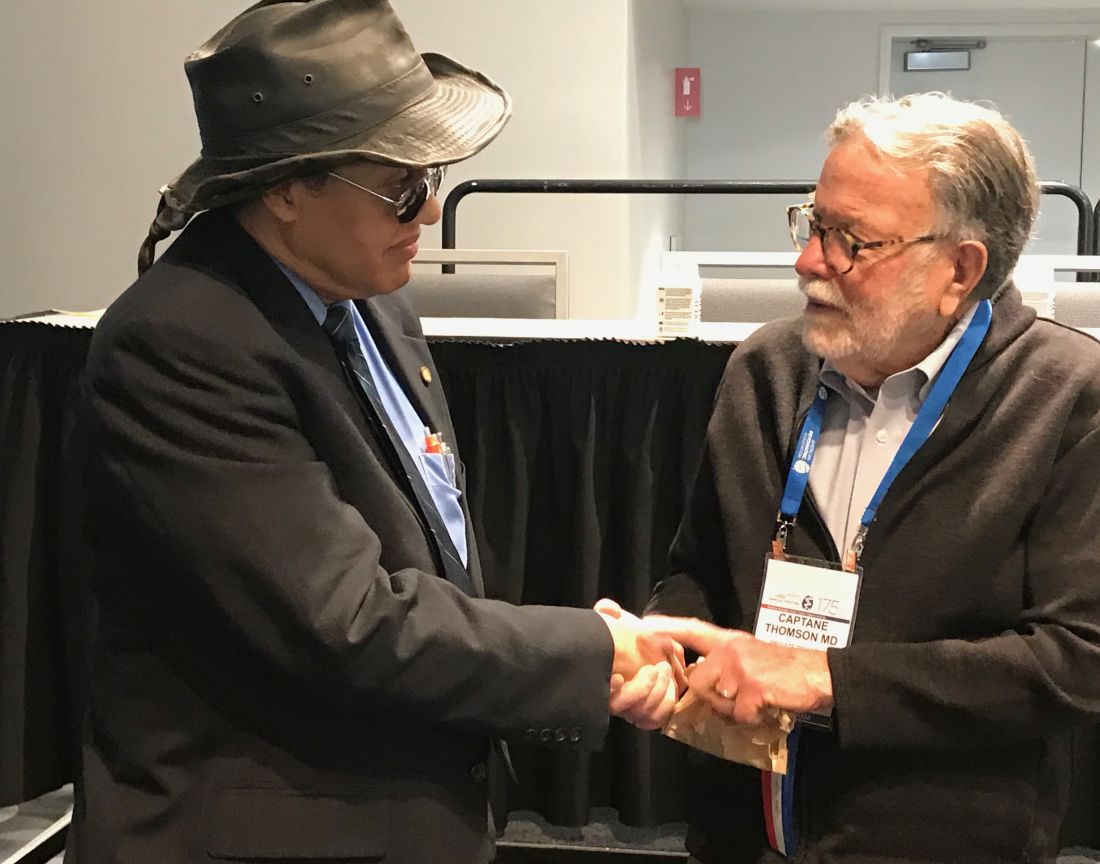
But, what was most meaningful about his productivity was his generosity of spirit. Any conversation was an opportunity for him to thoughtfully and respectfully share his knowledge. For example, once, we were discussing a clinical case that included the differential diagnosis of a patient, who happened to be African American, who was having auditory hallucinations. Dr. Bell might have been the first psychiatrist to alert the medical community about the misdiagnosis of schizophrenia among African Americans with bipolar disorder (J Nat Med Assoc. 1980 Feb 72[2]:141-5).
Contrary to my expectation that he was going to remind me of the tendency to misdiagnose, he instead offered, “You know, there are 40 reasons for auditory hallucinations.” Not what I had expected, yet, a response that reflected his continually giving nature and sharing of his abundance of gems. He was always teaching.
I later learned that his workday at Jackson Park Hospital usually ended at 2 p.m. He had treated patients, and supervised medical students and residents there for more than 40 years. The afternoons afforded him time to read, write, listen to music (Ella Fitzgerald), watch movies, and spend time with his adult children, to whom he was quite devoted. Dr. Bell was an avid martial artist and enjoyed sharing this practice with his son, William.
He was a longtime active member of the National Medical Association, recently receiving its prestigious Distinguished Service Award in Hawaii for his “exceptional work in medical service, medical research, and academic medicine.” It would be his last professional talk, though his delivery would belie his numbered days.
He was a former vice president of the Black Psychiatrists of America (BPA) and for 10 years had been the editor of the BPA Newsletter. Conversations were often peppered with anecdotes from time spent with other pioneering ancestors, such as Chester “Chet” Pierce, MD, Jeanne Spurlock, MD, Robert Phillips, MD, PhD, Charles Prudhomme, MD, Frances Cress Welsing, MD, and others. Dr. Bell was at the tail end of a generation of African American psychiatrists who had experienced firsthand the transition from segregation to federally mandated integration of our society.
Dr. Bell and his peers applied their education and training to improve clinical care for all, to decrease health inequities, and to eliminate disparities. It is evident that he loved his people and committed his life to addressing the needs of marginalized communities, those without the benefit of abundant resources, and those disproportionately affected by violence and trauma. As he stated in his last book, Fetal Alcohol Exposure in the African-American Community:
“I should add, my main concern is African American people living within the United States of America where in one community the rate of Fetal Alcohol Exposure is 388/1,000 people. ... However, this problem extends much further. Fetal Alcohol Exposure (FAE) is increasingly being found to (be) problematic in people of color around the world: Native Americans in Canada ... Aboriginal people in Australia ... and various tribes of people on the continent of Africa. ... Lastly, while the problem of Fetal Alcohol Exposure seems to be disproportionately affecting people of color, it also affects people who lack pigment in their skin. For example, FAE is a problem in Russia. From a public health perspective, so often people of color are like the proverbial “canary in a coal mine,” i.e., if there is poisonous gas in the coal mine, the canary will die first, warning the miners that they need to do something about it.”
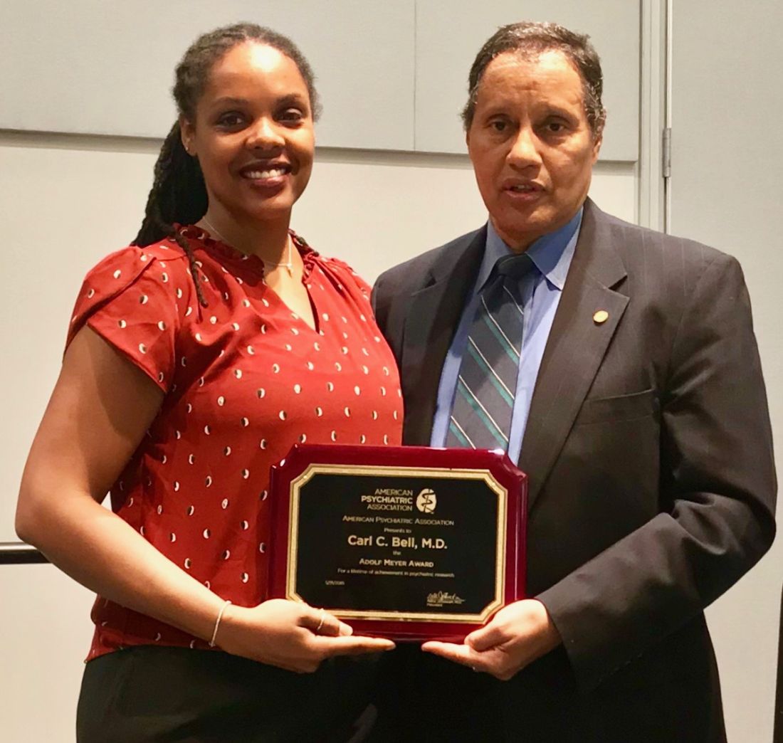
However, Dr. Bell was a Distinguished Lifetime Fellow of the American Psychiatric Association and a Lifetime Fellow of the American College of Psychiatrists. He was a founding member of the board of directors of the National Commission on Correctional Health, and a member of prominent work groups of the National Institutes of Mental Health and the National Academy of Medicine (formerly the Institute of Medicine). And Dr. Bell’s mentees and students transcend generations, race, religions, professional disciplines, and national boundaries.
Because Dr. Bell was grounded and never forgot his roots, it was in these professional society circles that he ensured that clinicians with more privilege and limited or no exposure to communities of color were educated about the needs of those he treated. Without exposure to Carl Bell, it is likely that many of our psychiatric colleagues would remain unaware of both the brilliant dynamic resources and enormous challenges that are found in the black community and communities of color. By sharing his work with the house of medicine, he obviated the excuse of doing nothing because of ignorance.
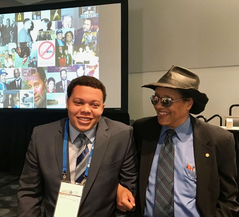
I last saw Dr. Bell in San Francisco toward the end of the 2019 annual APA meeting. He had received the APA’s Adolf* Meyer Award for lifetime achievement. Afterward, I joined him for a dim sum lunch in Chinatown with two of his colleagues and Joseph Calhoun, his mentee in the APA’s Black Men in Psychiatry Early Pipeline Program. As we walked back to our respective hotels, we paused at what is now the Chinese Affirmative Action Center. We learned that this site had been the home of one of Dr. Bell’s former martial arts instructors. As Dr. Bell recounted his martial arts training, the reverence for his sensei was evident in his eyes.
When I reflect on how much I learned from and about Carl Bell in such a short period of time, I realize that he was one of those people who was so present and so astute that he allowed you to know him while he was giving.
So, how do we honor someone who gave so much of himself? When I now think of the lyric from “Hamilton” – “Why do you write like you’re running out of time?” – I realize that we get it twisted when we associate running out of time with our elders and their phase of life. It was not Carl Bell who was running out of time. He had been extraordinarily respectful of the space, time, and energy allotted to him in his lifetime. He would say, “People squander their personal resources.” He certainly had not squandered his.
As we reflect and mourn his passing, we will hear about his candor, authenticity, integrity, discipline, reliability, dedication, and serving spirit. This is called character.
Dr. Bell was beyond generous with his life, and it is going to take decades, if not more, for us to digest the compendium of knowledge that he left behind. I ask you: How will you use that knowledge to advance the causes he so diligently devoted his life to solving?
To Carl Compton Bell, I say, Well done. Thank you. And, rest now my dear brother.
Dr. Dunlap, a psychiatrist and psychoanalyst who practices in Washington, is a Washington Psychiatric Society representative to the APA Assembly, a past president of the Washington Psychiatric Society, and clinical professor of psychiatry and behavioral sciences at George Washington University, Washington. She is interested in the role “difference” – race, culture, and ethnicity – plays in interpersonal relationships and group dynamics.
*This column was updated 9/3/2019.
Psychiatrist educated the field with his work on gun violence, prenatal alcohol exposure
Psychiatrist educated the field with his work on gun violence, prenatal alcohol exposure
With the heart of a child and the spirit of a warrior, Carl Bell always spoke his truth. And, he did so in his own inimitable way. Sporting his signature dark brown wide-brim leather cowboy hat or NMA (National Medical Association) baseball cap, aviator sunglasses, and accompanying Superman belt buckle, Carl Compton Bell, MD, – psychiatrist, researcher, mental health advocate, father, grandfather, friend, colleague, pioneer, and servant – was driven by a deep commitment to serve others.
As those who truly knew him can attest, it is not hyperbole to say that Carl Compton Bell was one of the most genuine, brilliant, and humble physicians of our professional community and time.
My collaboration and friendship began with Dr. Bell began during the summer of 2016 as I was preparing for the 2017 Washington Psychiatric Society’s (WPS) Presidential Symposium at Saint Elizabeths Hospital. As president of WPS, I had chosen gun violence as my topic and sought out Dr. Bell because of his work on the South Side of Chicago, where he had devoted himself, becoming an internationally known clinician, researcher, and mental health advocate for those personally affected by violence and trauma. He immediately accepted.
In his presentation, “Gun Violence, Urban Youth and Mental Illness,” he reviewed his research on the neurocognitive behavioral effects of prenatal exposure to alcohol and its relation to the neurodevelopmental dynamics of youth violence, intimate partner violence, and mass shootings. Dr. Bell suggested that the relationship between prenatal exposure to alcohol and the diagnosis of numerous psychiatric conditions had been underestimated in the medical community. He eventually summarized his work in Fetal Alcohol Exposure in the African-American Community, published by Third World Press (2018). This vital resource not only summarizes in plain language the scope of the problem of prenatal alcohol exposure but is a narrative of Carl Bell’s life journey.
After the symposium, he would send articles, while warning, “I can bombard you with stuff.” Sometimes we would not speak for weeks at a time while I digested the resources he had shared. However, whenever I picked up the phone to call and respond to what he had provided, he would answer the phone, “Yessssss?” – as if he were anticipating my call and was ready to address any queries or comments I might have. Even when he were about to board a plane or charting – after making rounds on his patients while listening to the music of James Brown – he would answer the phone, even if only to coordinate a more mutually convenient time to connect.
During the process of digesting the plethora of articles and resources he provided on prenatal fetal alcohol exposure, including the 1996 Institute of Medicine’s report and the American Medical Association’s 2017 resolution supporting the addition of adequate amounts of choline to prenatal vitamins, I found myself immersed in neuroscience topics, such as the role of neuronal acetylcholine receptor subunit alpha-7 in the formation of neurotransmitters, the strengthening of cell membranes, and the promotion of proper brain and spinal cord development. Dr. Bell spoke authoritatively about the neuroscience and the public health implications. One of his mantras was “Where is the data? You’ve got to have data.”
The information that he shared became the foundation of the action paper calling for the American Psychiatric Association (APA) to endorse the AMA’s resolution supporting the addition of adequate amounts (450 mg/d for pregnant women) of phosphatidylcholine to prenatal vitamins. The APA Assembly passed this action paper in May 2018.
He was also responsible for a second action paper, “Psychiatric Management of the Impact of Racism on Social and Clinical Events,” which passed at the same May 2018 assembly. Dr. Bell agreed to coauthor this paper, which was only fitting since the paper was a further elaboration of his efforts with the APA Caucus of Black Psychiatrists to implore the APA to acknowledge the deleterious effects of racism on both the victim and perpetrator.
While researching this topic, I had come across his 1980 article, Racism: A Symptom of the Narcissistic Personality Disorder (J Nat Med Assoc. 1980 Jul;72[7]:661-5), in which Dr. Bell applied psychoanalytic theory to posit that racism is one psychic derivative through which narcissism may manifest itself.
Although he was not formally trained as a psychoanalyst, he had benefited from strong psychoanalytic supervision at the Illinois State Psychiatric Institute, a training program of the University of Illinois at Chicago. He wrote confidently and clearly, applying self-psychology principles. He had the gravitas to write and speak about a range of topics, from neuroscience, psychotherapy, medical management of illness, and mental health advocacy. His 387-page curriculum vitae of 500+ articles, chapters, and books on mental health issues is a catalog of evidence that he had given thought to just about any topic along the spectrum of psychiatry and beyond.
In July 2018, after leaving a performance of “Hamilton” at the Kennedy Center, a lyric from the song “Non-Stop” stayed with me:
Why do you write like you’re running out of time?
(Why do you write like you’re running out of time?)
Write day and night like you’re running out of time?
The pace at which he read, wrote, lectured, researched, collaborated, and served on committees reminded me of the prodigious work of the former Secretary of the Treasury. When I shared this with Dr. Bell, he volunteered that he wrote to clear his mind. I suggested that, like other true writers, it seemed that he had to write. He did not disagree.
But, what was most meaningful about his productivity was his generosity of spirit. Any conversation was an opportunity for him to thoughtfully and respectfully share his knowledge. For example, once, we were discussing a clinical case that included the differential diagnosis of a patient, who happened to be African American, who was having auditory hallucinations. Dr. Bell might have been the first psychiatrist to alert the medical community about the misdiagnosis of schizophrenia among African Americans with bipolar disorder (J Nat Med Assoc. 1980 Feb 72[2]:141-5).
Contrary to my expectation that he was going to remind me of the tendency to misdiagnose, he instead offered, “You know, there are 40 reasons for auditory hallucinations.” Not what I had expected, yet, a response that reflected his continually giving nature and sharing of his abundance of gems. He was always teaching.
I later learned that his workday at Jackson Park Hospital usually ended at 2 p.m. He had treated patients, and supervised medical students and residents there for more than 40 years. The afternoons afforded him time to read, write, listen to music (Ella Fitzgerald), watch movies, and spend time with his adult children, to whom he was quite devoted. Dr. Bell was an avid martial artist and enjoyed sharing this practice with his son, William.
He was a longtime active member of the National Medical Association, recently receiving its prestigious Distinguished Service Award in Hawaii for his “exceptional work in medical service, medical research, and academic medicine.” It would be his last professional talk, though his delivery would belie his numbered days.
He was a former vice president of the Black Psychiatrists of America (BPA) and for 10 years had been the editor of the BPA Newsletter. Conversations were often peppered with anecdotes from time spent with other pioneering ancestors, such as Chester “Chet” Pierce, MD, Jeanne Spurlock, MD, Robert Phillips, MD, PhD, Charles Prudhomme, MD, Frances Cress Welsing, MD, and others. Dr. Bell was at the tail end of a generation of African American psychiatrists who had experienced firsthand the transition from segregation to federally mandated integration of our society.
Dr. Bell and his peers applied their education and training to improve clinical care for all, to decrease health inequities, and to eliminate disparities. It is evident that he loved his people and committed his life to addressing the needs of marginalized communities, those without the benefit of abundant resources, and those disproportionately affected by violence and trauma. As he stated in his last book, Fetal Alcohol Exposure in the African-American Community:
“I should add, my main concern is African American people living within the United States of America where in one community the rate of Fetal Alcohol Exposure is 388/1,000 people. ... However, this problem extends much further. Fetal Alcohol Exposure (FAE) is increasingly being found to (be) problematic in people of color around the world: Native Americans in Canada ... Aboriginal people in Australia ... and various tribes of people on the continent of Africa. ... Lastly, while the problem of Fetal Alcohol Exposure seems to be disproportionately affecting people of color, it also affects people who lack pigment in their skin. For example, FAE is a problem in Russia. From a public health perspective, so often people of color are like the proverbial “canary in a coal mine,” i.e., if there is poisonous gas in the coal mine, the canary will die first, warning the miners that they need to do something about it.”
However, Dr. Bell was a Distinguished Lifetime Fellow of the American Psychiatric Association and a Lifetime Fellow of the American College of Psychiatrists. He was a founding member of the board of directors of the National Commission on Correctional Health, and a member of prominent work groups of the National Institutes of Mental Health and the National Academy of Medicine (formerly the Institute of Medicine). And Dr. Bell’s mentees and students transcend generations, race, religions, professional disciplines, and national boundaries.
Because Dr. Bell was grounded and never forgot his roots, it was in these professional society circles that he ensured that clinicians with more privilege and limited or no exposure to communities of color were educated about the needs of those he treated. Without exposure to Carl Bell, it is likely that many of our psychiatric colleagues would remain unaware of both the brilliant dynamic resources and enormous challenges that are found in the black community and communities of color. By sharing his work with the house of medicine, he obviated the excuse of doing nothing because of ignorance.
I last saw Dr. Bell in San Francisco toward the end of the 2019 annual APA meeting. He had received the APA’s Adolf* Meyer Award for lifetime achievement. Afterward, I joined him for a dim sum lunch in Chinatown with two of his colleagues and Joseph Calhoun, his mentee in the APA’s Black Men in Psychiatry Early Pipeline Program. As we walked back to our respective hotels, we paused at what is now the Chinese Affirmative Action Center. We learned that this site had been the home of one of Dr. Bell’s former martial arts instructors. As Dr. Bell recounted his martial arts training, the reverence for his sensei was evident in his eyes.
When I reflect on how much I learned from and about Carl Bell in such a short period of time, I realize that he was one of those people who was so present and so astute that he allowed you to know him while he was giving.
So, how do we honor someone who gave so much of himself? When I now think of the lyric from “Hamilton” – “Why do you write like you’re running out of time?” – I realize that we get it twisted when we associate running out of time with our elders and their phase of life. It was not Carl Bell who was running out of time. He had been extraordinarily respectful of the space, time, and energy allotted to him in his lifetime. He would say, “People squander their personal resources.” He certainly had not squandered his.
As we reflect and mourn his passing, we will hear about his candor, authenticity, integrity, discipline, reliability, dedication, and serving spirit. This is called character.
Dr. Bell was beyond generous with his life, and it is going to take decades, if not more, for us to digest the compendium of knowledge that he left behind. I ask you: How will you use that knowledge to advance the causes he so diligently devoted his life to solving?
To Carl Compton Bell, I say, Well done. Thank you. And, rest now my dear brother.
Dr. Dunlap, a psychiatrist and psychoanalyst who practices in Washington, is a Washington Psychiatric Society representative to the APA Assembly, a past president of the Washington Psychiatric Society, and clinical professor of psychiatry and behavioral sciences at George Washington University, Washington. She is interested in the role “difference” – race, culture, and ethnicity – plays in interpersonal relationships and group dynamics.
*This column was updated 9/3/2019.
Genomic Medicine and Genetic Counseling in the Department of Veterans Affairs and Department of Defense (FULL)
Vickie Venne, MS. What is the Genomic Medicine Service (GMS) at the US Department of Veterans Affairs (VA)?
Renee Rider, JD, MS, LCGC. GMS is a telehealth service. We are part of central office and field stationed at the George E. Wahlen VA Medical Center (VAMC) in Salt Lake City, Utah. We provide care to about 90 VAMCs and their associated clinics. Veterans are referred to us by entering an interfacility consult in the VA Computerized Patient Record System (CPRS). We review the consult to determine whether the patient needs to be seen, whether we can answer with an e-consult, or whether we need more information. For the patients who need an appointment, the telehealth department at the veteran’s VA facility will contact the patient to arrange a visit with us. At the time of the appointment, the facility has a staff member available to seat the patient and connect them to us using video equipment.
We provide genetic care for all specialties, including cancer, women’s health, cardiology and neurology. In today’s discussion, we are focusing on cancer care.
Vickie Venne. What do patients do at facilities that don’t get care through GMS?
Renee Rider. There are a handful of facilities that provide their own genetic care in-house. For example, VA Boston Healthcare System in Massachusetts and the Michael E. DeBakey VAMC in Houston, Texas each have their own programs. For veterans who are not at a VA facility that has an agreement with GMS and do not have a different genetics program, their providers need to make referrals to community care.
Vickie Venne. How do patients get referred and what happens at their facility when the patients return to the specialty and primary care providers (PCP)? Ishta, who do you refer to GMS and how do you define them initially?
Ishta Thakar, MD, FACP. Referrals can come at a couple of points during a veteran’s journey at the VA. The VA covers obstetrics care for women veterans. Whenever a PCP or a women’s health provider is doing the initial history and physical on a new patient, if the female veteran has an extensive family history of breast, ovarian, colon, or endometrial cancer, then we take more history and we send a consult to GMS. The second instance would be if she tells us that she has had a personal history of breast, ovarian, or endometrial cancer and she has never had genetic testing. The third instance would be whenever we have a female veteran who is diagnosed with breast, ovarian, endometrial, or colon cancer. We would definitely talk to her about genetic counseling and send a referral to GMS. We would ask for a GMS consult for a patient with advanced maternal age, with exposure to some kind of teratogens, with an abnormal ultrasound, a family history of chromosomal disorders, or if she’s seeing an obstetrician who wants her to be tested. And finally, if a patient has a constellation of multiple cancers in the family and we don’t know what’s going on, we would also refer the patient to GMS.
Vickie Venne. That would be why GMS fields over 150 referrals every week. It is a large list. We also see veterans with personal or family histories of neurologic or cardiologic concerns as well.
Renee, as somebody who fields many of these referrals from unaffected individuals, what is the family history process?
Renee Rider. We don’t expect the referring provider to be a genetic expert. When a provider is seeing a constellation of several different cancers and he or she doesn’t know if there’s anything going on genetically or even if it’s possible, absolutely they should put in a referral to GMS. We have a triage counselor who reviews every consult that comes into our service within 24 hours.
Many cancers are due to exposures that are not concerning for a genetic etiology. We can let you know that it is not concerning, and the PCP can counsel the patient that it is very unlikely to be genetic in nature. We still give feedback even if it’s not someone who is appropriate for genetic counseling and testing. It is important to reach out to GMS even if you don’t know whether a cancer is genetic in nature.
It also is important to take your time when gathering family histories. We get a lot of patients who say, “There’s a lot of cancer in my family. I have no idea who had cancer, but I know a lot of people had cancer.” That’s not the day to put in a referral to GMS. At that point, providers should tell the patient to get as much information as they can about the family history and then reassess. It’s important for us to have accurate information. We’ve had several times where we receive a referral because the veteran says that their sister had ovarian cancer. And then when our staff calls, they later find out it was cervical cancer. That’s not a good use of the veteran’s time, and it’s not a good use of VA resources.
The other important thing about family histories is keeping the questions open-ended. Often a PCP or specialist will ask about a certain type of cancer: “Does anyone in your family have breast cancer, ovarian cancer?” Or if the veteran
is getting a colonoscopy, they ask, “Does anybody have colon cancer?” Where really, we need to be a little bit more open-ended. We prefer questions like, “Has anyone in your family
had cancer?” because that’s the question that prompts a response of, “Yes, 3 people in my family have had thyroid cancer.” That’s very important for us to know, too.
If you do get a positive response, probe a little bit more: what kind of cancer did someone have, how old were they when they had their cancer? And how are they related? Is this an aunt on your mom’s side or on your dad’s side? Those are the types of information that we need to figure out if that person needs a referral.
Vickie Venne. It’s a different story when people already have a cancer diagnosis. Which hematology or oncology patients are good referrals and why?
Lisa Arfons, MD. When patients come in with newly diagnosed cancer, breast for example, it is an emotional diagnosis and psychologicallydistressing. Oftentimes, they want to know why this happened to them. The issues surrounding
genetic testing also becomes very emotional. They want to know whether their children are at risk as well.
Genetic discussions take a long time. I rarely do that on the first visit. I always record for myself in my clinic note if something strikes me regarding the patient’s diagnosis. I quickly run through the National Comprehensive Cancer Network (NCCN) guidelines to remind myself of what I need to go over with the patient at our next meeting. Most patients don’t need to be referred to GMS, and most patients don’t need to be tested once they’re seen.
I often save the referral discussion for after I have established a rapport with a patient, we have a treatment plan, or they already have had their first surgery. Therefore, we are not making decisions about their first surgery based on the genetic medicine results.
If I’m considering a referral, I do a deeper dive with the patient. Is the patient older or younger than 45 years? I pull up NCCN guidelines and we go through the entire checklist.
We have male breast cancer patients at the VA—probably more than the community—so we refer those patients. At the Louis Stokes Cleveland VAMC in Ohio, we have had some in-depth discussions about referring male breast cancer patients for genetic testing and whether it was beneficial to older patients with male breast cancer. Ultimately, we decided that it was important for our male veterans to be tested because it empowered them to have better understanding of their medical conditions that may not just have effect on them but on their offspring, and that that can be a source of psychological and emotional support.
I don’t refer most people to GMS once I go through the checklist. I appreciate the action for an e-consult within the CPRS telemedicine consult itself, as Renee noted. If it is not necessary, GMS makes it an e-consult. I try to communicate that I don’t know whether it is necessary or not so that GMS understands where I’m coming from.
Vickie Venne. In the US Department of Defense (DoD) the process is quite different. Mauricio, can you explain the clinical referral process, who is referred, and how that works from a laboratory perspective?
Maj De Castro, MD, FACMG, USAF. The VA has led the way in demonstrating how to best provide for the medical genetic needs of a large, decentralized population distributed all over the country. Over the last 5 to 10 years, the DoD has made strides in recognizing the role genetics plays in the practice of everyday medicine and redoubling efforts to meet the needs of servicemembers.
The way that it traditionally has worked in the DoD is that military treatment facilities (MTFs) that have dedicated geneticists and genetic counselors: Kessler Medical Center in Mississippi, Walter Reed National Military Medical
Center in Maryland, Tripler Army Medical Center in Hawaii, Madigan Army Medical Center in Washington, Brooke Army Medical Center in Texas, Naval Medical Center San Diego in California, and Naval Medical Center Portsmouth in Virginia. A patient seeking genetic evaluation, counseling, or testing in those larger facilities would be referred to the genetics service by their primary care manager. Wait times vary, but it would usually be weeks, maybe months. However, the great majority of MTFs do not have dedicated genetics support. Most of the time, those patients would have to be referred to the local civilian community—there was no process for them to be seen in in the military healthcare system—with wait times that exceed 6 to 8 months in some cases. This is due to just not a military but a national shortage of genetics professionals (counselors and physicians).
Last year we started the telegenetics initiative, which is small compared to the VA—it is comprised of 2 geneticists and 1 genetic counselor—but with the full intent of growing it over time. Its purpose is to extend the resources we
had to other MTFs. Genetics professionals stationed state-side can provide care to remote facilities with limited access to local genetics support such as Cannon Air Force Base (AFB) or overseas facilities such as Spangdahlem AFB in Germany.
We recognize there are military-specific needs for the DoD regarding the genetic counseling process that have to take into account readiness, genetic discrimination, continued ability to serve and fitness for duty. For this important reason, we are seeking to expand our telegenetics initiative. The goal is to be able to provide 100% of all genetic counseling in-house, so to speak.
Currently, providers at the 4 pilot sites (Cannon AFB, Fort Bragg, Spangdahlem AFB, and Guantanamo Bay) send us referrals. We triage them and assign the patient to see a geneticist or a counselor depending on the indication.
On the laboratory side, it has been a very interesting experience. Because we provide comprehensive germline cancer testing at very little cost to the provider at any MTF, we have had high numbers of test requests over the years.
In addition to saving the DoD millions of dollars in testing, we have learned some interesting lessons in the process. For instance, we have worked closely with several different groups to better understand how to educate providers on the genetic counseling and testing process. This has allowed us to craft a thorough and inclusive consent form that addresses the needs of the DoD. We have also learned valuable lessons about population-based screening vs evidence-based testing, and lessons surrounding narrow-based testing (BRCA1 and BRCA2 only testing) vs ordering a more comprehensive panel that includes other genes supported by strong evidence (such as PALB2, CHEK2, or TP53).
For example, we have found that in a significant proportion of individuals with and without family history, there are clinically relevant variants in genes other than BRCA1 or BRCA2. And so, we have made part of our consent process,
a statement on secondary findings. If the patient consents, we will report pathogenic variants in other genes known to be associated with cancer (with strong evidence) even if the provider ordered a narrow panel such as BRCA1 and BRCA2 testing only. In about 1% to 4% of patients that would otherwise not meet NCCN guidelines, we’ve reported variants that were clinically actionable and changed the medical management of that patient.
We feel strongly that this is a conversation that we need to have in our field, and we realize it’s a complex issue, maybe we need to expand who gets testing. Guideline based testing is missing some patients out there that could benefit from it.
Vickie Venne. There certainly are many sides to the conversation of population-based vs evidence-based genetic testing. Genetic testing policies are changing rapidly. There are teams exploring comprehensive gene sequencing for
newborns and how that potential 1-time test can provide information will be reinterpreted as a person goes from cradle to grave. However, unlike the current DoD process, in the VA there are patients who we don’t see.
Renee Rider. I want to talk about money. When we order a genetic test, that test is paid for by the pathology department at the patient’s VAMC. Most of the pathology departments we work with are clear that they only can provide
genetic testing that is considered medically necessary. Thus, we review each test to make sure it meets established guidelines for testing. We don’t do population genetic screening as there isn’t evidence or guidelines to support offering it. We are strict about who does and does not get genetic testing, partly because we have a responsibility to pathology departments and to the taxpayers.
GMS focuses on conditions that are inherited, that is to say, we deal with germline genetics. Therefore, we discontinue referrals for somatic requests, such as when an OncotypeDX test is requested. It is my understanding that pharmacogenetic referrals may be sent to the new PHASeR initiative, which is a joint collaboration between the VA and Sanford Health and is headed by Deepak Voora, MD.
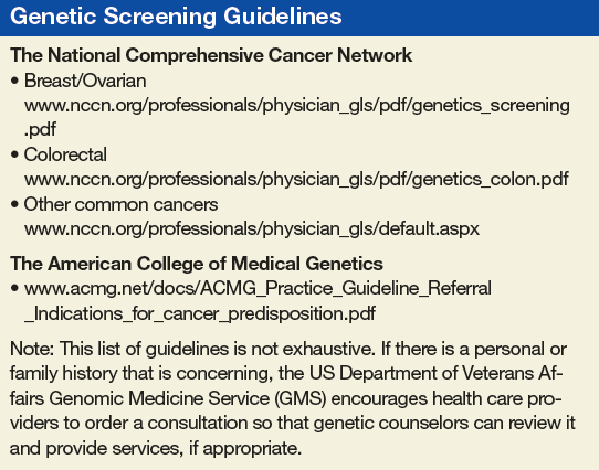
We generally don’t see patients who still are having diagnostic procedures done. For example, if a veteran has a suspicious breast mass, we recommend that the provider workup the mass before referring to GMS. Regardless of a genetic test result, a suspicious mass needs to be worked up. And, knowing if the mass is cancerous could change how we would proceed with the genetic workup. For example, if the mass were not cancerous, we may recommend that an affected relative have the first genetic evaluation. Furthermore, knowing if the patient has cancer changes how we interpret negative test results.
Another group of patients we don’t see are those who already had genetic testing done by the referring provider. It’s a VA directive that if you order a test, you’re the person who is responsible for giving the results. We agree with
this directive. If you don’t feel comfortable giving back test results, don’t order the test. Often, when a provider sends a patient to us after the test was done, we discover that the patient didn’t have appropriate pretest counseling. A test result, such as a variant of uncertain significance (VUS), should never be a surprise to either the provider or the patient.
Ishta Thakar. For newly diagnosed cancers, the first call is to the patient to inform them that they have cancer. We usually bring up genetic counseling or testing, if applicable, when they are ready to accept the diagnosis and have a conversation about it. All our consults are via telehealth, so none of our patients physically come to GMS in Salt Lake City. All the consults are done virtually.
For newly diagnosed patients, we would send a consult in within a couple of weeks. For patients who had a family history, the referral would not be urgent: They can be seen within about 3 months. The turnaround times for GMS are so much better than what we have available in the community where it’s often at least 6 months, as previously noted.
Vickie Venne. Thank you. We continue to work on that. One of the interesting things that we’ve done, which is the brainchild of Renee, is shared medical appointments.
Renee Rider. We have now created 4 group appointments for people who have concerns surrounding cancer. One group is for people who don’t have cancer but have family members who have cancer who may be the best testing candidate. For example, that might be a 30-year old who tells you that her mother had breast cancer at age 45 years. Her mother is still living, but she’s never had genetic testing. We would put her in a group where we discuss the importance of talking to the family members and encouraging them to go get that first genetic evaluation in the family.
Our second group is for people who don’t have cancer themselves, but have a family history of cancer and those affected relatives have passed away. The family needs a genetic evaluation, and the veteran is the best living testing candidate.
That group is geared towards education about the test and informed consent.
The third group is for people with cancer who qualify for genetic testing. We provide all of the information that they need to make an informed decision on having (or not having) genetic testing.
The final group is for people who have family histories of known genetic mutations in cancer genes. Again, we provide them with all of the information that they need to make an informed decision regarding genetic testing.
With the shared medical appointments, we have been able to greatly increase the number of patients that we can see. Our first 3 groups all meet once a week and can have 10 or 12 veterans. Our last group meets every other week and has a maximum of 6 veterans. Wait times for our groups are generally ≤ 2 weeks. All veterans can choose to have an individual appointment if they prefer. We regularly get unsolicited feedback from veterans that they learn a lot during our groups and appreciate it.
Our group appointments have lowered the wait time for the people in the groups. And, they’ve lowered the wait time for the people who are seen individually. They’ve allowed us to address the backlog of patients waiting to see us in a more timely manner. Our wait time for individual appointment had been approaching 6 months, and it is now about 1.5 months.
We also think that being in a group normalizes the experience. Most people don’t know anyone who has had genetic testing. Now, they are in a group with others going through the same experience. In one of my groups, a male veteran talked about his breast cancer being really rare. Another male in the group volunteer that he had breast cancer, too. They both seemed to appreciate not feeling alone.
Vickie Venne. I want to move to our final piece. What do the referring providers tell the patients about a genetics referral and what should they expect?
Lisa Arfons. First and foremost, I tell the patient that it is a discussion with a genetic counselor. I make it clear that they understand that it is a discussion. They then can agree or not agree to accept genetic testing if it’s recommended.
I talk in general terms about why I think it can be important for them to have the discussion, but that we don’t have great data for decisionmaking. We understand that there are more options for preventive measures but then it ultimately will be a discussion between the PCP, the patient, and their family members about how they proceed about the preventive measures. I want them to start thinking about how the genetic test results, regardless of if they are positive, negative, or a variant that is not yet understood, can impact their offspring.
Probably I am biased, as my mom had breast cancer and she underwent genetic testing. So, I have a bit of an offspring focus as well. I already mentioned that you must discuss about whether or not it’s worth screening or doing any preventive measures on contralateral breast, or screening for things like prostate cancer at age 75 years. And so I focus more on the family members.
I try to stay in my lane. I am extremely uncomfortable when I hear about someone in our facility sending off a blood test and then asking someone else to interpret the results and discuss it with the patient. Just because it’s a blood test and it’s easy to order doesn’t mean that it is easy to know what to do with it, and it needs to be respected as such.
Ishta Thakar. Our PCPs let the patients know that GMS will contact the patient to schedule a video appointment and that if they want to bring any family members along with them, they’re welcome to. We also explain that certain cancers are genetically based and that if they have a genetic mutation, it can be passed on to their offspring. I also explain that if they have certain mutations, then we would be more vigilant in screening them for other kinds of cancers. That’s the reason that we refer that they get counseled. After counseling if they’re ready for the testing, then the counselor orders the test and does the posttest discussion with the patient.
Vickie Venne. In the VA, people are invited to attend a genetic counseling session but can certainly decline. Does the the DoD have a different approach?
Maj De Castro. I would say that the great majority of active duty patients have limited knowledge of what to expect out of a genetics appointment. One of the main things we do is educate them on their rights and protections and the potential risks associated with performing genetic testing, in particular when it comes to their continued ability to serve. Genetic testing for clinical purposes is not mandatory in the DoD, patients can certainly decline testing. Because genetic testing has the potential to alter someone’s career, it is critical we have a very thorough and comprehensive pre- and posttest counseling sessions that includes everything from career implications to the Genetic Information Nondiscrimination Act (GINA) and genetic discrimination in the military, in addition to the standard of care medical information.
Scenarios in which a servicemember is negatively impacted by pursuing a genetic diagnosis are very rare. More than 90% of the time, genetic counseling and/or testing has no adverse career effect. When they do, it is out of concern for the safety and wellbeing of a servicemember. For instance, if we diagnosis a patient with a genetic form of some arrhythmogenic disorder, part of the treatment plan can be to limit that person’s level of exertion, because it could potentially lead to death. We don’t want to put someone in a situation that may trigger that.
Vickie Venne. We also have a certain number of veterans who ask us about their service disability pay and the impact of genetic testing on it. One example is veterans with prostate cancer who were exposed to Agent Orange, which has been associated with increased risk for developing prostate cancer. I have had men who have been referred for genetic evaluation ask, “Well, if I have an identifiable mutation, how will that impact my service disability?” So we discuss the carcinogenic process that may include an inherited component as well as the environmental risk factors. I think that’s a unique issue for a population we’re honored to be able to serve.
Renee Rider. When we are talking about how the population of veterans is unique, I think it is also important to acknowledge mental health. I’ve had several patients tell me that they have posttraumatic stress disorder or anxiety and the idea of getting an indeterminant test result, such as VUS, would really weigh on them.
In the community, a lot of providers order the biggest panel they can, but for these patients who are worried about getting those indeterminant test results, I’ve been able to work with them to limit the size of the panel. I order a small panel that only has genes that have implications for that veteran’s clinical management. For example, in a patient with ductal breast cancer, I remove the genes that cause lobular breast cancer. This takes a bit of knowledge and critical thinking that our VA genetic counselors have because they have experience with veterans and their needs.
As our time draws to a close, I have one final thought. This has been a heartwarming conversation today. It is really nice to hear that GMS services are appreciated. We in GMS want to partner with our referring providers. Help us help you! When you enter a referral, please let us know how we can help you. The more we understand why you are sending your veteran to GMS, the more we can help meet your needs. If there are any questions or problems, feel free to send us an email or pick up the phone and call us.
Vickie Venne, MS. What is the Genomic Medicine Service (GMS) at the US Department of Veterans Affairs (VA)?
Renee Rider, JD, MS, LCGC. GMS is a telehealth service. We are part of central office and field stationed at the George E. Wahlen VA Medical Center (VAMC) in Salt Lake City, Utah. We provide care to about 90 VAMCs and their associated clinics. Veterans are referred to us by entering an interfacility consult in the VA Computerized Patient Record System (CPRS). We review the consult to determine whether the patient needs to be seen, whether we can answer with an e-consult, or whether we need more information. For the patients who need an appointment, the telehealth department at the veteran’s VA facility will contact the patient to arrange a visit with us. At the time of the appointment, the facility has a staff member available to seat the patient and connect them to us using video equipment.
We provide genetic care for all specialties, including cancer, women’s health, cardiology and neurology. In today’s discussion, we are focusing on cancer care.
Vickie Venne. What do patients do at facilities that don’t get care through GMS?
Renee Rider. There are a handful of facilities that provide their own genetic care in-house. For example, VA Boston Healthcare System in Massachusetts and the Michael E. DeBakey VAMC in Houston, Texas each have their own programs. For veterans who are not at a VA facility that has an agreement with GMS and do not have a different genetics program, their providers need to make referrals to community care.
Vickie Venne. How do patients get referred and what happens at their facility when the patients return to the specialty and primary care providers (PCP)? Ishta, who do you refer to GMS and how do you define them initially?
Ishta Thakar, MD, FACP. Referrals can come at a couple of points during a veteran’s journey at the VA. The VA covers obstetrics care for women veterans. Whenever a PCP or a women’s health provider is doing the initial history and physical on a new patient, if the female veteran has an extensive family history of breast, ovarian, colon, or endometrial cancer, then we take more history and we send a consult to GMS. The second instance would be if she tells us that she has had a personal history of breast, ovarian, or endometrial cancer and she has never had genetic testing. The third instance would be whenever we have a female veteran who is diagnosed with breast, ovarian, endometrial, or colon cancer. We would definitely talk to her about genetic counseling and send a referral to GMS. We would ask for a GMS consult for a patient with advanced maternal age, with exposure to some kind of teratogens, with an abnormal ultrasound, a family history of chromosomal disorders, or if she’s seeing an obstetrician who wants her to be tested. And finally, if a patient has a constellation of multiple cancers in the family and we don’t know what’s going on, we would also refer the patient to GMS.
Vickie Venne. That would be why GMS fields over 150 referrals every week. It is a large list. We also see veterans with personal or family histories of neurologic or cardiologic concerns as well.
Renee, as somebody who fields many of these referrals from unaffected individuals, what is the family history process?
Renee Rider. We don’t expect the referring provider to be a genetic expert. When a provider is seeing a constellation of several different cancers and he or she doesn’t know if there’s anything going on genetically or even if it’s possible, absolutely they should put in a referral to GMS. We have a triage counselor who reviews every consult that comes into our service within 24 hours.
Many cancers are due to exposures that are not concerning for a genetic etiology. We can let you know that it is not concerning, and the PCP can counsel the patient that it is very unlikely to be genetic in nature. We still give feedback even if it’s not someone who is appropriate for genetic counseling and testing. It is important to reach out to GMS even if you don’t know whether a cancer is genetic in nature.
It also is important to take your time when gathering family histories. We get a lot of patients who say, “There’s a lot of cancer in my family. I have no idea who had cancer, but I know a lot of people had cancer.” That’s not the day to put in a referral to GMS. At that point, providers should tell the patient to get as much information as they can about the family history and then reassess. It’s important for us to have accurate information. We’ve had several times where we receive a referral because the veteran says that their sister had ovarian cancer. And then when our staff calls, they later find out it was cervical cancer. That’s not a good use of the veteran’s time, and it’s not a good use of VA resources.
The other important thing about family histories is keeping the questions open-ended. Often a PCP or specialist will ask about a certain type of cancer: “Does anyone in your family have breast cancer, ovarian cancer?” Or if the veteran
is getting a colonoscopy, they ask, “Does anybody have colon cancer?” Where really, we need to be a little bit more open-ended. We prefer questions like, “Has anyone in your family
had cancer?” because that’s the question that prompts a response of, “Yes, 3 people in my family have had thyroid cancer.” That’s very important for us to know, too.
If you do get a positive response, probe a little bit more: what kind of cancer did someone have, how old were they when they had their cancer? And how are they related? Is this an aunt on your mom’s side or on your dad’s side? Those are the types of information that we need to figure out if that person needs a referral.
Vickie Venne. It’s a different story when people already have a cancer diagnosis. Which hematology or oncology patients are good referrals and why?
Lisa Arfons, MD. When patients come in with newly diagnosed cancer, breast for example, it is an emotional diagnosis and psychologicallydistressing. Oftentimes, they want to know why this happened to them. The issues surrounding
genetic testing also becomes very emotional. They want to know whether their children are at risk as well.
Genetic discussions take a long time. I rarely do that on the first visit. I always record for myself in my clinic note if something strikes me regarding the patient’s diagnosis. I quickly run through the National Comprehensive Cancer Network (NCCN) guidelines to remind myself of what I need to go over with the patient at our next meeting. Most patients don’t need to be referred to GMS, and most patients don’t need to be tested once they’re seen.
I often save the referral discussion for after I have established a rapport with a patient, we have a treatment plan, or they already have had their first surgery. Therefore, we are not making decisions about their first surgery based on the genetic medicine results.
If I’m considering a referral, I do a deeper dive with the patient. Is the patient older or younger than 45 years? I pull up NCCN guidelines and we go through the entire checklist.
We have male breast cancer patients at the VA—probably more than the community—so we refer those patients. At the Louis Stokes Cleveland VAMC in Ohio, we have had some in-depth discussions about referring male breast cancer patients for genetic testing and whether it was beneficial to older patients with male breast cancer. Ultimately, we decided that it was important for our male veterans to be tested because it empowered them to have better understanding of their medical conditions that may not just have effect on them but on their offspring, and that that can be a source of psychological and emotional support.
I don’t refer most people to GMS once I go through the checklist. I appreciate the action for an e-consult within the CPRS telemedicine consult itself, as Renee noted. If it is not necessary, GMS makes it an e-consult. I try to communicate that I don’t know whether it is necessary or not so that GMS understands where I’m coming from.
Vickie Venne. In the US Department of Defense (DoD) the process is quite different. Mauricio, can you explain the clinical referral process, who is referred, and how that works from a laboratory perspective?
Maj De Castro, MD, FACMG, USAF. The VA has led the way in demonstrating how to best provide for the medical genetic needs of a large, decentralized population distributed all over the country. Over the last 5 to 10 years, the DoD has made strides in recognizing the role genetics plays in the practice of everyday medicine and redoubling efforts to meet the needs of servicemembers.
The way that it traditionally has worked in the DoD is that military treatment facilities (MTFs) that have dedicated geneticists and genetic counselors: Kessler Medical Center in Mississippi, Walter Reed National Military Medical
Center in Maryland, Tripler Army Medical Center in Hawaii, Madigan Army Medical Center in Washington, Brooke Army Medical Center in Texas, Naval Medical Center San Diego in California, and Naval Medical Center Portsmouth in Virginia. A patient seeking genetic evaluation, counseling, or testing in those larger facilities would be referred to the genetics service by their primary care manager. Wait times vary, but it would usually be weeks, maybe months. However, the great majority of MTFs do not have dedicated genetics support. Most of the time, those patients would have to be referred to the local civilian community—there was no process for them to be seen in in the military healthcare system—with wait times that exceed 6 to 8 months in some cases. This is due to just not a military but a national shortage of genetics professionals (counselors and physicians).
Last year we started the telegenetics initiative, which is small compared to the VA—it is comprised of 2 geneticists and 1 genetic counselor—but with the full intent of growing it over time. Its purpose is to extend the resources we
had to other MTFs. Genetics professionals stationed state-side can provide care to remote facilities with limited access to local genetics support such as Cannon Air Force Base (AFB) or overseas facilities such as Spangdahlem AFB in Germany.
We recognize there are military-specific needs for the DoD regarding the genetic counseling process that have to take into account readiness, genetic discrimination, continued ability to serve and fitness for duty. For this important reason, we are seeking to expand our telegenetics initiative. The goal is to be able to provide 100% of all genetic counseling in-house, so to speak.
Currently, providers at the 4 pilot sites (Cannon AFB, Fort Bragg, Spangdahlem AFB, and Guantanamo Bay) send us referrals. We triage them and assign the patient to see a geneticist or a counselor depending on the indication.
On the laboratory side, it has been a very interesting experience. Because we provide comprehensive germline cancer testing at very little cost to the provider at any MTF, we have had high numbers of test requests over the years.
In addition to saving the DoD millions of dollars in testing, we have learned some interesting lessons in the process. For instance, we have worked closely with several different groups to better understand how to educate providers on the genetic counseling and testing process. This has allowed us to craft a thorough and inclusive consent form that addresses the needs of the DoD. We have also learned valuable lessons about population-based screening vs evidence-based testing, and lessons surrounding narrow-based testing (BRCA1 and BRCA2 only testing) vs ordering a more comprehensive panel that includes other genes supported by strong evidence (such as PALB2, CHEK2, or TP53).
For example, we have found that in a significant proportion of individuals with and without family history, there are clinically relevant variants in genes other than BRCA1 or BRCA2. And so, we have made part of our consent process,
a statement on secondary findings. If the patient consents, we will report pathogenic variants in other genes known to be associated with cancer (with strong evidence) even if the provider ordered a narrow panel such as BRCA1 and BRCA2 testing only. In about 1% to 4% of patients that would otherwise not meet NCCN guidelines, we’ve reported variants that were clinically actionable and changed the medical management of that patient.
We feel strongly that this is a conversation that we need to have in our field, and we realize it’s a complex issue, maybe we need to expand who gets testing. Guideline based testing is missing some patients out there that could benefit from it.
Vickie Venne. There certainly are many sides to the conversation of population-based vs evidence-based genetic testing. Genetic testing policies are changing rapidly. There are teams exploring comprehensive gene sequencing for
newborns and how that potential 1-time test can provide information will be reinterpreted as a person goes from cradle to grave. However, unlike the current DoD process, in the VA there are patients who we don’t see.
Renee Rider. I want to talk about money. When we order a genetic test, that test is paid for by the pathology department at the patient’s VAMC. Most of the pathology departments we work with are clear that they only can provide
genetic testing that is considered medically necessary. Thus, we review each test to make sure it meets established guidelines for testing. We don’t do population genetic screening as there isn’t evidence or guidelines to support offering it. We are strict about who does and does not get genetic testing, partly because we have a responsibility to pathology departments and to the taxpayers.
GMS focuses on conditions that are inherited, that is to say, we deal with germline genetics. Therefore, we discontinue referrals for somatic requests, such as when an OncotypeDX test is requested. It is my understanding that pharmacogenetic referrals may be sent to the new PHASeR initiative, which is a joint collaboration between the VA and Sanford Health and is headed by Deepak Voora, MD.
We generally don’t see patients who still are having diagnostic procedures done. For example, if a veteran has a suspicious breast mass, we recommend that the provider workup the mass before referring to GMS. Regardless of a genetic test result, a suspicious mass needs to be worked up. And, knowing if the mass is cancerous could change how we would proceed with the genetic workup. For example, if the mass were not cancerous, we may recommend that an affected relative have the first genetic evaluation. Furthermore, knowing if the patient has cancer changes how we interpret negative test results.
Another group of patients we don’t see are those who already had genetic testing done by the referring provider. It’s a VA directive that if you order a test, you’re the person who is responsible for giving the results. We agree with
this directive. If you don’t feel comfortable giving back test results, don’t order the test. Often, when a provider sends a patient to us after the test was done, we discover that the patient didn’t have appropriate pretest counseling. A test result, such as a variant of uncertain significance (VUS), should never be a surprise to either the provider or the patient.
Ishta Thakar. For newly diagnosed cancers, the first call is to the patient to inform them that they have cancer. We usually bring up genetic counseling or testing, if applicable, when they are ready to accept the diagnosis and have a conversation about it. All our consults are via telehealth, so none of our patients physically come to GMS in Salt Lake City. All the consults are done virtually.
For newly diagnosed patients, we would send a consult in within a couple of weeks. For patients who had a family history, the referral would not be urgent: They can be seen within about 3 months. The turnaround times for GMS are so much better than what we have available in the community where it’s often at least 6 months, as previously noted.
Vickie Venne. Thank you. We continue to work on that. One of the interesting things that we’ve done, which is the brainchild of Renee, is shared medical appointments.
Renee Rider. We have now created 4 group appointments for people who have concerns surrounding cancer. One group is for people who don’t have cancer but have family members who have cancer who may be the best testing candidate. For example, that might be a 30-year old who tells you that her mother had breast cancer at age 45 years. Her mother is still living, but she’s never had genetic testing. We would put her in a group where we discuss the importance of talking to the family members and encouraging them to go get that first genetic evaluation in the family.
Our second group is for people who don’t have cancer themselves, but have a family history of cancer and those affected relatives have passed away. The family needs a genetic evaluation, and the veteran is the best living testing candidate.
That group is geared towards education about the test and informed consent.
The third group is for people with cancer who qualify for genetic testing. We provide all of the information that they need to make an informed decision on having (or not having) genetic testing.
The final group is for people who have family histories of known genetic mutations in cancer genes. Again, we provide them with all of the information that they need to make an informed decision regarding genetic testing.
With the shared medical appointments, we have been able to greatly increase the number of patients that we can see. Our first 3 groups all meet once a week and can have 10 or 12 veterans. Our last group meets every other week and has a maximum of 6 veterans. Wait times for our groups are generally ≤ 2 weeks. All veterans can choose to have an individual appointment if they prefer. We regularly get unsolicited feedback from veterans that they learn a lot during our groups and appreciate it.
Our group appointments have lowered the wait time for the people in the groups. And, they’ve lowered the wait time for the people who are seen individually. They’ve allowed us to address the backlog of patients waiting to see us in a more timely manner. Our wait time for individual appointment had been approaching 6 months, and it is now about 1.5 months.
We also think that being in a group normalizes the experience. Most people don’t know anyone who has had genetic testing. Now, they are in a group with others going through the same experience. In one of my groups, a male veteran talked about his breast cancer being really rare. Another male in the group volunteer that he had breast cancer, too. They both seemed to appreciate not feeling alone.
Vickie Venne. I want to move to our final piece. What do the referring providers tell the patients about a genetics referral and what should they expect?
Lisa Arfons. First and foremost, I tell the patient that it is a discussion with a genetic counselor. I make it clear that they understand that it is a discussion. They then can agree or not agree to accept genetic testing if it’s recommended.
I talk in general terms about why I think it can be important for them to have the discussion, but that we don’t have great data for decisionmaking. We understand that there are more options for preventive measures but then it ultimately will be a discussion between the PCP, the patient, and their family members about how they proceed about the preventive measures. I want them to start thinking about how the genetic test results, regardless of if they are positive, negative, or a variant that is not yet understood, can impact their offspring.
Probably I am biased, as my mom had breast cancer and she underwent genetic testing. So, I have a bit of an offspring focus as well. I already mentioned that you must discuss about whether or not it’s worth screening or doing any preventive measures on contralateral breast, or screening for things like prostate cancer at age 75 years. And so I focus more on the family members.
I try to stay in my lane. I am extremely uncomfortable when I hear about someone in our facility sending off a blood test and then asking someone else to interpret the results and discuss it with the patient. Just because it’s a blood test and it’s easy to order doesn’t mean that it is easy to know what to do with it, and it needs to be respected as such.
Ishta Thakar. Our PCPs let the patients know that GMS will contact the patient to schedule a video appointment and that if they want to bring any family members along with them, they’re welcome to. We also explain that certain cancers are genetically based and that if they have a genetic mutation, it can be passed on to their offspring. I also explain that if they have certain mutations, then we would be more vigilant in screening them for other kinds of cancers. That’s the reason that we refer that they get counseled. After counseling if they’re ready for the testing, then the counselor orders the test and does the posttest discussion with the patient.
Vickie Venne. In the VA, people are invited to attend a genetic counseling session but can certainly decline. Does the the DoD have a different approach?
Maj De Castro. I would say that the great majority of active duty patients have limited knowledge of what to expect out of a genetics appointment. One of the main things we do is educate them on their rights and protections and the potential risks associated with performing genetic testing, in particular when it comes to their continued ability to serve. Genetic testing for clinical purposes is not mandatory in the DoD, patients can certainly decline testing. Because genetic testing has the potential to alter someone’s career, it is critical we have a very thorough and comprehensive pre- and posttest counseling sessions that includes everything from career implications to the Genetic Information Nondiscrimination Act (GINA) and genetic discrimination in the military, in addition to the standard of care medical information.
Scenarios in which a servicemember is negatively impacted by pursuing a genetic diagnosis are very rare. More than 90% of the time, genetic counseling and/or testing has no adverse career effect. When they do, it is out of concern for the safety and wellbeing of a servicemember. For instance, if we diagnosis a patient with a genetic form of some arrhythmogenic disorder, part of the treatment plan can be to limit that person’s level of exertion, because it could potentially lead to death. We don’t want to put someone in a situation that may trigger that.
Vickie Venne. We also have a certain number of veterans who ask us about their service disability pay and the impact of genetic testing on it. One example is veterans with prostate cancer who were exposed to Agent Orange, which has been associated with increased risk for developing prostate cancer. I have had men who have been referred for genetic evaluation ask, “Well, if I have an identifiable mutation, how will that impact my service disability?” So we discuss the carcinogenic process that may include an inherited component as well as the environmental risk factors. I think that’s a unique issue for a population we’re honored to be able to serve.
Renee Rider. When we are talking about how the population of veterans is unique, I think it is also important to acknowledge mental health. I’ve had several patients tell me that they have posttraumatic stress disorder or anxiety and the idea of getting an indeterminant test result, such as VUS, would really weigh on them.
In the community, a lot of providers order the biggest panel they can, but for these patients who are worried about getting those indeterminant test results, I’ve been able to work with them to limit the size of the panel. I order a small panel that only has genes that have implications for that veteran’s clinical management. For example, in a patient with ductal breast cancer, I remove the genes that cause lobular breast cancer. This takes a bit of knowledge and critical thinking that our VA genetic counselors have because they have experience with veterans and their needs.
As our time draws to a close, I have one final thought. This has been a heartwarming conversation today. It is really nice to hear that GMS services are appreciated. We in GMS want to partner with our referring providers. Help us help you! When you enter a referral, please let us know how we can help you. The more we understand why you are sending your veteran to GMS, the more we can help meet your needs. If there are any questions or problems, feel free to send us an email or pick up the phone and call us.
Portrayal of Federal Endoscopy Technology
To the Editor: I was excited to see that in the latest issue of Federal Practitioner there is an article titled “Unrelated Death After Colorectal Cancer Screening: Implications for Improving Colonoscopy Referrals.”1 In fact, it made the cover! But your cover image showed what appears to be an ancient (an ancient artifact, perhaps)—did I mention ancient?—fiber-optic endoscope. Fiber-optic endoscopes haven’t been used in maybe 20 years. High-definition endoscopy is the standard of care. Before that it was standard definition. The cover image suggests that federal endoscopists may be using museum-quality colonoscopes, which I know is not the case. I just wanted to point out what I found to be humorous.
Thank you for opportunity to share my opinion.
CDR R. Daniel Lawson, MD, MC, USN
Head, Endoscopy
Naval Medical Center San Diego
Owner, Lawson GI LLC
Gastroenterologist
Response: Dr. Lawson, thank you for your concern. The image in question was selected by myself and the art director and not the authors of the article in question, purely for its recognizable and iconic nature. The image was in no way meant to portray the current state of the technology used at federal facilities. We regret that it may have confused or misled any readers about the current standard of endoscopy care. In the future we will retire such images to the museums where they belong.
Reid A. Paul, MA
Editor
1. Gawron A, Bielefeldt K. Unrelated death after colorectal cancer screening: implications for improving colonoscopy referrals. Fed Pract. 2019;36(6):262-270.
To the Editor: I was excited to see that in the latest issue of Federal Practitioner there is an article titled “Unrelated Death After Colorectal Cancer Screening: Implications for Improving Colonoscopy Referrals.”1 In fact, it made the cover! But your cover image showed what appears to be an ancient (an ancient artifact, perhaps)—did I mention ancient?—fiber-optic endoscope. Fiber-optic endoscopes haven’t been used in maybe 20 years. High-definition endoscopy is the standard of care. Before that it was standard definition. The cover image suggests that federal endoscopists may be using museum-quality colonoscopes, which I know is not the case. I just wanted to point out what I found to be humorous.
Thank you for opportunity to share my opinion.
CDR R. Daniel Lawson, MD, MC, USN
Head, Endoscopy
Naval Medical Center San Diego
Owner, Lawson GI LLC
Gastroenterologist
Response: Dr. Lawson, thank you for your concern. The image in question was selected by myself and the art director and not the authors of the article in question, purely for its recognizable and iconic nature. The image was in no way meant to portray the current state of the technology used at federal facilities. We regret that it may have confused or misled any readers about the current standard of endoscopy care. In the future we will retire such images to the museums where they belong.
Reid A. Paul, MA
Editor
1. Gawron A, Bielefeldt K. Unrelated death after colorectal cancer screening: implications for improving colonoscopy referrals. Fed Pract. 2019;36(6):262-270.
1. Gawron A, Bielefeldt K. Unrelated death after colorectal cancer screening: implications for improving colonoscopy referrals. Fed Pract. 2019;36(6):262-270.
Of God and Country
Whoever seeks to set one religion against another seeks to destroy all religion.1
President Franklin D. Roosevelt
Recently, a US Department of Veterans Affairs (VA) colleague knowing of my background in religious studies asked me what I thought of the recent change in VA religious policy. VA Secretary Robert Wilke had announced on July 3 that VA was revising its policies on religious symbols at all VA facilities and religious and pastoral care in the Veterans Health Administration, respectively.2,3 A news release from the VA Office of Public and Intergovernmental Affairs designated the changes as an “overhaul.”4
The revisions in these VA directives are designed to address confusion and inconsistency regarding displays of religious matters, not just between different VA medical centers (VAMCs) but even within a single facility. From my decades as a federal practitioner and ethicist, I can attest to the confusion. I have heard or read from staff and leaders of VAMCs everything from “VA prohibits all religious symbols so take that Christmas tree down” to “it is fine to host holiday parties complete with decorations.” There certainly was a need for clarity, transparency, and fairness in VA policy regarding religious and spiritual symbolism. This editorial will discuss how, why, and whether the policy accomplishes this organizational ethics purpose.
The new policies have 3 aims: (1) to permit VA facilities to publicly display religious content in appropriate circumstances; (2) to allow patients and their guests to request and receive religious literature, sacred texts, and spiritual symbols during visits to VA chapels or episodes of treatment; and (3) to permit VA facilities to receive and dispense donations of religious literature, cards, and symbols to VA patrons under appropriate circumstances or when they ask for them.
Secretary Wilke announced the aim of the revised directives: “These important changes will bring simplicity and clarity to our policies governing religious and spiritual symbols, helping ensure we are consistently complying with the First Amendment to the US Constitution at thousands of facilities across the department.”4 As with most US Department of Defense (DoD) and VA decisions about potentially controversial issues, this one has a backstory involving 2 high-profile court cases that provide a deeper understanding of the subtext of the policy change.
In February 2019, the US Supreme Court heard oral arguments for The American Legion v American Humanist Association, the most recent of a long line of important cases about the First Amendment and its freedom of religion guarantee.5 This case involved veterans—although not the VA or DoD—and is of prima facie interest for those invested or interested in the VA’s position on religion. A 40-foot cross had stood in a veteran memorial park in Bladensburg, Maryland, for decades. In the 1960s the park became the property of the Maryland National Capital Park and Planning Commission (MNCPPC), which assumed the responsibility for upkeep for the cross at considerable expense. The American Humanist Association, an organization advocating for church-state separation, sued the MNCPPC on the grounds it violated the establishment clause of the First Amendment by promoting Christianity as a federally supported religion.
The US District Court found in favor of MNCPPC, but an appeals court reversed that decision. The American Legion, a major force in VA politics, joined MNCPPC to appeal the case to the Supreme Court. The Court issued a 7 to 2 decision, which ruled that the cross did not violate the establishment clause. Even though the cross began as religious symbol, with the passage of time the High Court opined that the cross had become a historic memorial honoring those who fought in the First World War, which rose above its purely Christian meaning.5
The American Legion website explicitly credited their success before the Supreme Court as the impetus for VA policy changes.6 Hence, from the perspective of VA leadership, this wider latitude for religious expression, which the revised policy now allows, renderings VA practice consonant with the authoritative interpreters of constitutional law—the highest court in the land.
Of course, on a question that has been so divisive for the nation since its founding, there are many who protest this extension of religious liberty in the federal health care system. Veterans stand tall on both sides of this divide. In May 2019 a US Air Force veteran filed a federal lawsuit against the Manchester VAMC director asking the court to remove a Christian Bible from a public display.
Air Force Times compared the resulting melee to actual combat!7 As with the first case, such legal battles are ripe territory for advocacy and lobbying organizations of all political stripes to weigh in while promoting their own ideologic agendas. The Military Religious Freedom Foundation assumed the mantle on behalf of the Air Force veteran in the Manchester suit. The news media reported that the plaintiff in the case identified himself as a committed Christian. According to the news reports, what worried this veteran was the same thing that troubled President Roosevelt in 1940: By featuring the Christian Bible, the VA excluded other faith groups.1 Other veterans and some veteran religious organizations objected just as strenuously to its removal, likely done to reduce potential for violence. Veterans opposing the inclusion of the Bible in the display also grounded their arguments in the First Amendment clause that prohibits the federal government from establishing or favoring any religion.8
Presumptively, displays of such religious symbols may well be supported in VA policy as a protected expression of religion, which Secretary Wilke stated was the other primary aim of the revisions. “We want to make sure that all of our veterans and their families feel welcome at VA, no matter their religious beliefs. Protecting religious liberty is a key part of how we accomplish that goal.”4
In the middle of this sensitive controversy are the many veterans and their families that third parties—for profit, for politics, for publicity—have far too often manipulated for their own purposes. If you want to get an idea of the scope of these diverse stakeholders, just peruse the amicus briefs submitted to the Supreme Court on both sides of the issues in The American Legion v American Humanist Association.8
VA data show that veterans while being more religious than the general public are religiously diverse: 2015 data on the religion of veterans in every state listed 13 different faith communities.9 My response to the colleague who asked me about my opinion of the VA policies changes was based on the background narrative recounted here. My rsponse, in light of Roosevelt’s concern and this snippet of a much larger swath of legal machinations, is the change in the VA policy is reasonable as long as it “has room for the expression of those whose trust is in God, in country, in neither, and in both.” We know from research that religion is a strength and a support to many veterans and that spirituality as an aspect of psychological therapies and pastoral counseling has shown healing power for the wounds of war.10 Yet we also know that religiously based hatred and discrimination are among the most divisive and destructive forces that threaten our democracy. Let’s all hope—and those who pray do so—that these policy changes deter the latter and promote the former.
1. Roosevelt FD. The Public Papers and Addresses of Franklin D. Roosevelt. 1940 volume, War-and Aid to Democracies: With a Special Introduction and Explanatory Notes by President Roosevelt [Book 1]. New York: Macmillan; 1941:537.
2. US Department of Veterans Affairs, Veterans Health Administration. VA Directive 0022: Religious symbols in VA facilities. https://www.va.gov/vapubs/viewPublication.asp?Pub_ID=849. Published July 3, 2019. Accessed July 18, 2019.
3. US Department of Veterans Affairs, Veterans Health Administration. SVA Directive 1111(1): Spiritual and pastoral care in the Veterans Health Administration. https://www.va.gov/vhapublications/ViewPublication.asp?pub_ID=4299. Published November 22, 2016. Amended July 3, 2019. Accessed July 22, 2019.
4. VA Office of Public and Intergovernmental Affairs. VA overhauls religious and spiritual symbol policies to protect religious liberty. https://www.va.gov/opa/pressrel/pressrelease.cfm?id=5279. Updated July 3, 2019. Accessed July 22, 2019.
5. Oyez. The American Legion v American Humanist Association. www.oyez.org/cases/2018/17-1717. Accessed July 16, 2019.
6. The American Legion. Legion salutes VA policy change for religious freedom. https://www.legion.org/honor/246151/legion-salutes-va-policy-change-victory-religious-freedom. Published July 03, 2019. Accessed July 22, 2019.
7. Miller K. Lawsuit filed over Bible display at New Hampshire VA Hospital; uproar ensues. https://www.airforcetimes.com/news/your-military/2019/05/07/lawsuit-filed-over-bible-display-at-new-hampshire-va-hospital-uproar-ensues. Published May 7, 2019. Accessed July 22, 2019.
8. Scotusblog. The American Legion v American Humanist Association. https://www.scotusblog.com/case-files/cases/the-american-legion-v-american-humanist-association. Accessed July 22, 2019.
9. US Department of Veterans Affairs. Veterans religions by state 2015. https://www.va.gov/vetdata/docs/SpecialReports/Veterans_Religion_by_State.xlsx. Accessed July 22, 2019.
10. Smothers ZPW. Koenig HG. Spiritual interventions in veterans with PTSD: a systematic review. J Relig Health. 2018;57(5):2033-2048.
Whoever seeks to set one religion against another seeks to destroy all religion.1
President Franklin D. Roosevelt
Recently, a US Department of Veterans Affairs (VA) colleague knowing of my background in religious studies asked me what I thought of the recent change in VA religious policy. VA Secretary Robert Wilke had announced on July 3 that VA was revising its policies on religious symbols at all VA facilities and religious and pastoral care in the Veterans Health Administration, respectively.2,3 A news release from the VA Office of Public and Intergovernmental Affairs designated the changes as an “overhaul.”4
The revisions in these VA directives are designed to address confusion and inconsistency regarding displays of religious matters, not just between different VA medical centers (VAMCs) but even within a single facility. From my decades as a federal practitioner and ethicist, I can attest to the confusion. I have heard or read from staff and leaders of VAMCs everything from “VA prohibits all religious symbols so take that Christmas tree down” to “it is fine to host holiday parties complete with decorations.” There certainly was a need for clarity, transparency, and fairness in VA policy regarding religious and spiritual symbolism. This editorial will discuss how, why, and whether the policy accomplishes this organizational ethics purpose.
The new policies have 3 aims: (1) to permit VA facilities to publicly display religious content in appropriate circumstances; (2) to allow patients and their guests to request and receive religious literature, sacred texts, and spiritual symbols during visits to VA chapels or episodes of treatment; and (3) to permit VA facilities to receive and dispense donations of religious literature, cards, and symbols to VA patrons under appropriate circumstances or when they ask for them.
Secretary Wilke announced the aim of the revised directives: “These important changes will bring simplicity and clarity to our policies governing religious and spiritual symbols, helping ensure we are consistently complying with the First Amendment to the US Constitution at thousands of facilities across the department.”4 As with most US Department of Defense (DoD) and VA decisions about potentially controversial issues, this one has a backstory involving 2 high-profile court cases that provide a deeper understanding of the subtext of the policy change.
In February 2019, the US Supreme Court heard oral arguments for The American Legion v American Humanist Association, the most recent of a long line of important cases about the First Amendment and its freedom of religion guarantee.5 This case involved veterans—although not the VA or DoD—and is of prima facie interest for those invested or interested in the VA’s position on religion. A 40-foot cross had stood in a veteran memorial park in Bladensburg, Maryland, for decades. In the 1960s the park became the property of the Maryland National Capital Park and Planning Commission (MNCPPC), which assumed the responsibility for upkeep for the cross at considerable expense. The American Humanist Association, an organization advocating for church-state separation, sued the MNCPPC on the grounds it violated the establishment clause of the First Amendment by promoting Christianity as a federally supported religion.
The US District Court found in favor of MNCPPC, but an appeals court reversed that decision. The American Legion, a major force in VA politics, joined MNCPPC to appeal the case to the Supreme Court. The Court issued a 7 to 2 decision, which ruled that the cross did not violate the establishment clause. Even though the cross began as religious symbol, with the passage of time the High Court opined that the cross had become a historic memorial honoring those who fought in the First World War, which rose above its purely Christian meaning.5
The American Legion website explicitly credited their success before the Supreme Court as the impetus for VA policy changes.6 Hence, from the perspective of VA leadership, this wider latitude for religious expression, which the revised policy now allows, renderings VA practice consonant with the authoritative interpreters of constitutional law—the highest court in the land.
Of course, on a question that has been so divisive for the nation since its founding, there are many who protest this extension of religious liberty in the federal health care system. Veterans stand tall on both sides of this divide. In May 2019 a US Air Force veteran filed a federal lawsuit against the Manchester VAMC director asking the court to remove a Christian Bible from a public display.
Air Force Times compared the resulting melee to actual combat!7 As with the first case, such legal battles are ripe territory for advocacy and lobbying organizations of all political stripes to weigh in while promoting their own ideologic agendas. The Military Religious Freedom Foundation assumed the mantle on behalf of the Air Force veteran in the Manchester suit. The news media reported that the plaintiff in the case identified himself as a committed Christian. According to the news reports, what worried this veteran was the same thing that troubled President Roosevelt in 1940: By featuring the Christian Bible, the VA excluded other faith groups.1 Other veterans and some veteran religious organizations objected just as strenuously to its removal, likely done to reduce potential for violence. Veterans opposing the inclusion of the Bible in the display also grounded their arguments in the First Amendment clause that prohibits the federal government from establishing or favoring any religion.8
Presumptively, displays of such religious symbols may well be supported in VA policy as a protected expression of religion, which Secretary Wilke stated was the other primary aim of the revisions. “We want to make sure that all of our veterans and their families feel welcome at VA, no matter their religious beliefs. Protecting religious liberty is a key part of how we accomplish that goal.”4
In the middle of this sensitive controversy are the many veterans and their families that third parties—for profit, for politics, for publicity—have far too often manipulated for their own purposes. If you want to get an idea of the scope of these diverse stakeholders, just peruse the amicus briefs submitted to the Supreme Court on both sides of the issues in The American Legion v American Humanist Association.8
VA data show that veterans while being more religious than the general public are religiously diverse: 2015 data on the religion of veterans in every state listed 13 different faith communities.9 My response to the colleague who asked me about my opinion of the VA policies changes was based on the background narrative recounted here. My rsponse, in light of Roosevelt’s concern and this snippet of a much larger swath of legal machinations, is the change in the VA policy is reasonable as long as it “has room for the expression of those whose trust is in God, in country, in neither, and in both.” We know from research that religion is a strength and a support to many veterans and that spirituality as an aspect of psychological therapies and pastoral counseling has shown healing power for the wounds of war.10 Yet we also know that religiously based hatred and discrimination are among the most divisive and destructive forces that threaten our democracy. Let’s all hope—and those who pray do so—that these policy changes deter the latter and promote the former.
1. Roosevelt FD. The Public Papers and Addresses of Franklin D. Roosevelt. 1940 volume, War-and Aid to Democracies: With a Special Introduction and Explanatory Notes by President Roosevelt [Book 1]. New York: Macmillan; 1941:537.
2. US Department of Veterans Affairs, Veterans Health Administration. VA Directive 0022: Religious symbols in VA facilities. https://www.va.gov/vapubs/viewPublication.asp?Pub_ID=849. Published July 3, 2019. Accessed July 18, 2019.
3. US Department of Veterans Affairs, Veterans Health Administration. SVA Directive 1111(1): Spiritual and pastoral care in the Veterans Health Administration. https://www.va.gov/vhapublications/ViewPublication.asp?pub_ID=4299. Published November 22, 2016. Amended July 3, 2019. Accessed July 22, 2019.
4. VA Office of Public and Intergovernmental Affairs. VA overhauls religious and spiritual symbol policies to protect religious liberty. https://www.va.gov/opa/pressrel/pressrelease.cfm?id=5279. Updated July 3, 2019. Accessed July 22, 2019.
5. Oyez. The American Legion v American Humanist Association. www.oyez.org/cases/2018/17-1717. Accessed July 16, 2019.
6. The American Legion. Legion salutes VA policy change for religious freedom. https://www.legion.org/honor/246151/legion-salutes-va-policy-change-victory-religious-freedom. Published July 03, 2019. Accessed July 22, 2019.
7. Miller K. Lawsuit filed over Bible display at New Hampshire VA Hospital; uproar ensues. https://www.airforcetimes.com/news/your-military/2019/05/07/lawsuit-filed-over-bible-display-at-new-hampshire-va-hospital-uproar-ensues. Published May 7, 2019. Accessed July 22, 2019.
8. Scotusblog. The American Legion v American Humanist Association. https://www.scotusblog.com/case-files/cases/the-american-legion-v-american-humanist-association. Accessed July 22, 2019.
9. US Department of Veterans Affairs. Veterans religions by state 2015. https://www.va.gov/vetdata/docs/SpecialReports/Veterans_Religion_by_State.xlsx. Accessed July 22, 2019.
10. Smothers ZPW. Koenig HG. Spiritual interventions in veterans with PTSD: a systematic review. J Relig Health. 2018;57(5):2033-2048.