User login
Repeat LTBI testing best in patients taking biologics with new risk factors
ATLANTA – Patients taking biologics who received latent tuberculosis testing on an annual basis were unlikely to convert from a negative QuantiFERON test to a positive result, which suggests that the test may be unnecessary for patients without new tuberculosis risk factors, according to research presented at the annual meeting of the American College of Rheumatology.
In addition, nearly all of the cost of repeat testing for latent tuberculosis infection (LTBI) went to patients who were not diagnosed with or treated for LTBI, noted Urmi Khanna, MD, a dermatologist with the Cleveland Clinic.
“All in all, about $1.4 million U.S. dollars was spent just on additional QuantiFERON testing, and only 1% of this additional cost was actually spent on testing patients who were diagnosed with and treated for latent tuberculosis,” Dr. Khanna said in her presentation at the meeting.
“Based on this study, we would like to propose that, in low incidence TB regions such as the United States, repeat LTBI testing in patients on biologic therapies should be focused on patients who have new risk factors for TB infection since their last screening,” she said.
The National Psoriasis Foundation has recommended patients be screened annually for LTBI, and the Centers for Disease Control and Prevention and the ACR have recommended patients taking biologics be screened annually for LTBI if they have new risk factors for TB, such as coming into contact with immigrants, a person infected with TB, immunosuppressed individuals, or persons working in areas where TB might be present. Annual screening was also recently added to the Medicare Merit-Based Incentive Payment System (MIPS), which will affect physician reimbursement. “Based on [the addition of this quality outcome measure], we expect that more and more physicians will adopt this practice of annual LTBI screening in all patients on biologics,” Dr. Khanna said.
She and her colleagues examined QuantiFERON tuberculosis test (QFT) results of 10,914 patients from the Cleveland Clinic Foundation between August 2007 and March 2019 where patients were receiving systemic biologic therapy for inflammatory or autoimmune conditions, including nearly 32% with inflammatory bowel disease, 29% with rheumatoid arthritis, and 25% with psoriatic disease. Overall, 5,212 patients were included in the final analysis, and patients had a median of three QFT results. Patients had a median age of 41 years, had taken an average of 1.80 biologics during follow-up, and had a median biologic therapy duration of about 49 months. The most common biologics used were adalimumab (33%), etanercept (17%), and infliximab (17%).
Of these patients, 4,561 patients had negative QFTs (88%), 172 patients had one or more positive QFTs (3%), and 479 patients had one or more indeterminate QFTs (9%). For patients who converted from a negative QFT to a positive QFT, the most common risk factors were exposure to someone with TB (26%), immigrating or traveling to an endemic area (26%), and occupational exposure (16%).
Within the group with one or more positive QFTs, there were 108 patients with baseline positive QFTs prior to starting biologic therapy (2.1%), 61 patients who converted from a baseline negative QFT to a positive QFT (1.2%), and 3 patients where a positive result overlapped with a negative result (0.1%). The majority of patients who converted to a positive QFT result had borderline positive results (70.5%), defined as 0.35 to 1 IU/mL, compared with 29.5% of converters who had a positive QFT result of more than 1.0 IU/mL.
Among the 61 patients who converted to a positive QFT result, 28 patients with LTBI (46%) and 1 patient with an active case of TB (2%) were diagnosed and treated. The active TB case was a 29-year-old patient with inflammatory bowel disease and ankylosing spondylitis receiving adalimumab who had recently traveled to India.
The researchers also examined the cost of additional QFTs in each group. Among negative QFTs, the cost of an additional 9,611 tests was $1,201,375. The cost of additional tests for indeterminate QFTs was $136,200, but Dr. Khanna noted that 99.99% of additional tests in this group were for patients never diagnosed with or treated for LTBI. Additional tests for positive QFTs cost another $47,700, and 26.1% of patients in this group were diagnosed and received treatment for LTBI, compared with 73.9% who did not receive an LTBI diagnosis or treatment.
In the discussion session following the presentation, Dr. Khanna emphasized that discontinuing annual screening in low-risk patients was not standard of care at the Cleveland Clinic, and this study was conducted to raise awareness of focusing testing on patients with new TB risk factors.
Dr. Khanna reported no relevant financial disclosures. A few of her coauthors reported financial relationships with pharmaceutical companies.
SOURCE: Khanna U et al. Arthritis Rheumatol. 2019;71(suppl 10), Abstract 1802.
REPORTING FROM ACR 2019
TULIP trials show clinical benefit of anifrolumab for SLE
ATLANTA –
In TULIP-1, which compared intravenous anifrolumab at doses of 300 or 150 mg and placebo given every 4 weeks for 48 weeks, the primary endpoint of SLE Responder Index (SRI) in the 300 mg versus the placebo group was not met, but in post hoc analyses, numeric improvements at thresholds associated with clinical benefit were observed for several secondary outcomes, Richard A. Furie, MD, a professor of medicine at the Hofstra University/Northwell, Hempstead, N.Y., reported during a plenary session at the annual meeting of the American College of Rheumatology.
The findings were published online Nov. 11 in Lancet Rheumatology.
TULIP-2 compared IV anifrolumab at a dose of 300 mg versus placebo every 4 weeks for 48 weeks and demonstrated the superiority of anifrolumab for multiple efficacy endpoints, including the primary study endpoint of British Isles Lupus Assessment Group (BILAG)-based Composite Lupus Assessment (BICLA), Eric F. Morand, MD, PhD, reported during a late-breaking abstract session at the meeting.
The double-blind, phase 3 TULIP trials each enrolled seropositive SLE patients with moderate to severe active disease despite standard-of-care therapy (SOC). All patients met ACR criteria, had a SLE Disease Activity Index (SLEDAI)-2K of 6 or greater, and BILAG index scoring showing one or more organ systems with grade A involvement or two or more with grade B. Both trials required stable SOC therapy throughout the study except for mandatory attempts at oral corticosteroid (OCS) tapering for patients who were receiving 10 mg/day or more of prednisone or its equivalent at study entry.
The trials followed a phase 2 trial, reported by Dr. Furie at the 2015 ACR meeting and published in Arthritis & Rheumatology in 2017, which showed “very robust” efficacy of anifrolumab in this setting.
“The burning question for the last 20 years has been, ‘Can type 1 interferon inhibitors actually reduce lupus clinical activity?’ ” Dr. Furie said. “The problem here [is that] you can inhibit interferon-alpha, but there are four other subtypes capable of binding to the interferon receptor.”
Anifrolumab, which was first studied in scleroderma, inhibits the interferon (IFN) receptor, thereby providing broader inhibition than strategies that specifically target interferon-alpha, he explained.
In the phase 2 trial, the primary composite endpoint of SRI response at day 169 and sustained reduction of OCS dose between days 85 and 169 was met by 51.5% of patients receiving 300 mg of anifrolumab versus 26.6% of those receiving placebo.
TULIP-1
The TULIP-1 trial, however, failed to show a significant difference in the primary endpoint of week 52 SRI, although initial analyses showed some numeric benefit with respect to BICLA, OCS dose reductions, and other organ-specific endpoints.
The percentage of SRI responders at week 52 in the double-blind trial was 36.2% in 180 patients who received 300 mg anifrolumab vs. 40.4% in 184 who received placebo (nominal P value = .41), and in a subgroup of patients who had high IFN gene signature (IFNGS) test results, the rates were 35.9% and 39.3% (nominal P value = 0.55), respectively.
Sustained OCS reduction to 7.5 mg/day or less occurred in 41% of anifrolumab and 32.1% of placebo group patients, and a 50% or greater reduction in Cutaneous Lupus Erythematosus Disease Activity Severity Index (CLASI) activity from baseline to week 12 occurred in 41.9% and 24.9%, respectively. The annualized flare rate to week 52 was 0.72 for anifrolumab and 0.60 for placebo.
BICLA response at week 52 was 37.1% with anifrolumab versus 27% with placebo, and a 50% or greater reduction in active joints from baseline to week 52 occurred in 47% versus 32.5% of patients in the groups, respectively.
The 150-mg dose, which was included to provide dose-response data, did not show efficacy in secondary outcomes.
“We see a delta of about 10 percentage points [for BICLA], and about a 15-percentage point change [in swollen and tender joint count] in favor of anifrolumab,” Dr. Furie said. “So why the big difference between phase 2 results and phase 3 results? Well, that led to a year-long interrogation of all the data ... [which revealed that] about 8% of patients were misclassified as nonresponders for [NSAID] use.”
The medication rules in the study automatically required any patient who used a restricted drug, including NSAIDs, to be classified as a nonresponder. That means a patient who took an NSAID for a headache at the beginning of the study, for example, would have been considered a nonresponder regardless of their outcome, he explained.
“This led to a review of all the restricted medication classification rules, and after unblinding, a meeting was convened with SLE experts and the sponsors to actually revise the medication rules just to make them clinically more appropriate. The key analyses were repeated post hoc,” he said.
The difference between the treatment and placebo groups in terms of the week 52 SRI didn’t change much in the post hoc analysis (46.9% vs. 43% of treatment and placebo patients, respectively, met the endpoint). Similarly, SRI rates in the IFNGS test–high subgroup were 48.2% and 41.8%, respectively.
However, more pronounced “shifts to the right,” indicating larger differences favoring anifrolumab over placebo, were seen for OCS dose reduction (48.8% vs. 32.1%), CLASI response (43.6% vs. 24.9%), and BICLA response (48.1% vs. 29.8%).
“For BICLA response, we see a fairly significant change ... with what appears to be a clinically significant delta (about 16 percentage points), and as far as the change in active joints, also very significant in my eyes,” he said.
Also of note, the time to BICLA response sustained to week 52 was improved with anifrolumab (hazard ratio, 1.93), and CLASI response differences emerged early, at about 12 weeks, he said.
The type 1 IFNGS was reduced by a median of 88% to 90% in the anifrolumab groups vs. with placebo, and modest changes in serologies were also noted.
Serious adverse events occurred in 13.9% and 10.8% of patients in the anifrolumab 300- and 150-mg arms, compared with 16.3% in the placebo arm. Herpes zoster was more common in the anifrolumab groups (5.6% for 300 mg and 5.4% for 150 mg vs. 1.6% for placebo).
“But other than that, no major standouts as far as the safety profile,” Dr. Furie said.
The findings, particularly after the medication rules were amended, suggest efficacy of anifrolumab for corticosteroid reductions, skin activity, BICLA, and joint scores, he said, noting that corticosteroid dose reductions are very important for patients, and that BICLA is “actually a very rigorous composite.”
Importantly, the findings also underscore the importance and impact of medication rules, and the critical role that endpoint selection plays in SLE trials.
“We’ve been seeing discordance lately between the SRI and BICLA ... so [there is] still a lot to learn,” he said. “And I think it’s important in evaluating the drug effect to look at the totality of the data.”
TULIP-2
BICLA response, the primary endpoint of TULIP-2, was achieved by 47.8% of 180 patients who received anifrolumab, compared with 31.5% of 182 who received placebo, said Dr. Morand, professor and head of the School of Clinical Sciences at Monash University, Melbourne.
“The effect size was 16.3 percentage points with an adjusted p value of 0.001. Therefore, the primary outcome of this trial was attained,” said Dr. Morand, who also is head of the Monash Health Rheumatology Unit. “Separation between the treatment arms occurred early and was maintained across the progression of the trial.”
Anifrolumab was also superior to placebo for key secondary endpoints, including OCS dose reduction to 7.5 mg/day or less (51.5% vs. 30.2%) and CLASI response (49.0% vs. 25.0%).
“Joint responses did not show a significant difference between the anifrolumab and placebo arms,” he said, adding that the annualized flare rate also did not differ significantly between the groups, but was numerically lower in anifrolumab-treated patients (0.43 vs. 0.64; rate ratio, 0.67; P = .081).
Numeric differences also favored anifrolumab for multiple secondary endpoints, including SRI responses, time to onset of BICLA-sustained response, and time to first flare, he noted.
Further, in patients with high baseline IFNGS, anifrolumab induced neutralization of IFNGS by week 12, with a median suppression of 88.0%, which persisted for the duration of the study; no such effect was seen in the placebo arm.
Serum anti–double stranded DNA also trended toward normalization with anifrolumab.
The safety profile of anifrolumab was similar to that seen in previous trials, including TULIP-1, with herpes zoster occurring more often in those receiving anifrolumab (7.2% vs. 1.1% in the placebo group), Dr. Morand said, noting that “all herpes zoster episodes were cutaneous, all responded to antiviral therapy, and none required [treatment] discontinuation.”
Serious adverse events, including pneumonia and SLE worsening, occurred less frequently in the anifrolumab arm (8.3% vs. 17.0%, respectively), as did adverse events leading to treatment discontinuation (2.8% and 7.1%). One death occurred in the anifrolumab group from community-acquired pneumonia, and few patients (0.6%) developed antidrug antibodies.
No new safety signals were identified, he said, noting that “the findings add to cumulative evidence identifying anifrolumab as a potential new treatment option for SLE.”
“In conclusion, TULIP-2 was a positive phase 3 trial in lupus, and there aren’t many times that that sentence has been spoken,” he said.
The TULIP-1 and -2 trials were sponsored by AstraZeneca. Dr. Furie And Dr. Morand both reported grant/research support and consulting fees from AstraZeneca, as well as speaker’s bureau participation for AstraZeneca.
SOURCES: Furie RA et al. Arthritis Rheumatol. 2019;71(suppl 10), Abstract 1763; Morand EF et al. Arthritis Rheumatol. 2019;71(suppl 10), Abstract L17.
REPORTING FROM ACR 2019
Glucocorticoids linked to damage accrual in SLE regardless of disease activity
, according to recent findings published in The Lancet Rheumatology.
“Disentangling the potential contribution of glucocorticoid use to organ damage in SLE is confounded, in most studies, by the fact that glucocorticoid use is usually associated with active disease. Our study showed that organ damage accrual occurred in a similar proportion of patients without disease activity as in the overall cohort, and that glucocorticoid use was a significant risk factor for damage,” wrote Diane Apostolopoulos, MBBS, of Monash University, Melbourne, and colleagues.
The longitudinal cohort study prospectively enrolled 1,707 patients with SLE from May 2013 to December 2016. Study participants were recruited from 13 institutions throughout Australia and Asia. The researchers defined glucocorticoid use as any exposure and cumulative exposure to prednisolone, in addition to mean time-adjusted daily prednisolone dose. Follow-up assessment occurred at least once every 6 months and varied depending on clinical necessity. At baseline, the researchers collected various demographic information, including smoking status, age, and education level, among others. The primary endpoint measured was organ damage accrual, which was assessed at baseline and annually thereafter. In addition, disease activity was evaluated in two multivariable models using Physician Global Assessment (PGA) and Systemic Lupus Erythematosus Disease Activity Index 2000 (SLEDAI-2K) scores.
After a median duration of 2.2 years, the researchers found that 82.3% of patients were exposed to prednisolone, and 14.9% of patients had experienced a damage accrual event. In the PGA model, mean time-adjusted PGA score (based on a 0- to 1-unit increase) was associated with damage accrual independent of clinical or serologic disease activity (hazard ratio, 1.05; P = .0012).
In the SLEDAI-2K model, baseline damage scores were independently associated with damage accrual (hazard ratio, 1.32; P = .0427).
In both models, ethnicity (Asian vs. non-Asian), age at study enrollment, and mean time-adjusted prednisolone dose were parameters independently associated with damage accrual.
“Novel findings from this study were that Asian ethnicity was protective when compared with any non-Asian ethnicity, and that antimalarial usage was not protective for damage accrual,” the researchers wrote.
Despite the novel results, the researchers acknowledged that the protective effects of antimalarials could have gone undetected because of the short duration of follow-up.
In a related editorial, Guillermo Ruiz-Irastorza, MD, PhD, of University of the Basque Country in Bizkaia, Spain, said that the study provides novel data highlighting that glucocorticoid use has the potential to negatively affect the clinical progression of patients with SLE (Lancet Rheumatol. 2019 Nov 25. doi: 10.1016/S2665-9913(19)30132-8).
One question that remains from the current study is how to effectively reduce glucocorticoid-related adverse events, while still managing the disease, he further explained.
“These findings suggest that unnecessary use of glucocorticoids should be avoided in the management of the disease where possible,” the investigators concluded.
The study was funded by UCB Pharma, GlaxoSmithKline, Janssen, Bristol-Myers Squibb, and AstraZeneca. The authors reported financial affiliations with Abbott, AbbVie, Astellas, Ayumi Pharmaceutical, Bristol-Myers Squibb, Novartis, Pfizer, and several others.
SOURCE: Apostolopoulos D et al. Lancet Rheumatol. 2019 Nov 25. doi: 10.1016/S2665-9913(19)30105-5.
FROM THE LANCET RHEUMATOLOGY
Have earlier diagnosis and treatment advances cut deaths from systemic sclerosis?
This abstract highlights a promising result for our younger patients diagnosed with systemic sclerosis. The fact that patients aged 44 years and younger with systemic sclerosis now have a death rate similar to that of the general population should be celebrated. Practicing clinicians can speculate why this trend has occurred. It is well known that these patients many times suffer from cardiopulmonary involvement.
Thus, the advances in the treatment of interstitial lung disease and pulmonary hypertension may account for the decreased mortality in this population.
Earlier detection has certainly helped this group. In addition, using immune modulators earlier at disease onset is likely another contributor to this positive trend. We are entering a decade and beyond filled with hope to change the course of this particular autoimmune disease.

Dr. Oberstein is a practicing rheumatologist at the University of Miami Health System and is senior medical director of musculoskeletal at Modernizing Medicine in Boca Raton, Fla. She has no relevant disclosures to report.
This abstract highlights a promising result for our younger patients diagnosed with systemic sclerosis. The fact that patients aged 44 years and younger with systemic sclerosis now have a death rate similar to that of the general population should be celebrated. Practicing clinicians can speculate why this trend has occurred. It is well known that these patients many times suffer from cardiopulmonary involvement.
Thus, the advances in the treatment of interstitial lung disease and pulmonary hypertension may account for the decreased mortality in this population.
Earlier detection has certainly helped this group. In addition, using immune modulators earlier at disease onset is likely another contributor to this positive trend. We are entering a decade and beyond filled with hope to change the course of this particular autoimmune disease.
Dr. Oberstein is a practicing rheumatologist at the University of Miami Health System and is senior medical director of musculoskeletal at Modernizing Medicine in Boca Raton, Fla. She has no relevant disclosures to report.
Fewer people are dying from systemic sclerosis at younger ages
ATLANTA – Patients with systemic sclerosis aged 44 years and younger in the United States now have mortality comparable to that of the general population in that age group, according to recent results presented at the annual meeting of the American College of Rheumatology.
“Mortality for scleroderma has steadily decreased in younger ages for the last 5 decades,” Ram R. Singh, MD, professor of medicine, pathology, and laboratory medicine at the University of California, Los Angeles, said in his presentation.
Using the Centers for Disease Control and Prevention’s National Vital Statistics System database, Dr. Singh and colleagues analyzed data of adults with systemic sclerosis (SSc) and identified 46,798 adults who died between 1968 and 2015. They divided the adults with and without SSc into three different age groups: 44 and younger, 45-64, and 65 and older. The researchers performed a joinpoint trend analysis, calculating the annual percent change (APC) and average APC as well as the age-standardized mortality rate (ASMR) in each age group.
In 1968, 466 deaths were attributed to SSc, compared with 1,195 deaths in 2015. Between 1968 and 2015, there was a 19% cumulative percentage increase in SSc-related deaths, compared with a 44% decrease in mortality not attributed to SSc; when the researchers analyzed the ratio of SSc-related ASMR to non-SSc-related ASMR, there was an increase of 112%, Dr. Singh said.
When analyzing the mortality of adults with SSc by age group during 1968-2015 using the CDC’s database, Dr. Singh and colleagues found 5,457 deaths in adults 44 and younger (11.7%), 18,395 deaths in adults aged 45-64 (39.3%), and 22,946 deaths in adults aged 65 and older (49.0%), compared with totals for the general population of 10.3 million deaths in adults aged 44 and younger (9.7%), 20.8 million deaths in adults 45-64 years (19.6%), and 74.8 million deaths in adults aged 65 and older (70.6%).
Over the 48-year period, there were three major trends in SSc-related ASMR, Dr. Singh noted. In the first trend period between 1968 and 1988, there was a 1.0% increase per year (95% confidence interval, 0.6%-1.4%). The second trend period, lasting until 2000, saw a 2.2% increase per year (95% CI, 1.6%-2.7%), while the SSc-related ASMR declined by 2.6% per year in the third trend period from 2001 to 2015 (95% CI, –3.1% to –2.2%).
The percentage of annual deaths for adults with SSc decreased between 1968 and 2015, from 23.4% to 5.7%, and the average APC was greater among adults aged 44 and younger with SSc (–2.2%; 95% CI, –2.4% to –2.0%) than for adults without SSc in the same age group (–1.5%; 95% CI, –1.9% to –1.1%).
There was a cumulative 60% decrease in the ASMR of adults with SSc aged 44 and younger between 1968 and 2015 from an ASMR of 1.0 per million (95% CI, 0.8%-1.2%) to an ASMR of 0.4 per million (0.3-0.5). Adults aged 45-64 years with SSc had a cumulative 20.3% decrease in ASMR over the same time period, with an ASMR of 5.9 per million in 1968 (95% CI, 5.2-6.7) and an ASMR of 4.7 per million in 2015 (95% CI, 4.2-5.2). However, adults aged 65 and older with SSc had a 187% cumulative increase in ASMR, with an ASMR of 5.4 per million in 1968 (95% CI, 4.4-6.5) and an ASMR of 15.5 per million in 2015 (95% CI, 14.3-16.6). Adults with non-SSc-related deaths between 1968 and 2015 had a 50.0% cumulative decrease in ASMR in the group aged 44 and under, a 48.0% cumulative decrease in the 45-year to 64-year-old group, and a 42.1% decrease in the 65-year-old or older group.
The ratio of SSc to non-SSc ASMRs between 1968 and 2015 in the group aged 44 and younger declined 20.0%, whereas there was a 53.1% cumulative increase in the 45- to 64-year-old group and a 395.4% cumulative increase in the 65-year-old and older group. In the oldest group, the APC increased by 3.9% each year for 33 years (95% CI, 3.7%-4.1%) before declining by 1.6% until 2015 (95% CI, –2.0 to –1.3). In contrast, the APC for adults 44 and younger never significantly increased over the 48 years, Dr. Singh noted.
“Increasing scleroderma mortality in older age could be due to improving survival and/or increasing age of onset of scleroderma,” he said.
Dr. Singh reported no conflicts of interest.
SOURCE: Yen E et al. Arthritis Rheumatol. 2019;71(suppl 10), Abstract 825.
REPORTING FROM ACR 2019
Rituximab tops azathioprine for relapsing ANCA-associated vasculitis remission maintenance
ATLANTA – Rituximab (Rituxan) is superior to azathioprine (Imuran) for preventing ANCA-associated vasculitis relapses in patients with histories of previous relapses, according to a randomized trial of 170 patients presented at the annual meeting of the American College of Rheumatology.
Rituximab has been previously shown to be the superior remission maintenance option in the French MAINRITSAN trial (N Engl J Med. 2014 Nov 6;371[19]:1771-80), but mostly in newly diagnosed patients after cyclophosphamide induction. The results expand the finding to those with relapsing disease who previously had remission induced with rituximab, said lead investigator Rona Smith, MD, a clinical lecturer at Cambridge (England) University.
Subjects in the RITAZAREM trial (rituximab versus azathioprine as therapy for maintenance of remission for antineutrophil cytoplasmic antibody [ANCA]–associated vasculitis) were enrolled during a relapse of either granulomatosis with polyangiitis or microscopic polyangiitis and underwent remission induction with rituximab 375 mg/m2 per week for 4 weeks coupled with prednisone, either 1 or 0.5 mg/kg per day at provider discretion.
After successful induction – defined as a Birmingham Vasculitis Activity Score for Wegener’s Granulomatosis of 1 point or less on no more than 10 mg/day prednisone – 85 patients were randomized to rituximab 1 g every 4 months for 20 months and 85 to azathioprine 2 mg/kg per day for 20 months, followed by a taper. Prednisone was tapered per protocol to discontinuation at 20 months.
Eleven rituximab patients (13%) relapsed during the 20-month maintenance phase; two relapses were major. There were 32 relapses (38%) in the azathioprine group, 12 of them (38%) major (hazard ratio for rituximab versus azathioprine, 0.3; 95% CI, 0.15-0.60; P less than .001). ANCA type, glucocorticoid induction regimen, and severity of the enrollment relapse did not affect the outcomes.
Also, “there was an increase in the proportion of patients who became ANCA negative” in the rituximab arm, while “there was really no change” with azathioprine. In short, “rituximab is superior to azathioprine for prevention of disease relapse,” Dr. Smith said.
Her audience had a few questions about the rituximab regimen. The French MAINRITSAN trial dosed rituximab every 6 months instead of every 4, for a cumulative dose of 2.5 g, not 5 g, which an audience member said is the standard approach.
Dr. Smith explained that she and her colleagues have seen relapses with rituximab maintenance at 5 and 6 months, so they wanted to move to a shorter schedule. As for the higher dose, they wanted to “achieve complete B-cell depletion for the duration of the treatment period” to see if it translates into longer lasting remissions. “We will hopefully be able to address that question” with further analysis, she said.
There were no new safety signals; 19 rituximab patients (22%) and 31 azathioprine subjects (36%) had at least one serious adverse event; an infection requiring hospitalization occurred in 7 rituximab patients (8%) and 11 azathioprine patients (13%). Twenty-five (29%) rituximab and 21 (25%) azathioprine subjects developed hypogammaglobulinemia (IgG less than 5 g/L), and about half of each group developed an infection that required antibiotics, but not hospitalization.
There was one death in the azathioprine arm from malignancy, and three in the rituximab group, one from infection and two as of yet unclassified.
The groups were well balanced. Subjects were a median of 59 years old, with a median disease duration of 5.3 years. Refractory patients – those who had not achieved remission during a previous relapse – were excluded, as were patients who had received a B cell–depleting treatment in the previous 6 months and those with eosinophilic granulomatosis with polyangiitis or a malignancy in the past 5 years. Among patients in the study, 72% had tested positive for anti–proteinase 3 ANCA, and 28% for myeloperoxidase ANCA.
The work was funded by Versus Arthritis (formerly Arthritis Research UK), the National Institutes of Health, and the makers of rituximab, Roche/Genentech. Dr. Smith reported ties to Roche, Sanofi, and MedImmune.
SOURCE: Smith R et al. Arthritis Rheumatol. 2019;71(suppl 10), Abstract 806.
REPORTING FROM ACR 2019
Novel antibody looks promising in lupus nephritis
WASHINGTON – A novel antibody, obinutuzumab, enhances renal responses in patients with lupus nephritis, through more complete B-cell depletion, compared with standard immunotherapy, and is well tolerated, according to results from the phase 2 NOBILITY trial.
“We know from our previous trials with anti–B-cell antibodies that results were mixed and we felt that these variable results were possibly due to variability in B-cell depletion with a type 1 anti-CD20 antibody such as rituximab,” Brad Rovin, MD, director, division of nephrology, Ohio State University in Columbus, told a press briefing here at Kidney Week 2019: American Society of Nephrology annual meeting.
“So we hypothesized that if we could deplete B cells more efficiently and completely, we would achieve better results. At week 52, 35% of patients in the obinutuzumab-treated group achieved a complete renal response, compared to 23% in the standard-of-care arm.”
And by week 76, the difference between obinutuzumab and the standard of care was actually larger at 40% vs. 18%, respectively, “and this was statistically significant at a P value of .01,” added Dr. Rovin, who presented the full findings of the study at the conference.
Obinutuzumab, a highly engineered anti-CD20 antibody, is already approved under the brand name Gazyva for use in certain leukemias and lymphomas. The NOBILITY study was funded by Genentech-Roche, and Dr. Rovin reported being a consultant for the company.
Asked by Medscape Medical News to comment on the study, Duvuru Geetha, MBBS, noted that with standard-of-care mycophenolate mofetil (MMF) plus corticosteroids, “the remissions rates we achieve [for lupus nephritis] are still not great,” ranging from 30% to 50%, depending on the patient population.
“This is why there is a need for alternative agents,” added Dr. Geetha, who is an associate professor of medicine, Johns Hopkins University, Baltimore.
With obinutuzumab, “the data look very promising because there is a much more profound and sustained effect on B-cell depletion and the renal response rate is much higher [than with MMF and corticosteroids],” she noted.
Dr. Geetha added, however, that she presumes patients were all premedicated with prophylactic agents to prevent infectious events, as they are when treated with rituximab.
“I think what is definitely different about this drug is that it induces direct cell death more efficiently than rituximab and that is probably what’s accounting for the higher efficacy seen with it,” said Dr. Geetha, who disclosed having received honoraria from Genentech a number of years ago.
“So yes, I believe the results are clinically meaningful,” she concluded.
NOBILITY study design
The NOBILITY trial randomized 125 patients with Class III or IV lupus nephritis to either obinutuzumab plus MMF and corticosteroids, or to MMF plus corticosteroids alone, for a treatment interval of 104 weeks.
Patients in the obinutuzumab group received two infusions of the highly engineered anti-CD20 antibody at week 0 and week 2 and another two infusions at 6 months.
“The primary endpoint was complete renal response at week 52,” the authors wrote, “while key secondary endpoints included overall renal response and modified complete renal response.”
Both at week 52 and week 76, more patients in the obinutuzumab group achieved an overall renal response as well as a modified complete renal response, compared with those treated with immunosuppression alone.
“If you look at the complete renal response over time, you can see that the curves separate after about 6 months but the placebo group starts to decline as you go further out, whereas the obinutuzumab group continues to separate, so my prediction is that we are going to see this trend continue because of the mechanism of action of obinutuzumab,” Dr. Rovin explained.
Phase 3 trials to start early 2020
All of the serologies relevant to lupus and lupus nephritis “including C3 and C4 improved while antidoubled stranded DNA levels declined, as did the urine protein-to-creatinine ratio, although the decline was more rapid and more profound in the obinutuzumab-treated patients,” Dr. Rovin said.
Importantly as well, despite the profound B-cell depletion produced by obinutuzumab, “the adverse event profile of this drug was very similar to the placebo group,” he stressed.
As expected, rates of infusion reactions were slightly higher in the experimental group than the immunosuppression alone group, but rates of serious adverse events were the same between groups, as were adverse infectious events, he noted.
Investigators have now initiated a global phase 3 trial, scheduled to start in early 2020, to evaluate the same treatment protocol in a larger group of patients.
Kidney Week 2019. Abstract #FR-OR136. Presented Nov. 8, 2019.
This story first appeared on Medscape.com.
Heavy metals linked with autoimmune liver disease
BOSTON – Exposure to heavy metals from natural and man-made sources may contribute to development of autoimmune liver disease, according to a recent U.K. study involving more than 3,500 patients.
Coal mines were particularly implicated, as they accounted for 39% of the risk of developing primary biliary cholangitis (PBC), reported lead author Jessica Dyson, MBBS, of Newcastle (England) University, and colleagues.
“We know that the etiology of autoimmune liver disease remains unclear, but we’re increasingly coming to understand that it’s likely to be a complex interplay between genetic and environmental factors,” Dr. Dyson said during a presentation at the annual meeting of the American Association for the Study of Liver Diseases. Showing a map of England, she pointed out how three autoimmune liver diseases – PBC, primary sclerosing cholangitis (PSC), and autoimmune hepatitis (AIH) – each have unique clusters of distribution. “This implies that environmental exposure may have a role in disease pathogenesis.”
To investigate this possibility, Dr. Dyson and colleagues used structural equation modeling to look for associations between the above three autoimmune liver diseases, socioeconomic status, and environmental factors. Specific environmental factors included soil concentrations of heavy metals (cadmium, arsenic, lead, manganese, and iron), coal mines, lead mines, quarries, urban areas, traffic, stream pH, and landfills.
The study was conducted in the northeast of England, where migration rates are low, Dr. Dyson said. From this region, the investigators identified patients with PBC (n = 2,150), AIH (n = 963), and PSC (n = 472). Conceptual models were used to examine relationships between covariates and prevalence of disease, with good models exhibiting a root-mean-square error of association less than 0.05 and a 95% covariate significance. After adjusting for population density, comparative fit was used to measure variation within each model.
The best model for PBC revealed the aforementioned link with coal mines, proximity to which accounted for 39% of the pathogenesis of PBC. High levels of cadmium in soil had an interactive role with coal mines, and itself directly contributed 22% of the risk of PBC; however, Dr. Dyson noted that, while many cadmium-rich areas had high rates of PBC, not all did.
“This demonstrates the complexity of causality of disease, and we certainly can’t say that cadmium, in its own right, is a direct cause and effect,” Dr. Dyson said. “But I think [cadmium] certainly potentially is one of the factors at play.”
For AIH, coal mines contributed less (6%), although cadmium still accounted for 22% of variation of disease, as did alkaline areas. Finally, a significant link was found between PSC and regions with high arsenic levels.
“To conclude, our data suggest that heavy metals may be risk factors for autoimmune liver disease,” Dr. Dyson said. “There are a number of exposure routes that may be pertinent to patients, from heavy metals occurring via natural sources, and also via virtue of human activity, such as burning of fossil fuels, heavy-metal production, and pesticides.” Dr. Dyson emphasized this latter route, as some rural areas, where pesticide use is common, had high prevalence rates of autoimmune liver disease.
Dr. Dyson went on to put her findings in context. “Heavy metals are a well-recognized cause of immune dysregulation and epithelial injury and are actually actively transported into the bile, and that may be particularly relevant in terms of cholangiopathies. And this leads us to the possibility of interventions to reduce toxic exposure that may modify risk of disease.”
Looking to the future, Dr. Dyson described plans to build on this research with measurements of heavy metals in tissues, serum, and urine.
The investigators reported no relevant disclosures.
SOURCE: Dyson J et al. The Liver Meeting 2019, Abstract 48.
REPORTING FROM THE LIVER MEETING 2019
Apremilast for Behçet’s oral ulcers: Benefits maintained at 64 weeks
MADRID – of the long-term extension phase of the pivotal RELIEF trial, Alfred Mahr, MD, PhD, reported at the annual congress of the European Academy of Dermatology and Venereology.
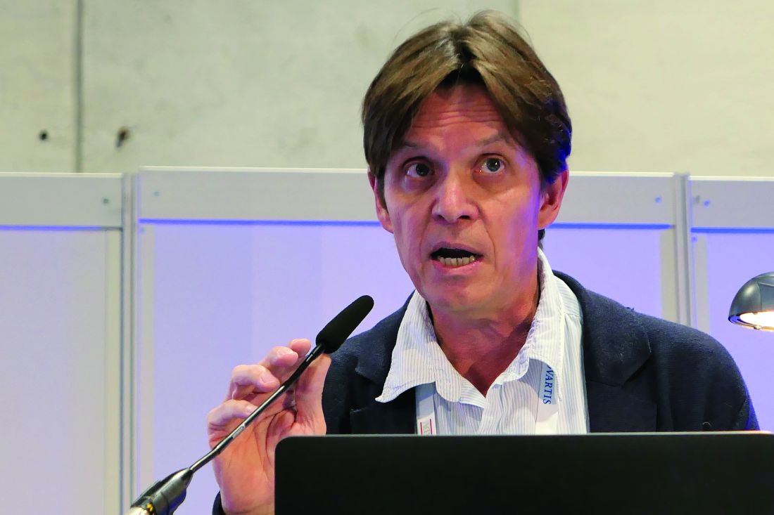
“We now have strong evidence that apremilast is an effective and safe therapy to treat oral ulcers in patients with Behçet’s syndrome. I think this is a major advance in the field,” declared Dr. Mahr, a rheumatologist at St. Gallen (Switzerland) Cantonal Hospital.
Based largely upon the results of the 12-week, double-blind portion of the phase 3 RELIEF trial, the Food and Drug Administration approved apremilast (Otezla) for the treatment of oral ulcers in patients with Behçet’s disease in the summer of 2019.
The safety profile of the oral phosphodiesterase-4 inhibitor was as seen in other studies, including in patients with psoriatic arthritis, an FDA-approved indication for the drug since 2014. The main side effects in the long-term extension of RELIEF were diarrhea and nausea, typically mild or moderate in nature and roughly twice as frequent as in placebo-treated controls in the double-blind study phase.
“At the end of the day, at week 64, only 12% of patients treated with apremilast during the entire 64 weeks discontinued the drug due to a treatment-emergent adverse event, which I believe is a good indicator of the safety of this medication,” the rheumatologist said. “The overall feeling is that the benefit-to-risk ratio is very good and it’s a safe drug to prescribe.”
At the close of the initial 12-week, double-blind phase of RELIEF, 178 of the original 207 participants elected to enter the long-term extension, either staying on apremilast at 30 mg twice a day for an additional 52 weeks or switching to that regimen from placebo.
The focus of the long-term extension was on disease activity and quality of life outcomes. The results in patients who had switched from placebo to apremilast after 12 weeks proved to be reassuringly similar to outcomes in patients on the drug for the full duration. For example, the mean improvement on the patient-reported Behçet’s Syndrome Activity Scale was 18.6 points after 12 weeks of double-blind apremilast, 16.9 points after 64 weeks of continuous apremilast, and 16.8 points with 12 weeks of placebo followed by 52 weeks of active therapy.
After 12 weeks of double-blind apremilast, patients averaged a 3.4-point improvement on the Behçet’s Disease Quality of Life measure. After 64 weeks on the drug, the improvement over baseline was 3.6 points, while in the switch group it was 3.4 points. Similarly, on all three components of the SF-36 quality of life metric, the continuous apremilast group showed maintenance of effect from week 12 to week 64, while the placebo-to-apremilast group caught up. The same was true with regards to the Behçet’s Disease Current Activity Index, which encompasses measures of both the patient’s and clinician’s perception of disease activity.
At the outset of the RELIEF trial, participants averaged four oral ulcers. At week 64, the continuous apremilast group averaged 1.4 and the switch group 0.8, a nonsignificant difference.
Asked if apremilast had a favorable impact upon other manifestations of Behçet’s disease besides the oral ulcers, Dr. Mahr replied, “This is a very good question. People often wonder about it. We do, too. But this trial was not designed to capture less common manifestations of Behçet’s syndrome, such as genital ulcers. There have been some analyses done, but the number of patients who had genital ulcers at 12 weeks were very few. The same was true for eye manifestations. There was sort of a signal that it works, but we can’t prove it in a placebo-controlled trial.”
Dr. Mahr reported receiving research funding from and serving as a consultant to Celgene, the study sponsor.
MADRID – of the long-term extension phase of the pivotal RELIEF trial, Alfred Mahr, MD, PhD, reported at the annual congress of the European Academy of Dermatology and Venereology.
“We now have strong evidence that apremilast is an effective and safe therapy to treat oral ulcers in patients with Behçet’s syndrome. I think this is a major advance in the field,” declared Dr. Mahr, a rheumatologist at St. Gallen (Switzerland) Cantonal Hospital.
Based largely upon the results of the 12-week, double-blind portion of the phase 3 RELIEF trial, the Food and Drug Administration approved apremilast (Otezla) for the treatment of oral ulcers in patients with Behçet’s disease in the summer of 2019.
The safety profile of the oral phosphodiesterase-4 inhibitor was as seen in other studies, including in patients with psoriatic arthritis, an FDA-approved indication for the drug since 2014. The main side effects in the long-term extension of RELIEF were diarrhea and nausea, typically mild or moderate in nature and roughly twice as frequent as in placebo-treated controls in the double-blind study phase.
“At the end of the day, at week 64, only 12% of patients treated with apremilast during the entire 64 weeks discontinued the drug due to a treatment-emergent adverse event, which I believe is a good indicator of the safety of this medication,” the rheumatologist said. “The overall feeling is that the benefit-to-risk ratio is very good and it’s a safe drug to prescribe.”
At the close of the initial 12-week, double-blind phase of RELIEF, 178 of the original 207 participants elected to enter the long-term extension, either staying on apremilast at 30 mg twice a day for an additional 52 weeks or switching to that regimen from placebo.
The focus of the long-term extension was on disease activity and quality of life outcomes. The results in patients who had switched from placebo to apremilast after 12 weeks proved to be reassuringly similar to outcomes in patients on the drug for the full duration. For example, the mean improvement on the patient-reported Behçet’s Syndrome Activity Scale was 18.6 points after 12 weeks of double-blind apremilast, 16.9 points after 64 weeks of continuous apremilast, and 16.8 points with 12 weeks of placebo followed by 52 weeks of active therapy.
After 12 weeks of double-blind apremilast, patients averaged a 3.4-point improvement on the Behçet’s Disease Quality of Life measure. After 64 weeks on the drug, the improvement over baseline was 3.6 points, while in the switch group it was 3.4 points. Similarly, on all three components of the SF-36 quality of life metric, the continuous apremilast group showed maintenance of effect from week 12 to week 64, while the placebo-to-apremilast group caught up. The same was true with regards to the Behçet’s Disease Current Activity Index, which encompasses measures of both the patient’s and clinician’s perception of disease activity.
At the outset of the RELIEF trial, participants averaged four oral ulcers. At week 64, the continuous apremilast group averaged 1.4 and the switch group 0.8, a nonsignificant difference.
Asked if apremilast had a favorable impact upon other manifestations of Behçet’s disease besides the oral ulcers, Dr. Mahr replied, “This is a very good question. People often wonder about it. We do, too. But this trial was not designed to capture less common manifestations of Behçet’s syndrome, such as genital ulcers. There have been some analyses done, but the number of patients who had genital ulcers at 12 weeks were very few. The same was true for eye manifestations. There was sort of a signal that it works, but we can’t prove it in a placebo-controlled trial.”
Dr. Mahr reported receiving research funding from and serving as a consultant to Celgene, the study sponsor.
REPORTING FROM EADV 2019
Weekly tocilizumab provides durable benefits for giant cell arteritis patients
ATLANTA –
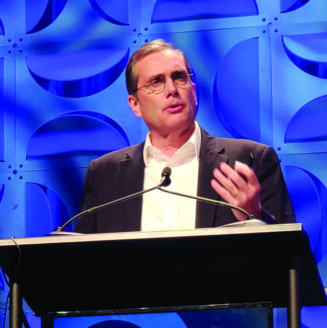
Of 250 patients treated in the double-blind portion of the trial, 215 entered the extension period (part 2 of the trial), and 197 (92%) completed all 3 years of the trial. A total of 38 of 81 (47%) extension participants in clinical remission (CR) at week 52 after receiving tocilizumab (Actemra) weekly maintained it throughout the extension, and 13 of 36 (36%) treated every other week maintained it, John Stone, MD, reported at the annual meeting of the American College of Rheumatology.
Notably, more of those 51 patients treated with tocilizumab who maintained CR throughout the extension were treatment free at the end of the extension, compared with those originally assigned to the placebo groups (65% vs. 45%), and their median time to first flare after stopping treatment was longer, said Dr. Stone, director of clinical rheumatology at Massachusetts General Hospital and associate professor of medicine in the division of rheumatology at Harvard Medical School, both in Boston.
For example, times to first flare were 575 and 428 days for the weekly and biweekly tocilizumab dosing groups – which each had 26-week prednisone tapering – but they were only 162 days in a placebo group with 26-week prednisone tapering and 295 days for patients in another placebo group with 52-week prednisone tapering, Dr. Stone explained. Retreatment with tocilizumab restored CR in patients who experienced flares after discontinuing treatment, he added.
Importantly, cumulative glucocorticoid doses over the 3 years were also lower in the tocilizumab groups (2,647 and 3,782 mg/day with weekly and biweekly tocilizumab and 5,248, and 5,323 mg/day in the placebo groups, respectively), Dr. Stone said.
No new safety signals were observed during the extension. Rates of serious adverse events (SAEs) and serious infections per 100 patient-years during the entire 3 years of the study were comparable among those who never received tocilizumab and those who received at least 1 dose (23.2 and 25.4 for SAEs and 4.6 and 3.5 for serious infections).
Detailed results of the extension period
GiACTA enrolled patients with GCA and randomized them to receive weekly or biweekly treatment with the interleukin-6 receptor–alpha inhibitor tocilizumab at doses of 162 mg delivered subcutaneously with a 26-week prednisone taper, or to the placebo groups with a 26- or 52-week prednisone taper. The results, reported in 2017, demonstrated the superiority of tocilizumab over placebo for providing sustained glucocorticoid-free remission and ultimately led to approval from Food and Drug Administration of the biologic agent for the treatment of GCA.
The current analysis was carried out to determine long-term safety, to explore maintenance of efficacy after discontinuation, and to “gain insight into the long-term glucocorticoid-sparing effects of tocilizumab,” Dr. Stone said.
He noted that the extension period was not randomized because of the way that patients in the “original four randomized groups had sorted themselves out into very different groups by virtue of their response in part 1 [of the trial],” which made randomization impossible because of the varying categories of patients.
Patients in the extension were also treated at investigators’ discretion to prevent “catastrophic GCA flares, vision loss, and other problems” that could potentially occur with discontinuation of the blinded injections in part 1, he said.
Nevertheless, the analysis “provides some incredibly important information about the management of GCA now,” he said, adding that the dramatic effect of the original randomization was still profound at 3 years “despite the fact that investigators could treat the patients as they wished.”
“Even at 3 years there was still a profound difference in the cumulative glucocorticoid [use], compared to those patients who were randomized to one of the placebo versus prednisone groups,” he explained.
However, the most important question from this study relates to what happened when weekly tocilizumab was stopped: Of the 81 patients in part 2 who were in CR on tocilizumab after part 1 of the study, 59 (73%) were not started on any treatment by their investigator, and of those, 25 (42%) maintained treatment-free remission for 2 full years following discontinuation of tocilizumab, he said.
What happened when prednisone was stopped?
In the absence of a direct comparison group (since the extension wasn’t a randomized, controlled part of the trial), the best comparison to that is what happens when prednisone is stopped, he noted.
“And we did that experiment in part 1” of the trial, he said. Overall, 68% of patients in the prednisone/placebo groups flared in 1-year of follow-up, and most of those did so even before they stopped prednisone. In the group that received placebo plus a 52-week prednisone taper, 49% of patients flared and “every single one of those patients flared before they got off prednisone,” he said.
“So in that context, 2 years of treatment-free remission following weekly tocilizumab discontinuation seems very good,” he added.
Of the patients who flared in part 2 of the trial, 11 whose investigator elected to treat them with only tocilizumab reentered remission in a median of 15.8 days, 13 treated with tocilizumab and glucocorticoids reentered remission in a median of 8.5 days (1 didn’t reenter remission at all), and 15 treated with only glucocorticoids reentered remission in a median of 38 days.
“There are a number of biases in these data, but they do point out the fact that some patients can be treated with tocilizumab alone,” Dr. Stone said. “I would be very careful doing that.”
The trial was not powered for the comparison of weekly versus biweekly dosing, he said. However, “multiple lines of investigation and analysis support the idea that weekly tocilizumab is more effective at controlling disease,” he added. But “if you want to be assured that you are doing everything you can to control the disease, weekly tocilizumab would be better than every-other-week tocilizumab,” he said.
GiACTA was funded by Roche. Dr. Stone reported research grants from Roche/Genentech and consulting fees from Chugal, Roche/Genentech, and Xencor.
SOURCE: Stone J et al. Arthritis Rheumatol. 2019;71(suppl 10), Abstract 808.
ATLANTA –
Of 250 patients treated in the double-blind portion of the trial, 215 entered the extension period (part 2 of the trial), and 197 (92%) completed all 3 years of the trial. A total of 38 of 81 (47%) extension participants in clinical remission (CR) at week 52 after receiving tocilizumab (Actemra) weekly maintained it throughout the extension, and 13 of 36 (36%) treated every other week maintained it, John Stone, MD, reported at the annual meeting of the American College of Rheumatology.
Notably, more of those 51 patients treated with tocilizumab who maintained CR throughout the extension were treatment free at the end of the extension, compared with those originally assigned to the placebo groups (65% vs. 45%), and their median time to first flare after stopping treatment was longer, said Dr. Stone, director of clinical rheumatology at Massachusetts General Hospital and associate professor of medicine in the division of rheumatology at Harvard Medical School, both in Boston.
For example, times to first flare were 575 and 428 days for the weekly and biweekly tocilizumab dosing groups – which each had 26-week prednisone tapering – but they were only 162 days in a placebo group with 26-week prednisone tapering and 295 days for patients in another placebo group with 52-week prednisone tapering, Dr. Stone explained. Retreatment with tocilizumab restored CR in patients who experienced flares after discontinuing treatment, he added.
Importantly, cumulative glucocorticoid doses over the 3 years were also lower in the tocilizumab groups (2,647 and 3,782 mg/day with weekly and biweekly tocilizumab and 5,248, and 5,323 mg/day in the placebo groups, respectively), Dr. Stone said.
No new safety signals were observed during the extension. Rates of serious adverse events (SAEs) and serious infections per 100 patient-years during the entire 3 years of the study were comparable among those who never received tocilizumab and those who received at least 1 dose (23.2 and 25.4 for SAEs and 4.6 and 3.5 for serious infections).
Detailed results of the extension period
GiACTA enrolled patients with GCA and randomized them to receive weekly or biweekly treatment with the interleukin-6 receptor–alpha inhibitor tocilizumab at doses of 162 mg delivered subcutaneously with a 26-week prednisone taper, or to the placebo groups with a 26- or 52-week prednisone taper. The results, reported in 2017, demonstrated the superiority of tocilizumab over placebo for providing sustained glucocorticoid-free remission and ultimately led to approval from Food and Drug Administration of the biologic agent for the treatment of GCA.
The current analysis was carried out to determine long-term safety, to explore maintenance of efficacy after discontinuation, and to “gain insight into the long-term glucocorticoid-sparing effects of tocilizumab,” Dr. Stone said.
He noted that the extension period was not randomized because of the way that patients in the “original four randomized groups had sorted themselves out into very different groups by virtue of their response in part 1 [of the trial],” which made randomization impossible because of the varying categories of patients.
Patients in the extension were also treated at investigators’ discretion to prevent “catastrophic GCA flares, vision loss, and other problems” that could potentially occur with discontinuation of the blinded injections in part 1, he said.
Nevertheless, the analysis “provides some incredibly important information about the management of GCA now,” he said, adding that the dramatic effect of the original randomization was still profound at 3 years “despite the fact that investigators could treat the patients as they wished.”
“Even at 3 years there was still a profound difference in the cumulative glucocorticoid [use], compared to those patients who were randomized to one of the placebo versus prednisone groups,” he explained.
However, the most important question from this study relates to what happened when weekly tocilizumab was stopped: Of the 81 patients in part 2 who were in CR on tocilizumab after part 1 of the study, 59 (73%) were not started on any treatment by their investigator, and of those, 25 (42%) maintained treatment-free remission for 2 full years following discontinuation of tocilizumab, he said.
What happened when prednisone was stopped?
In the absence of a direct comparison group (since the extension wasn’t a randomized, controlled part of the trial), the best comparison to that is what happens when prednisone is stopped, he noted.
“And we did that experiment in part 1” of the trial, he said. Overall, 68% of patients in the prednisone/placebo groups flared in 1-year of follow-up, and most of those did so even before they stopped prednisone. In the group that received placebo plus a 52-week prednisone taper, 49% of patients flared and “every single one of those patients flared before they got off prednisone,” he said.
“So in that context, 2 years of treatment-free remission following weekly tocilizumab discontinuation seems very good,” he added.
Of the patients who flared in part 2 of the trial, 11 whose investigator elected to treat them with only tocilizumab reentered remission in a median of 15.8 days, 13 treated with tocilizumab and glucocorticoids reentered remission in a median of 8.5 days (1 didn’t reenter remission at all), and 15 treated with only glucocorticoids reentered remission in a median of 38 days.
“There are a number of biases in these data, but they do point out the fact that some patients can be treated with tocilizumab alone,” Dr. Stone said. “I would be very careful doing that.”
The trial was not powered for the comparison of weekly versus biweekly dosing, he said. However, “multiple lines of investigation and analysis support the idea that weekly tocilizumab is more effective at controlling disease,” he added. But “if you want to be assured that you are doing everything you can to control the disease, weekly tocilizumab would be better than every-other-week tocilizumab,” he said.
GiACTA was funded by Roche. Dr. Stone reported research grants from Roche/Genentech and consulting fees from Chugal, Roche/Genentech, and Xencor.
SOURCE: Stone J et al. Arthritis Rheumatol. 2019;71(suppl 10), Abstract 808.
REPORTING FROM ACR 2019