User login
Demyelinating diseases, especially MS, disrupt normal brain development in children
BERLIN – Demyelinating diseases appear to disrupt white matter development in children, slowing the trajectory of brain growth to almost unmeasurable levels, based on results from a single-center comparison study of 213 individuals.

While children with multiple sclerosis (MS) showed the most severe slowing, even a single demyelinating event slowed white matter growth, Robert A. Brown, PhD, said at the annual congress of the European Committees for Treatment and Research in Multiple Sclerosis.
Dr. Brown of the Montreal Neurological Institute at McGill University, Montreal, employed a signal mass correction of consecutive brain MRIs enhanced with magnetization transfer. Magnetization transfer ratio (MTR) quantifies myelin more effectively than does other imaging enhancement modalities, Dr. Brown said.
“It labels the macromolecules of myelin and correlates almost perfectly with Luxol fast blue stain on histology,” he said. And by measuring myelin instead of whole-brain volume, MTR sidesteps the confounders of inflammation and edema. “When tissue swells, the water dilutes the myelin. MRI is really sensitive to density, so dilution with water lowers that signal.”
But MTR isn’t failsafe either, he said, especially in teens. “A cautionary note: In healthy adolescents, white matter MTR can actually decrease, not increase, not because they are losing myelin but because the axons in brain tissue are growing so fast that they outstrip the production of new myelin. So, we can get another dilution effect here, except that instead of water, axons are diluting the myelin. We have to take that into account when using MTR.”
A volume-corrected MTR calculates both mass and volume to give what Dr. Brown termed signal mass. “We have demonstrated previously that signal mass is about twice as powerful as volume change alone for measuring the differences [in brain volume] between adults with MS and healthy controls.”
The study he presented at ECTRIMS used this technique to examine the trajectory of white matter change in a cohort of children from the Canadian Pediatric Demyelinating Disease Study who were all scanned at the same site in the same center. He compared brain volume at baseline and 1 year in 102 children with a monophasic demyelinating disease, 87 with MS, and 24 healthy, age-matched controls.
The children with MS were a median of about 17 years old at baseline, while those with a monophasic event and healthy controls were a median of about 12 years old. Median follow-up was 1 year in the healthy controls, 2 years in the MS cohort, and 4 years in the monophasic group. The investigators adjusted their comparisons for sex, since both bioavailable testosterone and androgen-receptor activity correlate with decreased MTR in young men. This doesn’t mean, though, that testosterone decreases myelination. Rather, it’s postulated that testosterone increases axonal caliber, which would decrease the number of neurons in each imaging voxel and, thus, the MTR signal (J Neurosci. 2008 Sep 17;28[38]:9519-24).
In the volume-only assessment, white matter in healthy controls increased at a rate of about 0.5% per year. White matter growth was about 0.2% per year in children with monophasic demyelination, which was significantly lower than in healthy controls.
“The MS children had no white matter growth that we could measure,” with an annual change of about 0.01%, Dr. Brown said. “It looks like a failure of normal development and was significantly lower than what we saw in the children with a demyelination event.”
MTR showed the expected age-associated decreases, which were highest among those with MS: –0.8% per year in healthy controls, –0.6% per year in those with a monophasic event, and –0.9% per year in those with MS.
The signal mass change showed the whole picture, Dr. Brown said. Signal mass declined 0.3% per year in healthy controls, 0.5% per year in the monophasic group, and 0.9% per year in the MS group – a significantly worse trajectory than either the control subjects or those with a monophasic event.
“Signal mass puts it all together and gives us the total picture of tissue loss, with quite severe loss in children with MS. It seems as though both monophasic insult and pediatric-onset MS disrupt brain development.”
Dr. Brown has been a consultant for NeuroRx Research and Biogen.
SOURCE: Brown RA et al. Mult Scler. 2018;24(S2):27-8, Abstract 63.
BERLIN – Demyelinating diseases appear to disrupt white matter development in children, slowing the trajectory of brain growth to almost unmeasurable levels, based on results from a single-center comparison study of 213 individuals.
While children with multiple sclerosis (MS) showed the most severe slowing, even a single demyelinating event slowed white matter growth, Robert A. Brown, PhD, said at the annual congress of the European Committees for Treatment and Research in Multiple Sclerosis.
Dr. Brown of the Montreal Neurological Institute at McGill University, Montreal, employed a signal mass correction of consecutive brain MRIs enhanced with magnetization transfer. Magnetization transfer ratio (MTR) quantifies myelin more effectively than does other imaging enhancement modalities, Dr. Brown said.
“It labels the macromolecules of myelin and correlates almost perfectly with Luxol fast blue stain on histology,” he said. And by measuring myelin instead of whole-brain volume, MTR sidesteps the confounders of inflammation and edema. “When tissue swells, the water dilutes the myelin. MRI is really sensitive to density, so dilution with water lowers that signal.”
But MTR isn’t failsafe either, he said, especially in teens. “A cautionary note: In healthy adolescents, white matter MTR can actually decrease, not increase, not because they are losing myelin but because the axons in brain tissue are growing so fast that they outstrip the production of new myelin. So, we can get another dilution effect here, except that instead of water, axons are diluting the myelin. We have to take that into account when using MTR.”
A volume-corrected MTR calculates both mass and volume to give what Dr. Brown termed signal mass. “We have demonstrated previously that signal mass is about twice as powerful as volume change alone for measuring the differences [in brain volume] between adults with MS and healthy controls.”
The study he presented at ECTRIMS used this technique to examine the trajectory of white matter change in a cohort of children from the Canadian Pediatric Demyelinating Disease Study who were all scanned at the same site in the same center. He compared brain volume at baseline and 1 year in 102 children with a monophasic demyelinating disease, 87 with MS, and 24 healthy, age-matched controls.
The children with MS were a median of about 17 years old at baseline, while those with a monophasic event and healthy controls were a median of about 12 years old. Median follow-up was 1 year in the healthy controls, 2 years in the MS cohort, and 4 years in the monophasic group. The investigators adjusted their comparisons for sex, since both bioavailable testosterone and androgen-receptor activity correlate with decreased MTR in young men. This doesn’t mean, though, that testosterone decreases myelination. Rather, it’s postulated that testosterone increases axonal caliber, which would decrease the number of neurons in each imaging voxel and, thus, the MTR signal (J Neurosci. 2008 Sep 17;28[38]:9519-24).
In the volume-only assessment, white matter in healthy controls increased at a rate of about 0.5% per year. White matter growth was about 0.2% per year in children with monophasic demyelination, which was significantly lower than in healthy controls.
“The MS children had no white matter growth that we could measure,” with an annual change of about 0.01%, Dr. Brown said. “It looks like a failure of normal development and was significantly lower than what we saw in the children with a demyelination event.”
MTR showed the expected age-associated decreases, which were highest among those with MS: –0.8% per year in healthy controls, –0.6% per year in those with a monophasic event, and –0.9% per year in those with MS.
The signal mass change showed the whole picture, Dr. Brown said. Signal mass declined 0.3% per year in healthy controls, 0.5% per year in the monophasic group, and 0.9% per year in the MS group – a significantly worse trajectory than either the control subjects or those with a monophasic event.
“Signal mass puts it all together and gives us the total picture of tissue loss, with quite severe loss in children with MS. It seems as though both monophasic insult and pediatric-onset MS disrupt brain development.”
Dr. Brown has been a consultant for NeuroRx Research and Biogen.
SOURCE: Brown RA et al. Mult Scler. 2018;24(S2):27-8, Abstract 63.
REPORTING FROM ECTRIMS 2018
Key clinical point: Demyelinating disorders disrupt brain growth in children.
Major finding: Children with MS had virtually no white matter growth, and those with ADM lagged significantly behind controls.
Study details: The prospective imaging study comprised 24 controls, 102 with an ADM, and 87 with MS.
Disclosures: Dr. Brown has been a consultant for NeuroRx Research and Biogen Idec.
Source: Brown RA et al. Mult Scler. 2018;24(S2):27-8, Abstract 63
Ganglion stimulation boosts cerebral blood flow, improves stroke outcomes
MONTREAL – Stimulation of the sphenopalatine ganglion (SPG) using a small, implanted electrode for 5 days in patients who had just had an acute ischemic stroke led to statistically significant and clinically meaningful improvements in the subset of patients with confirmed cortical involvement in a pivotal, sham-controlled trial.
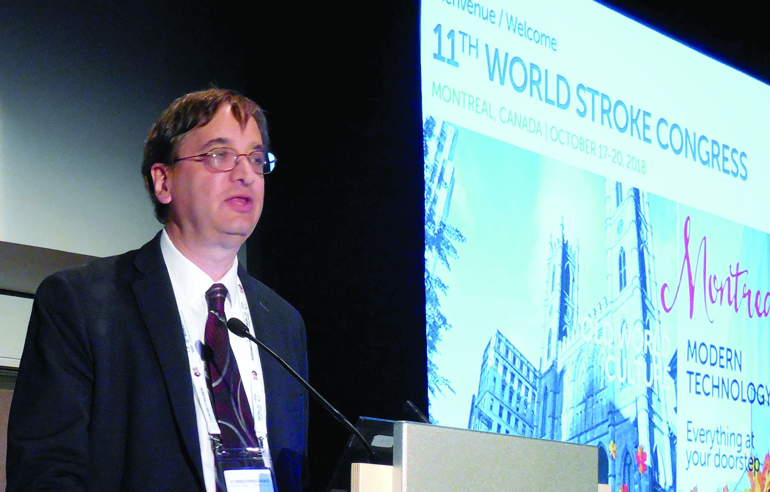
SPG stimulation started within 24 hours of an acute ischemic stroke “reduced poststroke disability over the entire outcome range and increased the proportion of patients who were alive and independent 3 months after their stroke” in the subgroup with a confirmed cortical infarction (CCI), Jeffrey L. Saver, MD, said at the World Stroke Congress. Five days of SPG stimulation, done once daily starting within 24 hours of stroke onset, “enhances ipsilateral collateral blood flow” and may also stabilize the blood brain barrier, explained Dr. Saver, professor of neurology and director of the stroke center at the University of California, Los Angeles. The study included a prespecified primary endpoint analysis that focused exclusively on the CCI subgroup, 52% of the total enrolled population.
If the reported data result in Food and Drug Administration marketing approval for the system, Dr. Saver said that he anticipated “substantial uptake” of the strategy, which he tested in patients who had not undergone thrombectomy or thrombolysis treatment. In current U.S. practice, there is “a large group of patients with a missed opportunity for recanalization” who would be candidates for treatment with SPG stimulation, a treatment that appeared to provide benefits beyond current standard care, he said in an interview.
Ongoing studies are also testing whether SPG stimulation can benefit acute ischemic stroke patients who have already undergone treatment with thrombectomy or thrombolysis, he added. The same SPG stimulation device is additionally undergoing U.S. testing as a treatment for headache and has regulatory approval in the European Union for treating headache and migraine.
The ImpACT-24B (Implant for Augmentation of Cerebral Blood Flow Trial, Effectiveness and Safety in a 24-Hour Window) trial involved 1,000 patients at 73 centers in 18 countries, including the United States. The investigators enrolled acute ischemic stroke patients 8-24 hours after stroke onset who had a National Institutes of Health Stroke Scale (NIHSS) score of 7-18.
Each patient received an implant of a short, thin metal electrode placed through the soft palate at the rear roof of the mouth, near the SPG. Neurologists primarily performed the implants in a procedure that had a “skin to skin” time of less than 5 minutes. Patients received either electrical stimulation or a sham stimulation through the electrode immediately after placement and then daily for the next 4 days. The investigators titrated the strength of the treatment stimulation in each patient to maximize its strength while maintaining patient comfort. Subsequent analysis of the results showed that the stronger the tolerated stimulation, the bigger the treatment effect in a clear dose-response pattern, Dr. Saver reported.
The study’s primary endpoint was improvement in the modified Rankin scale (mRS) score at 90 days after the index stroke when measured against historical expectations. By this measure, the overall study cohort showed a small, statistically insignificant improvement in actively treated patients, compared with sham-treated patients. However, in the prespecified, coprimary endpoint cohort of patients with a CCI, active treatment resulted in 50% of patients having a better-than-expected 90-day outcome, compared with 40% of controls, a 48% relative improvement in this measure that met the prespecified definition of statistical significance. The results also showed about a 50% relative improvement in each of three secondary outcomes in the CCI cohort: the percentage of patients with a mRS score of 0-2 after 90 days, the percentage with a mRS score of 0-3 after 90 days, and average stroke-related quality of life at 90 days.
Dr. Saver also reported results of a meta-analysis that combined the results he reported from 520 patients with CCI with results from 87 CCI patients enrolled in the preceding pilot study of this treatment strategy, ImpACT-1. The pilot findings were completely consistent and when combined with the current results strengthened the statistical significance of the primary and secondary endpoints.
“There is a compelling story” of efficacy based on the study results, the dose-response relationship, and the meta-analysis results, Dr. Saver said. “I think it’s a very strong case.”
He also reported “no safety concerns” raised in the new study, with no serious adverse effects seen in or experienced by the patients on active treatment.
“The data are compelling” for safety and efficacy, for this novel approach for treating acute ischemic stroke, commented Pooja Khatri, MD, professor of neurology and director of the acute stroke program at the University of Cincinnati.
The study was sponsored by BrainsGate, the company developing the tested device. Dr. Saver has been a consultant to BrainsGate. Dr. Khatri has been a consultant to Biogen, Greenwich, Lumosa, and PTC Therapeutics.
SOURCE: Saver J et al. Int J. Stroke. 2018 Oct;13(2S):28, Abstract 104.
MONTREAL – Stimulation of the sphenopalatine ganglion (SPG) using a small, implanted electrode for 5 days in patients who had just had an acute ischemic stroke led to statistically significant and clinically meaningful improvements in the subset of patients with confirmed cortical involvement in a pivotal, sham-controlled trial.
SPG stimulation started within 24 hours of an acute ischemic stroke “reduced poststroke disability over the entire outcome range and increased the proportion of patients who were alive and independent 3 months after their stroke” in the subgroup with a confirmed cortical infarction (CCI), Jeffrey L. Saver, MD, said at the World Stroke Congress. Five days of SPG stimulation, done once daily starting within 24 hours of stroke onset, “enhances ipsilateral collateral blood flow” and may also stabilize the blood brain barrier, explained Dr. Saver, professor of neurology and director of the stroke center at the University of California, Los Angeles. The study included a prespecified primary endpoint analysis that focused exclusively on the CCI subgroup, 52% of the total enrolled population.
If the reported data result in Food and Drug Administration marketing approval for the system, Dr. Saver said that he anticipated “substantial uptake” of the strategy, which he tested in patients who had not undergone thrombectomy or thrombolysis treatment. In current U.S. practice, there is “a large group of patients with a missed opportunity for recanalization” who would be candidates for treatment with SPG stimulation, a treatment that appeared to provide benefits beyond current standard care, he said in an interview.
Ongoing studies are also testing whether SPG stimulation can benefit acute ischemic stroke patients who have already undergone treatment with thrombectomy or thrombolysis, he added. The same SPG stimulation device is additionally undergoing U.S. testing as a treatment for headache and has regulatory approval in the European Union for treating headache and migraine.
The ImpACT-24B (Implant for Augmentation of Cerebral Blood Flow Trial, Effectiveness and Safety in a 24-Hour Window) trial involved 1,000 patients at 73 centers in 18 countries, including the United States. The investigators enrolled acute ischemic stroke patients 8-24 hours after stroke onset who had a National Institutes of Health Stroke Scale (NIHSS) score of 7-18.
Each patient received an implant of a short, thin metal electrode placed through the soft palate at the rear roof of the mouth, near the SPG. Neurologists primarily performed the implants in a procedure that had a “skin to skin” time of less than 5 minutes. Patients received either electrical stimulation or a sham stimulation through the electrode immediately after placement and then daily for the next 4 days. The investigators titrated the strength of the treatment stimulation in each patient to maximize its strength while maintaining patient comfort. Subsequent analysis of the results showed that the stronger the tolerated stimulation, the bigger the treatment effect in a clear dose-response pattern, Dr. Saver reported.
The study’s primary endpoint was improvement in the modified Rankin scale (mRS) score at 90 days after the index stroke when measured against historical expectations. By this measure, the overall study cohort showed a small, statistically insignificant improvement in actively treated patients, compared with sham-treated patients. However, in the prespecified, coprimary endpoint cohort of patients with a CCI, active treatment resulted in 50% of patients having a better-than-expected 90-day outcome, compared with 40% of controls, a 48% relative improvement in this measure that met the prespecified definition of statistical significance. The results also showed about a 50% relative improvement in each of three secondary outcomes in the CCI cohort: the percentage of patients with a mRS score of 0-2 after 90 days, the percentage with a mRS score of 0-3 after 90 days, and average stroke-related quality of life at 90 days.
Dr. Saver also reported results of a meta-analysis that combined the results he reported from 520 patients with CCI with results from 87 CCI patients enrolled in the preceding pilot study of this treatment strategy, ImpACT-1. The pilot findings were completely consistent and when combined with the current results strengthened the statistical significance of the primary and secondary endpoints.
“There is a compelling story” of efficacy based on the study results, the dose-response relationship, and the meta-analysis results, Dr. Saver said. “I think it’s a very strong case.”
He also reported “no safety concerns” raised in the new study, with no serious adverse effects seen in or experienced by the patients on active treatment.
“The data are compelling” for safety and efficacy, for this novel approach for treating acute ischemic stroke, commented Pooja Khatri, MD, professor of neurology and director of the acute stroke program at the University of Cincinnati.
The study was sponsored by BrainsGate, the company developing the tested device. Dr. Saver has been a consultant to BrainsGate. Dr. Khatri has been a consultant to Biogen, Greenwich, Lumosa, and PTC Therapeutics.
SOURCE: Saver J et al. Int J. Stroke. 2018 Oct;13(2S):28, Abstract 104.
REPORTING FROM THE WORLD STROKE CONGRESS
Key clinical point: Sphenopalatine ganglion stimulation of acute ischemic stroke patients boosted cerebral blood flow and improved 90-day outcomes in patients with confirmed cortical infarctions.
Major finding: For confirmed cortical infarctions ganglion stimulation led to a 48% higher rate of better-than-expected outcomes, compared with controls.
Study details: ImpACT-24B, a multicenter pivotal trial with 1,000 acute ischemic stroke patients.
Disclosures: The study was sponsored by BrainsGate, the company developing the tested device. Dr. Saver has been a consultant to BrainsGate. Dr. Khatri has been a consultant to Biogen, Greenwich, Lumosa, and PTC Therapeutics.
Source: Saver J et al. Int J. Stroke. 2018 Oct;13(2S):28, Abstract 104.
Increased risk of atrial fibrillation with migraine aura
The presence of visual aura during migraine is associated with an increased risk of atrial fibrillation, a study in Neurology has found.

Researchers reported an analysis of data from the longitudinal, community-based Atherosclerosis Risk in Communities (ARIC) Study, which included 11,939 individuals with no history of atrial fibrillation or stroke. Of these, 426 experienced migraines with visual aura, 1,090 experienced migraines without aura, 1,018 experienced nonmigraine headache, and 9,405 experienced no headache.
After adjustment for age and sex, individuals who had migraine with visual aura showed a significant 46% increase in the risk of incident atrial fibrillation when compared with those who experienced migraine without aura and a 39% increased risk when compared with individuals who did not experience headache (P = .004). After adjustment for risk factors such as hypertension, smoking, coronary artery disease, and congestive heart failure, the hazard ratio of incident atrial fibrillation was 1.30 for migraineurs with aura, compared with people without headache. In addition, the hazard ratio of incident atrial fibrillation was 1.39 for migraineurs with aura, compared with migraineurs without aura.
In contrast, individuals who experienced migraines without aura did not show a significantly increased risk of atrial fibrillation.
“This finding has important clinical implications and may help us better understand the atrial fibrillation mediation of the migraine-stroke link,” wrote Souvik Sen, MD, MPH, a professor in the department of neurology at the University of South Carolina, Columbia, and his coauthors. “A randomized clinical trial may help ascertain whether patients with migraine with visual aura may benefit from atrial fibrillation detection and subsequent anticoagulation or antiplatelet therapy as a primary stroke prevention strategy.”
The study also showed a significant interaction with age and sex. While men who experienced migraine with aura had an 89% higher risk of atrial fibrillation, women with aura showed no increase in risk, compared with individuals who experienced no headache. Similarly, only individuals aged 60 years or older who experienced migraine with aura showed an increased risk of atrial fibrillation, while those younger than 60 years did not.
The authors noted that previous case reports have recorded the incidence of atrial fibrillation during a migraine attack. Autonomic dysfunction influences the pathophysiology of atrial fibrillation and migraine.
“Cardiac arrhythmia recordings have been shown to be present in ECGs of patients while experiencing migraine headaches as compared with migraine-free phases,” they wrote. “This hypothesis is further supported by atrial fibrillation ablation procedures that have shown tendencies to reduce migraine symptoms and frequencies.”
In regard to the role that migraine aura played in this, they speculated as to whether migraine aura could be the result of cardioembolic stroke that might have occurred because of the atrial fibrillation.
Overall, 167 patients had incident cardioembolic strokes, and researchers suggested strokes in 87% of these cases could be attributed to the atrial fibrillation that came before the stroke.
The stroke incidence rate also was around twice as high in individuals who experienced migraine with aura, compared with those who experienced migraine without aura (4.1 per 1,000 person-years vs. 2.07 per 1,000 person-years).
The study authors acknowledged that patent foramen ovale, which was not assessed in ARIC, is a possible confounder. Previous studies have showed that patent foramen ovale is more common in younger individuals with migraine and particularly in patients who experience migraine with aura.
However, they also noted that trials of patent foramen ovale closures as a treatment for migraine have not shown success in reducing migraine frequency and, therefore, argued against patent foramen ovale as being a major confounder.
The study was supported by the National Heart, Lung, and Blood Institute and the American Heart Association. One author declared grants from the National Institutes of health, one declared research support from Tian Medical, and one author is an associate editor for Neurology. No other conflicts of interest were declared.
SOURCE: Sen S et al. Neurology. 2018;91:1-9.
This article was updated 12/12/18.
The presence of visual aura during migraine is associated with an increased risk of atrial fibrillation, a study in Neurology has found.
Researchers reported an analysis of data from the longitudinal, community-based Atherosclerosis Risk in Communities (ARIC) Study, which included 11,939 individuals with no history of atrial fibrillation or stroke. Of these, 426 experienced migraines with visual aura, 1,090 experienced migraines without aura, 1,018 experienced nonmigraine headache, and 9,405 experienced no headache.
After adjustment for age and sex, individuals who had migraine with visual aura showed a significant 46% increase in the risk of incident atrial fibrillation when compared with those who experienced migraine without aura and a 39% increased risk when compared with individuals who did not experience headache (P = .004). After adjustment for risk factors such as hypertension, smoking, coronary artery disease, and congestive heart failure, the hazard ratio of incident atrial fibrillation was 1.30 for migraineurs with aura, compared with people without headache. In addition, the hazard ratio of incident atrial fibrillation was 1.39 for migraineurs with aura, compared with migraineurs without aura.
In contrast, individuals who experienced migraines without aura did not show a significantly increased risk of atrial fibrillation.
“This finding has important clinical implications and may help us better understand the atrial fibrillation mediation of the migraine-stroke link,” wrote Souvik Sen, MD, MPH, a professor in the department of neurology at the University of South Carolina, Columbia, and his coauthors. “A randomized clinical trial may help ascertain whether patients with migraine with visual aura may benefit from atrial fibrillation detection and subsequent anticoagulation or antiplatelet therapy as a primary stroke prevention strategy.”
The study also showed a significant interaction with age and sex. While men who experienced migraine with aura had an 89% higher risk of atrial fibrillation, women with aura showed no increase in risk, compared with individuals who experienced no headache. Similarly, only individuals aged 60 years or older who experienced migraine with aura showed an increased risk of atrial fibrillation, while those younger than 60 years did not.
The authors noted that previous case reports have recorded the incidence of atrial fibrillation during a migraine attack. Autonomic dysfunction influences the pathophysiology of atrial fibrillation and migraine.
“Cardiac arrhythmia recordings have been shown to be present in ECGs of patients while experiencing migraine headaches as compared with migraine-free phases,” they wrote. “This hypothesis is further supported by atrial fibrillation ablation procedures that have shown tendencies to reduce migraine symptoms and frequencies.”
In regard to the role that migraine aura played in this, they speculated as to whether migraine aura could be the result of cardioembolic stroke that might have occurred because of the atrial fibrillation.
Overall, 167 patients had incident cardioembolic strokes, and researchers suggested strokes in 87% of these cases could be attributed to the atrial fibrillation that came before the stroke.
The stroke incidence rate also was around twice as high in individuals who experienced migraine with aura, compared with those who experienced migraine without aura (4.1 per 1,000 person-years vs. 2.07 per 1,000 person-years).
The study authors acknowledged that patent foramen ovale, which was not assessed in ARIC, is a possible confounder. Previous studies have showed that patent foramen ovale is more common in younger individuals with migraine and particularly in patients who experience migraine with aura.
However, they also noted that trials of patent foramen ovale closures as a treatment for migraine have not shown success in reducing migraine frequency and, therefore, argued against patent foramen ovale as being a major confounder.
The study was supported by the National Heart, Lung, and Blood Institute and the American Heart Association. One author declared grants from the National Institutes of health, one declared research support from Tian Medical, and one author is an associate editor for Neurology. No other conflicts of interest were declared.
SOURCE: Sen S et al. Neurology. 2018;91:1-9.
This article was updated 12/12/18.
FROM NEUROLOGY
Key clinical point: Aura in migraine is associated with an increased risk of atrial fibrillation.
Major finding: Individuals who experience migraine with aura have a 39% higher risk of atrial fibrillation than do those without aura or without migraine.
Study details: The longitudinal, community-based Atherosclerosis Risk in Communities Study in 11,939 individuals.
Disclosures: The study was supported by the National Heart, Lung, and Blood Institute and the American Heart Association. One author declared grants from the National Institutes of health, one declared research support from Tian Medical, and one author is an associate editor for Neurology. No other conflicts of interest were declared.
Source: Sen S et al. Neurology. 2018;91:1-9.
Children with headache disorders may benefit from anti-CGRP mAb treatment
Use of anti–calcitonin gene-related peptide (CGRP) monoclonal antibodies (mAbs) may benefit children with more than 8 headache days each month, a high Pediatric Migraine Disability Assessment (PedMIDAS) score, and failure of other treatments; however, researchers cautioned that long-term safety outcomes for the treatment are not yet known, according to a recent set of recommendations published in the journal Headache.

Christina L. Szperka, MD, MSCE, of the division of neurology at the Children’s Hospital of Philadelphia and members of the Pediatric and Adolescent Headache special interest group of the American Headache Society discussed the topic of anti-CGRP mAbs at the 2018 Annual Scientific Meeting of the American Headache Society. They noted clinical outcomes for anti-CGRP mAbs in pediatric patients will likely not be available for several years and created a set of recommendations based on expert opinion of anti-CGRP mAb use in children and adolescents.
Their recommendations support using anti-CGRP mAbs for children with migraine if patients meet the following criteria: headache frequency exceeding 8 headache days per month; a PedMIDAS score of 30 or greater; failure of two or more therapies, such as pharmacologic, nonpharmacologic, or nutraceutical ones; and in patients who are past puberty.
The special interest group recommended against use of anti-CGRP mAbs in children and adolescents with recent meningitis, recent peripheral nerve injury, neurosurgery, or a central nervous system injury caused by a potentially compromised blood-brain barrier. Children and adolescents with immunodeficiency, receiving immunosuppressive medications, with structural heart defects, with pulmonary hypertension, with coronary artery disease, with cardiomyopathy, or at risk for stroke should also avoid use of anti-CGRP mAbs. Anti-CGRP mAbs are also potentially teratogenic and should not be used by adolescents or women who are pregnant, breastfeeding, or have a pregnancy wish.
Pediatric patients with significant osteoporosis or bone disease should be monitored when prescribed anti-CGRP mAbs, and the recommendations specified monitoring height and linear growth or waiting until after puberty to prescribe anti-CGRP mAbs. Although there is currently no evidence that use of anti-CGRP mAb requires pituitary hormone monitoring, the recommendations noted that weight and body mass index should also be observed.
“Pediatric and adolescent trials of anti-CGRP mAbs should be designed to maximize the chances of determining efficacy in these age groups and should focus on those who have not been successful with current multidisciplinary care,” Dr. Szperka and her colleagues wrote in the recommendations. “In the interim, the use of anti-CGRP mAbs for the treatment of headache disorders in children and adolescents may be considered in appropriate cases but should be done with close follow-up and attention to patient characteristics such as age, pubertal state, and medical comorbidities.”
Dr. Szperka receives grant support from Pfizer and Amgen and research funding from NIH. Other authors have reported grants, consulting fees, speaking fees, royalties, advisory board memberships, speaker’s bureau memberships and travel funds from Alder, Allergan, American Academy of Neurology, Amgen, Aralez, Avanir, Autonomic Technologies Inc., Biohaven, Cambridge University Press, Curelator, Depomed, Dr. Reddy’s Laboratories, Electrocore, eNeura, Genentech, Healint, Impax, JAMA Neurology, Journal Watch, Lilly, Massachusetts Medical Society, MedDay, MedicoLegal, Merck, NIH, Novartis, Oxford University Press, Quest Diagnostics, Scion, Supernus, Teva, Trigemina Inc., Upsher-Smith, UpToDate, Wolters Kluwer and Zosano.
SOURCE: Szperka CL et al. Headache. 2018. doi: 10.1111/head.13414.
Use of anti–calcitonin gene-related peptide (CGRP) monoclonal antibodies (mAbs) may benefit children with more than 8 headache days each month, a high Pediatric Migraine Disability Assessment (PedMIDAS) score, and failure of other treatments; however, researchers cautioned that long-term safety outcomes for the treatment are not yet known, according to a recent set of recommendations published in the journal Headache.
Christina L. Szperka, MD, MSCE, of the division of neurology at the Children’s Hospital of Philadelphia and members of the Pediatric and Adolescent Headache special interest group of the American Headache Society discussed the topic of anti-CGRP mAbs at the 2018 Annual Scientific Meeting of the American Headache Society. They noted clinical outcomes for anti-CGRP mAbs in pediatric patients will likely not be available for several years and created a set of recommendations based on expert opinion of anti-CGRP mAb use in children and adolescents.
Their recommendations support using anti-CGRP mAbs for children with migraine if patients meet the following criteria: headache frequency exceeding 8 headache days per month; a PedMIDAS score of 30 or greater; failure of two or more therapies, such as pharmacologic, nonpharmacologic, or nutraceutical ones; and in patients who are past puberty.
The special interest group recommended against use of anti-CGRP mAbs in children and adolescents with recent meningitis, recent peripheral nerve injury, neurosurgery, or a central nervous system injury caused by a potentially compromised blood-brain barrier. Children and adolescents with immunodeficiency, receiving immunosuppressive medications, with structural heart defects, with pulmonary hypertension, with coronary artery disease, with cardiomyopathy, or at risk for stroke should also avoid use of anti-CGRP mAbs. Anti-CGRP mAbs are also potentially teratogenic and should not be used by adolescents or women who are pregnant, breastfeeding, or have a pregnancy wish.
Pediatric patients with significant osteoporosis or bone disease should be monitored when prescribed anti-CGRP mAbs, and the recommendations specified monitoring height and linear growth or waiting until after puberty to prescribe anti-CGRP mAbs. Although there is currently no evidence that use of anti-CGRP mAb requires pituitary hormone monitoring, the recommendations noted that weight and body mass index should also be observed.
“Pediatric and adolescent trials of anti-CGRP mAbs should be designed to maximize the chances of determining efficacy in these age groups and should focus on those who have not been successful with current multidisciplinary care,” Dr. Szperka and her colleagues wrote in the recommendations. “In the interim, the use of anti-CGRP mAbs for the treatment of headache disorders in children and adolescents may be considered in appropriate cases but should be done with close follow-up and attention to patient characteristics such as age, pubertal state, and medical comorbidities.”
Dr. Szperka receives grant support from Pfizer and Amgen and research funding from NIH. Other authors have reported grants, consulting fees, speaking fees, royalties, advisory board memberships, speaker’s bureau memberships and travel funds from Alder, Allergan, American Academy of Neurology, Amgen, Aralez, Avanir, Autonomic Technologies Inc., Biohaven, Cambridge University Press, Curelator, Depomed, Dr. Reddy’s Laboratories, Electrocore, eNeura, Genentech, Healint, Impax, JAMA Neurology, Journal Watch, Lilly, Massachusetts Medical Society, MedDay, MedicoLegal, Merck, NIH, Novartis, Oxford University Press, Quest Diagnostics, Scion, Supernus, Teva, Trigemina Inc., Upsher-Smith, UpToDate, Wolters Kluwer and Zosano.
SOURCE: Szperka CL et al. Headache. 2018. doi: 10.1111/head.13414.
FROM HEADACHE
Key clinical point: Treatment of headache disorders with anti–calcitonin gene-related peptide (CGRP) monoclonal antibodies (mAbs) in children and adolescents may be indicated in some cases.
Major finding: Pediatric patients with more than eight headache days per month, PedMIDAS score of 30 or higher, and failure of other pharmacological, nonpharmacological, and nutraceutical treatments may benefit from anti-CGRP mAb treatment.
Study details: Expert opinion from the members of the Pediatric and Adolescent Headache special interest group based on recommendations made at the 2018 Annual Scientific Meeting of the American Headache Society.
Disclosures: Dr. Szperka receives grant support from Pfizer and Amgen and research funding from NIH. Other authors have reported grants, consulting fees, speaking fees, royalties, advisory board memberships, speaker’s bureau memberships, and travel funds from Alder, Allergan, American Academy of Neurology, Amgen, Aralez, Avanir, Autonomic Technologies, Biohaven, Cambridge University Press, Curelator, Depomed, Dr. Reddy’s Laboratories, Electrocore, eNeura, Genentech, Healint, Impax, JAMA Neurology, Journal Watch, Lilly, Massachusetts Medical Society, MedDay, MedicoLegal, Merck, NIH, Novartis, Oxford University Press, Quest Diagnostics, Scion, Supernus, Teva, Trigemina, Upsher-Smith, UpToDate, Wolters Kluwer, and Zosano.
Source: Szperka CL et al. Headache. 2018. doi: 10.1111/head.13414.
AAP advises moderate physical, cognitive activity after sports concussion
according to a new clinical report from the American Academy of Pediatrics.

The update to the 2010 guidelines was needed to reflect the latest research “and it was necessary to provide this new information to guide pediatricians in evaluating and treating concussions they may see in their practice,” Mark Halstead, MD, of Washington University, St. Louis, said in an interview.
The biggest changes to the guidelines involve management of concussion, noted Dr. Halstead, who was a coauthor of the AAP clinical report. “The previous recommendation called for cognitive and physical rest, which unfortunately was interpreted as complete removal from all physical activity and limiting many other things including electronic use.
“Because of research that has been conducted since the original report, it has been shown that starting some light physical activity to increase heart rate, provided it does not worsen symptoms, can be beneficial in recovery. Also, the recommendation for complete removal of electronics and computer use has unfortunately created some issues with kids getting socially isolated,” he added.
“For better or for worse, kids are connected through their electronic devices. Removing them, with no evidence that it worsens the concussion, essentially punishes kids for their injury. We also are trying to discourage prolonged removal of kids from school,” Dr. Halstead emphasized.
The new recommendations emphasize the unique nature of sports-related concussion (SRC) from one individual to another, and the need for individualized management.
Symptoms of SRC fall into five categories, according to the guidelines: somatic, vestibular, oculomotor, cognitive, and emotional/sleep. Pediatric health care providers should rule out more severe head injuries and recognize that concussion symptoms are nonspecific and may reflect preexisting conditions, such as migraine or headache disorders, learning disorders, ADHD, mental health conditions, or sleep disorders.
Use of assessments such as the Sport Concussion Management Tool (SCAT5 for 13 years and older or Child SCAT5 for 5-12 years) can help guide clinicians, but should not be used in isolation to diagnose a concussion, the guideline authors wrote.
Strategies for injury prevention are included in the guidelines as well, such as the use of appropriate headgear. As for management, computerized neurocognitive testing can play a role in decisions regarding return to play, but should not be used in isolation.

“The biggest thing we are lacking is an objective diagnostic test to determine the presence of a concussion or its resolution,” coauthor Kody A. Moffatt, MD, of Creighton University, Omaha, Nebraska, said in an interview.
“Mandatory baseline and postinjury computerized neurocognitive testing is not recommended,” he added.
Clinicians can best manage SRC with prompt recognition and diagnosis using the available tools, followed by relative rest and return to school, then noncontact physical activities, and eventually a return to sport if appropriate.
“Most concussions in children and adolescents will resolve within 4 weeks as long as there is not additional injury to the brain during that time,” Dr. Moffat said.
More research is needed in particular about concussions in elementary and middle school children, Dr. Halstead added.
In the meantime, the take-home message to pediatricians for managing SRC is one of common sense. “Extremes of removing all stimulus from a child is not likely to get them better sooner and research suggests may take them longer to get better,” Dr. Halstead noted. “That doesn’t mean they don’t have to reduce anything, as it is important to reduce physical activity and modify school workload while recovering but we should be avoiding the blanket recommendation to ‘stay home and do nothing until you are better’ approach to concussion management.”
Dr. Halstead and Dr. Moffatt reported no relevant financial conflicts to disclose; the same was true for the other report coauthors. There was no external funding for the report.
SOURCE: Halstead M et al. Pediatrics. 2018 Nov 12. doi: 10.1542/peds.2018-3074.
according to a new clinical report from the American Academy of Pediatrics.
The update to the 2010 guidelines was needed to reflect the latest research “and it was necessary to provide this new information to guide pediatricians in evaluating and treating concussions they may see in their practice,” Mark Halstead, MD, of Washington University, St. Louis, said in an interview.
The biggest changes to the guidelines involve management of concussion, noted Dr. Halstead, who was a coauthor of the AAP clinical report. “The previous recommendation called for cognitive and physical rest, which unfortunately was interpreted as complete removal from all physical activity and limiting many other things including electronic use.
“Because of research that has been conducted since the original report, it has been shown that starting some light physical activity to increase heart rate, provided it does not worsen symptoms, can be beneficial in recovery. Also, the recommendation for complete removal of electronics and computer use has unfortunately created some issues with kids getting socially isolated,” he added.
“For better or for worse, kids are connected through their electronic devices. Removing them, with no evidence that it worsens the concussion, essentially punishes kids for their injury. We also are trying to discourage prolonged removal of kids from school,” Dr. Halstead emphasized.
The new recommendations emphasize the unique nature of sports-related concussion (SRC) from one individual to another, and the need for individualized management.
Symptoms of SRC fall into five categories, according to the guidelines: somatic, vestibular, oculomotor, cognitive, and emotional/sleep. Pediatric health care providers should rule out more severe head injuries and recognize that concussion symptoms are nonspecific and may reflect preexisting conditions, such as migraine or headache disorders, learning disorders, ADHD, mental health conditions, or sleep disorders.
Use of assessments such as the Sport Concussion Management Tool (SCAT5 for 13 years and older or Child SCAT5 for 5-12 years) can help guide clinicians, but should not be used in isolation to diagnose a concussion, the guideline authors wrote.
Strategies for injury prevention are included in the guidelines as well, such as the use of appropriate headgear. As for management, computerized neurocognitive testing can play a role in decisions regarding return to play, but should not be used in isolation.
“The biggest thing we are lacking is an objective diagnostic test to determine the presence of a concussion or its resolution,” coauthor Kody A. Moffatt, MD, of Creighton University, Omaha, Nebraska, said in an interview.
“Mandatory baseline and postinjury computerized neurocognitive testing is not recommended,” he added.
Clinicians can best manage SRC with prompt recognition and diagnosis using the available tools, followed by relative rest and return to school, then noncontact physical activities, and eventually a return to sport if appropriate.
“Most concussions in children and adolescents will resolve within 4 weeks as long as there is not additional injury to the brain during that time,” Dr. Moffat said.
More research is needed in particular about concussions in elementary and middle school children, Dr. Halstead added.
In the meantime, the take-home message to pediatricians for managing SRC is one of common sense. “Extremes of removing all stimulus from a child is not likely to get them better sooner and research suggests may take them longer to get better,” Dr. Halstead noted. “That doesn’t mean they don’t have to reduce anything, as it is important to reduce physical activity and modify school workload while recovering but we should be avoiding the blanket recommendation to ‘stay home and do nothing until you are better’ approach to concussion management.”
Dr. Halstead and Dr. Moffatt reported no relevant financial conflicts to disclose; the same was true for the other report coauthors. There was no external funding for the report.
SOURCE: Halstead M et al. Pediatrics. 2018 Nov 12. doi: 10.1542/peds.2018-3074.
FROM PEDIATRICS
More acute flaccid myelitis cases confirmed by CDC
Acute flaccid myelitis (AFM) has stricken 90 patients in the United States this year and another 252 cases are being investigated, according to new data from the Centers for Disease Control and Prevention.

The number of confirmed cases is triple that seen in 2017.
Nearly all of the patients (90%) were children aged 2-8 years, and 99% experienced a fever and /or respiratory illness 7-10 days before the onset of symptoms. But although the prodrome and seasonality of AFM suggest an infective process, only 54% of the patients tested positive for the virus, Nancy Messonnier, MD, said during a briefing held by CDC officials. The most common findings were the enteroviruses EV-A71 (29%) and EV-D68 (37%); other viruses were recovered in the remaining pathogen-positive cases.
It’s not at all clear that these were causative agents, said Dr. Messonnier, director of the National Center for Immunization and Respiratory Diseases.
“At this time of year lots of children have a fever and respiratory infections,” she said. AFM may be caused by one of the identified viruses, a still-undetected pathogen, or a pathogen hiding in untested tissue. “Or, it could be an infection that’s kicking off an immune process,” attacking gray matter in the spinal cord.
The reported increase in cases must be viewed cautiously, Dr. Messonnier said. Physicians are becoming more aware of AFM, so the spike could represent an increase in reporting as well as actual incidence.
It’s not clear why the disease manifests almost exclusively in children, Dr. Messonnier said. Nor do health officials have much of a grasp on AFM’s long-term sequelae.
“We know that patients can recover fully, but at least half don’t, and some of those have serious sequelae. Unfortunately, we have not been following every patient, so this is a gap in our knowledge.”
A newly created national task force will examine AFM’s long-term effects, Dr. Messonnier said. The task force will also look at mortality; health departments across the country will examine mortality records to identify any past deaths preceded by AFM-like symptoms.
“One of the reasons we have convened this task force is to think about this hypothesis [of an autoimmune syndrome]. We have not backed off on the idea of an infectious organism causing it, but we are thinking more broadly,” Dr. Messonnier said.
Some anti-immunization groups are blaming vaccines for the disease, noting that several childhood vaccines list encephalomyelitis and transverse myelitis as possible adverse events.
“We are investigating every one of the cases in this and prior years and have a list of hypotheses based on the epidemiology,” Dr. Messonnier said. “I would say toxins are low on that list. Many of the children may have been vaccinated [before developing AFM] and that is something we will look at, but for now we recommend that all children should be vaccinated” according to the recommended schedule.
Additional details were published on 80 of the cases. Patients’ mean age was 4 years; 59% were male. Symptoms suggesting a viral illness occurred in 99%; these included fever (81%), cough, rhinorrhea, and congestion (78%), and vomiting and diarrhea (38%).
AFM symptoms varied; 47% had only upper limb involvement, 9% only lower limb, 15% two or three upper, and 29% all four limbs. All the patients with confirmed AFM were hospitalized, and 59% treated in intensive care units. There were no deaths (MMWR. 2018;ePub:13 November. DOI: http://dx.doi.org/10.15585/mmwr.mm6745e1).
AFM remains extremely rare, Dr. Messonnier said. But physicians should be alert for any signs of sudden limb weakness in children and report those immediately. The workup should include questions about recent fever with or without respiratory or gastrointestinal symptoms. Prompt collection of viral testing samples (cerebrospinal fluid, serum, respiratory, and stool specimens) is critical.
Additional information for health care professionals is available on the CDC AFM web page.
Acute flaccid myelitis (AFM) has stricken 90 patients in the United States this year and another 252 cases are being investigated, according to new data from the Centers for Disease Control and Prevention.
The number of confirmed cases is triple that seen in 2017.
Nearly all of the patients (90%) were children aged 2-8 years, and 99% experienced a fever and /or respiratory illness 7-10 days before the onset of symptoms. But although the prodrome and seasonality of AFM suggest an infective process, only 54% of the patients tested positive for the virus, Nancy Messonnier, MD, said during a briefing held by CDC officials. The most common findings were the enteroviruses EV-A71 (29%) and EV-D68 (37%); other viruses were recovered in the remaining pathogen-positive cases.
It’s not at all clear that these were causative agents, said Dr. Messonnier, director of the National Center for Immunization and Respiratory Diseases.
“At this time of year lots of children have a fever and respiratory infections,” she said. AFM may be caused by one of the identified viruses, a still-undetected pathogen, or a pathogen hiding in untested tissue. “Or, it could be an infection that’s kicking off an immune process,” attacking gray matter in the spinal cord.
The reported increase in cases must be viewed cautiously, Dr. Messonnier said. Physicians are becoming more aware of AFM, so the spike could represent an increase in reporting as well as actual incidence.
It’s not clear why the disease manifests almost exclusively in children, Dr. Messonnier said. Nor do health officials have much of a grasp on AFM’s long-term sequelae.
“We know that patients can recover fully, but at least half don’t, and some of those have serious sequelae. Unfortunately, we have not been following every patient, so this is a gap in our knowledge.”
A newly created national task force will examine AFM’s long-term effects, Dr. Messonnier said. The task force will also look at mortality; health departments across the country will examine mortality records to identify any past deaths preceded by AFM-like symptoms.
“One of the reasons we have convened this task force is to think about this hypothesis [of an autoimmune syndrome]. We have not backed off on the idea of an infectious organism causing it, but we are thinking more broadly,” Dr. Messonnier said.
Some anti-immunization groups are blaming vaccines for the disease, noting that several childhood vaccines list encephalomyelitis and transverse myelitis as possible adverse events.
“We are investigating every one of the cases in this and prior years and have a list of hypotheses based on the epidemiology,” Dr. Messonnier said. “I would say toxins are low on that list. Many of the children may have been vaccinated [before developing AFM] and that is something we will look at, but for now we recommend that all children should be vaccinated” according to the recommended schedule.
Additional details were published on 80 of the cases. Patients’ mean age was 4 years; 59% were male. Symptoms suggesting a viral illness occurred in 99%; these included fever (81%), cough, rhinorrhea, and congestion (78%), and vomiting and diarrhea (38%).
AFM symptoms varied; 47% had only upper limb involvement, 9% only lower limb, 15% two or three upper, and 29% all four limbs. All the patients with confirmed AFM were hospitalized, and 59% treated in intensive care units. There were no deaths (MMWR. 2018;ePub:13 November. DOI: http://dx.doi.org/10.15585/mmwr.mm6745e1).
AFM remains extremely rare, Dr. Messonnier said. But physicians should be alert for any signs of sudden limb weakness in children and report those immediately. The workup should include questions about recent fever with or without respiratory or gastrointestinal symptoms. Prompt collection of viral testing samples (cerebrospinal fluid, serum, respiratory, and stool specimens) is critical.
Additional information for health care professionals is available on the CDC AFM web page.
FROM A CDC BRIEFING
Opiate-prescribing standard decreases opiate use in hospitalized patients
Clinical question: Can an opiate-prescribing standard that favors oral and subcutaneous over intravenous administration reduce exposure to intravenous opiates for hospitalized adults?
Background: IV opiates, while effective for analgesia, may have a higher addictive potential because of the rapid and intermittent rises of peak concentrations. Subcutaneous and/or oral administration is a proven method of opioid delivery with similar bioavailability and efficacy of intravenous administration with more favorable pharmacokinetics.
Study design: Intervention-based quality improvement project.

Setting: Adult general medicine inpatient unit in an urban academic center.
Synopsis: Clinical leadership of the study unit collaborated to create an opiate-prescribing standard recommending oral over parenteral opioids and subcutaneous over IV if parental administration was required. The standard was promoted and reinforced with prescriber and nurse education, and prescribers were able to order intravenous opiates per usual protocol.
After a 6-month preintervention control period of 4,500 patient-days, the 3-month intervention period included 2,459 patient-days and led to a 84% decrease in IV opiate doses (0.06 vs. 0.39; P less than .001) and a 55% decrease in parenteral doses (0.18 vs. 0.39; P less than .001). Surprisingly there was a 23% decrease in overall doses of opiates (0.73 vs. 0.95; P = .02). Pain scores were similar between the two groups during hospital days 1-3 and improved in the intervention group between days 4 and 5.
This study was limited by a narrow focus, unblinded participants, and nursing-reported pain scores. While promising, more information is needed before establishing conclusions on a broader scale.
Bottom line: Establishing and promoting an opioid prescribing standard on a single unit led to a decrease in intravenous, parenteral, and overall opiates prescribed with similar or improved pain scores.
Citation: Ackerman AL et al. Association of an opioid standard of practice intervention with intravenous opioid exposure in hospitalized patients. JAMA Intern Med. 2018 Jun 1;178(6):759-63.
Dr. Chowdury is an assistant professor in the division of hospital medicine, University of Colorado, Denver.
Clinical question: Can an opiate-prescribing standard that favors oral and subcutaneous over intravenous administration reduce exposure to intravenous opiates for hospitalized adults?
Background: IV opiates, while effective for analgesia, may have a higher addictive potential because of the rapid and intermittent rises of peak concentrations. Subcutaneous and/or oral administration is a proven method of opioid delivery with similar bioavailability and efficacy of intravenous administration with more favorable pharmacokinetics.
Study design: Intervention-based quality improvement project.
Setting: Adult general medicine inpatient unit in an urban academic center.
Synopsis: Clinical leadership of the study unit collaborated to create an opiate-prescribing standard recommending oral over parenteral opioids and subcutaneous over IV if parental administration was required. The standard was promoted and reinforced with prescriber and nurse education, and prescribers were able to order intravenous opiates per usual protocol.
After a 6-month preintervention control period of 4,500 patient-days, the 3-month intervention period included 2,459 patient-days and led to a 84% decrease in IV opiate doses (0.06 vs. 0.39; P less than .001) and a 55% decrease in parenteral doses (0.18 vs. 0.39; P less than .001). Surprisingly there was a 23% decrease in overall doses of opiates (0.73 vs. 0.95; P = .02). Pain scores were similar between the two groups during hospital days 1-3 and improved in the intervention group between days 4 and 5.
This study was limited by a narrow focus, unblinded participants, and nursing-reported pain scores. While promising, more information is needed before establishing conclusions on a broader scale.
Bottom line: Establishing and promoting an opioid prescribing standard on a single unit led to a decrease in intravenous, parenteral, and overall opiates prescribed with similar or improved pain scores.
Citation: Ackerman AL et al. Association of an opioid standard of practice intervention with intravenous opioid exposure in hospitalized patients. JAMA Intern Med. 2018 Jun 1;178(6):759-63.
Dr. Chowdury is an assistant professor in the division of hospital medicine, University of Colorado, Denver.
Concussion/TBI linked to suicide risk, meta-analysis suggests
Risk of suicide was doubled in persons who experienced a concussion or mild traumatic brain injury (TBI) earlier in life, according to results of a meta-analysis of 17 studies representing nearly 7 million patients.
However, the absolute risk of suicide remained quite low, according to Michael Fralick, MD, of the University of Toronto, and co-investigators.
“Nearly all patients diagnosed with concussion and/or mild TBI did not die by suicide,” Dr. Fralick and colleagues said in their report on the study, which appears in JAMA Neurology.
Nevertheless, the meta-analysis illustrates evidence for an increased risk of suicide, suicide attempts, and suicidal ideation for persons with a history of these injuries, they said in the report.
The meta-analysis included 10 cohort studies, 5 cross-sectional studies, and 2 case-control studies looking at the risk of suicide, suicide attempts, or suicidal ideation after a concussion or mild TBI. Those studies included a roughly 714,000 individuals with a concussion and/or TBI diagnosis, and 6,236,000 without a diagnosis.
For people diagnosed with at least one concussion and/or mild TBI, the risk of suicide was 2-fold higher (relative risk, 2.03; 95% CI, 1.47-2.80; P less than 0.001), according to the report.
The risk was “slightly stronger,” investigators said, when the analysis was limited to studies adjusting for factors associated with those brain injuries and with suicide (RR, 2.10; 95% CI, 1.40-3.13; P less than 0.01).
Four of the 5 cohort studies reported absolute risk of suicide, according to Dr. Fralick and coauthors. In one study with a median follow-up of 3.6 years, 0.50% of individuals with a concussion and/or TBI subsequently died of suicide, while similarly, 0.59% died in a study with 4.0 years of follow-up, 0.28% in a study with 9.3 years follow-up, and 0.49% in one with a 12.3 year median follow-up.
Most of the studies in the meta-analysis reported an increased risk of suicide attempt after concussion and/or mild TBI, according to Dr. Fralick and his collaborators, while the eight studies looking at suicidal ideation all reported heightened risk after those brain injuries.
The researchers acknowledged some limitations of their analysis. Recall bias could have led to an overestimation of the association between concussion and suicide risk, since suicide attempts may affect reporting of concussion history, they said.
Furthermore, most of the studies were retrospective, and did not include an active comparator group, such as individuals with non-neurologic injuries, they added.
“Until large prospective studies with sufficiently large durations of follow-up are available, we have to rely on the currently available data,” they said in the report.
Dr. Fralick and co-authors reported no conflict of interest disclosures related to the study.
SOURCE: Fralick M, et al. JAMA Neurol. 2018 Nov 12.
This meta-analysis provides a comprehensive review of medical science that suggests a significant association between concussions and later suicide, according to Donald A Redelmeier, MD, and Junaid A. Bhatti, MBBS.
In an editorial, Dr. Redelmeier and Dr. Bhatti noted “media speculation” on the link between concussion and suicide, and commented that medical science progresses more slowly than the news cycle.
“A meta-analysis always has limitations and these authors maintained a thoughtful approach to avoid overstatements,” they said in their editorial.
Although the absolute risks of suicide are modest, this meta-analysis highlights that a concussion could contribute to long-term neuropsychiatric illness, they added.
Health care should aim to prevent concussions, while clinicians need to avoid language such as “dinged” that trivializes the effects of concussion, according to the authors.
In particular, they said neurologists should be aware of the suicide risks highlighted in this meta-analysis, and may want to screen concussion patients for other factors such as mood disorders, substance use, or past suicide attempts, since there is some evidence that concussions may amplify latent psychiatric illnesses.
Likewise, they said, psychiatrists should look for a concussion history when evaluating a particular patient’s risk of suicide.
“We should all recognize that a concussion, in its own way, can be lethal,” the authors concluded.
Dr. Redelmeier and Dr. Bhatti are with the Departments of Medicine and of Surgery, University of Toronto. Their editorial was published in JAMA Neurology. Dr. Redelmeier reported support from the Canada Research Chair in Medical Decision Sciences, the Canadian Institutes of Health Research, and the BrightFocus Foundation, while Dr. Bhatti reported support from the Sunnybrook Research Institute.
Risk of suicide was doubled in persons who experienced a concussion or mild traumatic brain injury (TBI) earlier in life, according to results of a meta-analysis of 17 studies representing nearly 7 million patients.
However, the absolute risk of suicide remained quite low, according to Michael Fralick, MD, of the University of Toronto, and co-investigators.
“Nearly all patients diagnosed with concussion and/or mild TBI did not die by suicide,” Dr. Fralick and colleagues said in their report on the study, which appears in JAMA Neurology.
Nevertheless, the meta-analysis illustrates evidence for an increased risk of suicide, suicide attempts, and suicidal ideation for persons with a history of these injuries, they said in the report.
The meta-analysis included 10 cohort studies, 5 cross-sectional studies, and 2 case-control studies looking at the risk of suicide, suicide attempts, or suicidal ideation after a concussion or mild TBI. Those studies included a roughly 714,000 individuals with a concussion and/or TBI diagnosis, and 6,236,000 without a diagnosis.
For people diagnosed with at least one concussion and/or mild TBI, the risk of suicide was 2-fold higher (relative risk, 2.03; 95% CI, 1.47-2.80; P less than 0.001), according to the report.
The risk was “slightly stronger,” investigators said, when the analysis was limited to studies adjusting for factors associated with those brain injuries and with suicide (RR, 2.10; 95% CI, 1.40-3.13; P less than 0.01).
Four of the 5 cohort studies reported absolute risk of suicide, according to Dr. Fralick and coauthors. In one study with a median follow-up of 3.6 years, 0.50% of individuals with a concussion and/or TBI subsequently died of suicide, while similarly, 0.59% died in a study with 4.0 years of follow-up, 0.28% in a study with 9.3 years follow-up, and 0.49% in one with a 12.3 year median follow-up.
Most of the studies in the meta-analysis reported an increased risk of suicide attempt after concussion and/or mild TBI, according to Dr. Fralick and his collaborators, while the eight studies looking at suicidal ideation all reported heightened risk after those brain injuries.
The researchers acknowledged some limitations of their analysis. Recall bias could have led to an overestimation of the association between concussion and suicide risk, since suicide attempts may affect reporting of concussion history, they said.
Furthermore, most of the studies were retrospective, and did not include an active comparator group, such as individuals with non-neurologic injuries, they added.
“Until large prospective studies with sufficiently large durations of follow-up are available, we have to rely on the currently available data,” they said in the report.
Dr. Fralick and co-authors reported no conflict of interest disclosures related to the study.
SOURCE: Fralick M, et al. JAMA Neurol. 2018 Nov 12.
FROM JAMA NEUROLOGY
Key clinical point:
Major finding: For people diagnosed with at least one concussion and/or mild TBI, the risk of suicide was 2-fold higher.
Study details: A meta-analysis of 17 studies representing nearly 7 million individuals with or without a concussion diagnosis.
Disclosures: The authors reported no conflicts of interest.
Source: Fralick M, et al. JAMA Neurol. 2018 Nov 12.
Pediatric migraine linked to multiple comorbidities
Pediatric headache and migraine are associated with a variety of conditions including respiratory, gastrointestinal, neurologic, and mood disorders, according to findings published in the Journal of Pediatrics.

In a study of 9,329 pediatric patients from the Philadelphia Neurodevelopmental Cohort, at the University of Pennsylvania, children with any headache type were more likely to have cardiovascular (odds ratio, 1.4; 95% confidence interval, 1.1-1.7) and gastrointestinal (OR, 1.2; 95% CI, 1.1-1.4) problems than did those without headache. In addition, they were more likely to have attention deficit hyperactivity disorder (OR, 1.2; 95% CI, 1.1-1.4), wrote Tarannum M. Lateef, MD, MPH, of the Center for Neuroscience and Behavioral Medicine at Children’s National Medical Center in Washington, and her coauthors.
Study participants were aged 8-21 years and were enrolled in the cohort from November 2009 to November 2011. Physical conditions were identified using EMRs and interviews and were later grouped together by general category.
Patients were asked about lifetime headache and migraine symptoms including sensitivity to light and noise, gastrointestinal symptoms, unilateral pain, throbbing and pulsation, and interference at school or work. Migraine was defined as headache accompanied by any three of these symptoms. Mental disorders were identified using an abbreviated version of the Kiddie-Schedule for Affective Disorders and Schizophrenia, reported Dr. Lateef and her associates.
Lifetime prevalence of any headache was 45.5%, and 22.6% for migraine. Migraine was more common in female patients (25.5%) than in male patients (19.4%).
Compared with nonmigraine headache patients, those with migraine more frequently had neurologic/central nervous system disorders (OR, 1.7; 95% CI, 1.4-2.0), developmental disorders (OR, 1.3; 95% CI, 1.1-1.6), respiratory problems (OR, 1.3; 95% CI, 1.1-1.6), anxiety (OR, 1.6; 95% CI, 1.3-2), mood disorders (OR, 2; 95% CI, 1.6-2.3), and behavioral disorders (OR, 1.3; 95% CI, 1.1-1.6).
The results suggest that “headache, particularly migraine, is associated with respiratory and other neurologic and developmental disorders, as well as with anxiety and mood disorders,” the authors wrote.
“Comorbidity may be an important index of heterogeneity … that can guide clinical management, genetic investigation, and future research on shared pathophysiology” with other disorders, they concluded.
No disclosures were reported.
SOURCE: Lateef T et al. J Pediatr. 2018 Oct 29. doi: 10.1016/j.jpeds.2018.09.033.
Pediatric headache and migraine are associated with a variety of conditions including respiratory, gastrointestinal, neurologic, and mood disorders, according to findings published in the Journal of Pediatrics.
In a study of 9,329 pediatric patients from the Philadelphia Neurodevelopmental Cohort, at the University of Pennsylvania, children with any headache type were more likely to have cardiovascular (odds ratio, 1.4; 95% confidence interval, 1.1-1.7) and gastrointestinal (OR, 1.2; 95% CI, 1.1-1.4) problems than did those without headache. In addition, they were more likely to have attention deficit hyperactivity disorder (OR, 1.2; 95% CI, 1.1-1.4), wrote Tarannum M. Lateef, MD, MPH, of the Center for Neuroscience and Behavioral Medicine at Children’s National Medical Center in Washington, and her coauthors.
Study participants were aged 8-21 years and were enrolled in the cohort from November 2009 to November 2011. Physical conditions were identified using EMRs and interviews and were later grouped together by general category.
Patients were asked about lifetime headache and migraine symptoms including sensitivity to light and noise, gastrointestinal symptoms, unilateral pain, throbbing and pulsation, and interference at school or work. Migraine was defined as headache accompanied by any three of these symptoms. Mental disorders were identified using an abbreviated version of the Kiddie-Schedule for Affective Disorders and Schizophrenia, reported Dr. Lateef and her associates.
Lifetime prevalence of any headache was 45.5%, and 22.6% for migraine. Migraine was more common in female patients (25.5%) than in male patients (19.4%).
Compared with nonmigraine headache patients, those with migraine more frequently had neurologic/central nervous system disorders (OR, 1.7; 95% CI, 1.4-2.0), developmental disorders (OR, 1.3; 95% CI, 1.1-1.6), respiratory problems (OR, 1.3; 95% CI, 1.1-1.6), anxiety (OR, 1.6; 95% CI, 1.3-2), mood disorders (OR, 2; 95% CI, 1.6-2.3), and behavioral disorders (OR, 1.3; 95% CI, 1.1-1.6).
The results suggest that “headache, particularly migraine, is associated with respiratory and other neurologic and developmental disorders, as well as with anxiety and mood disorders,” the authors wrote.
“Comorbidity may be an important index of heterogeneity … that can guide clinical management, genetic investigation, and future research on shared pathophysiology” with other disorders, they concluded.
No disclosures were reported.
SOURCE: Lateef T et al. J Pediatr. 2018 Oct 29. doi: 10.1016/j.jpeds.2018.09.033.
FROM THE JOURNAL OF PEDIATRICS
Key clinical point: Pediatric headache and migraine are associated with respiratory, neurologic, developmental, anxiety, and mood disorders.
Major finding: Patients with migraine more frequently had neurologic/central nervous system disorders (odds ratio, 1.7; 95% confidence interval, 1.4-2), developmental disorders (OR, 1.3; 95% CI, 1.1-1.6), respiratory problems (OR, 1.3; 95% CI, 1.1-1.6), anxiety (OR, 1.6; 95% CI, 1.3-2), mood disorders (OR, 2; 95% CI, 1.6-2.3), and behavioral disorders (OR, 1.3; 95% CI, 1.1-1.6).
Study details: A study of 9,329 patients aged 8-21 years from the Philadelphia Neurodevelopmental Cohort.
Disclosures: No disclosures were reported.
Source: Lateef T et al. J Pediatr. 2018 Oct 29. doi: 10.1016/j.jpeds.2018.09.033.
Can biomarkers detect concussions? It’s complicated
A series of three studies in college students showed that some serum markers are associated with concussion but the background level of the markers can vary considerably. There was no association between the markers and history of concussion, and they markers varied significantly by sex and race.

The work, published in Neurology, suggests that there is hope for finding biomarkers for concussion, but much more work needs to be done.
Serum levels of amyloid beta 42 (Abeta42), total tau, and S100 calcium binding protein B (S100B) were associated with concussion, especially when tests were performed within 4 hours of the injury. However, the varying background levels indicate that these biomarkers are not yet ready for clinical application.
All three studies looked at serum levels of Abeta42, total tau, S100B, ubiquitin carboxyl-terminal hydrolase L1 (UCH-L1), glial fibrillary acidic protein (GFAP), microtubule-associated protein 2 (MAP2), and 2’,3’-cyclic-nucleotide 3’-phosphodiesterase (CNPase).
In the first study, researchers recruited 415 college athletes without a concussion (61% male, 40% white). The researchers took measurements outside of the athletes’ competitive sports season to maximize the odds that the levels would represent a true baseline. The median time between blood draw and the last risk of head impact was 80 days (mean, 98.4 days; interquartile range, 38-204 days).
Males had higher levels of UCH-L1 (Cohen d = 0.75; P less than .001) and S100B (Cohen d = 0.56; P less than .001), while females had higher levels of CNPase (Cohen d = 0.43; P less than .001). White subjects had higher levels of Abeta42 (Cohen d = .28; P = .005) and CNPase (Cohen d = 0.46; P less than .001). Black subjects had higher levels of UCH-L1 (Cohen d = 0.61; P less than .001) and S100 B (Cohen d = 1.1; P less than .001).
The measurements were not particularly reliable, with retests over 6- to 12-month periods yielding varying results such that none of the test/retest cutoff points reached the cutoff for acceptable reliability.
The second study was an observational cohort study of the same 415 subjects. The researchers assessed the self-reported concussion history and the cumulative exposure to collision sports with serum levels of the above biomarkers. The only relationship between a biomarker history and self-reported concussions was higher baseline Abeta42, but that had a small effect size (P = .005). Among football players, there was no association between approximate number of head impacts and any baseline biomarker.
The third study looked at 31 subjects who had experienced a sports-related concussion, 29 of whom had had both a baseline and a postconcussion blood draw, and compared them with nonconcussed, demographically matched athletes.
Of all the biomarkers studied, only levels of S100B rose following a concussion, with 67% of concussed subjects experiencing such a change (P = .003). When the researchers restricted the analysis to subjects who had a blood draw within 4 hours of the concussion, 88% of the tests showed an increase (P = .001). UCH-L1 also rose in 86% of subjects, but this change was not significant after adjustment for multiple comparisons (P greater than .007).
Compared with controls, concussed individuals had significantly higher levels of Abeta42, total tau, S100B, and GFAP. Of the concussed patients, 79.4% had Abeta42 levels higher than the median of controls, 67.6% had higher levels of total tau than the median of controls, and 83.3% had higher levels of S100B. Restriction of analysis to blood drawn within 4 hours of the injury yielded values of 81.3%, 75.0%, and 88.2%, respectively.
When limited to blood draws taken within 4 hours of injury, the researchers found fair diagnostic accuracy for measurements of Abeta42 (area under the curve, 0.75; 95% confidence interval, 0.59-0.91), total tau (AUC, 0.74; 95% CI, 0.58-0.90), and S100B (AUC, 0.75; 95% CI, 0.64-0.85). Abeta42 concentrations higher than 13.7 pg/mL were 75.0% sensitive and 82.4% specific to a sports-related concussion. Total tau concentrations higher than 1.7 pg/mL detected sports-related concussions at 75.0% sensitivity and 66.3% specificity, with acceptable diagnostic accuracy for white subjects (AUC, 0.82, 95% CI, 0.72-0.93). Also for white participants, S100B concentrations higher than 53 pg/mL predicted sports-related concussions with 83.3% sensitivity and 74.6% specificity.
The researchers found no associations between biomarkers and performance on clinical tests or time away from sports.
SOURCE: BM Asken et al. Neurology. 2018. doi: 10.1212/WNL.0000000000006613.
Concussion diagnosis has been constrained by reliance on subjective evidence, particularly in mild cases. Concussions also often result from a wide range of injuries, but focusing on sports-related concussions offers a chance to study biomarkers in a more controlled way.
These three studies represent the most comprehensive sports-related concussion biomarker work to date. The message may be that, for sports-related concussions, serum biomarkers may be able to detect the occurrence of a concussion, but they cannot predict motor, neurobehavioral, or neurocognitive outcome measures.
The study results also underline the need for larger, more complex prospective studies.
Erin Bigler, PhD, is a professor of psychology and neuroscience at Brigham Young University. Ellen Deibert, MD, is a neurologist in York, Pa. These comments were taken from an accompanying editorial (Neurology. 2018. doi: 10.1212/WNL.0000000000006609 ). Dr. Bigler and Dr. Deibert have no relevant conflicts of interest.
A series of three studies in college students showed that some serum markers are associated with concussion but the background level of the markers can vary considerably. There was no association between the markers and history of concussion, and they markers varied significantly by sex and race.
The work, published in Neurology, suggests that there is hope for finding biomarkers for concussion, but much more work needs to be done.
Serum levels of amyloid beta 42 (Abeta42), total tau, and S100 calcium binding protein B (S100B) were associated with concussion, especially when tests were performed within 4 hours of the injury. However, the varying background levels indicate that these biomarkers are not yet ready for clinical application.
All three studies looked at serum levels of Abeta42, total tau, S100B, ubiquitin carboxyl-terminal hydrolase L1 (UCH-L1), glial fibrillary acidic protein (GFAP), microtubule-associated protein 2 (MAP2), and 2’,3’-cyclic-nucleotide 3’-phosphodiesterase (CNPase).
In the first study, researchers recruited 415 college athletes without a concussion (61% male, 40% white). The researchers took measurements outside of the athletes’ competitive sports season to maximize the odds that the levels would represent a true baseline. The median time between blood draw and the last risk of head impact was 80 days (mean, 98.4 days; interquartile range, 38-204 days).
Males had higher levels of UCH-L1 (Cohen d = 0.75; P less than .001) and S100B (Cohen d = 0.56; P less than .001), while females had higher levels of CNPase (Cohen d = 0.43; P less than .001). White subjects had higher levels of Abeta42 (Cohen d = .28; P = .005) and CNPase (Cohen d = 0.46; P less than .001). Black subjects had higher levels of UCH-L1 (Cohen d = 0.61; P less than .001) and S100 B (Cohen d = 1.1; P less than .001).
The measurements were not particularly reliable, with retests over 6- to 12-month periods yielding varying results such that none of the test/retest cutoff points reached the cutoff for acceptable reliability.
The second study was an observational cohort study of the same 415 subjects. The researchers assessed the self-reported concussion history and the cumulative exposure to collision sports with serum levels of the above biomarkers. The only relationship between a biomarker history and self-reported concussions was higher baseline Abeta42, but that had a small effect size (P = .005). Among football players, there was no association between approximate number of head impacts and any baseline biomarker.
The third study looked at 31 subjects who had experienced a sports-related concussion, 29 of whom had had both a baseline and a postconcussion blood draw, and compared them with nonconcussed, demographically matched athletes.
Of all the biomarkers studied, only levels of S100B rose following a concussion, with 67% of concussed subjects experiencing such a change (P = .003). When the researchers restricted the analysis to subjects who had a blood draw within 4 hours of the concussion, 88% of the tests showed an increase (P = .001). UCH-L1 also rose in 86% of subjects, but this change was not significant after adjustment for multiple comparisons (P greater than .007).
Compared with controls, concussed individuals had significantly higher levels of Abeta42, total tau, S100B, and GFAP. Of the concussed patients, 79.4% had Abeta42 levels higher than the median of controls, 67.6% had higher levels of total tau than the median of controls, and 83.3% had higher levels of S100B. Restriction of analysis to blood drawn within 4 hours of the injury yielded values of 81.3%, 75.0%, and 88.2%, respectively.
When limited to blood draws taken within 4 hours of injury, the researchers found fair diagnostic accuracy for measurements of Abeta42 (area under the curve, 0.75; 95% confidence interval, 0.59-0.91), total tau (AUC, 0.74; 95% CI, 0.58-0.90), and S100B (AUC, 0.75; 95% CI, 0.64-0.85). Abeta42 concentrations higher than 13.7 pg/mL were 75.0% sensitive and 82.4% specific to a sports-related concussion. Total tau concentrations higher than 1.7 pg/mL detected sports-related concussions at 75.0% sensitivity and 66.3% specificity, with acceptable diagnostic accuracy for white subjects (AUC, 0.82, 95% CI, 0.72-0.93). Also for white participants, S100B concentrations higher than 53 pg/mL predicted sports-related concussions with 83.3% sensitivity and 74.6% specificity.
The researchers found no associations between biomarkers and performance on clinical tests or time away from sports.
SOURCE: BM Asken et al. Neurology. 2018. doi: 10.1212/WNL.0000000000006613.
FROM NEUROLOGY
Key clinical point: Serum biomarkers show promise in concussion diagnosis, but much work remains.
Major finding: Serum levels of Abeta42, total tau, and S100B were elevated after concussions.
Study details: Prospective studies on 415 college athletes.
Disclosures: The study was funded by the Head Health Initiative, Banyan Biomarkers, and the United States Army Medical Research and Materiel Command.
Sources: BM Asken et al. Neurology. 2018. doi: 10.1212/WNL.0000000000006613.