User login
Walking Fast May Help Prevent Type 2 Diabetes
Walking is a simple, cost-free form of exercise that benefits physical, social, and mental health in many ways. Several clinical trials have shown that walking regularly is associated with a lower risk for cardiovascular events and all-cause mortality, and having a higher daily step count is linked to a decreased risk for premature death.
Walking and Diabetes
In recent years, the link between walking speed and the risk for multiple health problems has sparked keen interest. Data suggest that a faster walking pace may have a greater physiological response and may be associated with more favorable health advantages than a slow walking pace. A previous meta-analysis of eight cohort studies suggested that individuals in the fastest walking-pace category (median = 5.6 km/h) had a 44% lower risk for stroke than those in the slowest walking-pace category (median = 1.6 km/h). The risk for the former decreased by 13% for every 1 km/h increment in baseline walking pace.
Type 2 diabetes (T2D) is one of the most common metabolic diseases in the world. People with this type of diabetes have an increased risk for microvascular and macrovascular complications and a shorter life expectancy. Approximately 537 million adults are estimated to be living with diabetes worldwide, and this number is expected to reach 783 million by 2045.
Physical activity is an essential component of T2D prevention programs and can favorably affect blood sugar control. A meta-analysis of cohort studies showed that being physically active was associated with a 35% reduction in the risk of acquiring T2D in the general population, and regular walking was associated with a 15% reduction in the risk of developing T2D.
However, no studies have investigated the link between different walking speeds and the risk for T2D. A team from the Research Center at the Semnan University of Medical Sciences in Iran carried out a systematic review of the association between walking speed and the risk of developing T2D in adults; this review was published in the British Journal of Sports Medicine.
10 Cohort Studies
This systematic review used publications (1999-2022) available in the usual data sources (PubMed, Scopus, CENTRAL, and Web of Science). Random-effects meta-analyses were used to calculate relative risk (RR) and risk difference (RD) based on different walking speeds. The researchers rated the credibility of subgroup differences and the certainty of evidence using the Instrument to assess the Credibility of Effect Modification ANalyses (ICEMAN) and Grading of Recommendations Assessment, Development, and Evaluation (GRADE) tools, respectively.
Of the 508,121 potential participants, 18,410 adults from 10 prospective cohort studies conducted in the United States, Japan, and the United Kingdom were deemed eligible. The proportion of women was between 52% and 73%, depending on the cohort. Follow-up duration varied from 3 to 11.1 years (median, 8 years).
Five cohort studies measured walking speed using stopwatch testing, while the other five used self-assessed questionnaires. To define cases of T2D, seven studies used objective methods such as blood glucose measurement or linkage with medical records, and in three cohorts, self-assessment questionnaires were used (these were checked against patient records). All studies controlled age, sex, and tobacco consumption in the multivariate analyses, and some controlled just alcohol consumption, blood pressure, total physical activity volume, body mass index, time spent walking or daily step count, and a family history of diabetes.
The Right Speed
The authors first categorized walking speed into four prespecified levels: Easy or casual (< 2 mph or 3.2 km/h), average or normal (2-3 mph or 3.2-4.8 km/h), fairly brisk (3-4 mph or 4.8-6.4 km/h), and very brisk or brisk/striding (> 4 mph or > 6.4 km/h).
Four cohort studies with 6,520 cases of T2D among 160,321 participants reported information on average or normal walking. Participants with average or normal walking were at a 15% lower risk for T2D than those with easy or casual walking (RR = 0.85 [95% CI, 0.70-1.00]; RD = 0.86 [1.72-0]). Ten cohort studies with 18,410 cases among 508,121 participants reported information on fairly brisk walking. Those with fairly brisk walking were at a 24% lower risk for T2D than those with easy or casual walking (RR = 0.76 [0.65-0.87]; I2 = 90%; RD = 1.38 [2.01-0.75]).
There was no significant or credible subgroup difference by adjustment for the total physical activity or time spent walking per day. The dose-response analysis suggested that the risk for T2D decreased significantly at a walking speed of 4 km/h and above.
Study Limitations
This meta-analysis has strengths that may increase the generalizability of its results. The researchers included cohort studies, which allowed them to consider the temporal sequence of exposure and outcome. Cohort studies are less affected by recall and selection biases compared with retrospective case–control studies, which increase the likelihood of causality. The researchers also assessed the credibility of subgroup differences using the recently developed ICEMAN tool, calculated both relative and absolute risks, and rated the certainty of evidence using the GRADE approach.
Some shortcomings must be considered. Most of the studies included in the present review were rated as having a serious risk for bias, with the most important biases resulting from inadequate adjustment for potential confounders and the methods used for walking speed assessment and diagnosis of T2D. In addition, the findings could have been subject to reverse causality bias because participants with faster walking speed are more likely to perform more physical activity and have better cardiorespiratory fitness, greater muscle mass, and better health status. However, the subgroup analyses of fairly brisk and brisk/striding walking indicated that there were no significant subgroup differences by follow-up duration and that the significant inverse associations remained stable in the subgroup of cohort studies with a follow-up duration of > 10 years.
The authors concluded that While current strategies to increase total walking time are beneficial, it may also be reasonable to encourage people to walk at faster speeds to further increase the health benefits of walking.”
This article was translated from JIM, which is part of the Medscape Professional Network. A version of this article appeared on Medscape.com.
FROM THE BRITISH JOURNAL OF SPORTS MEDICINE
FDA Issues Warning About Counterfeit Ozempic
Clinicians and patients are advised to check the product packages they have received and not to use those labeled with lot number NAR0074 and serial number 430834149057. Some of these counterfeit products may still be available for purchase, the FDA said in a statement.
Together with Ozempic manufacturer Novo Nordisk, the FDA is investigating “thousands of units” of the 1-mg injection product. Information is not yet available regarding the drugs’ identity, quality, or safety. However, the pen needles have been confirmed as fake — thereby raising the potential risk for infection — as have the pen labels, accompanying health care professional and patient label information, and carton.
“FDA takes reports of possible counterfeit products seriously and works closely with other federal agencies and the private sector to help protect the nation’s drug supply. FDA’s investigation is ongoing, and the agency is working with Novo Nordisk to identify, investigate, and remove further suspected counterfeit semaglutide injectable products found in the US,” the statement says.
Patients are advised to only obtain Ozempic with a valid prescription through state-licensed pharmacies and to check the product before using for any signs of counterfeiting. There are several differences between the genuine and counterfeit products in the way the pen needle is packaged. The most obvious is that the paper tab covering the fake needle says “Novofine®” whereas the genuine one says “Novofine® Plus.”
There have been at least five adverse events reported from this lot; none were serious and all were consistent with gastrointestinal issues known to occur with the genuine product.
Counterfeit products should be reported to the FDA ‘s consumer complaint coordinator or to the criminal activity division.
A version of this article first appeared on Medscape.com.
Commentary: Examining CGRP Antagonists for Migraine Relief, January 2024

The recent 3-month double-blind study by Schwedt and colleagues included 580 participants and compared the effects of galcanezumab (Emgality) with those of rimegepant (Nurtec ODT). These medications are administered by different methods when used for migraine prevention; galcanezumab is given subcutaneously (SC) every month, whereas rimegepant is taken by mouth every other day. To blind the study, participants were randomly assigned to receive either 120 mg galcanezumab SC per month (after a 240 mg loading dose) and a placebo oral disintegrating tablet every other day or every-other-day 75 mg rimegepant as oral disintegrating tablets and a monthly SC placebo. According to the study authors, 62% of the patients receiving galcanezumab vs 61% of those receiving rimegepant achieved ≥ 50% reduction in monthly migraine headache days after 3 months, with no statistically significant difference between the groups. Comparisons between CGRP receptor antagonists are scarce. The studies tend to be of a short duration and to include small sample sizes — and most are retrospective. To date, physicians who treat patients with these drugs do not have information about the distinguishing characteristics between these treatments that could be used to guide drug selection for subtypes of migraine or different patient populations. As further research emerges, we may see distinctions between these therapies, or we might continue to see that their effects are similar in terms of benefits, duration of action, and patient characteristics.
Many patients who are prescribed these new medications have already been treated with a variety of other previously available migraine therapies, with varying degrees of improvement. Physicians who prescribe treatments for migraine patients often move on to new therapeutic options when patients only experience partial relief, but recent research suggests that even these incomplete responses could be beneficial for patients.
Researchers at the Headache Centre — Neurology Clinic at the Spedali Civili di Brescia in Brescia, Italy, conducted a retrospective study to examine whether previous treatment with onabotulinumtoxinA affected patent response to anti-CGRP mAb (Ceccardi et al). These treatments have differing mechanisms. OnabotulinumtoxinA is an exotoxin produced by Clostridium botulinum that blocks the acetylcholine release from nerve endings temporarily disabling postsynaptic action. Anti-CGRP mAb work by inhibiting the inflammatory receptor, thereby inhibiting the pain sensation.2
Several studies have examined the effects of combining onabotulinumtoxinA (Botox) with anti-CGRP mAb, with varying results. For example, researchers of a study designed to compare the two treatments concluded, "In patients with chronic migraine who have only had a partial response to Botox, adjunctive preventative therapy with a CGRP-mAb drug is safe and effective."2 A review examining several small studies that evaluated the response of dual therapy included a few studies that found no significant differences between an anti-CGRP mAb monotherapy and dual therapy with onabotulinumtoxinA, as well as some studies that noted improvement with dual therapy over either therapy alone. The review authors concluded that a real-life application is not yet determined and that "Further sufficiently powered, placebo-controlled studies are warranted to shed light on potential additive or synergistic effects of combining onabotulinumtoxin A with a CGRP antagonist."3
The Brescia study was designed to examine the effect of previous onabotulinumtoxinA treatment on subsequent anti-CGRP mAb response. The researchers enrolled 128 patients, of whom 51 (39.9%) had previously been treated with onabotulinumtoxinA, with the last dose 3 months before preventive treatment with an anti-CGRP mAb was started. The study was conducted between November 2018 and May 2023. The outcomes noted included monthly headache days, monthly migraine days, mean analgesic consumption, and clinical disability according to the Migraine Disability Assessment test (MIDAS). Participants received 3 months of treatment with an anti-CGRP mAb.
In addition to comparing patients who had previously received onabotulinumtoxinA with those who did not, the researchers also "aimed to evaluate whether the clinical response to anti-CGRP mAb was affected by the number of previous Onabotulinumtoxin-A administrations.
The documented baseline prior to treatment with an anti-CGRP mAb was as follows: mean monthly headache days 23.7 (SD 5.7), monthly migraine days 13.9 (SD 8.0); mean MIDAS score 108.9 (SD 76.1); and mean analgesic consumption 24.8 (SD 18.8). After 3 months of treatment with an anti-CGRP mAb, both groups experienced significant improvement in all these parameters. Furthermore, after 3 months of treatment with an anti-CGRP mAb, the patients who received at least three onabotulinumtoxinA administrations prior to the study experienced lower MMD compared with those who had received fewer cycles.
For physicians and patients, this outcome provides validation that patients can potentially gain long-term benefits from migraine treatment, even if such interventions do not provide sufficient migraine relief. The conclusion cannot be generalized to other migraine treatment sequences, and the authors did not suggest deliberately postponing any treatment or using any treatment as "priming" for another treatment. Yet physicians may be able to give patients some reassurance that an incomplete response in migraine therapy is not futile.
Migraine treatment can be very effective, but sometimes it is not clear whether patients should take their medication before or during a migraine episode, or whether the signal to take medication should be based on specific symptoms. Many patients wait to take their migraine treatment until they are sure that they will have a migraine, especially if they frequently have prodromal symptoms that do not consistently lead to a migraine. Additionally, some of the new CGRP receptor antagonists are expensive, and many payers only approve a limited amount per month. Patients might not want to waste their CGRP receptor antagonist supply in case they run out before their next refill authorization.
AbbVie, the makers of ubrogepant (Ubrelvy), a CGRP receptor antagonist approved for acute treatment of migraine, conducted a phase 3, multicenter, randomized, double-blind, placebo-controlled, crossover trial of ubrogepant at 75 research centers and headache clinics in the US (Dodick et al). According to the manufacturer, the aim of the trial was to evaluate the efficacy, safety, and tolerability of 100 mg ubrogepant for the acute treatment of migraine when administered during the prodrome of a migraine attack. The study included 518 participants age 18-75 years who had at least a 1-year history of migraine and had had two to eight migraine attacks per month that included symptoms of a moderate to severe headache in each of the 3 months before the study. Because this was a crossover trial, the participants were randomly assigned to either receive placebo for treatment of the first qualifying prodrome event and 100 mg ubrogepant for treatment of the second qualifying prodrome event or to receive 100 mg ubrogepant to treat the first qualifying prodrome event and placebo to treat the second qualifying prodrome event.
According to AbbVie's news release following publication of the study, "Absence of moderate or severe intensity headache within 24 hours was achieved following 46% of qualifying prodrome events when treated with UBRELVY vs 29% of placebo-treated events" and "absence of moderate or severe intensity headache within 48 hours was achieved following 41% of qualifying prodrome events when treated with UBRELVY vs 25% of placebo-treated events" (both P < .0001).4 Safety and tolerability of treatment during the prodromal period were also established.
In clinical practice, these results hold promise because patients can gain some assurance in knowing that taking their migraine treatment during their early prodromal symptoms is safe and could potentially improve the outcome of the event, preventing migraine symptoms for 48 hours. Even for patients who do not have an ample supply of ubrogepant or another CGRP antagonist, taking a treatment that is approved by their doctor at the onset of prodromal symptoms can provide relief compared with waiting until symptoms worsen.
Additional References
1. Waliszewska-Prosół M, Vuralli D, Martelletti P. What to do with non-responders to CGRP(r) monoclonal antibodies: Switch to another or move to gepants? J Headache Pain. 2023;24:163. doi: 10.1186/s10194-023-01698-8
2. Pallapothu MR, Quintana Mariñez MG, Chakkera M, et al. Long-term management of migraine with OnabotulinumtoxinA (Botox) vs calcitonin gene-related peptide antibodies (Anti-CGRP). Cureus. 2023;15:e46696. doi: 10.7759/cureus.46696
3. Pellesi L. Combining onabotulinumtoxin A with a CGRP antagonist for chronic migraine prophylaxis: Where do we stand? Front Pain Res (Lausanne). 2023;4:1292994. doi: 10.3389/fpain.2023.1292994
4. AbbVie. Results published in The Lancet show UBRELVY® (ubrogepant) reduces the headache phase of a migraine attack when dosed during the prodrome of migraine. November 16, 2023. Source
Elevate Your Career: AGA Women in GI Regional Workshops Await
As a woman in a dynamic and ever-changing profession, balancing life as a powerhouse physician or scientist is no easy feat.
Expanded to six workshops in 2024, AGA is pleased to offer regionally curated workshops with distinguished speakers at all experience levels to fuel your professional and personal growth. Participate in candid discussions regarding the distinct challenges you face as a woman navigating the 21st century healthcare environment. Derive inspiration from your community and cultivate meaningful connections that will carry you beyond the workshop.
Join us in-person or virtually, whatever fits into your busy schedule. We are also pleased to offer travel grants of up to $300 (per workshop) to help offset the costs of attending this program for one selected individual per region. The travel grant is to support travel and registration fees for early-career women. Additional details for the Maria Leo-Lieber Travel Award may be found in your confirmation email.
Ready to thrive? Register today to attend one of our first workshops or stay tuned for an additional workshop coming near you.
This program is supported by Janssen.
Midwest Regional Workshop
Saturday, Feb. 24, 2024
8 a.m.-3 p.m. CT
University of Chicago, Gleacher Center, Chicago, IL
Deadline to apply for a travel grant: Feb. 9, 2024 Deadline to register: Feb. 16, 2024
Click here to register.
Western Regional Workshop
Saturday, April 27, 2024
8 a.m.-3 p.m. PT
UCLA Luskin Conference Center, Los Angeles, CA
Meet fellow attendees at our pre-workshop networking event on Friday, Apr. 26 from 8 p.m. to 10:30 p.m.
Deadline to apply for a travel grant: April 12, 2024 Deadline to register: April 19, 2024
Click here to register.
ALL: Asparaginase Tx Boosts Survival in AYA Patients
“These findings of a large cohort of adolescents and young adults treated at a variety of U.S. centers confirm the findings of the clinical trial and also provide confidence that patients remaining on this regimen have very excellent 3-year outcomes,” senior author Lori S. Muffly, MD, associate professor of medicine at Stanford University in the Division of Blood and Marrow Transplantation and Cellular Therapy, in Stanford, California, said in an interview.
The study was presented at the American Society of Hematology annual meeting. In the Cancer and Leukemia Group B 10403 trial, the intensive asparaginase pediatric regimen, used in the adult oncology treatment setting, showed benefits in the adolescent and young adult population, with a 3-year event-free survival (EFS) rate of 59% and an overall survival rate of 73%.
Based on the results, the regimen has gained widespread utilization in the United States. However, evidence of the therapy’s safety and efficacy in real-world practice, outside of the controlled clinical trial setting, has been lacking.
To investigate, first author Dr. Muffly, along with coauthor Brandon DaSilva, MD, and colleagues at Stanford University School of Medicine conducted a retrospective analysis of 101 adolescent and young adult patients aged 17 to 40 with newly diagnosed Philadelphia chromosome (Ph)-negative B-cell ALL (B-ALL) or T-cell ALL (T-ALL).
The patients had been treated with the C10403 regimen off-trial at five U.S. centers between October 2012 and July 2020.
The study excluded Ph-positive or Burkitt-type ALL patients, in addition to those who were previously treated, with the exception of treatment with hydroxyurea, steroids, one dose of single-agent therapy, or rituximab for CD20-positive B-ALL. Of the patients, about half, 54%, were between the ages of 20 and 29; 69% were male and 55% were White. Most patients (70%) had B-cell immunophenotype, and among them, 49% had CD20 expression.
Forty percent of patients had normal karyotype; 3% were hypodiploid, 7% were KMT2a-rearranged, and 30% of the 27 patients assessed had Ph-like ALL. CNS involvement was present at diagnosis among 20% (9% with CNS2, 11% with CNS3) and 14% of patients had a mediastinal mass.
Of 71 patients with B-ALL, 16 (23%) received at least one dose of rituximab.
Among the 101 patients who started induction with C10403, 72 (71%) completed induction and continued to consolidation; 51 (50%) continued beyond consolidation, while only 31 (31%) completed the entire C10403 regimen through the end of maintenance.
For the primary outcomes, overall, the rate of induction response, defined as achieving <5% blasts on bone marrow by the end of induction or extended induction, was 91% of whom 54% were measurable residual disease [MRD]-negative (threshold of at least 10–4).
The co-primary endpoint of 3-year event-free survival was 65% and 3-year overall survival was 82.7%.
Two deaths occurred (2%) among patients who were in remission and still receiving treatment.
Overall, 44 patients (44%) were taken off C10403 while in complete remission, including 20 (20%) to receive an allogeneic hematopoietic cell transplant (HCT), 23 (23%) to receive non-HCT alternative treatments including Hyper-CVAD or blinatumomab, and 1 (1%) for patient preference.
Dr. Muffly noted that the 31% of treatment completion is about the same as that seen on the original C10403 trial.
“In clinical practice, there are a variety of reasons that these patients came off therapy — probably the most common reason is for MRD-directed therapy, such as with blinatumomab.”
“We are currently analyzing the results of the patients who came off therapy relative to those who stayed on therapy which will be interesting.”
The slightly higher real-world 3-year EFS and OS (65% and 82.7%, respectively) compared with the outcomes in the clinical trial (59% and 73%, respectively), were “very encouraging,” Dr. Muffly noted.
“A lot has changed and improved in B-ALL for adolescent/young adults since this trial closed to enrollment over 10 years ago,” she explained.
“We have better MRD methods, MRD-directed therapies, and a variety of targeted immunotherapies being used in a variety of ways,” Dr. Muffly said. “The overall outcomes for adolescent/young adult ALL patients are improving and we can see that in this data set.”
Commenting on the study, Catherine Bollard, MD, a pediatric oncologist at Children’s National Hospital in Washington, DC, noted that the study’s retrospective nature is “definitely a major caveat that needs to be considered when evaluating the impact of the data.”
Regarding the relatively low rate of regimen completion, Dr. Bollard said, “I do think the pros still outweigh the cons. But getting patients into a deep complete remission and then evaluating their outcomes after consolidation with HCT or alternative therapy is certainly an important consideration and needs to be studied further in a larger cohort.”
Overall, however, “this ‘real world’ experience validates the use of this regimen outside of the clinical trial setting,” she said.
Dr. Muffly and Dr. Bollard had no disclosures to report. Dr. Bollard is the editor-in-chief of ASH’s journal, Blood Advances.
FROM ASH 2023
AGA Tech Summit: Bridging the Gap Between Innovation, Industry, and Gastroenterologists
Medicine is transforming at a remarkable pace. It is therefore imperative for the future of the field that physicians understand innovation and collaborate with industry partners. Innovation can be defined as invention, adoption, and diffusion.1 During my training in gastroenterology and advanced fellowships, I learned about multiple endoscopic tools and techniques and became familiar with industry names that I frequently encountered in the endoscopy unit or clinic.
I was nominated to attend the AGA Tech Summit Fellows Program by my advanced endoscopy fellowship program director. A total of 22 fellows from around the United States at various stages of their training and interests in the field of gastroenterology and hepatology were selected for the program through an application process. The program included registration, travel, and accommodations to attend the AGA Tech Summit and Fellows Immersion Day at Medtronic.

The first event in the program was a visit to the Medtronic Santa Clara office, where our initial stop was at the research and development lab. We were introduced to design and biomedical engineers who reviewed with us the extensive testing that devices and endoscopy equipment undergo before coming to the market. These labs have a heavy focus on prototyping and experimentation and exist to promote in-house innovation and inventions.

During the day, we met physicians who shared their journeys on how they developed and advanced their careers in partnership with industry. Our visit also included a session with the business development and strategy manager at Medtronic, who discussed strategy and steps involved in product development — from the inception of an idea, institutional policies, and patents, to industry collaboration, and finally to successful commercialization. During medical school and training, we are focused on appropriately learning and applying medical knowledge to clinical care. The Medtronic Fellows Immersion Day experience offered a different perspective and showed other ways by which clinical knowledge and experience can be used to make an impact, in collaboration with industry and stakeholders. It also highlighted alternative career paths for medical professionals. The evening concluded with a meet and greet with the AGA Center for GI Innovation & Technology (CGIT) members and leadership.
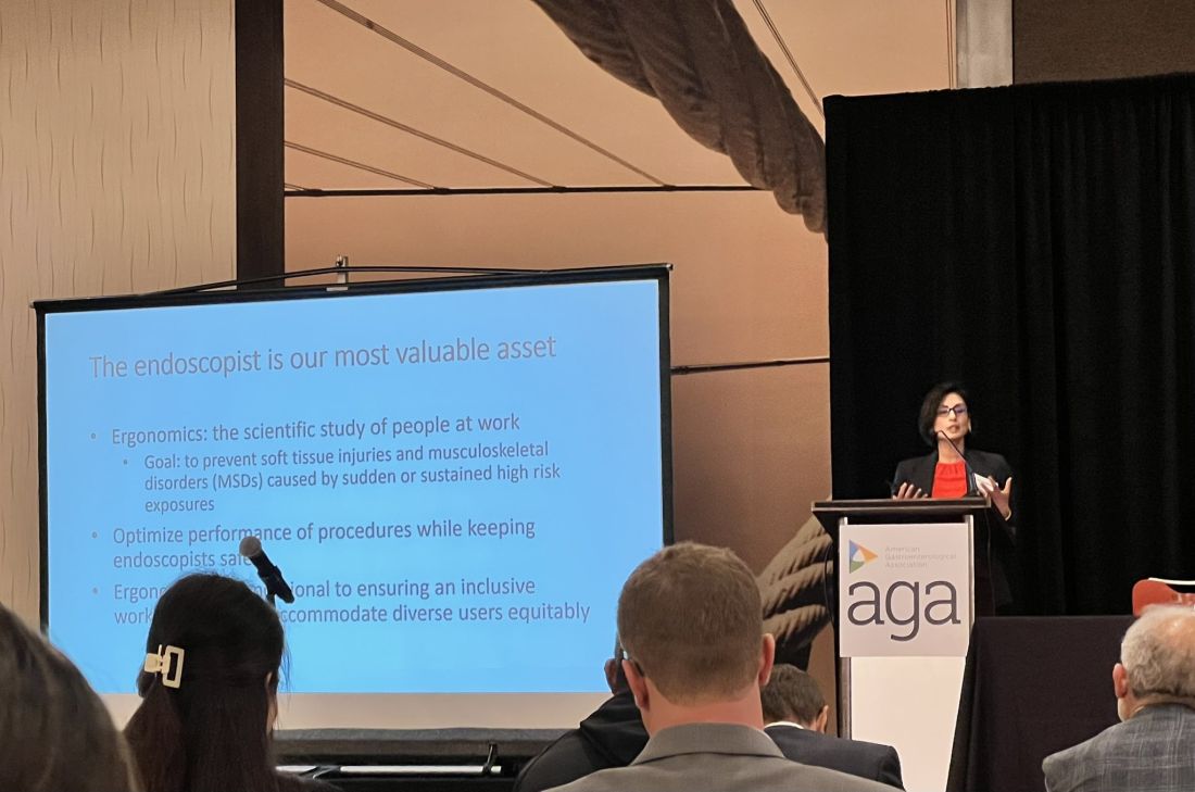
The AGA Tech Summit was unlike any conference I have been to in my 13 years of training in medicine (which included mostly clinically focused scientific meetings). Sessions involved ergonomics, applications of artificial intelligence, advances in imaging, environmental endoscopy, the role of the FDA, and innovations around the world. The audience included but was not limited to industry executives, AGA CGIT leadership, physician innovators, gastroenterologists, venture capitalists, and others. Attendees represented the diversity of our field in terms of organizational structures and backgrounds. This resulted in an opportunity to hear and learn different perspectives about products, emerging technology, and the costs involved for physicians, industry, and patients.
The final session of the summit, the AGA Shark Tank, was perhaps the most intriguing one of all. The session showcased landscape-changing technology to AGA investors and venture capitalists. The participants presented their own pitches and faced the sharks (judges). The winner received additional funding, tailored guidance from the AGA CGIT committee, partnering opportunities with interested parties, and the opportunity to represent AGA Shark Tank at the Digestive Disease Week (DDW).
The AGA Tech Summit Fellows Program is a learning platform that not only helps you find your niche in the world of GI innovation but also equips you with resources and connections to make an impact. It is also a great way to infuse new ideas into your practice or research. As healthcare professionals, we must create a culture where innovation can flourish, and where staff and patients feel empowered to contribute to the innovation process and help make change happen — to me, the AGA Tech Summit is one such avenue.
Reference
1. Kelly CJ and Young AJ. Promoting innovation in healthcare. Future Healthc J. 2017 Jun. doi: 10.7861/futurehosp.4-2-121.
Dr. Umar is Assistant Professor of Medicine, Section of Gastroenterology and Hepatology, Baylor College of Medicine, Houston, Texas, and a staff physician at Michael E. DeBakey VA Medical Center, Houston. Dr. Umar has no relevant financial conflicts and is on X, formerly Twitter, @shifaumarMD.
Medicine is transforming at a remarkable pace. It is therefore imperative for the future of the field that physicians understand innovation and collaborate with industry partners. Innovation can be defined as invention, adoption, and diffusion.1 During my training in gastroenterology and advanced fellowships, I learned about multiple endoscopic tools and techniques and became familiar with industry names that I frequently encountered in the endoscopy unit or clinic.
I was nominated to attend the AGA Tech Summit Fellows Program by my advanced endoscopy fellowship program director. A total of 22 fellows from around the United States at various stages of their training and interests in the field of gastroenterology and hepatology were selected for the program through an application process. The program included registration, travel, and accommodations to attend the AGA Tech Summit and Fellows Immersion Day at Medtronic.
The first event in the program was a visit to the Medtronic Santa Clara office, where our initial stop was at the research and development lab. We were introduced to design and biomedical engineers who reviewed with us the extensive testing that devices and endoscopy equipment undergo before coming to the market. These labs have a heavy focus on prototyping and experimentation and exist to promote in-house innovation and inventions.
During the day, we met physicians who shared their journeys on how they developed and advanced their careers in partnership with industry. Our visit also included a session with the business development and strategy manager at Medtronic, who discussed strategy and steps involved in product development — from the inception of an idea, institutional policies, and patents, to industry collaboration, and finally to successful commercialization. During medical school and training, we are focused on appropriately learning and applying medical knowledge to clinical care. The Medtronic Fellows Immersion Day experience offered a different perspective and showed other ways by which clinical knowledge and experience can be used to make an impact, in collaboration with industry and stakeholders. It also highlighted alternative career paths for medical professionals. The evening concluded with a meet and greet with the AGA Center for GI Innovation & Technology (CGIT) members and leadership.
The AGA Tech Summit was unlike any conference I have been to in my 13 years of training in medicine (which included mostly clinically focused scientific meetings). Sessions involved ergonomics, applications of artificial intelligence, advances in imaging, environmental endoscopy, the role of the FDA, and innovations around the world. The audience included but was not limited to industry executives, AGA CGIT leadership, physician innovators, gastroenterologists, venture capitalists, and others. Attendees represented the diversity of our field in terms of organizational structures and backgrounds. This resulted in an opportunity to hear and learn different perspectives about products, emerging technology, and the costs involved for physicians, industry, and patients.
The final session of the summit, the AGA Shark Tank, was perhaps the most intriguing one of all. The session showcased landscape-changing technology to AGA investors and venture capitalists. The participants presented their own pitches and faced the sharks (judges). The winner received additional funding, tailored guidance from the AGA CGIT committee, partnering opportunities with interested parties, and the opportunity to represent AGA Shark Tank at the Digestive Disease Week (DDW).
The AGA Tech Summit Fellows Program is a learning platform that not only helps you find your niche in the world of GI innovation but also equips you with resources and connections to make an impact. It is also a great way to infuse new ideas into your practice or research. As healthcare professionals, we must create a culture where innovation can flourish, and where staff and patients feel empowered to contribute to the innovation process and help make change happen — to me, the AGA Tech Summit is one such avenue.
Reference
1. Kelly CJ and Young AJ. Promoting innovation in healthcare. Future Healthc J. 2017 Jun. doi: 10.7861/futurehosp.4-2-121.
Dr. Umar is Assistant Professor of Medicine, Section of Gastroenterology and Hepatology, Baylor College of Medicine, Houston, Texas, and a staff physician at Michael E. DeBakey VA Medical Center, Houston. Dr. Umar has no relevant financial conflicts and is on X, formerly Twitter, @shifaumarMD.
AGA Legacy Society Members Sustain GI Research
Research creates successful practices. Patients benefit from GI research daily in practices. Scientists are working hard to develop new treatments, therapies and discover cures to advance the field and better patient care. But they can’t do this without research funding.
They recognize the value that research has had in their profession, both in academic medicine and in private practice, and are showing their appreciation by giving back.
“I give back because I have a firsthand knowledge of what it will mean to a young investigator’s career,” said Shrikant Anant, PhD, AGAF, University of Kansas, AGA Legacy Society member. “I was propelled in my career when I received the 2002 AGA Research Scholar Award from the AGA Research Foundation. The funds helped me develop my independent research that led to many NIH grants and, associated with it, career advancement. I still vividly remember the day I received the notice of award and how my whole life changed. Today, I am proud to be a donor myself because I know it is making a difference on yet another young investigator.”
The AGA Legacy Society boasts 161 members. AGA Legacy Society members see the promise the future holds and are committed to furthering research in gastroenterology and hepatology through their generous donations.
AGA members who make gifts at the AGA Legacy Society level anytime before Digestive Disease Week® (DDW) 2024 will receive an invitation to the AGA Research Foundation Benefactor’s Event in Washington, D.C. Individuals interested in learning more about the AGA Legacy Society membership may contact [email protected] or visit https://foundation.gastro.org/our-donors/aga-legacy-society/ for more information about the AGA Legacy Society.
What’s the Real Prevalence of Conjunctivitis in AD Patients Treated With Dupilumab?
Those are key findings from an analysis of published trials of dupilumab for AD and other conditions that study author Matthew Zirwas, MD, presented during a late-breaking abstract session at the Revolutionizing Atopic Dermatitis (RAD) Virtual Conference.
Adults with AD have a significant and disease severity–dependent increased risk of developing ocular surface diseases, including conjunctivitis and keratitis, compared with the general population and independent of any drug effect, according to Dr. Zirwas, a dermatologist with Probity Medical Research of Columbus, Ohio.
Dupilumab inhibits signaling of interleukin (IL)-4 and IL-13, which drive type 2 inflammatory diseases such as AD, asthma, chronic rhinosinusitis with nasal polyposis (CRSwNP), eosinophilic esophagitis (EoE), prurigo nodularis (PN), and chronic spontaneous urticaria (CSU).
In randomized, placebo-controlled trials of dupilumab in patients with moderate to severe AD, conjunctivitis was reported in more patients who received dupilumab treatment than in placebo-treated patients.
“When it comes to dupilumab-induced conjunctivitis, we have a good idea of the etiology, but the question of how frequently it occurs versus how frequently the conjunctivitis is unrelated to dupilumab is an interesting one,” he said. “How often is it clinically meaningful? What is it that is so unique about AD patients? We’ve all heard that it is a unique adverse event that only happens to people with AD and not to people using dupilumab for other indications. Where it gets interesting to me is how do we differentiate the cases that are dupilumab induced versus the cases that are just part of the underlying AD process?”
For their analysis, Dr. Zirwas and co-authors reviewed the incidence of conjunctivitis adverse events in patients from 15 completed, randomized, double-blind placebo-controlled trials evaluating dupilumab in AD, asthma, CRSwNP, EoE, PN, and CSU, along with the severity and resolution of conjunctivitis events in adults with AD.
Of the 15 trials, 7 were conducted in patients with AD: 4 in adults, 1 in adolescents, 1 in school-aged children, and 1 in preschoolers. One of the AD trials, LIBERTY AD CHRONOS, extends 52 weeks. The remaining eight trials of patients with asthma, CRSwNP, EoE, PN, and CSU lasted 24-52 weeks.
In the non-AD trials, the researchers observed that conjunctivitis rates were generally in the 1%-3% range, with less pronounced or no differences between the dupilumab and placebo groups. In the AD trials, conjunctivitis rates were higher in patients receiving dupilumab, compared with those receiving placebo across age groups.
In the 16-week SOLO 1 & 2 and AD-1021 monotherapy trials, 12 conjunctivitis events occurred in 517 patients who received placebo (2%), compared with 103 of 1047 patients (9.84%) who received dupilumab. Of the dupilumab-associated conjunctivitis cases, 80 (78%) patients recovered by the end of the trials. Of the 23 cases of conjunctivitis that did not recover or dropped out of the trial, 15 were among the 529 patients who received 300 mg dupilumab once every 2 weeks (q2w) (3%) and eight were among the 518 who received 300 mg dupilumab once weekly (qw) (2%).
In the 52-week LIBERTY AD CHRONOS trial, 29 conjunctivitis events occurred in 315 patients who received placebo plus topical corticosteroid (TCS) (9%), compared with 113 of 425 patients (27%) who received dupilumab plus TCS. Of these, 103 (91%) recovered by the end of the trial. Of the 11 patients with conjunctivitis who did not recover or dropped out of the trial, 3 were among the 110 patients who received 300 mg dupilumab q2w plus TCS (3%), and 8 were among the 315 who received 300 mg dupilumab qw plus TCS (3%).
“When I look at all of this data, I think that about 2% of people treated with dupilumab are going to get clinically very meaningful conjunctivitis that may be therapy limiting,” Dr. Zirwas concluded. “The vast majority of those cases appear to happen in the first 16 weeks. This is just not something we see in cases of patients treated with dupilumab treated for other reasons.”
Following his presentation, a meeting attendee asked Dr. Zirwas if conjunctivitis occurs more often in patients with facial dermatitis. “My perception is that it happens more often in people who have facial or eyelid dermatitis, but I’ve seen it in plenty of people who didn’t have any facial or eyelid dermatitis,” he said. “I have seen more conjunctivitis in people with more severe AD and more severe atopic comorbidities. That is anecdotal. The data that I have seen has been back and forth on this topic.”
Dr. Zirwas disclosed that he is a speaker and consultant for Sanofi, Regeneron, Leo, Lilly, Galderma, Pfizer, and AbbVie. Several of his co-authors work for Regeneron Pharmaceuticals.
FROM RAD 2023
2024 Gut Microbiota for Health World Summit Explores the Clinical Impacts of the Microbiome
Join global experts in-person or online as they gather for the 2024 Gut Microbiota for Health World Summit (GMFH) on March 23-24, 2024, in Washington, DC.
This meeting brings together an international and multidisciplinary community of GI clinicians, dietitians, and researchers to discuss personalized approaches to modifying the gut microbiome to improve health and treat disease.
This year’s program will explore:
- Better health through the gut microbiome.
- Big data and the gut microbiome.
- Human-derived to synthetic communities.
- Bringing new microbiome-based products to market.
Early-career faculty and trainees are encouraged to submit abstracts for presentation during the reception. Five $1,000 abstract prizes are available for top-scoring submissions.
Register here.
Announcing AGA Journal Social Media Editors
AGA journals have welcomed new social media editors for Clinical Gastroenterology and Hepatology (CGH), Cellular and Molecular Gastroenterology and Hepatology (CMGH), Techniques and Innovations in Gastrointestinal Endoscopy (TIGE) and Gastro Hep Advances (GHA).
Clinical Gastroenterology and Hepatology (CGH)
Joseph Sleiman, MD
University of Pittsburgh Medical Center
Dr. Sleiman’s research interests include inflammatory bowel disease (IBD), immunotherapy-induced colitis, Lynch Syndrome surveillance strategies and machine learning for GI research purposes.
Follow Dr. Sleiman
Cellular and Molecular Gastroenterology and Hepatology (CMGH)
Lindsey Kennedy, PhD
Indiana University School of Medicine
Dr. Kennedy’s research interests include the cellular crosstalk and pathological mechanisms regulating biliary and liver damage in cholestatic disorders, such as primary sclerosing cholangitis (PSC) and primary biliary cholangitis (PBC).
Follow Dr. Kennedy
Techniques and Innovations in Gastrointestinal Endoscopy (TIGE)
Judy Trieu, MD, MPH
Washington University Physicians
Dr. Trieu specializes in interventional endoscopy and general gastroenterology.
Follow Dr. Trieu
Gastro Hep Advances (GHA)
Shida Haghighat, MD, MPH
University of Miami
Dr. Haghighat’s research interests center around the prevention and screening of gastrointestinal cancers.
Follow Dr. Haghihat