User login
Opioids still overprescribed for postop pain management
Most patients use far less opioids than they are prescribed after hernia and other abdominal surgery, resulting in substantial waste and potential diversion, a prospective cohort study has found.
In an evaluation of 176 narcotic-naive patients who underwent surgery in a over the 14-day postdischarge study period was 30 morphine milligram equivalents (MME) but the median prescription was 150 MME, reported Wen Hui Tan, MD, and her research team at Washington University, St. Louis. The report was published in the Journal of the American College of Surgeons.
Overall, 76.7% of patients reported being satisfied or very satisfied with their postoperative pain management. Some patients (n = 31, 17.6%) reported not filling their prescription or not taking any of their prescribed opioid pain medications at all.
Sixty-nine percent of the surgeries were laparoscopic. A variety of abdominal procedures were represented, including hiatal hernia repair, inguinal hernia repair, and cholecystectomy. The median age was 60 years. Of postoperative pain prescriptions, 67% were for hydrocodone-acetaminophen and most of the remainder were for oxycodone-acetaminophen or oxycodone alone. The median prescription was for the equivalent of 20 5-mg oxycodone pills, while the median consumption in the first 7 postoperative days was 3.7 pills. Only 4.5% of patients received a refill.
The findings are consistent with numerous studies of different types of operations showing that patients often don’t use all of the opioid medications they are prescribed for pain control after surgery.
“Now that opioid pain medications can no longer be refilled with a pharmacy via telephone, overprescription may also be partially driven by a desire to prevent future inconvenience and workload of office staff from patients requesting refills. However, the rising numbers of opioid-related unintentional deaths over the last decade point to the fact that overprescription has serious potential consequences,” the researchers wrote.
They reported having no potential conflicts of interest.
SOURCE: Tan et al. J Am Coll Surg 2018 May 7. doi: 10.1016/j.jamcollsurg.2018.04.032.
FROM THE JOURNAL OF THE AMERICAN COLLEGE OF SURGEONS
Key clinical point: For postoperative recovery, a survey showed that far more opioids are prescribed than are consumed.
Major finding: On average, surgical patients went home with opioid prescriptions of 150 MME but took a median of 30 MME.
Study details: Prospective cohort study.
Disclosures: The authors report no potential conflicts of interest.
Source: Tan et al. J Am Coll Surg 2018 May 7. doi: 10.1016/j.jamcollsurg.2018.04.032.
Eleven on a scale of 1 to 10
I literally rode into the sunset recently as I finished my tour of duty as one of the Director examiners for the American Board of Surgery. I was heading out of St. Louis westward toward my home in Kansas. It was a 9-hour drive, which gave me plenty of time to reflect on the 6 years I shared the responsibility of administering the certifying exam known by most surgeons as “the oral exam.”
Over the last dozen years, Directors V. Suzanne Klimberg and Karen J. Brasel, along with former Executive Director Frank Lewis, a team of psychometricians at the Board, and members of the certification committee of the Board, worked tirelessly to create a testing instrument as fair and statistically sound as possible given the inherently qualitative exam. I believe they did a magnificent job. Gone are the legends of yesteryear where candidates were subjected to the whims of whatever crossed the mind of the examiners, including their prejudices about the “right” answer. The oral exam now represents a well-constructed survey of surgical judgment issues that have been thoroughly vetted.
Being an examiner for the orals means you arrive Sunday afternoon before the exams that are given over the next two and a half days. Each examiner undergoes an overview briefing on Sunday afternoon and then studies “the book” for that test’s content usually until late into the night. This book is an impressive document.
We arise around 0530 to attend a breakfast meeting, which includes breaking into our six-person teams and going over each question that will be given that day. At 0800, the first candidates for the first session walk into a room and meet the two surgeons who will make some of the most important decisions affecting that candidate’s career. If you rate the intensity of this moment on a scale of 1 to 10, this is an 11 for both candidates and examiners. No one in the room knows how it will turn out because every session has its own twists and turns. Everyone there wants to see a passing score, but the two examiners know that they must make a decision that is safe for the public and fair for the candidate.
Twelve exams are given per team per day except for the final day which has only six. So, each team examines 30 candidates over 3 days. I’ve opened a door and shaken the moist hand of 438 candidates. I’ve seen every sort of emotion during those sessions. I’ve had moments of great joy and times of profound sadness as candidates respond to the questions. I’ve always tried to be friendly, but just like surgery, it is a serious business and decisions have to be made. That means ignoring one’s hopes and acting on the best facts available at the moment. Most surgeons remember their oral examiners and what they were asked for a lifetime. I know I do.
I could write a book on this experience (I won’t, though). But as I reflect on my time as a Director, what stands out in my mind are the associate examiners with whom I’ve worked. These surgeons are invited to participate and receive no compensation. It’s 3 days out of their lives, and because they don’t give the exam as frequently as the Directors do, the amount of study and effort is greater for them. Each is selected because he or she is considered to be a thoughtful surgeon with high standards. These surgeons do this job because they care about quality in our profession.
Most of the associates I have worked with are far more accomplished than I. I was once paired with a renowned breast surgeon (okay, it was Kelly K. Hunt). My ego was at great risk because I knew how accomplished she was. But like all the other associates, she was gracious and hard working. We rarely work with another Director, but Anne G. Rizzo, who later became a Director, and I did an exam together. She was a dazzling questioner with very high standards. In other words, she was typical of the people I met. My first associate (also later a Director) was Reid Adams. He was great; I was nervous. My last associate was Marc L. Melcher, a transplant surgeon who asked penetrating questions in a calm manner. I wish I could name each of my associates and thank them for making my work so much better, for teaching me things I didn’t know, for deepening my own knowledge, and serving in a hard job with grace. This column can’t be that long, but you all know who you are. Thank you.
At the end of the day, I believe the oral exam to be a great thing for our profession. When you think about the number of patients potentially affected throughout a surgeon’s career, the impact of decisions made on the day of the exam can be enormous. Given that, over a 20-year career, a surgeon may operate on 25,000 patients, a summative check on a surgeon’s judgment and knowledge is important. Each year, the ABS adjudicates on some 1,100 surgeons. A single year’s set of surgeons over the following 20-year period translates into 27.5 million patients. I hope we never stop doing the orals because of cost, time, or convenience. The exam is just too important to our profession to risk forgoing this last, big step before a surgeon is presented to the world as “certified.”
Valbenazine Provides Long-Term Benefits for Tardive Dyskinesia
LOS ANGELES—Once-daily treatment with valbenazine for 48 weeks provides substantial improvements on clinician- and patient-reported outcomes in adults with tardive dyskinesia, according to data described at the 70th Annual Meeting of the American Academy of Neurology. The results are consistent with those of previous trials, said the researchers. Valbenazine is well tolerated and does not raise significant safety concerns.
Valbenazine was approved as a treatment for tardive dyskinesia on the basis of several short-term placebo-controlled trials, a blinded extension study, and the long-term KINECT 4 study. Stewart Factor, DO, Professor of Neurology at Emory University School of Medicine in Atlanta, and colleagues conducted a study to evaluate the long-term effects of once-daily valbenazine on tardive dyskinesia.

Eligible participants were adults with de novo tardive dyskinesia and those who had participated in prior trials of valbenazine. All participants received 48 weeks of open-label treatment with valbenazine. The initial dose was 40 mg. If an investigator judged that a patient had inadequate clinical response at week four, the dose was increased to 80 mg, based on tolerability. For patients who could not tolerate the 80-mg dose, the dose was decreased to 40 mg.
Dr. Factor and colleagues used the change from baseline in the Abnormal Involuntary Movement Scale (AIMS) total score to assess changes in tardive dyskinesia. Other efficacy assessments included the Patient Global Impression of Change (PGIC) and Clinical Global Impression of Change-Tardive Dyskinesia (CGI-TD) scales. The investigators applied standard safety methods, including treatment-emergent adverse event reporting.
The safety population included 163 participants. Of this group, 107 participants received and tolerated the 80-mg dose, 45 did not require escalation from the 40-mg dose, and 11 were escalated to 80-mg dose, but later required reduction to the 40-mg dose. The mean change from baseline to week 48 in AIMS total score indicated improvements in tardive dyskinesia in all dose groups. The 80-mg group had a decrease of 11.0 points, the 40-mg group had a decrease of 10.2 points, and the group whose dose was decreased from 80 mg to 40 mg had a decrease of 7.2 points.
At week 48, more than 75% of participants in each study arm had a PGIC score of 2 or lower (ie, much improved or very much improved). This outcome was achieved in 89.2% of the 80-mg group, 90.0% of the 40-mg group, and 77.8% of the group whose dose was decreased from 80 mg to 40 mg. Mean CGI-TD scores at week 48 were 1.6 for the 80-mg group, 1.7 for the 40-mg group, and 2.3 for the group whose dose was reduced from 80 mg to 40 mg. These scores indicated clinically meaningful long-term improvement for all dose groups.
Less than 15% of all participants had a serious treatment-emergent adverse event (12.9%) or treatment-emergent adverse event leading to discontinuation (14.7%).
The study was funded by Neurocrine Biosciences, the manufacturer of valbenazine.
LOS ANGELES—Once-daily treatment with valbenazine for 48 weeks provides substantial improvements on clinician- and patient-reported outcomes in adults with tardive dyskinesia, according to data described at the 70th Annual Meeting of the American Academy of Neurology. The results are consistent with those of previous trials, said the researchers. Valbenazine is well tolerated and does not raise significant safety concerns.
Valbenazine was approved as a treatment for tardive dyskinesia on the basis of several short-term placebo-controlled trials, a blinded extension study, and the long-term KINECT 4 study. Stewart Factor, DO, Professor of Neurology at Emory University School of Medicine in Atlanta, and colleagues conducted a study to evaluate the long-term effects of once-daily valbenazine on tardive dyskinesia.
Eligible participants were adults with de novo tardive dyskinesia and those who had participated in prior trials of valbenazine. All participants received 48 weeks of open-label treatment with valbenazine. The initial dose was 40 mg. If an investigator judged that a patient had inadequate clinical response at week four, the dose was increased to 80 mg, based on tolerability. For patients who could not tolerate the 80-mg dose, the dose was decreased to 40 mg.
Dr. Factor and colleagues used the change from baseline in the Abnormal Involuntary Movement Scale (AIMS) total score to assess changes in tardive dyskinesia. Other efficacy assessments included the Patient Global Impression of Change (PGIC) and Clinical Global Impression of Change-Tardive Dyskinesia (CGI-TD) scales. The investigators applied standard safety methods, including treatment-emergent adverse event reporting.
The safety population included 163 participants. Of this group, 107 participants received and tolerated the 80-mg dose, 45 did not require escalation from the 40-mg dose, and 11 were escalated to 80-mg dose, but later required reduction to the 40-mg dose. The mean change from baseline to week 48 in AIMS total score indicated improvements in tardive dyskinesia in all dose groups. The 80-mg group had a decrease of 11.0 points, the 40-mg group had a decrease of 10.2 points, and the group whose dose was decreased from 80 mg to 40 mg had a decrease of 7.2 points.
At week 48, more than 75% of participants in each study arm had a PGIC score of 2 or lower (ie, much improved or very much improved). This outcome was achieved in 89.2% of the 80-mg group, 90.0% of the 40-mg group, and 77.8% of the group whose dose was decreased from 80 mg to 40 mg. Mean CGI-TD scores at week 48 were 1.6 for the 80-mg group, 1.7 for the 40-mg group, and 2.3 for the group whose dose was reduced from 80 mg to 40 mg. These scores indicated clinically meaningful long-term improvement for all dose groups.
Less than 15% of all participants had a serious treatment-emergent adverse event (12.9%) or treatment-emergent adverse event leading to discontinuation (14.7%).
The study was funded by Neurocrine Biosciences, the manufacturer of valbenazine.
Long-Term Perampanel Treatment Is Safe and Effective for Adolescents
LOS ANGELES—Treatment for as long as four years with adjunctive perampanel is safe and effective for adolescents with secondarily generalized seizures or primary generalized tonic-clonic seizures, according to a post hoc study presented at the 70th Annual Meeting of the American Academy of Neurology. “These post hoc results are encouraging, given the refractory nature of these seizure types,” said the investigators.
Perampanel has regulatory approval for the treatment of partial seizures with or without secondarily generalized seizures, and for the adjunctive treatment of primary generalized tonic-clonic seizures in patients age 12 and younger with epilepsy. Phase II and III randomized, double-blind, placebo-controlled trials indicated that adjunctive perampanel (at doses of 12 mg/day or less) was effective and tolerable in patients with these types of seizures in idiopathic generalized epilepsy. The participants who completed these studies were eligible to enter one of four open-label extension studies.
Jesus Eric Piña-Garza, MD, a pediatric neurologist at the Children’s Hospital at TriStar Centennial in Nashville, and colleagues used data from these open-label extension studies to assess the long-term efficacy and safety of adjunctive perampanel in patients from ages 12 to 17 with secondarily generalized seizures or primary generalized tonic-clonic seizures.

The extension studies incorporated a blinded conversion period that lasted for six to 16 weeks, during which perampanel dose was optimized (at 12 mg/day or less), and a maintenance phase that lasted for 27 to 256 weeks. Total drug exposure was as long as five years. All patients received perampanel during these studies. Efficacy and safety assessments, which were performed for as long as four years, included median percent change in seizure frequency per 28 days and 50% and 75% responder and seizure-freedom rates. Investigators also monitored treatment-emergent adverse events.
The investigators included 129 adolescent patients in the safety analysis set, including 109 with secondarily generalized seizures and 19 with primary generalized tonic-clonic seizures. During the first year, median percent reductions in seizure frequency per 28 days were 62.8% for patients with secondarily generalized seizures and 84.0% for patients with primary generalized tonic-clonic seizures. During year four, median percent reductions in seizure frequency per 28 days were 73.2% for patients with secondarily generalized seizures and 100.0% for patients with primary generalized tonic-clonic seizures. The 50% responder rates were 56.9% for patients with secondarily generalized seizures and 63.2% for patients with primary generalized tonic-clonic seizures in year one, and 65.2% for patients with secondarily generalized seizures and 100.0% for patients with primary generalized tonic-clonic seizures in year four.
For each seizure type, the incidence of treatment-emergent adverse events was highest during the first year of perampanel exposure. The most common treatment-emergent adverse events were dizziness, somnolence, and nasopharyngitis.
The study was supported by Eisai.
LOS ANGELES—Treatment for as long as four years with adjunctive perampanel is safe and effective for adolescents with secondarily generalized seizures or primary generalized tonic-clonic seizures, according to a post hoc study presented at the 70th Annual Meeting of the American Academy of Neurology. “These post hoc results are encouraging, given the refractory nature of these seizure types,” said the investigators.
Perampanel has regulatory approval for the treatment of partial seizures with or without secondarily generalized seizures, and for the adjunctive treatment of primary generalized tonic-clonic seizures in patients age 12 and younger with epilepsy. Phase II and III randomized, double-blind, placebo-controlled trials indicated that adjunctive perampanel (at doses of 12 mg/day or less) was effective and tolerable in patients with these types of seizures in idiopathic generalized epilepsy. The participants who completed these studies were eligible to enter one of four open-label extension studies.
Jesus Eric Piña-Garza, MD, a pediatric neurologist at the Children’s Hospital at TriStar Centennial in Nashville, and colleagues used data from these open-label extension studies to assess the long-term efficacy and safety of adjunctive perampanel in patients from ages 12 to 17 with secondarily generalized seizures or primary generalized tonic-clonic seizures.
The extension studies incorporated a blinded conversion period that lasted for six to 16 weeks, during which perampanel dose was optimized (at 12 mg/day or less), and a maintenance phase that lasted for 27 to 256 weeks. Total drug exposure was as long as five years. All patients received perampanel during these studies. Efficacy and safety assessments, which were performed for as long as four years, included median percent change in seizure frequency per 28 days and 50% and 75% responder and seizure-freedom rates. Investigators also monitored treatment-emergent adverse events.
The investigators included 129 adolescent patients in the safety analysis set, including 109 with secondarily generalized seizures and 19 with primary generalized tonic-clonic seizures. During the first year, median percent reductions in seizure frequency per 28 days were 62.8% for patients with secondarily generalized seizures and 84.0% for patients with primary generalized tonic-clonic seizures. During year four, median percent reductions in seizure frequency per 28 days were 73.2% for patients with secondarily generalized seizures and 100.0% for patients with primary generalized tonic-clonic seizures. The 50% responder rates were 56.9% for patients with secondarily generalized seizures and 63.2% for patients with primary generalized tonic-clonic seizures in year one, and 65.2% for patients with secondarily generalized seizures and 100.0% for patients with primary generalized tonic-clonic seizures in year four.
For each seizure type, the incidence of treatment-emergent adverse events was highest during the first year of perampanel exposure. The most common treatment-emergent adverse events were dizziness, somnolence, and nasopharyngitis.
The study was supported by Eisai.
Multisensory Impairment Is Associated With Risk of Dementia
LOS ANGELES—Among older adults, the presence of a greater number of sensory impairments is associated with an increased risk of dementia, according to research presented at the 70th Annual Meeting of the American Academy of Neurology.
Sensory impairments are common among older adults and are associated with dementia, but no study has examined multisensory impairment and risk of dementia by incorporating measures of hearing, vision, smell, and touch, said Willa D. Brenowitz, PhD, MPH, a researcher in the Department of Epidemiology and Biostatistics at the University of California, San Francisco, and colleagues.
To evaluate whether the presence of multiple sensory impairments is associated with a greater risk of dementia, compared with a single or no sensory impairment, Dr. Brenowitz and colleagues studied 1,843 participants from the Health, Aging, and Body Composition Study. Participants were ages 70 to 79 and did not have dementia at baseline.
Investigators used sensory assessments that were conducted during the study years 3–5 to determine whether participants had visual impairment (ie, visual acuity ≤ 20/40 or log contrast sensitivity < 1.55), moderate to severe hearing loss (> 40 decibels hearing level based on pure tone average at 500 Hz, 1,000 Hz, 2,000 Hz, and 4,000 Hz), poor smell (lowest tertile of 12-item Cross Cultural Smell Identification Test), or impaired touch (peripheral nerve insensitivity based on a 10-g monofilament test or vibration detection threshold).
Investigators followed participants for up to 10 years. Incident dementia was based on hospitalization records, dementia medications, or a decline in Modified Mini-Mental State Exam of 1.5 standard deviations or greater. The researchers evaluated the association between number of sensory impairments and risk of dementia using Cox proportional hazard models adjusted for demographics and health conditions.
Sensory impairments were common, the researchers said. In all, 28% had visual impairments, 35% had hearing loss, 22% had poor smell, 12% had peripheral nerve insensitivity, and 25% had multiple impairments. An increasing number of impairments was associated with risk of dementia in a graded fashion. Compared with no sensory impairment, having one sensory impairment was associated with a 50% greater risk of dementia, two impairments was associated with a twofold greater risk of dementia, and three or four impairments was associated with a 2.8-fold greater risk of dementia.
“It is possible that sensory impairment, especially multiple [impairments], could limit an older adult’s engagement in protective lifestyle factors such as cognitive, physical, and social activity, thereby increasing dementia risk,” Dr. Brenowitz and colleagues said.
Clinical assessment of sensory function may help identify patients at high risk of dementia. Further studies are needed to determine whether multisensory impairment is a risk factor for dementia or an indicator of neurodegeneration, the researchers said.
—Jake Remaly
Sarcopenia had minor impact on hernia repair SSIs
according to findings from a prospective, single-institution study.
Steve R. Siegal, MD, and his colleagues at the Oregon Health & Science University, Portland, wrote in Hernia that risk factors for postoperative complications after hernia repair established in the literature include uncontrolled diabetes, active tobacco use, prior hernia repair, active infection, and obesity. Sarcopenia occurs in many physiological or pathological states, not only in the elderly but also in cases of immunosuppression, cirrhosis, trauma, prolonged immobility, and malignancy. Many opportunities exist for sarcopenia and hernia surgery to coincide but the role of sarcopenia in hernia repair outcomes has not been much studied.

The investigators began with the hypothesis that sarcopenia in hernia patients would lead to worse postoperative outcomes given the large metabolic requirement for postoperative healing of hernia defects and abdominal wall reconstruction.
The study involved 135 patients who underwent ventral hernia repair, 27% of whom had sarcopenia. The literature-based definition of sarcopenia was a muscle index cutoff of less than 52.4 cm2/m2 for men and less than 38.5 cm2/m2 for women. The investigators noted that the index cutoff was validated in oncology patients. Patients underwent a preoperative CT scan to assess muscle mass. The study group included patients with ventral hernia repair with or without mesh, component separation/abdominal wall reconstruction, and a hernia defect of 2 cm or larger.
With data on variables including gender, diabetes status, body mass index (BMI), chronic obstructive pulmonary disease (COPD), wound class, alcohol abuse status, and prior wound infections, the investigators created a multivariate model to look at primary outcomes of surgical site infection (SSI), surgical site occurrences (wound complications, other infections) and hernia recurrence. Secondary outcomes included length of hospital stay, morbidities, and other postoperative complications.
Patients with sarcopenia were more likely to have lower BMI (median 29.6 kg/m2 vs. 36.6 kg/m2), to be slightly older (median 63.1 vs. 59.2 years), and to have a history of immunosuppression (29.7% vs. 11.2%). The differences for the other variables (diabetes, tobacco use, COPD) were not significant.

The surprising finding was that sarcopenia was not significantly correlated with SSIs (P = 0.140), other complications (P = 0.113), or recurrence (P = 0.895) after ventral hernia repair. In-hospital morbidities and length of hospital stay did not differ significantly between the sarcopenic and nonsarcopenic patients. But sarcopenia combined with other factors did have an impact on outcomes. After adjustment for BMI and diabetes and critical care status, muscle mass as a continuous variable “was notable for a 1.44 increased odds [95% confidence interval, 1.00-2.07; P = 0.049] of inpatient morbidity with every decrease of 10 cm2/m2 of muscle index,” Dr. Siegal and his associates wrote.
The nonsignificant results of this study may be explained by the markedly lower BMI in the sarcopenic group, which could have been protective against worse outcomes, the investigators noted. However, they concluded, “Sarcopenia may still be a factor in adverse perioperative outcomes, but the established muscle index cutoff value may be inappropriately applied to benign patients, such as our ventral hernia cohort results in this study.”
The investigators declared no conflicts of interest and received no outside funding for this study.
SOURCE: Siegal SR et al. Hernia. 2018 May 11. doi: 10.1007/s10029-018-1770-8.
according to findings from a prospective, single-institution study.
Steve R. Siegal, MD, and his colleagues at the Oregon Health & Science University, Portland, wrote in Hernia that risk factors for postoperative complications after hernia repair established in the literature include uncontrolled diabetes, active tobacco use, prior hernia repair, active infection, and obesity. Sarcopenia occurs in many physiological or pathological states, not only in the elderly but also in cases of immunosuppression, cirrhosis, trauma, prolonged immobility, and malignancy. Many opportunities exist for sarcopenia and hernia surgery to coincide but the role of sarcopenia in hernia repair outcomes has not been much studied.
The investigators began with the hypothesis that sarcopenia in hernia patients would lead to worse postoperative outcomes given the large metabolic requirement for postoperative healing of hernia defects and abdominal wall reconstruction.
The study involved 135 patients who underwent ventral hernia repair, 27% of whom had sarcopenia. The literature-based definition of sarcopenia was a muscle index cutoff of less than 52.4 cm2/m2 for men and less than 38.5 cm2/m2 for women. The investigators noted that the index cutoff was validated in oncology patients. Patients underwent a preoperative CT scan to assess muscle mass. The study group included patients with ventral hernia repair with or without mesh, component separation/abdominal wall reconstruction, and a hernia defect of 2 cm or larger.
With data on variables including gender, diabetes status, body mass index (BMI), chronic obstructive pulmonary disease (COPD), wound class, alcohol abuse status, and prior wound infections, the investigators created a multivariate model to look at primary outcomes of surgical site infection (SSI), surgical site occurrences (wound complications, other infections) and hernia recurrence. Secondary outcomes included length of hospital stay, morbidities, and other postoperative complications.
Patients with sarcopenia were more likely to have lower BMI (median 29.6 kg/m2 vs. 36.6 kg/m2), to be slightly older (median 63.1 vs. 59.2 years), and to have a history of immunosuppression (29.7% vs. 11.2%). The differences for the other variables (diabetes, tobacco use, COPD) were not significant.
The surprising finding was that sarcopenia was not significantly correlated with SSIs (P = 0.140), other complications (P = 0.113), or recurrence (P = 0.895) after ventral hernia repair. In-hospital morbidities and length of hospital stay did not differ significantly between the sarcopenic and nonsarcopenic patients. But sarcopenia combined with other factors did have an impact on outcomes. After adjustment for BMI and diabetes and critical care status, muscle mass as a continuous variable “was notable for a 1.44 increased odds [95% confidence interval, 1.00-2.07; P = 0.049] of inpatient morbidity with every decrease of 10 cm2/m2 of muscle index,” Dr. Siegal and his associates wrote.
The nonsignificant results of this study may be explained by the markedly lower BMI in the sarcopenic group, which could have been protective against worse outcomes, the investigators noted. However, they concluded, “Sarcopenia may still be a factor in adverse perioperative outcomes, but the established muscle index cutoff value may be inappropriately applied to benign patients, such as our ventral hernia cohort results in this study.”
The investigators declared no conflicts of interest and received no outside funding for this study.
SOURCE: Siegal SR et al. Hernia. 2018 May 11. doi: 10.1007/s10029-018-1770-8.
FROM HERNIA
Key clinical point: Sarcopenia was not correlated with surgical site infections, other complications, or recurrence after ventral hernia repair.
Major finding: After adjustment for body mass index and diabetes and critical care status, muscle mass as a continuous variable was notable for a 1.44 increased odds of inpatient morbidity.
Study details: A prospective, single-institution study of 135 patients who had ventral hernia repair, 27% of whom were sarcopenic.
Disclosures: The investigators declared no conflicts of interest and received no outside funding for this study.
Source: Siegal SR et al. Hernia. 2018 May 11. doi: 10.1007/s10029-018-1770-8.
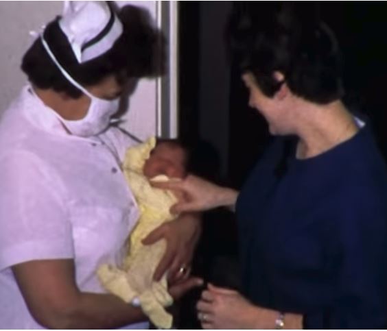
How more midwives may mean healthier mothers
Since ProPublica launched Lost Mothers, we’ve covered many facets of the U.S. maternal mortality crisis. Despite spending more per capita on health care than any other country, the U.S. has the highest rate of deaths related to pregnancy and childbirth in the industrialized world.
Source: Vox and ProPublica
But what makes maternal health care in other affluent countries look so different than the U.S.? Among other things, midwives. Midwives in the U.S. participate in less than 10 percent of births. But in Sweden, Denmark and France, they lead around three-quarters of deliveries. In Great Britain, they deliver half of all babies, including all three of Kate Middleton’s. So if the midwifery model works for royal babies, why not our own?
Check out the video to find out how midwives have been at the center of a culture war that’s deeply rooted in race and class in America.
Today we see vestiges of that history in states with restrictive midwifery laws and barriers to entry for midwives. Earlier this year, one study was the first systematic look at how and where they practice, offering new evidence that empowering them could significantly boost maternal and infant health. Many of the states with poor health outcomes and hostility to midwives also have large black populations. And with black mothers three to four times more likely to die in pregnancy or childbirth, the study raises the possibility that greater use of midwives could reduce racial disparities in maternal health.
This story makes up the eighth installment in Vox’s collaboration with ProPublica. You can find this video and all of Vox’s videos on YouTube. Subscribe and stay tuned for more from our partnership.
Teaching opportunities of live surgery broadcasts debated
SAN DIEGO – Two thoracic societies are being challenged to review their rejection of at medical meetings.
The American Association for Thoracic Surgery and Society of Thoracic Surgeons “should modify their stance on live surgery as an educational tool like almost all of their sister organizations,” said Joseph E. Bavaria, MD, FACS, of the University of Pennsylvania, Philadelphia. “Presently, it is effectively a ban on the practice. But there are no data to suggest that it does harm to patients if done with proper constraints.”

Live broadcasts of surgeries at medical meetings are both common and controversial. Dr. Bavaria listed numerous recent thoracic meetings in Europe and Canada that featured live broadcasts. But, he noted, the AATS and STS are not supportive of the trend.
In a policy amended in 2017, the AATS states, among other things, that “national and international cardiothoracic societies should consider prohibiting live surgery broadcasts to large audiences at their annual meetings.” The STS policy, amended in 2016, is nearly identical. However, it adds language specifying that “violation of these guidelines may lead to disciplinary action by the Society.”
Both the AATS and STS policies state that surgeons should not take part in live broadcasts of operations that are intended for the public. And both policies say “generally, recorded broadcasts, either edited or unedited, are preferable to live surgery broadcasts because recordings intended for later broadcast pose fewer risks of harm to patients.”
The policies don’t elaborate on how a live broadcast of an operation might be more harmful than one aired at a later time. “Where are the data to support this statement?” Dr. Bavaria asked. “Is this what we call the expert opinion of a few people with gray hair? Our European and Canadian colleagues certainly don’t agree with [the policies] and might take issue with our sanctimonious lecturing that’s not supported by any data whatsoever.”
He pointed to a 2011 study that examined 250 cardiac procedures that were broadcast live to 32 scientific meetings. Researchers found that “there is no evidence for an excess perioperative risk for patients operated under the conditions of live broadcasting” (Eur J Cardiothorac Surg. 2011;40[2]:367-71).
A member of the audience at the AATS session pointed out that the study may be misleading because only top surgeons may be willing to perform procedures live on television. Dr. Bavaria responded by noting that the outcomes of surgeries performed on live video were not worse.
Skeptics have also expressed concern about the potential for sensationalism in live broadcasts and the drive for participants to make a splash: “the demands of ‘the performance,’ including questions and discussion with the audience, may divert the surgeon’s attention and produce an inferior outcome,” wrote three physicians in 2011 (J Surg Educ. 2011 Jan-Feb; 68[1]:58-61).
The trio also expressed worry about safety risks posed by extra equipment and extra people in the operating room during a live broadcast. However, Dr. Bavaria said the crowded conditions of an operation observed in person – instead of via video – could be worse: “You don’t have a sterile environment, you have people hanging around all over. A moderated live session could be better for the surgeon and the patient, rather than having a bunch of people in the room.”
Dr. Bavaria was also asked why surgeries couldn’t be recorded for airing later instead of being broadcast live, as this approach “takes you out of the nexus of having two masters, the patient and the audience.” In response, Dr. Bavaria said editing takes extra time in order to capture the nuances of the live procedure.
Dr. Bavaria reports no relevant disclosures.

Peter Angelos, MD, FACS, is the Linda Kohler Anderson Professor of Surgery and Surgical Ethics, chief of endocrine surgery, and associate director of the MacLean Center for Clinical Medical Ethics at the University of Chicago. Dr. Angelos has no disclosures.
Peter Angelos, MD, FACS, is the Linda Kohler Anderson Professor of Surgery and Surgical Ethics, chief of endocrine surgery, and associate director of the MacLean Center for Clinical Medical Ethics at the University of Chicago. Dr. Angelos has no disclosures.
Peter Angelos, MD, FACS, is the Linda Kohler Anderson Professor of Surgery and Surgical Ethics, chief of endocrine surgery, and associate director of the MacLean Center for Clinical Medical Ethics at the University of Chicago. Dr. Angelos has no disclosures.
SAN DIEGO – Two thoracic societies are being challenged to review their rejection of at medical meetings.
The American Association for Thoracic Surgery and Society of Thoracic Surgeons “should modify their stance on live surgery as an educational tool like almost all of their sister organizations,” said Joseph E. Bavaria, MD, FACS, of the University of Pennsylvania, Philadelphia. “Presently, it is effectively a ban on the practice. But there are no data to suggest that it does harm to patients if done with proper constraints.”
Live broadcasts of surgeries at medical meetings are both common and controversial. Dr. Bavaria listed numerous recent thoracic meetings in Europe and Canada that featured live broadcasts. But, he noted, the AATS and STS are not supportive of the trend.
In a policy amended in 2017, the AATS states, among other things, that “national and international cardiothoracic societies should consider prohibiting live surgery broadcasts to large audiences at their annual meetings.” The STS policy, amended in 2016, is nearly identical. However, it adds language specifying that “violation of these guidelines may lead to disciplinary action by the Society.”
Both the AATS and STS policies state that surgeons should not take part in live broadcasts of operations that are intended for the public. And both policies say “generally, recorded broadcasts, either edited or unedited, are preferable to live surgery broadcasts because recordings intended for later broadcast pose fewer risks of harm to patients.”
The policies don’t elaborate on how a live broadcast of an operation might be more harmful than one aired at a later time. “Where are the data to support this statement?” Dr. Bavaria asked. “Is this what we call the expert opinion of a few people with gray hair? Our European and Canadian colleagues certainly don’t agree with [the policies] and might take issue with our sanctimonious lecturing that’s not supported by any data whatsoever.”
He pointed to a 2011 study that examined 250 cardiac procedures that were broadcast live to 32 scientific meetings. Researchers found that “there is no evidence for an excess perioperative risk for patients operated under the conditions of live broadcasting” (Eur J Cardiothorac Surg. 2011;40[2]:367-71).
A member of the audience at the AATS session pointed out that the study may be misleading because only top surgeons may be willing to perform procedures live on television. Dr. Bavaria responded by noting that the outcomes of surgeries performed on live video were not worse.
Skeptics have also expressed concern about the potential for sensationalism in live broadcasts and the drive for participants to make a splash: “the demands of ‘the performance,’ including questions and discussion with the audience, may divert the surgeon’s attention and produce an inferior outcome,” wrote three physicians in 2011 (J Surg Educ. 2011 Jan-Feb; 68[1]:58-61).
The trio also expressed worry about safety risks posed by extra equipment and extra people in the operating room during a live broadcast. However, Dr. Bavaria said the crowded conditions of an operation observed in person – instead of via video – could be worse: “You don’t have a sterile environment, you have people hanging around all over. A moderated live session could be better for the surgeon and the patient, rather than having a bunch of people in the room.”
Dr. Bavaria was also asked why surgeries couldn’t be recorded for airing later instead of being broadcast live, as this approach “takes you out of the nexus of having two masters, the patient and the audience.” In response, Dr. Bavaria said editing takes extra time in order to capture the nuances of the live procedure.
Dr. Bavaria reports no relevant disclosures.
REPORTING FROM AATS
Caution crucial for stem cell transplant in scleroderma, despite potential interest
SANDESTIN, FLA. – An expert called for restraint with the use of stem cell transplantation in scleroderma at the annual Congress of Clinical Rheumatology, emphasizing that the early mortality risk shows that the patient group that should be considered for the treatment is very narrow.
Results published earlier this year found a long-term benefit for myeloablative autologous stem cell transplantation, compared with cyclophosphamide, and were highly encouraging – but only in patients with severe disease (N Engl J Med. 2018;378:35-47). This might prompt patients to express interest in the treatment, many of whom are not suitable, said Janet Pope, MD, chair of rheumatology at St. Joseph’s Health Care in London, Ont.

Because of mortality seen in the first year in the stem cell transplant group, the benefit over cyclophosphamide was seen only in subsequent years, Dr. Pope stressed.
Consideration of the approach is only appropriate for patients with early diffuse scleroderma who have a 50% mortality risk over 5 years, Dr. Pope said. Patients in the SCOT trial had a modified Rodnan Skin Score (mRSS) of 30, with an average forced vital capacity of 74%, and 73 of the 75 patients in the trial had lung involvement. Essentially, Dr. Pope said, these are patients with some organ involvement that could be lethal, but not such severe organ involvement that it requires a transplant.
“These are our sick patients,” she said.
The results published this year are even more definitive, Dr. Pope said, considering that they largely meshed with results out of Europe, published in 2014, that showed that the stem cell transplant benefit was not seen after the first year because of early mortality (JAMA. 2014 Jun 25;311[24]:2490-8). In that trial, a 10% mortality risk was seen in the first year in the transplant group, and then a benefit to transplant in the long term over cyclophosphamide.
“These are very complementary positive trials,” Dr. Pope said. “We don’t want to kill 10% of our patients who don’t have perceived high chance of mortality in the next 5 years. ... These patients are highly selected, but I think it gives our patients hope and, for some of my patients, this will be a treatment for them.”
The long-term efficacy is also encouraging in that new but less potent treatments might yield good long-term results without the early deaths, she said. “It gives an idea and hope, and it helps us to understand that maybe with immune modulation, without the nuclear blast of stem cell transplant, maybe we can do better for our patients.”
Dr. Pope reported relevant financial relationships with Actelion, Bayer, Merck, Bristol-Myers Squibb, and Roche.
SANDESTIN, FLA. – An expert called for restraint with the use of stem cell transplantation in scleroderma at the annual Congress of Clinical Rheumatology, emphasizing that the early mortality risk shows that the patient group that should be considered for the treatment is very narrow.
Results published earlier this year found a long-term benefit for myeloablative autologous stem cell transplantation, compared with cyclophosphamide, and were highly encouraging – but only in patients with severe disease (N Engl J Med. 2018;378:35-47). This might prompt patients to express interest in the treatment, many of whom are not suitable, said Janet Pope, MD, chair of rheumatology at St. Joseph’s Health Care in London, Ont.
Because of mortality seen in the first year in the stem cell transplant group, the benefit over cyclophosphamide was seen only in subsequent years, Dr. Pope stressed.
Consideration of the approach is only appropriate for patients with early diffuse scleroderma who have a 50% mortality risk over 5 years, Dr. Pope said. Patients in the SCOT trial had a modified Rodnan Skin Score (mRSS) of 30, with an average forced vital capacity of 74%, and 73 of the 75 patients in the trial had lung involvement. Essentially, Dr. Pope said, these are patients with some organ involvement that could be lethal, but not such severe organ involvement that it requires a transplant.
“These are our sick patients,” she said.
The results published this year are even more definitive, Dr. Pope said, considering that they largely meshed with results out of Europe, published in 2014, that showed that the stem cell transplant benefit was not seen after the first year because of early mortality (JAMA. 2014 Jun 25;311[24]:2490-8). In that trial, a 10% mortality risk was seen in the first year in the transplant group, and then a benefit to transplant in the long term over cyclophosphamide.
“These are very complementary positive trials,” Dr. Pope said. “We don’t want to kill 10% of our patients who don’t have perceived high chance of mortality in the next 5 years. ... These patients are highly selected, but I think it gives our patients hope and, for some of my patients, this will be a treatment for them.”
The long-term efficacy is also encouraging in that new but less potent treatments might yield good long-term results without the early deaths, she said. “It gives an idea and hope, and it helps us to understand that maybe with immune modulation, without the nuclear blast of stem cell transplant, maybe we can do better for our patients.”
Dr. Pope reported relevant financial relationships with Actelion, Bayer, Merck, Bristol-Myers Squibb, and Roche.
EXPERT ANALYSIS FROM CCR 18
Keratinocyte carcinoma added no VTE risk in cohort study
Keratinocyte carcinoma patients were not at increased risk of venous thromboembolism (VTE) compared with controls in a recent population-based study of more than 700,000 insurance claims, according to investigators.
That finding suggests that clinicians should more carefully consider use of prophylactic anticoagulation in patients with squamous or basal cell carcinoma, said Shannon F. Rudy, MD, of Stanford (Calif.) University, and her coinvestigators. The report was published in JAMA Facial Plastic Surgery.
“While chemoprophylaxis is important when treating patients with an increased risk of VTE, it is equally important that such agents are not administered inappropriately because they can lead to perioperative complications,” wrote Dr. Rudy and her coauthors.
In current practice, patients with keratinocyte carcinomas (i.e., squamous cell carcinoma or basal cell carcinoma) are routinely classified at higher risk of thromboembolic events because of their diagnosis. That subsequently impacts treatment decisions regarding perioperative anticoagulation, the investigators noted.
Their population-based, retrospective analysis was based on insurance claims made between Jan. 1, 2007, and Dec. 31, 2014. The investigators identified three cohorts: 417,839 keratinocyte carcinoma patients, 314,736 controls at average risk of VTE, and 7,671 individuals considered to be at high risk of VTE because of a prior diagnosis of acute myeloid leukemia or pancreatic cancer.
In the keratinocyte carcinoma cohort, investigators found VTE risk was lower compared with the high-risk cohort in univariable analysis, multivariable analysis, and after matching patient characteristics and risk factors (odds ratio, 0.52; 95% confidence interval, 0.35-0.78; P = .001).
Compared with the control cohort, the keratinocyte carcinoma cohort had a higher risk of VTE in univariable analysis; however, the risk was lower in multivariable analysis, and not statistically different when patient characteristics and risk factors were matched (OR, 0.95; 95% CI, 0.89-1.01; P = .08).
“These results argue for careful consideration of risk assessment models, such as the Caprini score, when a surgical procedure is planned for a patient with keratinocyte carcinoma and no other risk factors for VTE in order to limit unnecessary exposure to the potential risk of VTE chemoprophylaxis,” Dr. Rudy and her coauthors wrote.
The Caprini score is a commonly used, validated, VTE risk stratification model that assigns points to specific risk factors, producing a score that can be used to decide on prophylaxis regimens, they noted.
A present or previous cancer diagnosis is worth 2 points in the Caprini system, which would put a patient at the upper end of the “low risk” category, while one additional risk factor such as planned minor surgery would indicate moderate risk.
“Recently, Caprini has begun to exclude basal cell carcinoma from this calculation, but no reference to evidence is given,” the researchers wrote.
Dr. Rudy and her coauthors had no conflicts of interest to disclose.
SOURCE: Rudy SF et al. JAMA Facial Plast Surg. 2018 May 24. doi: 10.1001/jamafacial.2018.0331.
FROM JAMA FACIAL PLASTIC SURGERY
Key clinical point: Keratinocyte patients had no increased risk of venous thromboembolism (VTE) versus controls, suggesting the need to carefully consider whether prophylactic anticoagulation is needed at the time of surgery in these patients.
Major finding: Risk of VTE in the keratinocyte carcinoma cohort was not different compared with controls after adjustment for patient characteristics and risk factors (OR, 0.95; 95% CI, 0.89-1.01; P = .08).
Study details: A population-based retrospective analysis of insurance claims made between Jan. 1, 2007, and Dec. 31, 2014, including 417,839 keratinocyte carcinoma patients, 314,736 controls, and a high-risk cohort of 7,671 individuals.
Disclosures: The authors declared no conflicts of interest.
Source: Rudy SF et al. JAMA Facial Plast Surg. 2018 May 24. doi: 10.1001/jamafacial.2018.0331.