User login
VA is First Hospital System to Release Opioid-Prescribing Rates
The VA has begun publicly posting information on opioids dispensed from VA pharmacies, becoming the only health care system in the country to do so.
An interactive map is available, showing data from 2012-2017, with opioid-dispensing rates for each facility and how much those rates have changed over time. The prescribing-rate information will be updated twice a year. According to the data, opioid prescribing rates dropped 41% between 2012-2017.
Nearly all (99%) facilities reduced their prescribing rates. El Paso, Texas, and Fayetteville, North Carolina are “most improved,” cutting prescribing rates by > 60% since 2012. San Juan, Puerto Rico, and Cleveland, Ohio, have the lowest prescribing rates, at 3%.
The map is available at https://www.data.va.gov/story/department-veterans-affairs-opioid-prescribing-data.
Farmer Flummoxed by Finger Lesion
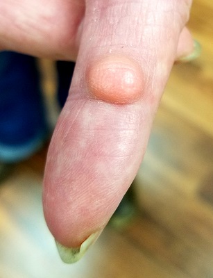
A 70-year-old man self-refers to dermatology for evaluation of a “risin’ in my finger,” which has existed for “at least 40 years.” While the lesion doesn’t really hurt, the patient wants it gone because he traumatizes it almost daily while working on his farm.
He has tried innumerable removal methods, including acids, blood root, and duct tape. Most recently, his primary care provider attempted treatment with cryotherapy.
The patient’s health is excellent in other respects, with no history of similar lesions elsewhere.
EXAMINATION
A dome-like pink nodule is seen on the palmar surface of the patient’s left index finger. The 1-cm lesion is smooth, moderately firm, and nontender. Although the majority of the lesion protrudes above the skin’s surface, there is an intradermal component. Normal skin lines on the surface are preserved, and there is no surface punctum. Palpation of the epitrochlear and axillary areas above the hand reveals no masses.
What is the diagnosis?
DISCUSSION
Traumatic puncture—especially of the palms and fingers—can invaginate the surface of skin, effectively burying the surface follicular infundibulum and associated sebaceous gland as the wound heals. These structures can continue to produce sebum and epidermal cells, which then accumulate in a space delineated by the lining of the infundibulum and form a sac called an epidermal inclusion cyst (EIC).
EICs differ from the more common epidermoid (or epidermal) cyst for several reasons. For one, epidermoid cysts are confined almost exclusively to oily skin (usually above the waist, with the back being the most common location). Unlike EICs, epidermoid cysts almost always have at least one central comedone, which acts as a plug, preventing the discharge of its contents.
The differential also includes pilar cysts—another common type, popularly known as “wens.” But these occur almost exclusively in the scalp and only rarely display a central punctum.
All of these cysts have an organized wall that contains their characteristic cheesy contents, and all can be removed surgically—preferably in one piece—under local anesthesia. Once excised, the lesion should always be submitted for pathologic examination, since cystic lesions can (albeit rarely) undergo malignant transformation.
In this case, excision was performed with anesthesia via digital block, supplemented with small amounts of lidocaine and epinephrine for hemostasis. The lesion was removed in one transverse elliptical piece and with special care to avoid trauma to underlying structures (visualized with the aid of a tourniquet). The wound was closed with interrupted sutures.
The pathology report confirmed the preoperative diagnosis, and the patient recovered uneventfully.
TAKE-HOME LEARNING POINTS
- Epidermal inclusion cysts (EICs) result from a puncture wound that buries surface follicular infundibula and their sebaceous glands under the skin, where they continue to produce material that collects in a sac.
- Unlike the more common epidermoid (epidermal) cysts, which affect the oily areas of the body (above the waist), EICs don’t have a central punctum.
- EICs can be left alone or excised, but removal must be done in one piece, lest they recur. The same is true for all aforementioned cysts.
SHM to induct new Masters in Hospital Medicine
The Society of Hospital Medicine will induct four new Masters in Hospital Medicine (MHM), the society’s highest professional honor, at HM18. Recipients are distinguished by the excellence and significance of their contributions to the field of hospital medicine and health care overall, said Larry Wellikson, MD, MHM, CEO of the Society of Hospital Medicine. They have been selected because of personal character; positions of honor; contributions toward furthering the society’s goals; distinction in practice, education, medical research; and other achievements in science or in the art of hospital medicine.
MHM nominees must be highly accomplished individuals in the hospital medicine specialty. Evidence of their achievements can come from many types of activities, such as excellence in clinical care, health care initiatives, education, research, writing and publication, volunteerism, and administrative positions. Current members of the society’s board are not eligible for nomination or selection.
This is truly the Hall of Fame for hospital medicine. Congratulations to this year’s MHMs.
Andrew Auerbach, MD, MPH, MHM, is professor of medicine in residence at the University of California, San Francisco, where he also serves as director of innovation research for the Center for Digital Health and Innovation. He was one of the first 200 members of the Society of Hospital Medicine when it was first called the National Association of Inpatient Physicians. “I have contributed to the field and society through my research and national role as a hospitalist in high-profile policy and guideline initiatives,” he said. These include formulating guidelines for the Institute of Medicine, American College of Cardiology, and Agency for Healthcare Research and Quality.
Dr. Auerbach has been deeply involved with the society through his role as chair of its Research Committee and Academic Hospitalist Committee for several years. Other accomplishments include being a founding framer of the Academic Hospitalist Academy, contributing to white papers outlining academic hospitalists’ needs, cofounding a national research network for hospitalists, and serving as editor-in-chief of the Journal of Hospital Medicine for 7 years.
“Being named an MHM is wonderful recognition,” he said. “I just hope it does not represent a ‘lifetime achievement’ award – I still have many things I want to accomplish in my career!”
Daniel J. Brotman, MD, MHM, professor of medicine, director of the hospitalist program at Johns Hopkins Hospital, Baltimore, and a member of the Society of Hospital Medicine since 2000, has served in many roles at the organization. These include being a member of the Annual Meeting Planning Committee (2007-2016), course director for the annual meeting (2013, 2014), chair of the Education Committee (2012-2016), and a member of the Research Committee (2008-2015). Dr. Brotman also won the society’s Research Award in 2015. He has been a staple of the editorial team at the Journal of Hospital Medicine since its founding in 2006.
“I am truly honored to be recognized for my participation and leadership in the field of hospital medicine and the society,” he said. “I am humbled to be included among the many luminaries who have won the award in prior years.”
He has been in his current role at Johns Hopkins since 2005, where he has more than tripled the program’s size and grown its academic profile.
“I have had the good fortune to work with a lot of talented faculty members and have helped them advance professionally and academically, while they pushed me to be a better leader,” Dr. Brotman said. “I encourage my team members to participate in the Society of Hospital Medicine; it is a fabulous way to gain leadership opportunities while staying abreast of the most important developments in the field.”
Bob Harrington, MD, MHM, is president and chief medical officer, SurveyVitals, an organization that provides digital patient experience and Consumer Assessment of Healthcare Providers and Systems (CAHPS) surveys nationwide to hospitals, health care systems, and physician practices. He believes he is an award recipient because he is a long-time advocate for hospitalists trained in family medicine (HTFMs). “I have always looked for ways to level the playing field for clinical and leadership opportunities for HTFMs,” he said.
Dr. Harrington first became involved with the Society of Hospital Medicine as chair of its Family Medicine Committee in the late 1990s. In that role, his committee looked at unique needs of HTFMs, including hiring practices, providing education to potential employers, and assisting HTFM members in career advancement.
“Our efforts resulted in tremendous growth of the HTFM membership, as these physicians began to view the society as their professional home and not as an internal medicine–dominant organization,” he said. He went on to become the first HTFM to serve on the society’s board and then became the first HTFM to be its president.
“This award is the greatest professional honor that I have received,” Dr. Harrington said. “It is especially important to me, because it is from a group of people whom I hold in the highest professional regard. They always put quality and patients first. I have been privileged to play a small part in that.”
Janet Nagamine, MD, BSN, MHM, is a hospitalist in the department of hospital-based specialty and inpatient palliative care at Kaiser Permanente Hospital in Santa Clara, Calif.
Dr. Nagamine was one of the first 200 people to join the Society of Hospital Medicine. Among her many roles within the society were serving on its board of directors from 2009 to 2014 and chairing the Quality and Safety committee for 5 years.
“I’m honored to be acknowledged in this formal and prestigious way, because my career path hasn’t always followed the traditional course of recipients who receive this type of award,” commented Dr. Nagamine, who has worked in a community hospital for the last 18 years. “This speaks volumes to the type of organization that the Society of Hospital Medicine is, and it makes me proud.”
She started her career as an ICU nurse and spent the last 30 years in various institutional and organizational roles trying to make hospitals a better place.
“At times, I was juggling family responsibilities and had to defer the fancy titles, but I always found a way to contribute meaningfully through my work with the Society of Hospital Medicine.”
HM18 satellite symposia schedule, information
From Hospital Admission to Home: New Standards for Extended Duration VTE Prophylaxis in Acutely Ill Medical Patients
Sunday, April 8
5:30 – 7:30 p.m., Canary Room 1-2
Dinner provided at 5:30 p.m.
Learning Objective: To educate on the risk of VTE in acutely ill medical patients, APEX clinical trial results, and Bevyxxa (betrixaban).
Overview:
- Review the burden of VTE in acutely ill medical patients.
- Provide an overview of the unmet need for extended-duration VTE prophylaxis from hospital admission to home.
- Review the APEX clinical trial data.
- Provide an overview of Bevyxxa (betrixaban) indication, safety information, dosing, and appropriate patient types.
Presenter: Hameed Ali, DO, FHM, clinical assistant professor of medicine and hospitalist, Baylor Scott and White Health Hospital, Temple, Tex.
This program is supported by Portola Pharmaceuticals.
Reducing COPD-related Readmissions through Individualized Maintenance Therapy and Increased Patient Engagement
Sunday, April 8
5:30 – 7:30 p.m., Canary Room 3-4
Dinner provided at 5:30 p.m.
Overview: Hospitals are a critical point of intervention in the care pathway of chronic obstructive pulmonary disease (COPD). Clinicians in this setting must be well versed in current treatment guidelines, as well as the full spectrum of medications and delivery devices, to provide disease management tailored to the physical and cognitive needs of each patient. Moreover, hospital clinicians also need to be adept at communicating with patients and engaging them in the management of their own disease. Collectively, these measures can significantly reduce symptom severity and the risk for future exacerbations, increase physical activity, and improve overall quality of life in patients with COPD. This program will improve the knowledge and competence of clinicians who care for patients with COPD.
Learning objectives: Upon completion of this educational activity, participants should be able to:
- Identify long-term treatment strategies to reduce hospital readmissions for COPD exacerbations.
- Review the clinical evidence regarding the efficacy and safety of long-acting maintenance regimens for COPD.
- Select medication delivery devices for patients with COPD based upon individual physical and cognitive characteristics.
- Outline a transitional care plan that promotes patient self-management to reduce the risk for future exacerbations and hospital readmissions.
Presenters: Stanley B. Fiel, MD, regional chair in the department of medicine, Atlantic Health System, and the deNeufville Professor, chairman of the department of medicine, Morristown (NJ) Medical Center; José Luis González, MD, assistant professor of internal medicine, department of internal medicine, University of Southern California, and primary care physician, department of primary care, LAC+USC Medical Center, Los Angeles.
Accreditation statement: Integrity Continuing Education, Inc. is accredited by the Accreditation Council for Continuing Medical Education to provide continuing medical education for physicians.
Credit designation: Integrity Continuing Education, Inc. designates this live activity for a maximum of 1.5 AMA PRA Category 1 Credits™. Physicians should claim only the credit commensurate with the extent of their participation in the activity.
A Physician’s Keys to Locking Out Lawsuits and Reducing Taxes
Monday, April 9
Noon – 1:00 p.m., Anaheim/Atlanta/Boston Room
Lunch provided at noon.
Objectives:
- Protect your license from negative reports to the National Practitioner Data Bank (NPDB) following a settlement from your insurance company. If there is no NPDB report, it’s unlikely that a board investigation into the legal matter will materialize. Preventing any sanctions from the state licensing board.
- Show how to structure: C-corps, S-corps, FLPs, LLCs, etc.
- Teach the use of legal tools that will protect their professional and personal assets from lawsuits. (Statistically, not even 1 in 100,000 are using these tools in the right way.)
- Learn how to protect business, property, and personal assets in the event of a judgment in excess of liability insurance.
- Learn how to protect your license from negative reports to the NPDB following a settlement from your insurance company. If there is no NPDB report, it’s unlikely that a board investigation into the legal matter will materialize. Preventing any sanctions from the state licensing board.
- Learn the best business structure for income tax reduction. Learn the new tax laws passed in 2017 and how they can benefit you.
Faculty: Art McOmber
Sponsored by Legally Mine.
Community-Acquired Bacterial Pneumonia (CABP) in the Hospital Setting: Why Are Patients Not Getting Better?
Monday, April 9
7:30 – 9:30 p.m., Canary Room 1-2
Dinner provided at 7:30 p.m.
Program Summary: Appropriate management of pneumonia in the hospital setting can have a substantial impact on patient outcomes and hospital measures such as readmission rates and length of stay. Community-acquired bacterial pneumonia (CABP) is one of the most common infectious diseases, one of the most frequent infections requiring antibiotics, and remains a leading cause of death in the United States. During this presentation, we will review current CABP guidelines from IDSA (the Infectious Diseases Society of America) and discuss the importance of appropriate antibiotic selection as it relates to the tenets of antimicrobial stewardship. The faculty will present two case studies and will solicit audience participation with a focus on antibiotic resistance and the importance of transition of care.
Chair: William Ford, MD, SFHM, Abington Jefferson Health, Abington, Penn.
Faculty: Mauricio Pinto, MD, St. David’s Round Rock Medical Center, Round Rock, Tex.; Sumeet Shetty, MD, MBA, FHM, Axel Health, Fort Meyers, Fla.
This program is supported by Nabriva Therapeutics, plc.
Register: [email protected] www.nabrivaevents.com/SHM/Symposium, or call 877-547-5640.
Direct Oral Anticoagulants (DOACs): Current Evidence for Extended VTE Prophylaxis in Medically Ill Patients and Reversal
Monday, April 9
7:30 – 9:30 p.m., Canary Room 3-4
Dinner provided at 7:30 p.m.
Overview: Patients hospitalized for an acute medical illness are at an increased risk for venous thromboembolism (VTE). With increasingly shortened hospital stays, acutely ill hospitalized patients are at an increased risk of developing VTE both in the hospital and after discharge. Outpatient VTE episodes often occur within 30 days of hospital discharge and fewer than half of those discharged patients receive VTE prophylaxis. Therefore, it is important for clinicians to be able to risk-stratify patients and provide extended prophylaxis for medical patients at increased risk for VTE.
This symposium will discuss the risk factors and burden of VTE and review ACCP guideline recommendations for VTE prophylaxis in acutely ill hospitalized medical patients. Faculty will assess the safety and efficacy of direct oral anticoagulants (DOACs) for extended VTE prophylaxis, explain the stratification of VTE and bleeding risk, as well as the process for devising evidence-based antithrombotic regimens. The presentation also will include an outline of current and emerging options for reversal of direct oral anticoagulants.
Learning objectives:
- Outline the risk factors and burden of VTE in medically ill patients post hospitalization.
- Review ACCP and ASH guideline recommendations for VTE prophylaxis in acutely ill hospitalized medical patients.
- Assess the safety and efficacy of DOACs for extended VTE prophylaxis in medically ill patients.
- Devise evidence-based antithrombotic regimens for medically ill patients taking into consideration patient-specific factors that impact VTE and bleeding risk
- Outline current and emerging options for reversal of DOACs
Faculty: Amir K. Jaffer, MD, MBA, chief medical officer, New York Presbyterian Queens Hospital, New York; Alex C. Spyropoulos, MD, FACP, FCCP, FRCPC, professor of medicine, Donald and Barbara Zucker School of Medicine at Hofstra/Northwell, system director – anticoagulation and clinical thrombosis services, Northwell Health at Lenox Hill Hospital, New York, NY; and Alan Jacobson, MD, FACC, assistant professor of medicine, Loma Linda University School of Medicine, director of anticoagulation services, Loma Linda VA Medical Center, Calif.
Target audience: Hospitalists, internists, nurse practitioners (NPs), physician assistants (PAs) who practice in a hospital setting.
Credit designation: Horizon CME designates this live activity for a maximum of 1.5 AMA PRA Category 1 Credit(s)™. Physicians should claim only the credit commensurate with the extent of their participation in the activity.
Accreditation statement: This activity has been planned and implemented in accordance with the accreditation requirements and policies of the Accreditation Council for Continuing Medical Education (ACCME) through the providership of Horizon CME. Horizon CME is accredited by the ACCME to provide continuing medical education for physicians.
ABIM MOC statement: Successful completion of this CME activity, which includes participation in the evaluation component, enables the participant to earn up to 1.5 Medical Knowledge MOC points in the American Board of Internal Medicine’s (ABIM) Maintenance of Certification (MOC) program.
Participants will earn MOC points equivalent to the amount of CME credits claimed for the activity. It is the CME activity provider’s responsibility to submit participant completion information to ACCME for the purpose of granting ABIM MOC credit.
Supporter statement: This activity is supported by an independent educational grant from Portola Pharmaceuticals.
Register: http://bit.ly/2EoP1tb
HM18 Special Interest Forums
The Society of Hospital Medicine presents a variety of special interest forums during its annual conference. The small-group sessions take place Monday, April 9, 4:30-5:25 p.m.
Academic and Research
Greg Seymann, MD, SFHM; Nicole Adler, MD, FHM
Grand Ballroom 12-14
The Academic and Research Forum brings together faculty and researchers to discuss topics of interest to the academic hospital medicine community, such as mentorship, research support, and professional development. Join this collaborative offering of the Academic and Research Committees.
Advocacy & Public Policy
Joshua Lenchus, DO, RPh, SFHM; Josh Boswell
Key Biscayne Room
During this forum with SHM’s Advocacy leaders and staff, you will learn about the direction of SHM’s Advocacy & Public Policy work and how you can help. Discussion will focus on SHM’s new Advocacy & Public Policy Section, its role, and how you can participate and share your own ideas.
Canadian Hospitalists
Serge Soolsma, MD
Key Largo Room
This forum provides a unique setting for hospitalists based in Canada to gather as an organized group, network with each other, and discuss the common issues with which they are faced.
Care for Vulnerable Populations
Mara Bann, MD; Pallabi Sanyal-Day, MD
Key West Room
SHM’s Caring for Vulnerable Populations Section aims to increase awareness and improve quality of care for vulnerable and underserved patient populations in the hospital setting. The principles and skills needed to care effectively for vulnerable patients span practitioners across all health systems, although they are important particularly for hospitalists practicing in safety-net and resource-limited settings.
Community-Based Hospitalists
Steve Behnke, MD; Jason Robertson, MD, SFHM
Sawgrass Room
This session provides a forum for sharing principles of successful clinical practices, quality care, and professional sustainability, as well as other “hot” topics of interest to the community-based hospitalist.
NEW: Critical Care
David Aymond, MD
Grand Ballroom 4-6
This special interest forum seeks to convene hospitalists charged with providing some level of critical care at their institution. Participants should come prepared to discuss and share their own experiences, including their current role in providing critical care, facing institutional barriers, and dealing with gaps in training.
NEW: Diversity and Inclusion
Marisha Burden, MD, SFHM; Flora Kisuule, MD, SFHM
Grand Ballroom 7A
SHM is committed to a diverse and inclusive membership that works to provide high-quality, equitable care to diverse populations. This forum invites hospitalists from any underrepresented group to discuss issues, concerns, and solutions to improve workforce diversity and their own career opportunities. In addition, this forum would be for HM leaders who would like to discuss strategies and opportunities for expanding the diversity and inclusion of their HM groups.
NEW: Ethics in Hospital Medicine
David Alfandre, MD, MSPH
West Indies Room
This forum serves as a resource for discussion, coaching, and mentorship regarding common and challenging ethical concerns that hospitalists face. We aim to support SHM members in collaborating on ethics scholarships and projects that address ethics in clinical care, education, and policy.
Global Health and Human Rights
Brett Hendel-Paterson, MD, FHM
Harbor Beach Room
SHM’s Global Health and Human Rights Section has been established to build interest and engagement in global health and human rights work among hospitalists so they can share their expertise. The section also plans to build long-term collaborations in the United States and abroad.
Hospitalists Trained in Family Medicine
David Goldstein, MD; Patricia Seymour, MD
Anaheim Room
Participants will network and discuss their training, how they’ve achieved recognition and access in the job market, as well as national trends related to hospitalists trained in family medicine.
Information Technology
Cheng-Kai Kao, MD; Andrew Young, DO
St. Thomas Room
This forum provides an opportunity for attendees to provide SHM and the IT Committee with input on what would be most beneficial regarding implementing, managing, and participating in health/hospital IT initiatives.
International Hospital Medicine
Guillherme Barcellos, MD, SFHM; Rafaela Dal Molin, MD, MEd, FHM; Nerea Fernandez, MEd, PhD
Crystal Ballroom G1/A&B
This forum is designed to provide an opportunity for attendees who practice hospital medicine outside of North America to share their ideas and discuss issues they’ve faced.
Leadership in Hospital Medicine
Thomas McIlraith, MD, SFHM, CLHM; Rob Zipper, MD, MMM, SFHM
Marco Island Room
Want to be a better leader? A better coach and mentor? Do you want to drive quality improvement (QI) at your hospital? Developing ourselves and our teams is what we are all here to do! We will review, discuss, and shape the resources and programmatic offerings that are needed to promote leadership skills development at all levels. We will also review SHMs existing programs, including the Leadership Academies, the Leadership Certificate Program, e-learning opportunities, and the HMX: Leadership Alumni Forum.
Med-Peds Hospitalists
Heather Toth, MD, SFHM; Carrie Herzke, MD, SFHM
Crystal Ballroom H
This special interest forum will explore the role of Med-Peds physicians in hospitalist medicine. Discussion items may include personal experiences, how to create more Med-Peds jobs, and how to succeed as a Med-Peds hospitalist.
Multi-Site HMG Leaders
Leslie Flores, MHA, SFHM; Ryan Brown, MD, FHM
Grand Ballroom 1-3
This forum is for physician and administrative leaders who are responsible for managing multiple hospitalist practice sites within the same health system. The number of people with this role has increased significantly in the last few years and comes with challenges that are different from those faced by the lead hospitalist at a single-practice site.
Nurse Practitioners and Physician Assistants
Emilie Davis, PA-C, FHM; Noam Shabani, MS, PA-C
Vinoy Room
Share best practices and challenges. Learn about SHM resources for NPs and PAs in practice, as well as onboarding and recruitment resources. Network with peers and help build membership engagement.
Oncology Hospitalists
Maria Campagna, MD, FHM; Barbara Egan, MD, SFHM; Kerry Reynolds, MD
Aruba Room
This special interest forum will explore the role of hospitalists in oncology services. Discussion items may include personal experiences and how to succeed as an oncology hospitalist.
Palliative Care
Rab Razzak, MBBS, MD; Jeffrey Frank, MD, MBA
Bahamas Room
This special interest forum seeks to convene hospitalists charged with providing some level of palliative care at their institution. Participants should come prepared to discuss and share their own experiences, including their current role in providing palliative care, facing institutional barriers, and dealing with gaps in training.
Patient Experience
Mark Rudolph, MD, SFHM; Patrick Kneeland, MD
Grand Ballroom 7B
Join the Patient Experience Forum to exchange ideas about how hospitalists can enhance patients’ care experiences while also improving professional satisfaction. Learn about the work of SHM’s Patient Experience Committee and opportunities for getting involved in SHM’s patient experience initiatives.
Pediatric Hospitalists
Sandy Gage, MD, SFHM
Grand Ballroom 9-11
This special interest forum will provide an opportunity for pediatric hospitalists to network, share, and discuss topics and issues of particular interest to them. Topics will include an updates on SHM’s pediatric activities, on potential paths to specialty certification, and about the relationships between SHM, AAP, APA, PRIS, and the Joint Council on Pediatric Hospital Medicine.
NEW: Perioperative Care
Steven Cohn, MD, SFHM; Kurt Pfeifer, MD
San Francisco Room
In this special interest forum, learn about guideline updates and recent literature while communicating controversial or difficult patient management issues around perioperative medicine.
Point-of-Care Ultrasound (POCUS)
Benji Mathews, MD, CLHM, SFHM; Gordon Johnson, MD, FHM
Atlanta Room
This special interest forum will discuss opportunities to collaborate and standardize processes for POCUS certification, including what resources already exist. In addition, discussion will revolve around privileging at your own institution, gaining skills, and the challenges and successes of procedural teams in the hospital.
Post-Acute Care Providers
Robert Reynolds, MD
Puerto Rico Room
This forum provides opportunities for hospitalists who practice in or are interested in learning more about working in or becoming more involved in the post-acute care arena, such as SNFs, LTACs, and rehab facilities.
Practice Administrators
Tiffani Panek, CLHM; Roberta Himebaugh, MBA, SFHM
Boston Room
Practice administrators are important members of the hospitalist team, providing key management and organizational skills. In this forum, administrators can voice their unique perspectives and hear from their peers.
Quality Improvement
Mangla Gulati, MD, MBBS, CPPS, SFHM; Jenna Goldstein
Grand Cayman Room
Hospitalists are at the center of the national quality and patient safety movement and are increasingly responsible for performance at their institutions. This forum provides a venue for connecting with SHM’s QI and patient safety community and for engaging with leaders, peers, and collaborators to share ideas and inform SHM’s QI efforts. Discussion during the forum will focus on what hospitalists need to know to become involved with QI at SHM or locally. Hear about SHM’s plans for future QI initiatives, and share your own ideas.
NEW: Residents & Medical Students
Aram Namavar, MS; Chris Bartlett, MD, MPH
New Orleans Room
This forum provides opportunities in networking and discussion for physicians in training who are contemplating a career in hospital medicine.
Rural Hospitalists
Ken Simone, DO, SFHM; Michael Sullivan, MD
Los Angeles Room
Hospital medicine groups in rural areas face some unique problems, from recruitment, night call, and staffing to communicating with geographically dispersed primary care physicians. Rural hospitalists may also face clinical challenges because of limited technological resources and/or limited access to specialists. This forum provides an opportunity for hospitalists in rural areas to share their issues and concerns and to see how others have solved similar problems.
Veterans Affairs Hospitalists
Kathlyn Fletcher, MD, FHM; Peter Kaboli, MD, FHM
Miami Room
This forum provides opportunities in networking and discussion for hospitalists who work at the VA. Issues unique to VA hospitalists will be discussed.
Women in Hospital Medicine
Melissa Mattison, MD, SFHM; Cory Ritter, MD, FHM
New York Room
This forum provides an opportunity to discuss issues relevant to women in hospital medicine and strategies for success/coping. Topics may include career satisfaction, occupational stresses, opportunities for change, promotion of leadership, and identification of resources.
SHM Loyalty Partners at HM18
The Society of Hospital Medicine is grateful for the loyal support of the following companies:
Platinum Level
Aurora Health Care
Baylor Scott & White Health
BMS/Pfizer
Boehringer Ingelheim Pharmaceuticals, Inc.
Community Hospitalists
CompHealth
D&Y
Emory
Envision Physician Services
In Compass Health, Inc.
Ingenious Med, Inc.
Kaiser Permanente
Lightning Bolt Solutions, Inc.
Medstaff National Medical Staffing
NEJM Group
PeaceHealth
PracticeLink
Schumacher Clinical Partners
Sound Physicians
Staff Care, Inc.
TeamHealth
The Delta Companies
Today’s Hospitalist
University of Tennessee Physician Executive MBA Program
VEP Healthcare
VISTA Staffing Solutions
Vituity
Gold Level
Allergan USA, Inc.
American Board of Internal Medicine (ABIM)
Billings Clinic; Billings, Montana
Hospitalists Plus
Jackson & Coker
Janssen Pharmaceuticals, Inc.
Locum Leaders
MedAptus
MediMobile
Mountain States Health Alliance
PatientKeeper
Sonosite FujiFilm
Sutter Health Sacramento Sierra Region
Texas Health Resources
The Doctors Company
Weatherby Healthcare
Silver Level
Adventist Health System
Agency for Healthcare Research and Quality
Apogee Physicians
Banner Health
Calmoseptine, Inc.
Community Health Systems
Geisinger
Global Medical Staffing
LocumTenens.com
Mayo Clinic
MedData, Inc.
Medicus Healthcare Solutions
Mint Physician Staffing
PracticeMatch
Providence St Joseph Health (aka Providence Health & Services)
QGenda, LLC
Quest Healthcare Solutions
ScribeAmerica
Shift Administrators
TIVA Healthcare, Inc.
VITAS Healthcare
Wellstar Health System
Bronze Level
AAPA Center for Healthcare Leadership & Management
Alteon Health
ApolloMD
Aspirus Clinics
BioFire Diagnostics, Inc.
Carnegie Mellon University Master of Medical Management
Carolinas HealthCare System
Center to Advance Palliative Care (CAPC)
DaVita Medical Group
Drwanted.com
ICON Medical Network
Indiana University Kelley School of Business, Business of Medicine MBA
Integrity Locums
Melinta Therapeutics
Mercy Health
Northwell
Nova Biomedical
Pinnacle Health Group Remedy Partners
The Ohio State University Division of Hospital Medicine
Stay on top of perioperative medicine
The fluid, evolving nature of perioperative medicine lends itself to a lively literature review and research update, according to Steven L. Cohn, MD, SFHM, a presenter of “ ‘A Whole New World’ of Perioperative Medicine: The 2018 Update” on Monday, April 9, at HM18.

The perioperative medicine update has been a fixture at the SHM annual conference for several years and provides an opportunity to highlight articles that may affect practice, said Dr. Cohn of the University of Miami in an interview. “Our goals are to familiarize the attendees with this new data so they are aware, so they can review the key articles in detail on their own, and so they can then apply the new information in their clinical practices,” he said.
The agenda for the session involves a presentation of 10-15 articles published since HM17. Dr. Cohn and presenter Paul J. Grant, MD, SFHM, the director of the consultative and perioperative medicine program and an associate chief medical information officer at Michigan Medicine, Ann Arbor, will briefly review the objectives and methods of each study before moving on to focus primarily on the result and conclusions.
“The top take-home message is to try to keep up with the perioperative literature, particularly in cardiology, because the evidence base keeps expanding and changing,” said Dr. Cohn. “This session is designed to deliver new information and also to stimulate the attendees to periodically review the literature on their own during the year,” he said.
This year’s collection of articles will address a range of topics, Dr. Cohn said, but he highlighted several articles “that will be thought provoking and potentially impact clinical practice regarding perioperative surveillance with the new fifth generation high sensitivity troponin assay and management of myocardial injury after noncardiac surgery. In particular, results from the MANAGE trial that was presented as a late-breaking trial at the American College of Cardiology meeting in March will be of interest to anyone involved in perioperative medicine,” he noted.
Dr. Cohn and Dr. Grant had no relevant financial conflicts to disclose.
“A Whole New World” of Perioperative Medicine: The 2018 Update
Monday, April 9, 2:00-2:40 p.m.
Crystal Ballroom G2/C-F
The fluid, evolving nature of perioperative medicine lends itself to a lively literature review and research update, according to Steven L. Cohn, MD, SFHM, a presenter of “ ‘A Whole New World’ of Perioperative Medicine: The 2018 Update” on Monday, April 9, at HM18.
The perioperative medicine update has been a fixture at the SHM annual conference for several years and provides an opportunity to highlight articles that may affect practice, said Dr. Cohn of the University of Miami in an interview. “Our goals are to familiarize the attendees with this new data so they are aware, so they can review the key articles in detail on their own, and so they can then apply the new information in their clinical practices,” he said.
The agenda for the session involves a presentation of 10-15 articles published since HM17. Dr. Cohn and presenter Paul J. Grant, MD, SFHM, the director of the consultative and perioperative medicine program and an associate chief medical information officer at Michigan Medicine, Ann Arbor, will briefly review the objectives and methods of each study before moving on to focus primarily on the result and conclusions.
“The top take-home message is to try to keep up with the perioperative literature, particularly in cardiology, because the evidence base keeps expanding and changing,” said Dr. Cohn. “This session is designed to deliver new information and also to stimulate the attendees to periodically review the literature on their own during the year,” he said.
This year’s collection of articles will address a range of topics, Dr. Cohn said, but he highlighted several articles “that will be thought provoking and potentially impact clinical practice regarding perioperative surveillance with the new fifth generation high sensitivity troponin assay and management of myocardial injury after noncardiac surgery. In particular, results from the MANAGE trial that was presented as a late-breaking trial at the American College of Cardiology meeting in March will be of interest to anyone involved in perioperative medicine,” he noted.
Dr. Cohn and Dr. Grant had no relevant financial conflicts to disclose.
“A Whole New World” of Perioperative Medicine: The 2018 Update
Monday, April 9, 2:00-2:40 p.m.
Crystal Ballroom G2/C-F
Embracing constant change in a growing practice
Although many physicians resist change, learning how to embrace change and making it work for you are keys to a long, successful practice in hospital medicine, said Thomas McIlraith, MD, SFHM, CLHM, immediate past chairman, department of hospital medicine, and immediate past treasurer of the board of directors of Mercy Medical Group in Sacramento, Calif.
During his Monday, April 9, session, “Scaling Up Your Hospital Medicine Group,” he will share what it was like to work through significant changes as the chair of a department that grew in 12 years from 12 hospitalists – averaging fewer than 100 patients per day – to 84 hospitalists averaging more than 500 patients per day.
“I will be discussing my experiences but relating some universal truths about leadership in hospital medicine that can be applied to all areas of hospital medicine and how to manage change in an environment where change is constant,” said Dr. McIlraith, who will share key lessons that he learned. “I will break it down into the cultural, personal, and structural aspects of thriving in this dynamic environment.”
Dr. McIlraith hopes that attendees will come away with a good understanding of what he dubs the “authority/accountability” equation in physician leadership. “This is a principle that is used to assess what is expected of you in order to be successful as a leader, and if your ‘authority’ is commensurate with your ‘accountability,’ ” he said. “The authority piece of the equation takes many forms: resources, personnel, reporting structure, clerical support, and so forth. The key is to make sure that the authority/accountably equation is balanced not only for you but also for your subordinates.”
One example of an imbalanced authority/accountability equation that he’ll discuss in some detail is becoming a figurehead, in which accountability exists without the necessary authority. “If you end up in that position, get out,” Dr. McIlraith said. He will also discuss an example of where the authority/accountability equation is tipped in the opposite direction, with disastrous consequences.
Dr. McIlraith and Kimberly Bell, MD, who will also present at this session, aim to make it interactive. Dr. Bell currently serves as the divisional director of the CHI Franciscan Inpatient Team located in Tacoma, Wash. “We both have a wealth of experience to offer, but we are not the only ones in attendance who have wisdom to share,” Dr. McIlraith said.
Another goal of this session is to help attendees identify other hospital medicine leaders in comparable situations for the purpose of networking outside of the meeting. The session, although aimed at physician leaders, will be relevant to all hospitalists.
Scaling Up Your Hospital Medicine Group
Monday, 1:35-2:35 p.m.
Grand Ballroom 7B
Speed mentoring returns to HM meeting
About 6 years ago, members of the Society of Hospital Medicine’s Academic Hospitalist Committee got together to try to answer a question: How do we meet the mentoring needs of junior hospitalists?
One answer was inspired by the singles social scene.
Joanna Bonsall, MD, PhD, SFHM, assistant professor of medicine at Emory University, Atlanta, has been a part of coordinating the event since the beginning.
“We tried it out for the first year, and it was wildly successful,” she said. “The feedback was enormous, so we’ve continued it ever since.”
Hospitalists looking for mentoring time with senior hospitalists submit their curriculum vitae and a question they’d like to have answered. Then they’re paired with three mentors who have expertise in that field, meeting with each for 15 minutes.
For instance, someone asking – How can I improve my teaching skills with medical students? – would be paired with veteran hospitalists known for their education skills. In its first year, there were 12 slots available. It has since expanded, and this year, there are 20 slots. Registration closed a month before the annual conference.
Dr. Bonsall said that mentoring, including the speed mentoring event, is all about matching up ambition and talent with ways to put them to use.
“I believe that our field is so full of talent, and I think that because we’re a relatively junior field, there is a lot of untapped talent out there – people who have energy and don’t know what to do with it,” she said. “I feel the same way about faculty development. Mentoring is so key to help connect the energy to things that they can do.”
Welcome to Hospital Medicine 2018
Welcome to Hospital Medicine 2018 (HM18), the largest conference ever held specifically for hospital medicine. The Society of Hospital Medicine (SHM) is proud that HM18 brings together a broad range of stakeholders in hospital medicine, including physicians of many specialties, acute care providers, administrators, pharmacists, C-suite executives, recruiters, and educators. Your decision to join your colleagues at HM18 demonstrates your commitment to not only the specialty of hospital medicine but also to the patients you serve.
This year’s renowned speakers will present important sessions addressing the rapidly evolving health care landscape. To open the main conference on April 9, Kate Goodrich, MD, MHS, chief medical officer and director of the Centers for Medicare & Medicaid Services’ Center for Clinical Standards and Quality, and a practicing hospitalist, will present her featured address, “Payment Reform is Here: What Hospitalists Need to Know.” You will also hear from the president of SHM, Ron Greeno, MD, FCCP, MHM, about the state of hospital medicine.
On April 9, be sure to attend one of SHM’s Special Interest Forums, where you can choose to network and connect with other hospitalists interested in the same things you are. There are 29 forums from which to choose. On April 10, SHM will open the first-ever International Hospital Medicine Lounge. We hope to welcome more than 100 hospitalists from around the world to HM18.
On April 11, HM18 concludes with the closing keynote, titled “Hospitalists as Drivers of Innovation and System Change: Are We Doing Enough?” presented by Robert M. Wachter, MD, MHM, chief of the department of medicine at the University of California, San Francisco, and the author of “Hospital Medicine” and “The Digital Doctor.”
Please make sure to download the HM18 At Hand meeting app, a wonderful resource for every HM18 attendee that puts the meeting at your fingertips. Create an individualized conference experience from your smartphone, tablet, or laptop.
Don’t miss the opportunity to meet one on one with members of SHM’s Board of Directors, who will be available in the SHM Pavilion, located in the Exhibit Hall, during scheduled visit times. Please consult the Meet the Board schedule in the HM18 At Hand app for further information.
On behalf of the SHM Board of Directors and staff, welcome to HM18 and to Orlando. Through this conference’s rich selection of educational opportunities, research offerings, and networking events, SHM continues to further its mission to promote excellence in the practice of hospital medicine. SHM remains at the forefront of health care today, continuing to empower hospitalists and revolutionize patient care.
Dr. Wellikson is CEO of SHM.
