User login
Empirical micafungin falls short for treating suspected fungal infection in the ICU
MILAN – Empirical antifungal treatment did not improve the rate of survival free of invasive fungal infection among high-risk colonized patients in the intensive care unit, based on results from the EMPIRICUS randomized controlled trial.
Trial participants were 260 nonneutropenic, nontransplanted critically ill patients with ICU-acquired sepsis, Candida colonization of at least one site, and multiple organ failure who were exposed to broad-spectrum antibacterial agents. They were randomized to 14 days of empirical treatment with micafungin (Mycamine, 100 mg once daily) or placebo.
By day 28, about two-thirds of patients overall remained alive and free of proven invasive fungal infection, with no significant difference between groups, according to data reported at the annual congress of the European Society of Intensive Care Medicine and simultaneously published online (JAMA. 2016 Oct 5. doi: 10.1001/jama.2016.14655). Results were similar in subsets of patients having established risk factors for candidemia.
The EMPIRICUS (Empirical Antifungal Treatment in ICUs) findings add to data from other studies suggesting that, in this patient population, sepsis is seldom a result of invasive fungal infection and Candida colonization status is not helpful for guiding treatment, according to the researchers, who were led by Dr. Jean-Francois Timsit of Inserm/Paris Diderot University and department of medical intensive care and infectious diseases, Hôpital Bichat-Claude-Bernard, Paris.
“Altogether, these results call into question the routine use of systematic surveillance for Candida colonization. Besides sparing unnecessary use of health care resources, it may also avoid inducing resistances to antifungals,” they maintain. “Whether this trial closes 3 decades of clinical research on Candida colonization deserves consideration.”
Patients were recruited to EMPIRICUS from 19 ICUs in France. On average, study participants had three Candida-colonized sites.
A modified intent-to-treat analysis showed that, by day 28 after enrollment, 68% of patients in the micafungin group and 60.2% in the placebo group were alive and free of invasive fungal infection, a nonsignificant difference.
Findings were similar in the subset of patients having high serum levels of (1-3)-beta-D-glucan and in the subset of patients having high Sepsis-Related Organ Failure Assessment (SOFA) scores – both risk factors for candidemia – and regardless of the number of colonized sites.
In analyses of secondary outcomes, empirical micafungin was associated with a lower rate of new invasive fungal infection when compared with placebo (3% vs. 12%; P = .008), but the rate of mortality was statistically indistinguishable (30% vs. 29.7%).
The groups were statistically indistinguishable with respect to the number of organ failure–free days and the rate of ventilator-acquired pneumonia.
Dr. Timsit disclosed that he receives lecture fees from Gilead, Pfizer, Merck, and Astellas; research grants to his university and research organization from Astellas, Gilead, Merck, and Pfizer companies; a consultancy honorarium from Bayer; and personal fees from Abbott for scientific board participation; additionally, he disclosed participation on a scientific committee of epidemiological studies organized by Astellas and Merck companies outside the submitted work. Astellas provided a research grant to the Grenoble Alpes University Hospital based on the final study protocol. The study was sponsored by the University of Grenoble 1/Albert Michallon University Hospital. The University of Grenoble provided compensation to the participating hospitals and universities for extra costs associated with the study.
Taken together, findings from EMPIRICUS and similar trials suggest that empirical antifungal treatment may reduce rates of invasive infection in critically ill patients, but does not improve survival.
These findings highlight two emerging themes in critical care medicine – less is more and targeted therapies are important when treating invasive fungal infection. In particular, the safety and efficacy of the newer antifungal agents are driving greater empirical use, yet this practice increases the cost of care and may contribute to antifungal resistance.
Guidelines have been implemented for empirical treatment of Candida and serial surveillance, yet there are no conclusive mortality benefits for this approach. Data have not ruled out the possibility that some subgroups of patients may see a survival benefit but, in light of the situation, guidelines concerning empirical treatment and surveillance should be revisited.
Like other prophylactic interventions, the risks and potential benefits of empirical echinocandin therapy for critically ill, immune-competent patients in the ICU need to be studied. Novel biomarkers or clinical risk assessment algorithms may help in identifying those patients who are at highest risk of infection-related morbidity and mortality and would benefit most from targeted preventive therapies.
Trishul Siddharthan, MD, Petros C. Karakousis, MD, and William Checkley, MD, PhD, are with the Johns Hopkins University in Baltimore. They made their remarks in an accompanying editorial in JAMA (2016 Oct 5. doi: 10.1001/jama.2016.13801).
MILAN – Empirical antifungal treatment did not improve the rate of survival free of invasive fungal infection among high-risk colonized patients in the intensive care unit, based on results from the EMPIRICUS randomized controlled trial.
Trial participants were 260 nonneutropenic, nontransplanted critically ill patients with ICU-acquired sepsis, Candida colonization of at least one site, and multiple organ failure who were exposed to broad-spectrum antibacterial agents. They were randomized to 14 days of empirical treatment with micafungin (Mycamine, 100 mg once daily) or placebo.
By day 28, about two-thirds of patients overall remained alive and free of proven invasive fungal infection, with no significant difference between groups, according to data reported at the annual congress of the European Society of Intensive Care Medicine and simultaneously published online (JAMA. 2016 Oct 5. doi: 10.1001/jama.2016.14655). Results were similar in subsets of patients having established risk factors for candidemia.
The EMPIRICUS (Empirical Antifungal Treatment in ICUs) findings add to data from other studies suggesting that, in this patient population, sepsis is seldom a result of invasive fungal infection and Candida colonization status is not helpful for guiding treatment, according to the researchers, who were led by Dr. Jean-Francois Timsit of Inserm/Paris Diderot University and department of medical intensive care and infectious diseases, Hôpital Bichat-Claude-Bernard, Paris.
“Altogether, these results call into question the routine use of systematic surveillance for Candida colonization. Besides sparing unnecessary use of health care resources, it may also avoid inducing resistances to antifungals,” they maintain. “Whether this trial closes 3 decades of clinical research on Candida colonization deserves consideration.”
Patients were recruited to EMPIRICUS from 19 ICUs in France. On average, study participants had three Candida-colonized sites.
A modified intent-to-treat analysis showed that, by day 28 after enrollment, 68% of patients in the micafungin group and 60.2% in the placebo group were alive and free of invasive fungal infection, a nonsignificant difference.
Findings were similar in the subset of patients having high serum levels of (1-3)-beta-D-glucan and in the subset of patients having high Sepsis-Related Organ Failure Assessment (SOFA) scores – both risk factors for candidemia – and regardless of the number of colonized sites.
In analyses of secondary outcomes, empirical micafungin was associated with a lower rate of new invasive fungal infection when compared with placebo (3% vs. 12%; P = .008), but the rate of mortality was statistically indistinguishable (30% vs. 29.7%).
The groups were statistically indistinguishable with respect to the number of organ failure–free days and the rate of ventilator-acquired pneumonia.
Dr. Timsit disclosed that he receives lecture fees from Gilead, Pfizer, Merck, and Astellas; research grants to his university and research organization from Astellas, Gilead, Merck, and Pfizer companies; a consultancy honorarium from Bayer; and personal fees from Abbott for scientific board participation; additionally, he disclosed participation on a scientific committee of epidemiological studies organized by Astellas and Merck companies outside the submitted work. Astellas provided a research grant to the Grenoble Alpes University Hospital based on the final study protocol. The study was sponsored by the University of Grenoble 1/Albert Michallon University Hospital. The University of Grenoble provided compensation to the participating hospitals and universities for extra costs associated with the study.
FROM ESICM CONGRESS 2016
Key clinical point:
Major finding: The day 28 invasive fungal infection–free survival was 68% with empirical micafungin and 60.2% with placebo, a nonsignificant difference.
Data source: A randomized controlled trial among 260 critically ill patients with ICU-acquired sepsis, Candida colonization, and multiple organ failure who were exposed to broad-spectrum antibacterial agents (EMPIRICUS trial).
Disclosures: Dr. Timsit disclosed that he receives lecture fees from Gilead, Pfizer, Merck, and Astellas; research grants to his university and research organization from Astellas, Gilead, Merck, and Pfizer companies; a consultancy honorarium from Bayer; and personal fees from Abbott for scientific board participation; additionally, he discloses participation on a scientific committee of epidemiological studies organized by Astellas and Merck companies outside the submitted work. Astellas provided a research grant to the Grenoble Alpes University Hospital based on the final study protocol. The study was sponsored by the University of Grenoble 1/Albert Michallon University Hospital. The University of Grenoble provided compensation to the participating hospitals and universities for extra costs associated with the study.
How will Donald Trump change health care?
Much speculation has already been written on what a Trump administration may look like, but comparatively little has been said about the potential impact of that administration on physicians, hospitals, and patients. While details will obviously not be available for some time, some early generalizations are possible, based on the position paper President-elect Donald Trump issued in March and the statements he has made since then.
One of Mr. Trump’s earliest and most repeated pledges was the one to repeal the Affordable Care Act and replace it with “something fantastic.” A repeal would be welcome news to many physicians, even without any idea of what its “fantastic” replacement might be; but Trump has hedged already. In mid-November, he told the Wall Street Journal that Obamacare would be “amended, or repealed and replaced,” which introduces considerable wiggle room.

In an interview with CNN, Mr. Trump indicated he would keep the individual mandate; but the next day, he tweeted – and then reiterated in his position paper – that he would remove the mandate and install a “backstop for preexisting conditions.” In the 1990s, when a few states tried to prohibit discrimination based on preexisting conditions without a corresponding mandate, premiums increased precipitously, driving away healthy customers, forcing insurers to stop selling policies in those states, and demonstrating that one cannot work without the other. Perhaps a more-informed Mr. Trump will come to see this.
Other proposals have been more enlightened. Mr. Trump has said that he favors portability of health insurance from state to state. In theory, this will introduce more competition into the system and drive premiums down. He has also proposed making health insurance premiums fully tax deductible for individuals, as they are now for businesses, further lowering premium costs.
He has proposed expanding the health savings account program, making contributions tax free, cumulative, and part of a patient’s estate. I have been a fan of HSAs since their inception because they eliminate the insurance “middleman,” which is good for physicians as well as for patients, who are more aware of what services they are receiving and what they are paying for them. Along the same lines, he has called for price transparency, so that patients can shop for the best prices for procedures and examinations, which now vary widely from one hospital or clinic to another.
Another good idea, in my opinion, is the removal of barriers to the sale of cheaper foreign-manufactured drugs in this country. Such barriers have kept drug prices higher here than anywhere else; consumers should be able to import their medications, from Canada, India, and elsewhere, as long as they are similarly safe and effective. Mr. Trump has also said that Medicare should be able to negotiate drug prices, which should have been true from the outset. The savings, particularly where biologics and other expensive new drugs are concerned, could be significant, since Medicare frequently sets the standard for prices in the industry. He also would raise the Medicare eligibility age, which I believe is a good idea as well.
Less inspired is his proposal to turn over administration of Medicaid to the states, supported by federal block grants. This plan does not take into account that Medicaid, an expensive and inefficient program with a narrow network of doctors, desperately needs an overhaul. Simply expanding it, and handing over full responsibility to the states, is not a viable solution, in my opinion.
Mr. Trump’s position paper also contains the dubious assumption that enforcing immigration laws will significantly decrease health care costs. Sealing the Southern border, he reasons, will help hospitals overburdened by the costs of services they provide to illegal immigrants who can’t pay for them and curtail the burgeoning heroin trade and its associated medical costs. There is little evidence to support either assumption – or even that the border can be effectively sealed in the first place.
So Mr. Trump has a health care plan, of sorts. How it will look by Inauguration Day, and what portion will be implemented, remains to be seen.
Dr. Eastern practices dermatology and dermatologic surgery in Belleville, N.J. He is the author of numerous articles and textbook chapters, and is a longtime monthly columnist for Dermatology News. Write to him at [email protected].
Much speculation has already been written on what a Trump administration may look like, but comparatively little has been said about the potential impact of that administration on physicians, hospitals, and patients. While details will obviously not be available for some time, some early generalizations are possible, based on the position paper President-elect Donald Trump issued in March and the statements he has made since then.
One of Mr. Trump’s earliest and most repeated pledges was the one to repeal the Affordable Care Act and replace it with “something fantastic.” A repeal would be welcome news to many physicians, even without any idea of what its “fantastic” replacement might be; but Trump has hedged already. In mid-November, he told the Wall Street Journal that Obamacare would be “amended, or repealed and replaced,” which introduces considerable wiggle room.
In an interview with CNN, Mr. Trump indicated he would keep the individual mandate; but the next day, he tweeted – and then reiterated in his position paper – that he would remove the mandate and install a “backstop for preexisting conditions.” In the 1990s, when a few states tried to prohibit discrimination based on preexisting conditions without a corresponding mandate, premiums increased precipitously, driving away healthy customers, forcing insurers to stop selling policies in those states, and demonstrating that one cannot work without the other. Perhaps a more-informed Mr. Trump will come to see this.
Other proposals have been more enlightened. Mr. Trump has said that he favors portability of health insurance from state to state. In theory, this will introduce more competition into the system and drive premiums down. He has also proposed making health insurance premiums fully tax deductible for individuals, as they are now for businesses, further lowering premium costs.
He has proposed expanding the health savings account program, making contributions tax free, cumulative, and part of a patient’s estate. I have been a fan of HSAs since their inception because they eliminate the insurance “middleman,” which is good for physicians as well as for patients, who are more aware of what services they are receiving and what they are paying for them. Along the same lines, he has called for price transparency, so that patients can shop for the best prices for procedures and examinations, which now vary widely from one hospital or clinic to another.
Another good idea, in my opinion, is the removal of barriers to the sale of cheaper foreign-manufactured drugs in this country. Such barriers have kept drug prices higher here than anywhere else; consumers should be able to import their medications, from Canada, India, and elsewhere, as long as they are similarly safe and effective. Mr. Trump has also said that Medicare should be able to negotiate drug prices, which should have been true from the outset. The savings, particularly where biologics and other expensive new drugs are concerned, could be significant, since Medicare frequently sets the standard for prices in the industry. He also would raise the Medicare eligibility age, which I believe is a good idea as well.
Less inspired is his proposal to turn over administration of Medicaid to the states, supported by federal block grants. This plan does not take into account that Medicaid, an expensive and inefficient program with a narrow network of doctors, desperately needs an overhaul. Simply expanding it, and handing over full responsibility to the states, is not a viable solution, in my opinion.
Mr. Trump’s position paper also contains the dubious assumption that enforcing immigration laws will significantly decrease health care costs. Sealing the Southern border, he reasons, will help hospitals overburdened by the costs of services they provide to illegal immigrants who can’t pay for them and curtail the burgeoning heroin trade and its associated medical costs. There is little evidence to support either assumption – or even that the border can be effectively sealed in the first place.
So Mr. Trump has a health care plan, of sorts. How it will look by Inauguration Day, and what portion will be implemented, remains to be seen.
Dr. Eastern practices dermatology and dermatologic surgery in Belleville, N.J. He is the author of numerous articles and textbook chapters, and is a longtime monthly columnist for Dermatology News. Write to him at [email protected].

VIDEO: Biologics: Proposed guideline addresses perioperative management
WASHINGTON – Biologic agents should be stopped prior to elective total knee or hip arthroplasty in patients with rheumatic diseases, according to a draft guideline developed by the American College of Rheumatology and the American Association of Hip and Knee Surgeons.
The guideline, which address the perioperative management of antirheumatic medications in patients with rheumatoid arthritis, spondyloarthritis, psoriatic arthritis, juvenile idiopathic arthritis (JIA), or lupus who are undergoing such surgery, is currently under review, Dr. Susan Goodman, MD, coprincipal investigator, reported at the annual meeting of the American College of Rheumatology.
The draft guideline was created because “guidance was needed for common clinical situations, even where data were sparse. We didn’t want to configure treatment mandates – that’s not what these are,” Dr. Goodman of Cornell University, New York, said.
The recommendations are conditional, she said, meaning that the benefits probably outweigh the harms, that the recommendations apply to most but not all patients, and that future research may lead to changes.
“They’re also preference sensitive,” she said, explaining that patients’ values and preferences should be carefully considered, as they might differ from those of the patient panel consulted during guideline development; the panel expressed greater concern about the risk of infection following surgery than about perioperative flares resulting from medication discontinuation.
Based on agreement by at least 80% of a voting panel which considered available evidence in the context of their clinical experience along with the input from the patient panel, the draft guideline states that:
• Current doses of methotrexate, leflunomide, hydroxychloroquine, and sulfasalazine should be continued in patients with rheumatic diseases undergoing elective hip and knee replacement. This recommendation is based on an extensive literature review that showed the infection rate is decreased in patients who continue these medications, Dr. Goodman said.
• All biologics should be withheld prior to surgery in patients with inflammatory arthritis, and surgery should be planned for the end of the dosing cycle. This matter wasn’t specifically addressed in the literature; however, numerous randomized controlled trials outside of the surgical setting demonstrate an increased risk of infection associated with their use, she noted.
“All of the biologic medications were found to be associated with an increased risk of infection,” she said. “Because of this and the level of importance patients place on minimizing infection risk, we’ve recommended that biologics be withheld prior to surgery.”
• Tofacitinib, which was considered in a separate oral, targeted therapy category, should be withheld for at least 7 days prior to surgery in patients with RA, spondyloarthritis, and JIA. Data from systematic reviews and meta-analyses showed an increased risk of infection with tofacitinib, although more research is needed in order to “firm up” this recommendation, Dr. Goodman said.
• In lupus patients, rituximab and belimumab should be withheld prior to surgery, and surgery should be planned for the end of the dosing period.
“Again, this was not answered in the literature. We depended on observational studies that we reviewed that did show that patients with severe active lupus were at much higher risk for adverse events. But since rituximab isn’t approved by the [Food and Drug Administration] for use in lupus, and belimumab isn’t approved for use in severe lupus – and those seem to be the high-risk patients – we thought withholding them was more prudent,” she said.
• Patients with severe lupus should continue on current doses of methotrexate, mycophenolic acid, azathioprine, mizoribine, cyclosporine, and tacrolimus through surgery. This recommendation is based on indirect data from experience in organ transplant patients.
• All medications should be discontinued in patients whose lupus is not severe.
“Our recommendation is to withhold for 7 days to 2-5 days after surgery in the absence of any wound healing complications or any other complications,” she said, noting that the literature does not directly address this; the recommendation is based on indirect evidence in patients with either active infection or who are at risk for infection.
“We thought that careful monitoring of the patient would permit us to identify flare and intervene quickly. … and that, for mild cases of lupus, the morbidity associated with infection might not be greater than the morbidity associated with the disease flare,” she said.
• Biologics should be restarted once surgical wounds show evidence of healing and there is no clinical evidence of infection. The literature does not directly address this; the recommendation is based on the rationale for use of these medications in patients with either active infection or risk for infection.
• Current daily doses of glucocorticoids, rather than supraphysiologic doses, should be continued in adults with RA, lupus, or inflammatory arthritis. A meta-analysis and systematic review of randomized controlled trial data and observational data showed no hemodynamic difference between daily doses and stress doses.
“In addition, there are abundant observational data demonstrating an increase in infection in patients on chronic steroids greater than 15 mg, and we thought that part of the optimization of the patient would be getting them on the lowest possible steroid dose,” she said, stressing that this refers only to adults receiving glucocorticoids for their rheumatic disease, and not to those with a history of JIA who may have received steroids during development, or to those receiving glucocorticoids for primary, adrenal, or hypothalamic disease.
According to Dr. Goodman, the time is right for the introduction of these recommendations, because the increased use of disease-modifying drugs and biologics means that most patients coming in for these surgeries will be taking these medications.
Further, despite the widespread use of the medications, the rate of total knee and hip arthroplasty surgeries among patients with rheumatic diseases is about the same as it was 20 or 30 years ago – and their risk for devastating complications, including infections, remains high, she said, noting that appropriate medication management provides an opportunity to mitigate risk.
Coprincipal investigator, Bryan Springer, MD, further emphasized the importance of the guideline, noting that the 5-year survival among rheumatic disease patients who develop certain perioperative complications is lower than for many common cancers, and that the literature offers little guidance on managing medications in the perioperative period.
“We now have a document that’s based on the available evidence, and also based on expert opinion, to help us manage these patients much more thoroughly in the perioperative period,” Dr. Springer, an orthopedic surgeon in Charlotte, N.C., said during a press briefing on the guideline.
Dr. Springer highlighted the value of the unique collaboration between the ACR and the AAHKS, calling the effort a win both for patients, and for “collaborative efforts, collaborative research, which we just really don’t do enough of,” he said. “I hope this is a huge step towards that direction.”
This guideline development process was funded by the ACR and AAHKS.
The video associated with this article is no longer available on this site. Please view all of our videos on the MDedge YouTube channel
AT THE ACR ANNUAL MEETING

Ebola research update: October 2016
The struggle to defeat Ebola virus disease continues globally, although it may not always make the headlines. To catch up on what you may have missed, here are some notable news items and journal articles published over the past few weeks that are worth a second look.
The National Institutes of Health has established a new program to strengthen the research capacity to study Ebola, Lassa fever, yellow fever, and other emerging viral diseases.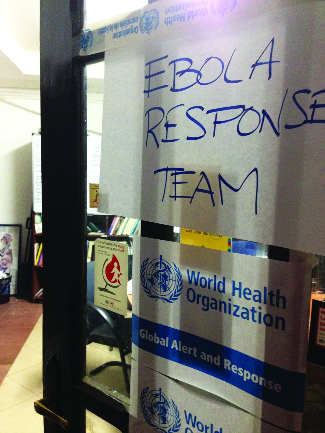
A special supplement on the Ebola outbreak in West Africa was released by The Journal of Infectious Diseases.
Investigators at Virginia Tech University, Blacksburg, released a study on the aerosolization of Ebola virus, which includes emission rates that could help inform risk assessment of inhalation exposure to Ebola virus.
Insights from a recent study into the molecular interactions behind the Ebola virus suggest new approaches for fighting the disease, specifically, blocking the viral protein from binding to Niemann-Pick C1 protein.
Despite challenges, the hazardous work with Ebola virus disease (EVD) patients in Sierra Leone was experienced as meaningful by health care workers and an important motivator, according to a study in the Journal of Advanced Nursing.
Researchers combined two common modeling methods – estimation of epidemic parameters using mathematical models, and estimation of associations using ecological regression models – to identify some factors predicting rapid and severe EVD epidemics in West African subnational regions.
A report in Nature showed how evolutionary analyses of Ebola virus genome sequences provided key insights into virus origins, evolution, and spread during the 2013-2016 West African epidemic.
Chinese researchers said there is no robust evidence that the 2014 Ebola virus outbreak in West Africa was fast-evolving and -adapting to humans, and that the unprecedented nature of the 2014 outbreak might be more likely related to nonvirological elements, such as environmental and sociological factors.
Several concerns were associated with new Ebola vaccine candidates, according to a recent analysis, including the safety profile in some particular populations, the immunization schedule for emergency vaccination, and the persistence of the protection.
Age younger than 5 years, bleeding, and high viral loads were poor prognostic indicators of children with EVD, according to results of a study in Liberia and Sierra Leone.
Fear, myth, and misconceptions were common among health care workers during the 2014 EVD outbreak in Nigeria, according to a study in PLOS One. The authors said strategies to allay fear are required to contain future outbreaks of EVD in Nigerian hospitals.
The risk of nosocomial transmission of the Ebola virus within Ebola Holding Units during the 2014 EVD epidemic in Sierra Leone was low, according to a recent analysis.
An unusual case of Ebola virus chain of transmission was uncovered in Conakry, Guinea in 2015, according to a research letter in Emerging Infectious Diseases.
[email protected]
On Twitter @richpizzi
Treatment withdrawal without prior liver biopsy found safe in well-controlled autoimmune hepatitis
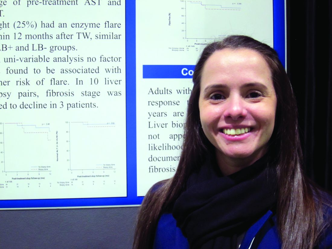
BOSTON – Although current guidance calls for a liver biopsy prior to treatment withdrawal in autoimmune hepatitis (AIH), a retrospective observational analysis conducted by researchers from the Cleveland Clinic offers a different view.
“Maybe not everyone needs a liver biopsy before withdrawing treatment,” Yilien Alonso, MD, an internist at the Cleveland Clinic in Weston, Fla., said in an interview at the annual meeting of the American Association for the Study of Liver Diseases.
Both the European Association for Study of the Liver and the AASLD recommend liver biopsy prior to treatment withdrawal in AIH, but the expensive procedure is not without risk of morbidity and mortality. Dr. Alonso and her coinvestigators reviewed the records of 508 AIH patients seen at their institution between January 2001 and April 2015. After excluding the records of patients who’d had juvenile onset of AIH, or who were treated with agents other than corticosteroids and azathioprine, the researchers found 34 adults with similar pretreatment profiles who’d had treatment withdrawal after 2 years of excellent response to treatment, 10 of whom had a liver biopsy prior to treatment withdrawal.
The outcomes at 1 year post treatment withdrawal for all 34 were similar, with no difference in flare rates or reinitiation of treatment. In those who’d had the second liver biopsy, the fibrosis stage was noted at 1 year to have declined in three patients.
“If you have a stable patient population that you are tracking every 3-6 months, we don’t see why you can’t stop the treatment without having to have another invasive procedure,” Dr. Alonso said.
[email protected]
On Twitter @whitneymcknight
AT THE LIVER MEETING 2016
Key clinical point:
Major finding: Flare rates were similar post treatment withdrawal at 1 year in autoimmune hepatitis patients with and without a prior liver biopsy.
Data source: Retrospective observational analysis of 34 adults with well-controlled autoimmune hepatitis given treatment withdrawal with and without liver biopsy in large academic practice.
Disclosures: Dr. Alonso did not have any relevant disclosures.
Hypervirulent Mycobacterium clone infecting cystic fibrosis patients worldwide
A recently evolved strain of Mycobacterium is circulating in hospitals worldwide, causing nearly impossible-to-treat lung infections among patients with cystic fibrosis.
A genome-wide study has determined that Mycobacterium abscessus is not transmitted through soil and water, as once thought, but is a nosocomial infection transmitted person to person through droplet and surface contamination, Andres Floto, MD, reported in Science (2016 Nov 11;354[6313]:751-7).
“The bug initially seems to have entered the patient population from the environment, but we think it has recently evolved to become capable of jumping from patient to patient, getting more virulent as it does so,” Dr. Floto of the University of Cambridge, England, wrote in a press statement.
The path of global transmission is not yet entirely clear, the authors noted. But since it first appeared, around 1978, M. abscessus has spread globally, strongly suggesting that asymptomatic carriers may be one source of transmission.
“We found no evidence of cystic fibrosis patients or of equipment moving between centers in different countries, indicating that the global spread of M. abscessus may be driven by alternative human, zoonotic, or environmental vectors of transmission,” the researchers wrote.
The team conducted whole-genome sequencing on 1,080 samples of M. abscessus obtained from 517 cystic fibrosis patients in clinics and hospitals within the United States, the United Kingdom, Europe, and Australia. They identified three subspecies, some of which contained nearly genetically identical strains, “suggesting widespread transmission of circulating clones within the global cystic fibrosis patient community.”
Most patients (74%) were infected with these genetically identical strains despite their diverse geographic locations. The isolates were amazingly similar, the authors noted: 90% differed by less than 20 single nucleotide polymorphisms.
Using these strains, the researchers were able to construct several possible transmission chains in most of the cystic fibrosis centers included in the study. The three dominant circulating clones were all observed in the United States, European, and Australian samples, indicating transcontinental transmission.
“We also detected numerous examples of identical or near-identical isolates infecting groups of patients in different cystic fibrosis centers and, indeed, across different countries, indicating the recent global spread of M. abscessus clones throughout the international cystic fibrosis patient community.”
The team also determined that the common ancestor of these strains probably emerged around 1978.
Another sequencing series tracked specific isolates among individual patients. This strongly suggests person-to-person transmission. Adding this to their previous work on M. abscessus transmission, the authors postulated that spread was probably by surface contamination by droplet contamination and by cough aerosol from infected patients.
The team then looked at clinical outcomes associated with the bacteria and treatment with amikacin and macrolides – antibiotics typically used to fight this very-challenging infection. “We did observe increased rates of chronic infection in individuals,” infected with the clones, which were resistant to both those medications, they said.
In immunodeficient mice, the strains were more likely to cause granulomatous and inflammatory lung changes. And the bacteria tended to survive even after being consumed by macrophages, “suggesting that the establishment of transmission chains may have permitted multiple rounds of within-host genetic adaptation to allow M. abscessus to evolve from an environmental organism to a true lung pathogen.”
The research was funded by the Wellcome Trust and the Cystic Fibrosis Trust in the United Kingdom. There were no financial disclosures.
[email protected]
On Twitter @alz_gal
Approximately 30,000 American adults, children, and infants have cystic fibrosis. Nontuberculous mycobacteria (NTM) are ubiquitous environmental microorganisms, and it has been known for some time that these infections can be transmitted person to person. Any patient, actually, who has preexisting lung disease – and especially those with poor mucociliary clearance – are at risk for a nontuberculous mycobacterial infection. This type of lung infection also can be difficult to diagnose and hard to treat. The U.S. Cystic Fibrosis Foundation in conjunction with the European Cystic Fibrosis Society has developed consensus guidelines for infection control, evaluation, and treatment of this problem. This executive summary was published last January (Floto et al. Thorax.2016;71:i1-i22).

At our center, in addition to the standard contact precautions we use for every CF patient, patients with confirmed NTM infections are seen at every clinic visit in an airborne infection isolation room. We also require all CF patients to wear an isolation mask when entering the hospital or clinic facility, when going to a laboratory, or even when going to the bathroom down the hall. Finally, we stress the significant importance of good hand hygiene.
Susan Millard, MD, FCCP, is a pediatric pulmonologist with Spectrum Health/Butterworth Hospital in Grand Rapids, Mich.
Approximately 30,000 American adults, children, and infants have cystic fibrosis. Nontuberculous mycobacteria (NTM) are ubiquitous environmental microorganisms, and it has been known for some time that these infections can be transmitted person to person. Any patient, actually, who has preexisting lung disease – and especially those with poor mucociliary clearance – are at risk for a nontuberculous mycobacterial infection. This type of lung infection also can be difficult to diagnose and hard to treat. The U.S. Cystic Fibrosis Foundation in conjunction with the European Cystic Fibrosis Society has developed consensus guidelines for infection control, evaluation, and treatment of this problem. This executive summary was published last January (Floto et al. Thorax.2016;71:i1-i22).
At our center, in addition to the standard contact precautions we use for every CF patient, patients with confirmed NTM infections are seen at every clinic visit in an airborne infection isolation room. We also require all CF patients to wear an isolation mask when entering the hospital or clinic facility, when going to a laboratory, or even when going to the bathroom down the hall. Finally, we stress the significant importance of good hand hygiene.
Susan Millard, MD, FCCP, is a pediatric pulmonologist with Spectrum Health/Butterworth Hospital in Grand Rapids, Mich.
A recently evolved strain of Mycobacterium is circulating in hospitals worldwide, causing nearly impossible-to-treat lung infections among patients with cystic fibrosis.
A genome-wide study has determined that Mycobacterium abscessus is not transmitted through soil and water, as once thought, but is a nosocomial infection transmitted person to person through droplet and surface contamination, Andres Floto, MD, reported in Science (2016 Nov 11;354[6313]:751-7).
“The bug initially seems to have entered the patient population from the environment, but we think it has recently evolved to become capable of jumping from patient to patient, getting more virulent as it does so,” Dr. Floto of the University of Cambridge, England, wrote in a press statement.
The path of global transmission is not yet entirely clear, the authors noted. But since it first appeared, around 1978, M. abscessus has spread globally, strongly suggesting that asymptomatic carriers may be one source of transmission.
“We found no evidence of cystic fibrosis patients or of equipment moving between centers in different countries, indicating that the global spread of M. abscessus may be driven by alternative human, zoonotic, or environmental vectors of transmission,” the researchers wrote.
The team conducted whole-genome sequencing on 1,080 samples of M. abscessus obtained from 517 cystic fibrosis patients in clinics and hospitals within the United States, the United Kingdom, Europe, and Australia. They identified three subspecies, some of which contained nearly genetically identical strains, “suggesting widespread transmission of circulating clones within the global cystic fibrosis patient community.”
Most patients (74%) were infected with these genetically identical strains despite their diverse geographic locations. The isolates were amazingly similar, the authors noted: 90% differed by less than 20 single nucleotide polymorphisms.
Using these strains, the researchers were able to construct several possible transmission chains in most of the cystic fibrosis centers included in the study. The three dominant circulating clones were all observed in the United States, European, and Australian samples, indicating transcontinental transmission.
“We also detected numerous examples of identical or near-identical isolates infecting groups of patients in different cystic fibrosis centers and, indeed, across different countries, indicating the recent global spread of M. abscessus clones throughout the international cystic fibrosis patient community.”
The team also determined that the common ancestor of these strains probably emerged around 1978.
Another sequencing series tracked specific isolates among individual patients. This strongly suggests person-to-person transmission. Adding this to their previous work on M. abscessus transmission, the authors postulated that spread was probably by surface contamination by droplet contamination and by cough aerosol from infected patients.
The team then looked at clinical outcomes associated with the bacteria and treatment with amikacin and macrolides – antibiotics typically used to fight this very-challenging infection. “We did observe increased rates of chronic infection in individuals,” infected with the clones, which were resistant to both those medications, they said.
In immunodeficient mice, the strains were more likely to cause granulomatous and inflammatory lung changes. And the bacteria tended to survive even after being consumed by macrophages, “suggesting that the establishment of transmission chains may have permitted multiple rounds of within-host genetic adaptation to allow M. abscessus to evolve from an environmental organism to a true lung pathogen.”
The research was funded by the Wellcome Trust and the Cystic Fibrosis Trust in the United Kingdom. There were no financial disclosures.
[email protected]
On Twitter @alz_gal
FROM SCIENCE
Key clinical point:
Major finding: Hypervirulent clones of Mycobacterium abscessus with apparent person-to-person transmission, are appearing in cystic fibrosis centers worldwide.
Data source: Gene sequencing was performed on 1,080 samples of M. abscessus from the United States, the United Kingdom, Europe, and Australia.
Disclosures: The research was funded by the Wellcome Trust and the Cystic Fibrosis Trust in the United Kingdom.

Pharmacists: Weight-loss drugs and diabetes can mix
SAN DIEGO –
In light of their side effects and other complications, weight-loss drugs haven’t reached their full diet-in-a-pill potential. Diet drugs remain controversial even as the Food and Drug Administration continues to approve new formulations. Such weight-loss medications can still play a role in the care of diabetic patient, according to a pair of diabetes educators.
The key, they say, is to understand the limits of their potential and their not-so-limited drawbacks. “There is no magic pill,” said Charmaine Rochester, PharmD, an associate professor of pharmacy with the University of Maryland School of Pharmacy, Baltimore. “Patients need to know that it is not an easy fix. They still have to change their lifestyles with the medications.”
Dr. Rochester and Lisa Meade, PharmD, CDE, an associate professor of pharmacy at Wingate (N.C.) University School of Pharmacy, spoke about weight loss medications and diabetes care at the annual meeting of the American Association of Diabetes Educators and in later interviews.
These medications produce weight loss in the 5%-10% range. Still, “weight loss as little as 5% can significantly improve glycemic control, and modest weight loss of 5% to under 10% has been associated with significant improvements in cardiovascular disease risk factors at 1 year – decreased inflammation, improvement in insulin resistance, improved blood pressure, and improved cholesterol,” Dr. Meade said. “Also, weight loss may decrease the amount of medication needed to achieve glycemic control.”
According to Dr. Meade, it’s common for diabetic patients to ask about weight-loss drugs. “They have heard about them from friends or have seen advertisements on TV or the Internet or in magazines,” she said. However, “in general we do not encourage the use of medications for weight loss until the patient has demonstrated weight loss on their own. Counseling is very important so patients realize they will have more weight loss if combined with diet and exercise.”
Dr. Meade noted that the newer weight-loss medications – Qsymia (phentermine and topiramate), Belviq (lorcaserin HCl), Contrave (naltrexone HCl/bupropion HCl), and Saxenda (liraglutide) – are indicated in obese adults with a body mass index of 30 or above or those with a BMI of 27 with at least one comorbid condition, such as hypertension, type 2 diabetes, or dyslipidemia. Contrave is unusual: It’s a mix of the antidepressant bupropion (Wellbutrin) and the addiction drug naltrexone (Revia).
There are several other weight-loss drugs, including orlistat, which is available both by prescription (Xenical) and over-the-counter (Alli). Orlistat is famously linked to “anal leakage” – GI problems and oily and fatty stools.
Weight-loss drugs are controversial. Consumer Reports has warned against using Alli, Xenical, Belviq, Qsymia, and Saxenda “because they don’t help most people lose much, if any, weight, and they all carry potentially serious risks.” Earlier this year, Consumer Reports took aim at Contrave, too: “Nearly 1 in 4 people in the clinical trials stopped taking the prescription weight loss pill because they couldn’t tolerate the common side effects, including nausea, headache, and vomiting.”
As for other side effects, Dr. Meade noted that weight loss can cause hypoglycemia, so diabetes medications may need to be reduced. “Educators need to know that phentermine [in Qsymia and other weight-loss medications] is contraindicated for patients with any type of coronary artery disease [uncontrolled hypertension, stroke, arrhythmias or heart failure],” she said. There are concerns about cardiac issues in patients who take other weight-loss drugs, too.
Also, Belviq is not recommended in patients who are taking certain medications for depression, Dr. Meade said, “and some medications for depression and smoking cessation would be contraindicated with Contrave.”
Dr. Meade cautioned that the new drugs often aren’t covered by insurance and can be too expensive for patients. Goodrx.com estimates the cash price for a month’s supply of Contrave at $264-$295, although discounts and coupons can reduce that amount.
If patients do want to try the weight-loss drugs, Dr. Rochester urges educators to inform them about “the reality of the weight loss expected and remind patients that the research was completed in patients who also incorporated diet and exercise.”
Dr. Rochester and Dr. Meade report no relevant disclosures.
EXPERT ANALYSIS FROM AADE 16
VIDEO: Novel agent inclisiran dramatically lowers LDL
NEW ORLEANS – A single 300-mg injection of inclisiran, a novel non–monoclonal antibody PCSK9 inhibitor, plus statin therapy, achieved a mean 51% reduction in LDL cholesterol, compared with placebo plus a statin, an effect sustained for 90 days.
Moreover, a second subcutaneous 300-mg dose given at day 90 resulted in a 57% reduction in LDL sustained at day 180 with a highly favorable safety profile in the phase II ORION-1 study, Kausik K. Ray, MD, reported at the American Heart Association scientific sessions.
“The efficacy, safety and dosing profile of inclisiran are likely to ensure significant and durable reductions in LDL-C and thus could potentially impact cardiovascular outcomes,” said Dr. Ray, professor of public health at Imperial College London.
Inclisiran is a synthetic RNA interference agent. After injection it selectively goes to the liver, bypassing other tissues and thereby limiting toxicity. Inclisiran halts PCSK9 protein synthesis in the liver, with a resultant sharp, steep, and sustained drop in LDL levels.
Inclisiran achieves LDL lowering of a magnitude similar to that of the PCSK9-inhibiting monoclonal antibodies alirocumab (Praluent) and evolocumab (Repatha), but it will be less expensive to produce and far less costly to administer than the monoclonal antibodies. Dosing is planned to be two or three injections per year, rather than every 2 or 4 weeks, as with the PCSK9 inhibitor monoclonal antibodies, which cost a hefty $14,000 per year. And the sustained efficacy of a dose of inclisiran should make patient adherence to LDL-lowering therapy less of an issue, Dr. Ray continued.
ORION-1 (Inhibition of PCSK9 Synthesis Via RNA Interference) was a double-blind, randomized, placebo-controlled trial comprising 501 patients already on maximally tolerated statin therapy for atherosclerotic cardiovascular disease or at high risk. Nevertheless, their baseline LDL was 125-135 mg/dL. Participants were randomized to one of six doses of inclisiran or placebo. At 90 days they received a second dose. Dr. Ray presented 90- and 180-day outcomes.
This was primarily a safety and dose-ranging study. Side effects at 90 days were no different from placebo regardless of whether patients received 100, 200, 300, or 500 mg of inclisiran. There was no signal of a problem with myalgia or elevated liver enzymes. Mild injection site reactions lasting for more than 4 hours occurred in 1.5%-3.3% of inclisiran-treated patients, but not in a dose-dependent fashion.
One 300-mg dose of inclisiran, in addition to achieving a mean 51% reduction in LDL from baseline at day 90, resulted in a mean 28% reduction in total cholesterol, a modest 10% drop in triglycerides, a 9% increase in HDL, a 37% drop in Apo-B, and a median 23% reduction in Lp(a). Plasma PCSK9 levels plunged in parallel with LDL.
The mean drop in LDL from baseline at day 180 after two 300-mg doses of inclisiran spaced 90 days apart was 65 mg/dL. The LDL reduction ranged from a minimum of 26.5 mg/dL to a maximum of 122 mg/dL.
Dr. Ray’s presentation of the ORION-1 findings followed on the heels of publication of a positive but much smaller phase I study of inclisiran (N Engl J Med. 2016 Nov 13. doi: 10.1056/NEJMoa1609243).
A large phase II clinical trial with cardiovascular outcomes is planned.
Discussant Borge G. Nordestgaard, MD, could find no fault with ORION-1 or inclisiran’s performance to date.
“A 50%-60% reduction in LDL lasting for 3-6 months, with two or three injections per year – it seems fantastic. And there don’t seem to be side effects except for mild injection site reactions. It looks really, really encouraging, I must say,” said Dr. Nordestgaard, professor of genetic epidemiology at the University of Copenhagen.
“I think the key question is, will the very impressive LDL reduction that we see now be sustainable over time in the longer phase III trials,” he added.
Dr. Ray reported serving as a consultant to the Medicines Company, which together with Alnylam Pharmaceuticals sponsored ORION-1, and receiving research grants from and/or serving as a consultant to more than half a dozen other pharmaceutical companies.
Dr. Ray discussed his findings in a video interview at the meeting.
The video associated with this article is no longer available on this site. Please view all of our videos on the MDedge YouTube channel
AT THE AHA SCIENTIFIC SESSIONS
Key clinical point:
Major finding: A single 300-mg subcutaneous injection of inclisiran quickly dropped elevated LDL-cholesterol levels by 51% and kept them there for 90 days.
Data source: ORION-1, a double-blind, randomized, placebo-controlled, phase II study conducted in 501 patients assigned to various doses of inclisiran or placebo on top of maximally tolerated statin therapy.
Disclosures: Dr. Ray reported serving as a consultant to the Medicines Company, which together with Alnylam Pharmaceuticals sponsored ORION-1, and receiving research grants from and/or serving as a consultant to more than half a dozen other pharmaceutical companies.
HIV research update: Late October 2016
A great volume of HIV and AIDS research enters the medical literature every month. It’s difficult to monitor everything, so here’s a quick look at some notable news items and journal articles published over the past few weeks.
A study in the American Journal of Pathology found that HIV impairs Mycobacterium tuberculosis antigen presentation in human dendritic cells, thereby suppressing an important cell-linking innate and adaptive immune response in TB.
A study of the uptake of HIV preexposure prophylaxis (PrEP) among commercially insured persons in the U.S. found that interventions are needed to increase PrEP use by women at substantial risk of acquiring HIV infection.
Implementation of proper and integrated malaria preventive measures as well as frequent monitoring of anemia on prescription of antiretroviral therapy could likely improve the health conditions of HIV-infected children, according to a study in Cameroon.
A Colombian study of the clinical outcomes of a generic version of abacavir/lamivudine and efavirenz in HIV treatment naive patients showed the expected safety and effectiveness profile of proprietary antiretroviral drugs.
Skin autofluorescence is increased in HIV-infected subjects and related with smoking, low nadir CD4 count, and men who have sex with men, according to a study in the journal AIDS, but researchers say larger studies are needed to confirm whether skin autofluorescence is an independent predictor of cardiovascular disease events.
Researchers at Vanderbilt University, Nashville, Tenn., said that, as the HIV-positive population ages, structured subjective cognitive assessment may be beneficial to identify the early signs of cognitive impairment, and to allow for earlier interventions to maintain cognitive performance.
An antiretroviral therapy roll-out in rural Malawi resulted in a sustained 10-year gain in adult life expectancy for HIV-positive individuals, according to a study in the International Journal of Epidemiology.
Increasing HIV virulence driven by universal testing and treatment is likely not a major public health concern, according to a modeling study, but should be monitored in sentinel surveillance, in a manner similar to transmitted resistance to antiretroviral drugs.
A South African study found no association between duration of tenofovir exposure in utero and early linear growth in HIV-exposed, uninfected infants.
In 2014, 17.9% of AIDS cases reported by 29 countries in the European Union and European Economic Area (EU/EEA) presented with TB as an AIDS-defining illness, indicating that TB/HIV coinfection is a substantial problem in the EU/EEA.
A study of malnourished antiretroviral therapy (ART)-eligible adults in sub-Saharan Africa found that pre-ART mortality was twice that in the early post-ART period, suggesting many early ART deaths represent advanced HIV disease rather than treatment-related events.
A study in Taiwan found that HIV patients had an increased risk of stroke, particularly those with cryptococcal meningitis, cytomegalovirus, or P. marneffei infection.
A study of older patients living with HIV found that the associations between cognitive impairment and patient-reported outcome measures were weak, possibly because impairment was mild and therefore largely asymptomatic.
[email protected]
On Twitter @richpizzi
Interatrial shunt benefits sustained for 1 year in HFpEF patients
NEW ORLEANS – An interatrial septal shunt device continued to provide “sustained and meaningful clinical benefit” at 1-year follow-up for 64 patients who had heart failure with preserved ejection fraction (HFpEF), David M. Kaye, MD, PhD, reported at the American Heart Association scientific sessions.
The device is implanted via cardiac catheterization and is intended to reduce elevated left atrial pressure, particularly that associated with exertion, by allowing a small amount but not excessive left-to-right shunting. Patients showed improvements in 6-minute walk distance, New York Heart Association class, and HF-related quality of life scores at 6 months, and those effects persisted at the most recent (12-month) follow-up, he said in a presentation that was simultaneously published online in Circulation (2016 Nov 16).

Overall survival at 1 year was 95%. Three patients died (one from combined pneumonia and renal failure, one from a fatal stroke, and one from an undetermined cause) and one was lost to follow-up. Thirteen patients required 17 hospitalizations for heart failure.
Six-minute walk distance improved from 331 meters at baseline to 363 meters. NYHA classification improved dramatically, as did quality of life scores as assessed by the Minnesota Living with HF questionnaire.
All 48 devices that were evaluable on echocardiographic imaging remained patent, showing continued left-to-right shunting. Left ventricular ejection fraction remained unchanged while right ventricular ejection fraction was significantly elevated over baseline levels. “In conjunction, there were modest but stable reductions in LV end-diastolic volume index with a concomitant rise in RV end-diastolic index,” he said.
A subset of 18 study participants underwent heart catheterization during both rest and exercise so that hemodynamics could be assessed. Exercise time increased significantly, from 8.2 minutes at baseline to 9.7 minutes at 6 months and to 10.4 minutes at 1 year. Similarly, peak work capacity during supine cycling increased from 48 watts at baseline to 60 watts at 6 months and 55 watts at 1 year. These benefits occurred without any increase in pulmonary capillary wedge pressure.
Systemic blood pressure did not change over time, either at rest or during exercise. Left and right atrial volumes also remained unchanged.
Perhaps most importantly, Dr. Kaye said, right-sided cardiac output increased significantly, while left-sided cardiac output remained unchanged. There was no evidence of increased pulmonary pressure or pulmonary vascular resistance. This meant that patients could do more physical activity for a given level of left atrial pressure, he said.
Furthermore, 1-year mortality was lower in this trial, at 4.6%, than in the placebo groups of the I-PRESERVE trial in irbesartan (5.2%) and the U.S. group of the TOPCAT trial in spironolactone (7.7%), said Dr. Kaye, professor and chief of cardiology at the University of Arizona, Tuscon.
Device therapy could have an enormous impact in carefully selected patients with HFpEF, for whom there are no medical treatments, despite the nonrandomized nature of the trial.
REDUCE LAP-HF was funded by Corvia Medical, maker of the shunt device. Dr. Kaye is an unpaid member of Corvia’s scientific advisory group. Dr. Sweitzer is an investigator in the ongoing randomized trial of the interatrial shunt.
NEW ORLEANS – An interatrial septal shunt device continued to provide “sustained and meaningful clinical benefit” at 1-year follow-up for 64 patients who had heart failure with preserved ejection fraction (HFpEF), David M. Kaye, MD, PhD, reported at the American Heart Association scientific sessions.
The device is implanted via cardiac catheterization and is intended to reduce elevated left atrial pressure, particularly that associated with exertion, by allowing a small amount but not excessive left-to-right shunting. Patients showed improvements in 6-minute walk distance, New York Heart Association class, and HF-related quality of life scores at 6 months, and those effects persisted at the most recent (12-month) follow-up, he said in a presentation that was simultaneously published online in Circulation (2016 Nov 16).
Overall survival at 1 year was 95%. Three patients died (one from combined pneumonia and renal failure, one from a fatal stroke, and one from an undetermined cause) and one was lost to follow-up. Thirteen patients required 17 hospitalizations for heart failure.
Six-minute walk distance improved from 331 meters at baseline to 363 meters. NYHA classification improved dramatically, as did quality of life scores as assessed by the Minnesota Living with HF questionnaire.
All 48 devices that were evaluable on echocardiographic imaging remained patent, showing continued left-to-right shunting. Left ventricular ejection fraction remained unchanged while right ventricular ejection fraction was significantly elevated over baseline levels. “In conjunction, there were modest but stable reductions in LV end-diastolic volume index with a concomitant rise in RV end-diastolic index,” he said.
A subset of 18 study participants underwent heart catheterization during both rest and exercise so that hemodynamics could be assessed. Exercise time increased significantly, from 8.2 minutes at baseline to 9.7 minutes at 6 months and to 10.4 minutes at 1 year. Similarly, peak work capacity during supine cycling increased from 48 watts at baseline to 60 watts at 6 months and 55 watts at 1 year. These benefits occurred without any increase in pulmonary capillary wedge pressure.
Systemic blood pressure did not change over time, either at rest or during exercise. Left and right atrial volumes also remained unchanged.
Perhaps most importantly, Dr. Kaye said, right-sided cardiac output increased significantly, while left-sided cardiac output remained unchanged. There was no evidence of increased pulmonary pressure or pulmonary vascular resistance. This meant that patients could do more physical activity for a given level of left atrial pressure, he said.
Furthermore, 1-year mortality was lower in this trial, at 4.6%, than in the placebo groups of the I-PRESERVE trial in irbesartan (5.2%) and the U.S. group of the TOPCAT trial in spironolactone (7.7%), said Dr. Kaye, professor and chief of cardiology at the University of Arizona, Tuscon.
Device therapy could have an enormous impact in carefully selected patients with HFpEF, for whom there are no medical treatments, despite the nonrandomized nature of the trial.
REDUCE LAP-HF was funded by Corvia Medical, maker of the shunt device. Dr. Kaye is an unpaid member of Corvia’s scientific advisory group. Dr. Sweitzer is an investigator in the ongoing randomized trial of the interatrial shunt.
AT THE AHA SCIENTIFIC SESSIONS 2016
Key clinical point: An interatrial septal shunt device continued to provide sustained and meaningful clinical benefit at 1-year follow-up for 64 patients who had heart failure with preserved ejection fraction.
Major finding: Six-minute walk distance improved from 331 meters at baseline to 363 meters at 1 year, NYHA classification improved dramatically, and HF-related quality of life scores also improved.
Data source: REDUCE LAP-HF, a multicenter, prospective, open-label study involving 64 patients followed for 1 year after transcatheter implantation of a shunt device.
Disclosures: REDUCE LAP-HF was funded by Corvia Medical, maker of the shunt device. Dr. Kaye is an unpaid member of Corvia’s scientific advisory group.