User login
Clinical Segment 4: You know more than you think about behavioral and mental health
The video associated with this article is no longer available on this site. Please view all of our videos on the MDedge YouTube channel
People in this video: Whitney McKnight, cohost and producer of Mental Health Consult; Dr. Lorenzo Norris, an editorial board member for Clinical Psychiatry News, and assistant professor of psychiatry and behavioral sciences, assistant dean of student affairs at G.W. University School of Medicine & Health Sciences, and the medical director of psychiatric and behavioral services at G.W.U. Hospital, Washington; Dr. Lillian Beard, pediatrician with Children’s National Hospital Network, Washington, and a Pediatric News editorial board member; Dr. David Pickar, adjunct professor of psychiatry at Johns Hopkins University School of Medicine in Baltimore and at the Uniformed Services University of the Health Sciences in Bethesda, Md.
Dr. Pickar: Let me just say one thing about that training issue and so forth. There is a common ground in primary care medicine and psychiatry and that is the patient. You guys in primary care, you know patients. We do not use – I use stethoscopes – to make me feel like an internist again.
However, we psychiatrists really do not have to.
Whitney: Is that like “I’m not a doctor but I play one on TV”?
Dr. Pickar: I do that one, too, but in fact the real first step of evidence-based medicine is the patient. You just described it beautifully. Sometimes, I feel badly if the primary physician does not give him or herself credit for that first line of clinical observation. It is huge. Affect is the feeling state. You observe the affect: “He looks down or agitated, anxious.” That is affect, whereas the symptoms are if he is feeling sad, feeling anxious. That is what you do for a living, you find out these things. You get that piece going. We know we psychiatrists are going to need help in that direction. The issue around reimbursement for psychiatry and so forth, I am going to take a deep breath on that one.
I have plenty of feelings about that, but I just want to make sure that the primary care physician that may be watching this understands that he or she is not just the first line but he or she has good skills at observing the first pass what is going on with a patient.
Dr. Norris: Not only are they the first line, but frequently, if you are the person the patient has the relationship with – Dr. Beard, Dr. Barbour – the patient is more inclined to listen to you than to just some random specialist you refer them to.
Dr. Pickar: On the other side of that, even when you have collaborated with a primary care doctor, and times are changing and the meds are tricky, I like to be able to talk to the primary care person and say “Look, I am thinking this way …” The primary doctor might say, “I saw them and they were not looking bad,” that is helpful to hear, or “Yeah, boy we need to ...” That is helpful.
Dr. Norris: Not just a digital note on a shared electronic medical records. Talk … dialogue. There is a difference. This is an important point, there is a difference between clinicians dialogue on a shared patient versus I am reading your notes and you are reading my notes. I do not consider that dialogue.
Policy Segment 3: When depression is the differential diagnosis for distress
The video associated with this article is no longer available on this site. Please view all of our videos on the MDedge YouTube channel
People in this video: Dr. James Griffith, the Leon M. Yochelson Professor of Psychiatry and Behavioral Sciences, and chair of psychiatry and psychosomatic medicine at George Washington University School of Medicine, Washington; Whitney McKnight, cohost and producer of Mental Health Consult.
Whitney: I think we need to step back and define mental illness. For that, I’m going to go to you, Griff, because I think it’s important that we remember not all primary care doctors really do have an understanding of the nuances to definitions of mental health.
You and I were having a discussion about “How do you define depression?” There’s clinical diagnosis of it, but then there are other ways that it gets used.
Dr. James Griffith: There’s a big push in medical education to shorten it, to do more in less time, but this is complex. There has not been much acknowledgment of the complexity. I’ll give you two difficult scenarios.
“Huge numbers of people treated in primary care who would have high scores on the PHQ-9 are in fact just lonely.” – Dr. James GriffithOne is disorder versus distress. If you simply download a Patient Health Questionnaire-9 off the Internet, give it to people: They have a high score; we say they’re depressed, give them an antidepressant. Huge numbers of people in primary care who would have high depression scores, in fact, are lonely; they’re in abusive relationships; they’re grieving losses; they are demoralized because their aspirations in life won’t take place – none of these problems are helped by an antidepressant.
Medical students, or for that matter, psychiatry residents, are not well taught in how to distinguish disorder from distress. All of these are solvable problems. There’s sort of a myth of the depressed patient that if only we would recognize depressed people, give them a prescription, everything would be okay, but it doesn’t.
Whitney: How do you teach that, then? What is missing in the curriculum?
Dr. Griffith: It’s a little bit like what Dr. Kirschner said about money and teams. You don’t have teams, if you don’t have funding. You don’t have teaching, if you don’t have time, and that’s one of our first issues.
Clinical Segment 3: Should you add a psychiatrist to your practice?
The video associated with this article is no longer available on this site. Please view all of our videos on the MDedge YouTube channel
People in this video: Whitney McKnight, cohost and producer of Mental Health Consult; Dr. Lorenzo Norris, editorial board member of Clinical Psychiatry News and cohost of Mental Health Consult, and assistant professor of psychiatry and behavioral sciences, assistant dean of student affairs at G.W. University School of Medicine & Health Sciences, and the medical director of psychiatric and behavioral services at G.W.U. Hospital, Washington; Dr. Lillian Beard, pediatrician with Children’s National Hospital Network, Washington, and a Pediatric News editorial board member; Dr. David Pickar, adjunct professor of psychiatry at Johns Hopkins University School of Medicine, Baltimore, and at the Uniformed Services University of the Health Sciences in Bethesda, Md.; Dr. April Barbour, an associate professor of medicine and the director of general internal medicine and of the primary care residency program at G.W.U. School of Medicine, Washington.
Dr. Beard: This is one of the major frustrations. You've hit it right on the head. I will take anywhere from 45-50 minutes to do this, and I will have others who are waiting in my reception area or I will have a tap on the door. It takes that kind of time and the unfortunate thing is, I am never adequately reimbursed for the time that it really takes. Often, what I do is ask my front desk to screen patients when they call. If they say it is a routine check-up, the front desk knows to ask, "Are there any particular concerns that you have this year. Anything you would like the doctor to focus on?" If they do, then what I have to do is block out three of my regular times and that is very costly.
Whitney: As we move into a world in which it is not fee-for-service—based and “I think if it’s possible to have a mental health professional on site [in your practice], it is a win-win situation.” – Dr. Lillian Beardwe have to create these new metrics, I say “we,” but the health care system is moving toward setting up new accountable care organizations or other sorts of bundle payments. When we have the new legislation take effect, the MACRA (Medicaid Access and CHIP Reauthorization Act) legislation, are you building into the metrics that you are going to be reimbursed through your third-party payers to include these 50-minute sessions or is there no way to do that?
Dr. Beard: I do not know of a way to do it. I really do not.
Whitney: How is that going to impact outcomes and reimbursement?
Dr. Beard: Well, it is definitely going to impact outcomes. One of the areas of interest that I have is the feasibility of having a mental health specialist in my actual primary care site. Even if it is for a few segments a week, it would be a tremendous help. Just having that individual present removes certain barriers. For example, there are times that, even during the primary care encounter, the mental health specialist is able to say to the patient’s parents, “We’ll be glad to make an appointment and discuss that with you at a future time, so we can go more in depth.” Just that introduction lowers the barrier. Otherwise, there is more resistance if I say, “I am going to refer you to Dr. Pickar he is an associate who…” They object, and want to know, “Well, what kind of doctor is Dr. Pickar? He is a psychiatrist?” It depends on what association they have with the word “psychiatrist.” The parents might object, “My kid’s not crazy.” I have to explain that this is a mental health disorder that we can do something about, and the psychiatrist is going to assist us with that.
Dr. Pickar: That is a great model.
Whitney: Yes but is it feasible with all the new legislation that is coming down the line?
Dr. Barbour: I think there are very dramatic differences between the pediatric model of care, which tends to be more wraparound care that you are describing; (should this be “that” or “than”?) and the adult model of care, which is more consumer driven and in which we expect a lot of our patients. We find particularly that young people transitioning to their early 20s often have a hard time understanding how to interact in the adult model of care. Particularly the patients that we have worked on have had significant health problems, many of which include mental health disorders. The program that we put in place has some psychiatric services available in the clinic. That is not feasible – I think – in our current payment structure to do that everywhere, in all adult medicine clinics.
I think these patients are particularly vulnerable. They do not understand the health care systems. They come in with these diagnoses. You bring up ADHD and that is something an internist is not as comfortable in providing care for as you are, and that I think causes a lot of roadblocks for patients to get the medicines they need. It has worked well for them, but the new doctor is not as comfortable prescribing the medicine or making the diagnosis. There are issues around that.
Dr. Norris: This is one of points of the roundtable. Who should be delivering this treatment? If you can create a team based atmosphere where what Dr. Beard illustrated, just the introduction. "I want to introduce you to my colleague so that we can start treatment." That one element, just starting that can make a huge difference, but how do you make that fiscally viable? In the George Washington University Hospital Thriving After Cancer clinic, we used resident psychiatrist in training. These are senior-level residents who are very good at that or are supervised by a psychiatrist. If you were to put a psychiatrist in the TAC clinic and bill for their hours, it just would not work, Dr. Barbour is shaking her head like no way.
Dr. Barbour: I could not afford it.
Dr. Beard: What I am thinking is that this other professional, be it a psychiatrist or psychologist, a licensed clinical social worker, whatever, will have the capability of billing for his or her services. I think if it is possible to have that professional in your site it is a win/win situation.
Relationship-Based Care: A novel approach for patients and providers
When I think of the word “relationship,” I imagine gazing into the loving eyes of my husband, playing hide and seek with my children, or texting my best friend for no good reason other than to just say hello.
There is a special comfort zone we expect from people who are close to us; a feeling of love and acceptance that we can’t find elsewhere.
But in a much broader sense, our important relationships extend far beyond our inner circle to include every single person who is involved with our health care team. Our team includes the hospital executives who create new safety initiatives, develop budgets, and oversee a host of other patient care and fiscal functions. The physical therapists who evaluate our patients and make recommendations on how to safely transition them out of the hospital are on our team. The housekeepers who scrub the toilets and wash the linens to prevent nosocomial infections are on our team. They, along with many others, play a pivotal role in our patients’ care, although many important players make their impact behind the scenes.
Yet, of course, our most important professional relationships are not with the CEO, the pharmacist, or even the nursing staff. Our most important relationships are with our patients and their families. I recently attended an all-day conference on a little-known gem called Relationship-Based Care (RBC), a culture transformation and operational model that is gaining steam globally. The RBC model focuses not only on well-known metrics, such as patient safety, quality care, and patient satisfaction; it also emphasizes staff satisfaction by improving each and every relationship. Specifically, it creates therapeutic relationships between caregivers and the patients and families they serve, strengthens relationships between members of the health care team, and last, but certainly not least, it nurtures each caregiver’s relationship with himself or herself. What a novel, and much needed concept!
Numerous hospitals that have implemented this training model have achieved impressive outcomes, including significant improvement in HCAHPS (Hospital Consumer Assessment of Healthcare Providers and Systems) scores, and staff satisfaction survey scores so high that one hospital gained national recognition as one of the best places to work in America.
I look forward to future training on RBC and am glad to see that addressing the needs of caregivers, not just care receivers, is starting to take center stage, as it rightfully should. After all, how can we give our all to our patients when we are not whole?
Dr. Hester is a hospitalist at Baltimore-Washington Medical Center in Glen Burnie, Md. She is the creator of the Patient Whiz, a patient-engagement app for iOS. Reach her at [email protected].
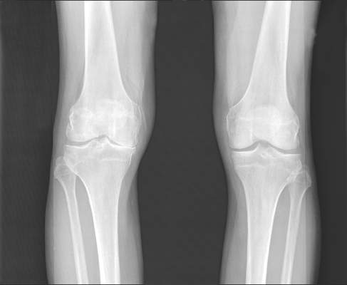
Radiography missed most clinical cases of hip osteoarthritis
Radiography detected up to 16% of cases of hip osteoarthritis among older patients with frequent hip pain in an analysis of participants in the Framingham Osteoarthritis Study and the Osteoarthritis Initiative.
“In older patients, inadequate recognition of osteoarthritis has consequences. Decreased functional status from osteoarthritis significantly increases morbidity from coronary heart disease, lung disease, diabetes, obesity, falls, frailty, and various other ailments,” said Dr. Chan Kim of Boston University and his associates. “Because many patients with hip pain do not have radiographic hip osteoarthritis, a health professional should continue with the evaluation and treatment of osteoarthritis, despite negative radiographic findings.”

Radiographic pathology often is detected late in the course of knee OA and correlates poorly with knee pain, but few studies have examined these trends for the hip. The researchers analyzed pelvic radiographs and hip pain among 946 participants in the Framingham Osteoarthritis Study and 4,366 participants in the Osteoarthritis Initiative. They defined radiographic hip OA as a Kellgren-Lawrence grade of 2 or more – that is, definite superolateral or superomedial joint space narrowing and a definite osteophyte. They used various clinical symptoms of hip OA for comparison. Participants in both studies were older than 45 years, and tended to be in their early 60s (BMJ 2015 Dec 2. doi: 10.1136/bmj.h5983).
The most sensitive criterion in the study was groin pain, for which radiography was positive in 37% of hips in the Framingham Study and 17% of hips in the Osteoarthritis Initiative, the researchers said. Other clinical criteria were less sensitive, including anterior thigh pain, frequent hip pain, and painful internal rotation. Moreover, about 21%-24% of hips with radiographic OA were frequently painful.
The study did not evaluate MRI findings, the investigators noted. They suggested that such results would resemble those for the knee, in which MRI is “more sensitive than radiography, [but] it is far less specific for abnormalities suggestive of osteoarthritis in most middle-aged and older people.”
The National Institute of Arthritis and Musculoskeletal and Skin Diseases funded the study. The Osteoarthritis Initiative is funded by the National Institutes of Health, Merck Research Laboratories, Novartis, GlaxoSmithKline, and Pfizer. The researchers had no disclosures.
Radiography detected up to 16% of cases of hip osteoarthritis among older patients with frequent hip pain in an analysis of participants in the Framingham Osteoarthritis Study and the Osteoarthritis Initiative.
“In older patients, inadequate recognition of osteoarthritis has consequences. Decreased functional status from osteoarthritis significantly increases morbidity from coronary heart disease, lung disease, diabetes, obesity, falls, frailty, and various other ailments,” said Dr. Chan Kim of Boston University and his associates. “Because many patients with hip pain do not have radiographic hip osteoarthritis, a health professional should continue with the evaluation and treatment of osteoarthritis, despite negative radiographic findings.”
Radiographic pathology often is detected late in the course of knee OA and correlates poorly with knee pain, but few studies have examined these trends for the hip. The researchers analyzed pelvic radiographs and hip pain among 946 participants in the Framingham Osteoarthritis Study and 4,366 participants in the Osteoarthritis Initiative. They defined radiographic hip OA as a Kellgren-Lawrence grade of 2 or more – that is, definite superolateral or superomedial joint space narrowing and a definite osteophyte. They used various clinical symptoms of hip OA for comparison. Participants in both studies were older than 45 years, and tended to be in their early 60s (BMJ 2015 Dec 2. doi: 10.1136/bmj.h5983).
The most sensitive criterion in the study was groin pain, for which radiography was positive in 37% of hips in the Framingham Study and 17% of hips in the Osteoarthritis Initiative, the researchers said. Other clinical criteria were less sensitive, including anterior thigh pain, frequent hip pain, and painful internal rotation. Moreover, about 21%-24% of hips with radiographic OA were frequently painful.
The study did not evaluate MRI findings, the investigators noted. They suggested that such results would resemble those for the knee, in which MRI is “more sensitive than radiography, [but] it is far less specific for abnormalities suggestive of osteoarthritis in most middle-aged and older people.”
The National Institute of Arthritis and Musculoskeletal and Skin Diseases funded the study. The Osteoarthritis Initiative is funded by the National Institutes of Health, Merck Research Laboratories, Novartis, GlaxoSmithKline, and Pfizer. The researchers had no disclosures.
FROM BMJ
Key clinical point: Radiographic hip osteoarthritis correlates poorly with hip pain, even among older patients with a high index of suspicion for hip OA.
Major finding: Radiography detected up to 16% of cases of hip OA among older patients with frequent hip pain.
Data source: An analysis of pelvic radiographs and hip pain reported by 946 participants in the Framingham Osteoarthritis Study and 4,366 participants in the Osteoarthritis Initiative.
Disclosures: The National Institute of Arthritis and Musculoskeletal and Skin Diseases funded the study. The Osteoarthritis Initiative is funded by the National Institutes of Health, Merck Research Laboratories, Novartis, GlaxoSmithKline, and Pfizer. The researchers had no disclosures.

Lateral wedge insoles provide minimal biomechanical help in knee OA
Lateral wedge insoles worn by people with medial knee osteoarthritis (OA) provide a limited amount of immediate biomechanical improvement during walking and may be best suited to people who have biomechanical phenotypes that would benefit the most, according to findings from a systematic review and meta-analysis of studies examining the intraindividual effects of the insoles.
“This review is ... the most definitive, up-to-date and comprehensive analysis on this issue to clarify the effects of lateral wedge insoles on biomechanical risk factors for knee OA progression,” wrote lead investigator John Arnold, Ph.D., of the University of South Australia, Adelaide, and his colleagues (Arthritis Care Res. 2015 Nov 25. doi: 10.1002/acr.22797).
The investigators reviewed 18 studies with a total of 534 participants and found small, but statistically significant reductions in estimates of knee joint loading based on the surrogate measures of external knee adduction moment (EKAM) and the knee adduction angular impulse (KAAI).
Most studies (14) tested full-length insoles, and the remaining four allowed a customized amount based on comfort and/or pain level. Another two used heel wedges, and two others tested both. The inclination angle of the insoles was most commonly 5 degrees, but ranged from 4 to 11 degrees. Some studies used a concomitant medial arch support; these were of a generic design in four studies and were made to order in another three. The lateral wedge insoles were compared against flat insoles, the patients’ own footwear, or standardized footwear.
The pooled effect sizes of both the first and second peak EKAM reductions were small, with standard mean differences of –0.20 to –0.25. For the first EKAM, the effect sizes did not vary according to whether studies used flat insoles or shoes only as comparators, whereas for the eight studies that reported second EKAM outcomes, there was a larger pooled effect size for comparisons against shoe-only than for one study that made flat insole comparisons. The pooled estimate for the standard mean difference in nine studies that reported KAAI was –0.14.
There was only weak evidence for publication bias in all the comparisons for the surrogate measures, and most had a low level of statistical heterogeneity between the outcomes of the studies.
The investigators noted that this meta-analysis of surrogate measures for knee joint loading does not take cumulative loading into account, so that even though the reduction in peak EKAM and KAAI was small, it may amount “to a large cumulative effect imparted on the knee over the course of the day. This should be considered when interpreting the findings of this review and future research on load modifying interventions in knee osteoarthritis.” They said that while EKAM has been associated with OA progression, KAAI has been thought to be a better measure of the duration and magnitude of loading in knee OA and has been associated with medial tibiofemoral cartilage loss over 1-2 years.
“Prescription [for lateral wedge insoles] based on biomechanical response and use of insoles only in individuals who show reductions in knee joint loading (biomechanical phenotypes) appears more appropriate to increase the likelihood of a favorable long-term response regarding the attenuation of structural changes. This would limit their application and benefit to a smaller number of individuals, but is still likely to be significant considering the overall prevalence of knee OA and projected rise due to population aging and rising obesity levels,” the authors concluded.
The investigators had no outside funding source for their systematic review. One of the authors may receive royalties from Salford Insole, a manufacturer of lateral wedge insoles.
FROM ARTHRITIS CARE & RESEARCH
Key clinical point: Make sure that patients with medial knee OA have an appropriate biomechanical phenotype to use lateral wedge insoles.
Major finding: The pooled effect sizes of both the first and second peak external knee adduction moment reductions were small, with standard mean differences of –0.20 to –0.25.
Data source: A systematic review and meta-analysis of 18 studies involving 534 patients with medial knee OA.
Disclosures: The investigators had no outside funding source for their systematic review. One of the authors may receive royalties from Salford Insole, a manufacturer of lateral wedge insoles.

CAR exhibits activity in resistant B-cell malignancies
Photo courtesy of ASH
ORLANDO, FL—Allogeneic chimeric antigen receptor (CAR) T cells directed against CD19 can have “significant” activity against resistant B-cell malignancies, even when given without prior chemotherapy, according to a presentation at the 2015 ASH Annual Meeting.
Nine of 20 patients responded to treatment with the CAR T cells, despite having failed prior allogeneic transplant. The best responses were observed in patients with acute lymphoblastic leukemia (ALL) and chronic lymphocytic leukemia (CLL).
“Malignancies that were resistant to allogeneic transplants and standard donor lymphocyte infusions regressed after infusions of allogeneic anti-CD19 CAR T cells,” said James N. Kochenderfer, MD, of the National Cancer Institute in Bethesda, Maryland.
“Allogeneic anti-CD19 CAR T cells seem to be particularly effective against ALL and CLL, suggesting a possible antigenic stimulation that may be more pronounced in these malignancies.”
Adverse events associated with these CAR T cells included severe but reversible cytokine release syndrome, mild aphasia, and muscle damage. There were no cases of acute graft-vs-host disease (GVHD).
Dr Kochenderfer presented these results at ASH as abstract 99.
For this phase 1 study, researchers tested a CAR T-cell therapy that was originally developed by Dr Kochenderfer and his colleagues. The therapy is now known as KTE-C19 and is under development by Kite Pharmaceuticals. However, the company did not sponsor this trial.
The study was open to patients with any CD19+ B-cell malignancy that persisted after allogeneic transplant.
All patients except those with ALL were required to have received at least one standard donor lymphocyte infusion. In addition, patients were only eligible if they had minimal or no GVHD and were not receiving any systemic immunosuppressive drugs.
The trial included 20 patients—5 each with ALL, CLL, mantle cell lymphoma (MCL), and diffuse large B-cell lymphoma (DLBCL).
All patients received a single infusion of CAR T cells derived from their original transplant donor. Production of these cells took 8 days. The highest dose of CAR T cells given was 107 cells/kg.
Four of the ALL patients obtained a minimal-residual disease-negative complete response (CR), but 2 of these patients subsequently relapsed. Of the other 2 patients, 1 remains in CR at 18 months of follow-up, and the other went on to receive a second allogeneic transplant. That patient remains in CR today.
Among the CLL patients, 1 achieved a CR, and 1 achieved a partial response (PR). One patient had stable disease (SD), and the other 2 progressed. Both the CR and the PR are ongoing at 36 and 18 months of follow-up, respectively.
One MCL patient achieved a CR, 1 had a PR, and 3 had SD. The CR is ongoing at 31 months. One DLBCL patient achieved a CR, 3 had SD, and 1 progressed.
Dr Kochenderfer noted that response was associated with higher blood CAR T-cell levels. There was a significant difference in CAR T-cell levels between responders and nonresponders (P=0.001).
In addition, the presence of blood B-cell levels before CAR T-cell infusion was associated with higher blood CAR T-cell levels. Patients with normal or high B lymphocytes had higher levels of CAR T cells in their blood (P=0.04).
Patients with high tumor burdens developed severe cytokine-release syndrome with fever, tachycardia, and hypotension. The ALL patients were particularly susceptible to cytokine-release syndrome.
Dr Kochenderfer said neurologic toxicity was rare and mild. There was 1 case of mild aphasia.
There were 2 patients with elevations in CPK, indicating muscle damage. Those patients also reported muscle pain, and 1 patient reported weakness.
“This is one of the first reports, I think, of muscle damage in CAR T-cell patients,” Dr Kochenderfer said.
None of the patients developed acute GVHD after CAR T-cell therapy. One patient had continued worsening of pre-existing chronic GVHD after treatment, and 1 patient developed mild chronic eye GVHD more than a year after CAR T-cell infusion.
Dr Kochenderfer said additional details from this trial will be published in an upcoming issue of the Journal of Clinical Oncology. ![]()
Mixing warfarin, sulfonylurea may cause serious events

A retrospective study published in The BMJ suggests the possibility of a significant drug interaction between warfarin and the sulfonylureas glipizide and glimepiride.
Taking either of these diabetes drugs in conjunction with warfarin was linked to increased hospitalizations for falls, altered mental state, and insulin shock among patients 65 and older.
Hospital admissions or emergency room visits were nearly 22% higher for patients who were taking warfarin with glipizide or glimepiride, compared to patients taking the diabetes drugs alone.
Clinical references warn doctors of a potential interaction between these drugs, but evidence of it has been thin, according to lead study author John Romley, PhD, of the University of Southern California (USC) in Los Angeles.
He and his colleagues said that, in their study, evidence of the drug-to-drug interaction was clear.
When taken with glipizide or glimepiride, warfarin can intensify their effects and send blood sugar levels crashing. Patients experiencing hypoglycemia may seem drunk, lightheaded, and confused, and are at risk of falling.
“The take-home message is simply that an interaction can occur that has clinical significance, so providers need to be aware in order to prevent a low blood sugar issue from occurring,” said Anne Peters, MD, also of USC.
“Sometimes this means having the patient monitor their blood sugar levels more often. There are many ways to deal with the issue if one is forewarned.”
Pharmacists don’t need to change patient instructions, added Bradley Williams, PharmD, of USC.
“What it does require is for pharmacists and other clinicians to be more vigilant when a sulfonylurea is added to a regimen that includes warfarin, as well as when a patient who is taking both has a change in their medical status,” Dr Williams said.
“I think additional research into the potential interactions between medications for diabetes and warfarin, as well as other drugs that affect blood clotting, is warranted because of the potential consequences of excessive bleeding.”
For the current study, the researchers analyzed a random sample of 465,918 Medicare beneficiaries with diabetes who filled a prescription for glipizide or glimepiride between 2006 and 2011. About 15% of these patients (n=71,895) also filled a prescription for warfarin.
The researchers found that hospital admissions or emergency department visits for hypoglycemia were more common with concurrent warfarin and glipizide/glimepiride use than with glipizide/glimepiride use alone. The adjusted odds ratio (AOR) was 1.22.
The risk of hypoglycemia associated with concurrent use was higher among patients taking warfarin for the first time, as well as in patients ages 65 to 74.
Concurrent use of warfarin and glipizide/glimepiride was also associated with hospital admission or emergency department visits for fall-related fractures (AOR=1.47) and altered consciousness/mental status (AOR=1.22).
The researchers said these findings may not be generalizable beyond the elderly Medicare population.
They also noted that their findings could be confounded by some unmeasured characteristics in patients that may be connected to warfarin use or a risk for hypoglycemia. Another limitation of this study is that the researchers did not measure drug use directly. ![]()
FDA approves recombinant product for VWD
The US Food and Drug Administration (FDA) has approved a recombinant von Willebrand factor product, Vonvendi (formerly BAX 111), for use in adults with von Willebrand disease (VWD).
Vonvendi is the first FDA-approved recombinant von Willebrand factor.
It is now approved for the on-demand treatment and control of bleeding episodes in VWD patients 18 years of age and older.
Vonvendi is expected to be broadly available in the US in late 2016.
Compared to other marketed von Willebrand factor concentrates, Vonvendi contains a more consistent concentration of ultra-large-molecular-weight multimers. And the product is developed using a plasma- and albumin-free manufacturing method.
Vonvendi is the first von Willebrand factor concentrate in the US that contains only trace amounts of factor VIII (FVIII), offering the flexibility to administer FVIII only when needed.
The FDA’s approval of Vonvendi was based on results from a phase 3 trial recently published in Blood. The study included 49 patients with WWD who received Vonvendi with and without recombinant FVIII.
All participants reported successful treatment of bleeding episodes. Most (96.9%) treated bleeds (n=192 bleeds in 22 patients) achieved an “excellent” efficacy rating.
Most bleeds (81.8%) were resolved with a single infusion of Vonvendi, and the treatment had a mean half-life of 21.9 hours.
There were 8 adverse events considered related to Vonvendi, but only 2 of these were serious. One patient experienced 2 simultaneous serious adverse events—chest discomfort and increased heart rate—but these were resolved.
There were no thrombotic events in this trial, no treatment-related binding or neutralizing antibodies against von Willebrand factor, and no neutralizing antibodies against FVIII.
Vonvendi is manufactured by Baxalta Incorporated. Baxalta said it is building a robust clinical development program to optimize patient access to Vonvendi worldwide. A series of clinical programs are planned to evaluate its use for prophylaxis, surgical, and pediatric indications.
Baxalta expects to file for regulatory approvals in Europe in 2017 and in other markets around the world.
For more details on Vonvendi, see the product information. ![]()
Antiplatelet agent proves ineffective in SCD

Photo courtesy of St. Jude
Children’s Research Hospital
Treatment with the antiplatelet agent prasugrel does not significantly reduce the rate of pain crises or severe lung complications in children with sickle cell disease (SCD), according to one of the largest and most geographically diverse international trials on SCD to date.
The DOVE trial was a double blind, randomized, placebo-controlled phase 3 study conducted at 51 sites across 13 nations in the Americas, Europe, the Middle East, Asia, and Africa.
“Although we were disappointed that prasugrel does not appear to ease the suffering of children with sickle cell disease, the fact that this study incorporated patients in the wide range of countries where the disease occurs is hugely significant,” said Carolyn Hoppe, MD, of UCSF Benioff Children’s Hospital Oakland in California.
“The logistical challenges that we addressed in designing and implementing the study can serve as a model for future research.”
The results of the study were published in NEJM. The research was sponsored by Daiichi Sankyo Company, Ltd. and Eli Lilly and Company.
The goal of the DOVE trial was to determine whether prasugrel could significantly reduce the rate of vaso-occlusive crises (VOCs)—defined as pain crises or acute chest syndrome—in children with SCD.
The trial enrolled 341 patients. About half of them (n=170) received daily oral prasugrel for between 9 and 24 months at individualized, blinded doses intended to maintain a selected target range of platelet activity. The remaining patients received a placebo.
All patients were monitored for VOCs prompting medical visits and for any increased risk of bleeding due to reduced platelet activity. In addition, patients 4 years old and older kept a daily electronic pain diary to aid in the reporting of painful events between visits.
At the end of the study period, there was no significant difference between the treatment arms in the overall rate of VOCs. The rate was 2.3 episodes per person-year in the prasugrel arm and 2.8 episodes per person-year in the placebo arm (P=0.12).
Similarly, prasugrel did not have a significant effect on the trial’s secondary outcome measures—VOC-related hospitalization rate, duration of hospitalization, time between VOCs, incidence of ischemic attack or ischemic stroke, transfusion rate, rate of pain, intensity of pain, use of analgesics, or missed school due to SCD-related pain.
The data did reveal a trend toward reduced VOC rates among patients ages 12 to 18 (P=0.06) and among patients who were not taking hydroxyurea (P=0.06). However, these trends were not statistically significant and require further research to determine their validity.
The investigators noted no adverse events related to prasugrel.
“Even though prasugrel did not achieve significant results, the findings should not detract from the fact that this team successfully completed a rigorous, sophisticated trial in a disease where the barriers to conducting large-scale international, patient-focused research are quite high,” said Matthew M. Heeney, MD, of the Dana–Farber/Boston Children’s Cancer and Blood Disorders Center in Massachusetts.
To conduct the study successfully on such a broad geographical scale, the investigators had to address several obstacles, such as varying standards of care, local technological resources, and infrastructure challenges, such as a lack of reliable electrical power. ![]()