User login
RSS feeds
In my last column, I mentioned RSS news feeds as a useful, versatile online tool. As my editor later reminded me, however, it has been over a decade since I’ve discussed RSS feeds – so an update is certainly in order.
The sheer volume of information on the web makes quick and efficient searching an indispensable skill, but once you become quick and efficient at finding the information you need, a new problem arises: The information changes! All the good medical, news, and other information-based websites change and update their content on a regular but unpredictable basis. And checking each one for new information can be very tedious, if you can remember to do it at all.

Many sites offer an email service to notify you of new content, but multiple email subscriptions clutter your inbox and often can’t select out the information you’re really interested in. RSS feeds are a more efficient and increasingly popular method of staying current on all the subjects that interest you – medical and otherwise. RSS (which stands for Rich Site Summary or Really Simple Syndication, depending on whom you ask) is a file format that websites use (or a similar one called Atom) to produce a summary file, or “feed,” of new content, along with links to full versions of that content. When you subscribe to a given website’s feed, you’ll receive a summary of new content each time the website is updated.
Thousands of websites now offer RSS feeds, including most of the large medical information services, all the major news organizations, and many web logs.
To subscribe to feeds, you must download a program called a feed reader, which is basically just a browser specializing in RSS and Atom files. Dozens of readers (also known as aggregators) are available. Some can be accessed through browsers, others are integrated into email programs, and still others run as standalone applications. With the rise of cloud computing, some cloud-based services offer feed aggregation as part of their service.
Many readers are free, but those with the most advanced features usually come with a fee of some sort. (As always, I have no financial interest in any enterprise discussed in this column.) A comprehensive list of available readers can be found in the Wikipedia article “Comparison of Feed Aggregators.”
It’s not always easy to find out whether a particular website offers a feed, because there is no universally recognized method of indicating its existence. Look for a link to “RSS” or “Syndicate This,” or an orange rectangle with the letters “RSS” or “XML” (don’t ask). These links are not always on the home page. You may need to consult the site map to find a link to a page explaining available feeds and how to find them.
Some of the major sites have multiple feeds to choose from. For example, you can generate a feed of current stories related to the page that you are following on Google News by clicking the RSS link on any Google News page.
Once you know the URL of the RSS feed you want, you provide it to your reader program, which will monitor the feed for you. (Many RSS aggregators come preconfigured with a list of feed URLs for popular news websites.)
In addition to notifying you of important news headlines, changes to your favorite websites, and new developments in any medical (or other) field of interest to you, RSS feeds have many other uses. Some will notify you of new products in a store or catalog, new newsletter issues (including email newsletters), weather and other changing-condition alerts, and the addition of new items to a database – or new members to a group.
It can work the other way as well: If you want readers of your website, blog, or podcast to receive the latest news about your practice, such as new treatments and procedures you’re offering – or if you want to know immediately anytime your name pops up in news or gossip sites – you can create your own RSS feed. In my next column, I’ll explain exactly how to do that.
Dr. Eastern practices dermatology and dermatologic surgery in Belleville, N.J. He is the author of numerous articles and textbook chapters, and is a longtime monthly columnist for Dermatology News. Write to him at [email protected].
In my last column, I mentioned RSS news feeds as a useful, versatile online tool. As my editor later reminded me, however, it has been over a decade since I’ve discussed RSS feeds – so an update is certainly in order.
The sheer volume of information on the web makes quick and efficient searching an indispensable skill, but once you become quick and efficient at finding the information you need, a new problem arises: The information changes! All the good medical, news, and other information-based websites change and update their content on a regular but unpredictable basis. And checking each one for new information can be very tedious, if you can remember to do it at all.
Many sites offer an email service to notify you of new content, but multiple email subscriptions clutter your inbox and often can’t select out the information you’re really interested in. RSS feeds are a more efficient and increasingly popular method of staying current on all the subjects that interest you – medical and otherwise. RSS (which stands for Rich Site Summary or Really Simple Syndication, depending on whom you ask) is a file format that websites use (or a similar one called Atom) to produce a summary file, or “feed,” of new content, along with links to full versions of that content. When you subscribe to a given website’s feed, you’ll receive a summary of new content each time the website is updated.
Thousands of websites now offer RSS feeds, including most of the large medical information services, all the major news organizations, and many web logs.
To subscribe to feeds, you must download a program called a feed reader, which is basically just a browser specializing in RSS and Atom files. Dozens of readers (also known as aggregators) are available. Some can be accessed through browsers, others are integrated into email programs, and still others run as standalone applications. With the rise of cloud computing, some cloud-based services offer feed aggregation as part of their service.
Many readers are free, but those with the most advanced features usually come with a fee of some sort. (As always, I have no financial interest in any enterprise discussed in this column.) A comprehensive list of available readers can be found in the Wikipedia article “Comparison of Feed Aggregators.”
It’s not always easy to find out whether a particular website offers a feed, because there is no universally recognized method of indicating its existence. Look for a link to “RSS” or “Syndicate This,” or an orange rectangle with the letters “RSS” or “XML” (don’t ask). These links are not always on the home page. You may need to consult the site map to find a link to a page explaining available feeds and how to find them.
Some of the major sites have multiple feeds to choose from. For example, you can generate a feed of current stories related to the page that you are following on Google News by clicking the RSS link on any Google News page.
Once you know the URL of the RSS feed you want, you provide it to your reader program, which will monitor the feed for you. (Many RSS aggregators come preconfigured with a list of feed URLs for popular news websites.)
In addition to notifying you of important news headlines, changes to your favorite websites, and new developments in any medical (or other) field of interest to you, RSS feeds have many other uses. Some will notify you of new products in a store or catalog, new newsletter issues (including email newsletters), weather and other changing-condition alerts, and the addition of new items to a database – or new members to a group.
It can work the other way as well: If you want readers of your website, blog, or podcast to receive the latest news about your practice, such as new treatments and procedures you’re offering – or if you want to know immediately anytime your name pops up in news or gossip sites – you can create your own RSS feed. In my next column, I’ll explain exactly how to do that.
Dr. Eastern practices dermatology and dermatologic surgery in Belleville, N.J. He is the author of numerous articles and textbook chapters, and is a longtime monthly columnist for Dermatology News. Write to him at [email protected].
The elephant in the room: Health care for the working poor
As I read the array of articles in medical newspapers and on websites, I am intrigued by various reports about medications, such as the article, “Antidepressants tied to lower dementia mortality.” This and other articles about new therapeutic approaches involving medications are useful and interesting. However, what is lacking in this coverage of health care are articles about the “elephant in the room.” That elephant is the sadly deficient state of health care availability for vast numbers of the population who might, in fact, benefit from these treatments. This problem obviously is not unique to psychiatry; I am sure this could be said for all branches of medicine.
I have been working for many years in the Medicaid/Medicare inner city sector of society and am continually appalled by the lack of support available to the people I serve. I am not talking about the problem of availability and access to doctors – which is another important issue. I am talking about people who “drop out” of treatment for 6 months or more because, as they have said: “I had trouble with my insurance changing!” or “They sent me a letter, and I didn’t know what it was for and discovered by not replying I lost my coverage,” and on and on.

Then there are those who confess that they didn’t fill the prescription “last month” or for several months because they could not afford the copay. Yes, that $3-$7 required copay added to other health medications plus food and shelter needs simply did not exist! And on and on go the stories. I am not talking about Ronald Reagan’s imaginary welfare queens or drug users who are diverting money elsewhere. These are people who might or might not even be on welfare. Contrary to myth, many people on Medicaid actually work but are still unable to earn enough money for food and rent, much less prescription copays.
In addition to this glaring deficiency in medicine as a whole, has anyone noticed the deafening silence regarding dental care? If we think about what we are trying to treat, how does giving an antidepressant serve the person with chronic dental problems, or the person who is reluctant to smile because of being self-conscious about not having dentures? How does the antidepressant help that patient? The field of psychiatry constantly includes discussions about addressing problems in the context of the whole individual. Does that not include the mouth? I am amazed that there is not more public discussion by psychiatrists and other physicians about this unconscionable state of affairs. We keep discovering new drugs and treatments, but so what if it is only for a select few?
Our publications and organizations like the American Psychiatric Association and the American Medical Association need to be producing blazing headlines about this terrible state of affairs. As physicians, we are in the business of helping people by treating and hopefully alleviating suffering. It shouldn’t be our problem to figure out how our patients can pay for this treatment. But if they can’t pay for it, what good is our training, intention, or business?
If our patients cannot reliably afford the treatments we prescribe, it seems our only choice as physicians is to become politically vocal: So when will we all unite to insist on accessible health care (including dental) for all? The article published within the past few days in the American Journal of Public Health from 2,000 doctors supporting a single-payer health care system is a wonderful start. I am sure that more than 2,000 doctors would agree. However, it is only a start! And if you object to the sound of “single payer,” that is fine as long as we create a situation in which there is a health care system that effectively provides truly comprehensive access for all.
Dr. Redstone is a psychiatrist who practices in Springfield, Mass.
As I read the array of articles in medical newspapers and on websites, I am intrigued by various reports about medications, such as the article, “Antidepressants tied to lower dementia mortality.” This and other articles about new therapeutic approaches involving medications are useful and interesting. However, what is lacking in this coverage of health care are articles about the “elephant in the room.” That elephant is the sadly deficient state of health care availability for vast numbers of the population who might, in fact, benefit from these treatments. This problem obviously is not unique to psychiatry; I am sure this could be said for all branches of medicine.
I have been working for many years in the Medicaid/Medicare inner city sector of society and am continually appalled by the lack of support available to the people I serve. I am not talking about the problem of availability and access to doctors – which is another important issue. I am talking about people who “drop out” of treatment for 6 months or more because, as they have said: “I had trouble with my insurance changing!” or “They sent me a letter, and I didn’t know what it was for and discovered by not replying I lost my coverage,” and on and on.
Then there are those who confess that they didn’t fill the prescription “last month” or for several months because they could not afford the copay. Yes, that $3-$7 required copay added to other health medications plus food and shelter needs simply did not exist! And on and on go the stories. I am not talking about Ronald Reagan’s imaginary welfare queens or drug users who are diverting money elsewhere. These are people who might or might not even be on welfare. Contrary to myth, many people on Medicaid actually work but are still unable to earn enough money for food and rent, much less prescription copays.
In addition to this glaring deficiency in medicine as a whole, has anyone noticed the deafening silence regarding dental care? If we think about what we are trying to treat, how does giving an antidepressant serve the person with chronic dental problems, or the person who is reluctant to smile because of being self-conscious about not having dentures? How does the antidepressant help that patient? The field of psychiatry constantly includes discussions about addressing problems in the context of the whole individual. Does that not include the mouth? I am amazed that there is not more public discussion by psychiatrists and other physicians about this unconscionable state of affairs. We keep discovering new drugs and treatments, but so what if it is only for a select few?
Our publications and organizations like the American Psychiatric Association and the American Medical Association need to be producing blazing headlines about this terrible state of affairs. As physicians, we are in the business of helping people by treating and hopefully alleviating suffering. It shouldn’t be our problem to figure out how our patients can pay for this treatment. But if they can’t pay for it, what good is our training, intention, or business?
If our patients cannot reliably afford the treatments we prescribe, it seems our only choice as physicians is to become politically vocal: So when will we all unite to insist on accessible health care (including dental) for all? The article published within the past few days in the American Journal of Public Health from 2,000 doctors supporting a single-payer health care system is a wonderful start. I am sure that more than 2,000 doctors would agree. However, it is only a start! And if you object to the sound of “single payer,” that is fine as long as we create a situation in which there is a health care system that effectively provides truly comprehensive access for all.
Dr. Redstone is a psychiatrist who practices in Springfield, Mass.
Myth of the Month: NPO good for people with pancreatitis?
A 60-year-old man presents to the emergency department with nausea and abdominal pain, and is admitted with pancreatitis due to alcohol. In the evening after receiving pain medication, his abdominal pain is diminished but still present. He has an appetite and asks for food.
What do you recommend?
A. Nil per os (NPO) until pain is resolved.
B. NPO until amylase/lipase have normalized.
C. Nasogastric tube placement.
D. Okay to start feeding.
Myth: Treatment of pancreatitis includes early avoidance of food.

The conventional management of acute pancreatitis involves an NPO regimen until the pain and nausea have resolved.1 This dogma is offered because of the concern that food intake will stimulate pancreatic enzyme release in an already inflamed/injured pancreas.
The approach of NPO and slowly reintroducing feeding after prolonged periods of being without food is associated with pain relapses and increased length of hospitalizations.2 Nasojejunal feedings have become well accepted in patients with severe pancreatitis requiring ICU care.3
Are there data to show that oral feeding of patients with mild pancreatitis causes worse outcomes?
Dr. Niels Teich and colleagues randomized 143 hospitalized patients with mild pancreatitis to eating when they felt ready to (69 patients) vs. a group that were kept NPO until lipase levels returned to normal.4 The patients who started eating when they were ready left the hospital a day earlier than the patients who were fed only when lipase levels normalized (7 days vs. 8 days). There was no difference in abdominal pain between the two groups.
Dr. Maxim Petrov and colleagues looked at whether nasogastric tube feeding was preferable to NPO in patients with mild to moderate pancreatitis.5 In a randomized trial of 35 patients with pancreatitis, 17 received nasogastric feedings within 24 hours of admission, and 18 were NPO. The patients who received early nasogastric feedings had lower pain scores at 72 hours, compared with the NPO group (1 vs. 3 on a visual analog 10-point scale, P = .036). The number of patients who did not require opiates at 48 hours was also significantly less in the nasogastric feeding group (9 vs. 3, P = .024).
I think the most striking difference was in patients’ ability to tolerate oral feeding. Patients in both groups received oral food at an average of 4 days; only 1 of 17 patients in the nasogastric feeding group could not tolerate feeding, compared with 9 of 18 patients in the NPO group.
Dr. Gunilla Eckerwall and colleagues studied the outcome of immediate oral feeding in patients with mild pancreatitis.6 Sixty patients with mild acute pancreatitis, defined by amylase greater than 3 times normal and APACHE scores less than 8, were randomized to either immediate oral feeding (30 patients) or fasting (30 patients). Key outcome measures in the study were amylase, systemic inflammatory response, and length of hospital stay.
There were no differences in amylase levels, labs measuring systemic inflammatory response, or gastrointestinal symptoms between the two groups. The immediate oral feeding group had a significantly shorter length of hospital stay than the fasting group (4 days vs. 6 days, P less than .05).
So, what does all this tell us about feeding patients with acute pancreatitis? For mild to moderate acute pancreatitis, the outcomes appear to be no worse when patients are fed early. There may be a trend to quicker hospital discharge in those who get fed earlier. The studies have all been small, and a large multicenter trial would be welcome.
References
1. Gastroenterology. 2007 May;132(5):2022-44.
3. Am J Gastroenterol. 2006 Oct;101(10):2379-400.
4. Pancreas. 2010 Oct;39(7):1088-92.
5. Clin Nutr. 2013 Oct;32(5):697-703.
6. Clin Nutr. 2007 Dec;26(6):758-63.
Dr. Paauw is professor of medicine in the division of general internal medicine at the University of Washington, Seattle, and he serves as third-year medical student clerkship director at the University of Washington. Contact Dr. Paauw at [email protected].
A 60-year-old man presents to the emergency department with nausea and abdominal pain, and is admitted with pancreatitis due to alcohol. In the evening after receiving pain medication, his abdominal pain is diminished but still present. He has an appetite and asks for food.
What do you recommend?
A. Nil per os (NPO) until pain is resolved.
B. NPO until amylase/lipase have normalized.
C. Nasogastric tube placement.
D. Okay to start feeding.
Myth: Treatment of pancreatitis includes early avoidance of food.
The conventional management of acute pancreatitis involves an NPO regimen until the pain and nausea have resolved.1 This dogma is offered because of the concern that food intake will stimulate pancreatic enzyme release in an already inflamed/injured pancreas.
The approach of NPO and slowly reintroducing feeding after prolonged periods of being without food is associated with pain relapses and increased length of hospitalizations.2 Nasojejunal feedings have become well accepted in patients with severe pancreatitis requiring ICU care.3
Are there data to show that oral feeding of patients with mild pancreatitis causes worse outcomes?
Dr. Niels Teich and colleagues randomized 143 hospitalized patients with mild pancreatitis to eating when they felt ready to (69 patients) vs. a group that were kept NPO until lipase levels returned to normal.4 The patients who started eating when they were ready left the hospital a day earlier than the patients who were fed only when lipase levels normalized (7 days vs. 8 days). There was no difference in abdominal pain between the two groups.
Dr. Maxim Petrov and colleagues looked at whether nasogastric tube feeding was preferable to NPO in patients with mild to moderate pancreatitis.5 In a randomized trial of 35 patients with pancreatitis, 17 received nasogastric feedings within 24 hours of admission, and 18 were NPO. The patients who received early nasogastric feedings had lower pain scores at 72 hours, compared with the NPO group (1 vs. 3 on a visual analog 10-point scale, P = .036). The number of patients who did not require opiates at 48 hours was also significantly less in the nasogastric feeding group (9 vs. 3, P = .024).
I think the most striking difference was in patients’ ability to tolerate oral feeding. Patients in both groups received oral food at an average of 4 days; only 1 of 17 patients in the nasogastric feeding group could not tolerate feeding, compared with 9 of 18 patients in the NPO group.
Dr. Gunilla Eckerwall and colleagues studied the outcome of immediate oral feeding in patients with mild pancreatitis.6 Sixty patients with mild acute pancreatitis, defined by amylase greater than 3 times normal and APACHE scores less than 8, were randomized to either immediate oral feeding (30 patients) or fasting (30 patients). Key outcome measures in the study were amylase, systemic inflammatory response, and length of hospital stay.
There were no differences in amylase levels, labs measuring systemic inflammatory response, or gastrointestinal symptoms between the two groups. The immediate oral feeding group had a significantly shorter length of hospital stay than the fasting group (4 days vs. 6 days, P less than .05).
So, what does all this tell us about feeding patients with acute pancreatitis? For mild to moderate acute pancreatitis, the outcomes appear to be no worse when patients are fed early. There may be a trend to quicker hospital discharge in those who get fed earlier. The studies have all been small, and a large multicenter trial would be welcome.
References
1. Gastroenterology. 2007 May;132(5):2022-44.
3. Am J Gastroenterol. 2006 Oct;101(10):2379-400.
4. Pancreas. 2010 Oct;39(7):1088-92.
5. Clin Nutr. 2013 Oct;32(5):697-703.
6. Clin Nutr. 2007 Dec;26(6):758-63.
Dr. Paauw is professor of medicine in the division of general internal medicine at the University of Washington, Seattle, and he serves as third-year medical student clerkship director at the University of Washington. Contact Dr. Paauw at [email protected].

ACO Insider: MACRA – don’t let indecision be your biggest decision
By now, most of us have heard of accountable care organizations and bundled payment. But for many of you, the shift to value-based population health management or compensation based on performance hasn’t affected your practice.
You still get paid fee for service. You’ve seen “the next big thing” in health care come and go; you don’t have the capital or spare intellectual bandwidth to make the transformation to value-based care – and as many of you have told me, at the end of the day, you just want to see patients.

There are a lot of reasons to sit on the sidelines a while longer. I get it. But that indecision could result in the biggest decision of your career. But it won’t be your decision – it will be defaulted to others. Why?
Welcome to MACRA – the Medicare Access and CHIP Reauthorization Act. On April 16, 2015, President Obama signed sweeping legislation irrevocably moving the American health care system to value-based payment. The United States Senate and House – Republicans and Democrats – came together to replace the Sustainable Growth Rate formula (SGR) with MACRA.
MACRA represents the end of a long history of perpetually delayed Medicare physician fee schedule cuts that were to be automatically triggered under the punitive SGR formula absent Congress’ annual postponement ritual. After providing for a series of annual physician payment increases, MACRA’s reimbursement methodology transitions to a value-based model that includes two pathways: 1) the Alternative Payment Model (APM), and 2) the Merit-Based Incentive Payment System (MIPS).
APMs include organizations that are focused on providing high-quality and cost-effective care, while also taking on significant financial risk (for example, an ACO).
MACRA highly incentivizes provider participation in APMs. For example, APM participants will receive 5% bonus payments from 2019 to 2024, if they receive a certain percentage of their Medicare revenue through APMs. In addition, providers qualifying as APM participants are excluded from participating in the MIPS model and are subject only to their own quality standards.
Under the MIPS model, provider performance will be evaluated according to established performance standards and used to calculate an adjustment factor that will then determine a provider’s payment for the year.
The performance standards will include the following weighted categories: 1. quality, 2. resource use, 3. clinical practice improvement activities, and 4. meaningful use. Depending on their performance in these categories, providers will receive either a positive adjustment, no adjustment, or a negative adjustment.
In 2022, these adjustments will range from a 9% negative adjustment to a similar positive adjustment. MIPS will apply to all Medicare services and items provided on or after Jan. 1, 2019.
What does this mean to you?
You are going to be reimbursed as if you have embraced value-based population health management, whether you really do or not. The MIPS formula could deny you north of 9% of your payments. Conversely, if you decide to get into an ACO or something similar, you not only don’t get dinged, you receive a 5% bump in fee-for-service compensation and the chance for additional savings payments. Of course, you have to decide to actually engage and lead this care improvement from your medical home. A fake ACO that lets costs rise will be responsible for those increases.
Readers of this column know that the statistics are bearing out the fact that primary care–led ACOs are the best model. The whole premise has changed. Instead of paying for volume and expensive procedures for very sick people, it rewards value – that is the highest quality at the lowest costs – through things in primary care’s wheelhouse: prevention, wellness, care coordination, complex patient management, and medical home care transition management.
In fact, CMS has recognized this by making primary care subspecialties the only ones required to be in the Medicare ACO program and the Medicare Shared Savings Program (MSSP), and recently with its ACO Investment Model, which prioritized ACO advanced infrastructure payments to physician- or small hospital-led ACOs in rural areas.
There are more physician-owned ACOs today than any other kind. If you are part of another type of ACO, such as one driven by a health system or multispecialty practice, don’t despair. They can work, too. But you need to step up and make sure they do.
The price of passivity
MACRA’s shifting of the annual flow of $3 trillion from rewarding volume to rewarding value will, in this author’s estimation, have MACRA easily eclipse the Affordable Care Act in significance. Indecision will not stop your placement in the value-based payment system. Why not control your destiny to achieve your professional and financial goals as leaders of health care? Through indecision, you will be both unprepared and defaulted into the quality and efficiency compensation measurements of MIPS.
MACRA has changed everything. You’ve been asked to lead American health care and get paid to do it. This is not a hard question. Please feel free to contact me directly with questions or comments on how to prepare.
Mr. Bobbitt is a head of the Health Law Group at the Smith Anderson law firm in Raleigh, N.C. He is president of Value Health Partners, LLC, a health care strategic consulting company. He has years of experience assisting physicians form integrated delivery systems. He has spoken and written nationally to primary care physicians on the strategies and practicalities of forming or joining ACOs. This article is meant to be educational and does not constitute legal advice. For additional information, readers may contact the author at [email protected] or 919-821-6612.
By now, most of us have heard of accountable care organizations and bundled payment. But for many of you, the shift to value-based population health management or compensation based on performance hasn’t affected your practice.
You still get paid fee for service. You’ve seen “the next big thing” in health care come and go; you don’t have the capital or spare intellectual bandwidth to make the transformation to value-based care – and as many of you have told me, at the end of the day, you just want to see patients.
There are a lot of reasons to sit on the sidelines a while longer. I get it. But that indecision could result in the biggest decision of your career. But it won’t be your decision – it will be defaulted to others. Why?
Welcome to MACRA – the Medicare Access and CHIP Reauthorization Act. On April 16, 2015, President Obama signed sweeping legislation irrevocably moving the American health care system to value-based payment. The United States Senate and House – Republicans and Democrats – came together to replace the Sustainable Growth Rate formula (SGR) with MACRA.
MACRA represents the end of a long history of perpetually delayed Medicare physician fee schedule cuts that were to be automatically triggered under the punitive SGR formula absent Congress’ annual postponement ritual. After providing for a series of annual physician payment increases, MACRA’s reimbursement methodology transitions to a value-based model that includes two pathways: 1) the Alternative Payment Model (APM), and 2) the Merit-Based Incentive Payment System (MIPS).
APMs include organizations that are focused on providing high-quality and cost-effective care, while also taking on significant financial risk (for example, an ACO).
MACRA highly incentivizes provider participation in APMs. For example, APM participants will receive 5% bonus payments from 2019 to 2024, if they receive a certain percentage of their Medicare revenue through APMs. In addition, providers qualifying as APM participants are excluded from participating in the MIPS model and are subject only to their own quality standards.
Under the MIPS model, provider performance will be evaluated according to established performance standards and used to calculate an adjustment factor that will then determine a provider’s payment for the year.
The performance standards will include the following weighted categories: 1. quality, 2. resource use, 3. clinical practice improvement activities, and 4. meaningful use. Depending on their performance in these categories, providers will receive either a positive adjustment, no adjustment, or a negative adjustment.
In 2022, these adjustments will range from a 9% negative adjustment to a similar positive adjustment. MIPS will apply to all Medicare services and items provided on or after Jan. 1, 2019.
What does this mean to you?
You are going to be reimbursed as if you have embraced value-based population health management, whether you really do or not. The MIPS formula could deny you north of 9% of your payments. Conversely, if you decide to get into an ACO or something similar, you not only don’t get dinged, you receive a 5% bump in fee-for-service compensation and the chance for additional savings payments. Of course, you have to decide to actually engage and lead this care improvement from your medical home. A fake ACO that lets costs rise will be responsible for those increases.
Readers of this column know that the statistics are bearing out the fact that primary care–led ACOs are the best model. The whole premise has changed. Instead of paying for volume and expensive procedures for very sick people, it rewards value – that is the highest quality at the lowest costs – through things in primary care’s wheelhouse: prevention, wellness, care coordination, complex patient management, and medical home care transition management.
In fact, CMS has recognized this by making primary care subspecialties the only ones required to be in the Medicare ACO program and the Medicare Shared Savings Program (MSSP), and recently with its ACO Investment Model, which prioritized ACO advanced infrastructure payments to physician- or small hospital-led ACOs in rural areas.
There are more physician-owned ACOs today than any other kind. If you are part of another type of ACO, such as one driven by a health system or multispecialty practice, don’t despair. They can work, too. But you need to step up and make sure they do.
The price of passivity
MACRA’s shifting of the annual flow of $3 trillion from rewarding volume to rewarding value will, in this author’s estimation, have MACRA easily eclipse the Affordable Care Act in significance. Indecision will not stop your placement in the value-based payment system. Why not control your destiny to achieve your professional and financial goals as leaders of health care? Through indecision, you will be both unprepared and defaulted into the quality and efficiency compensation measurements of MIPS.
MACRA has changed everything. You’ve been asked to lead American health care and get paid to do it. This is not a hard question. Please feel free to contact me directly with questions or comments on how to prepare.
Mr. Bobbitt is a head of the Health Law Group at the Smith Anderson law firm in Raleigh, N.C. He is president of Value Health Partners, LLC, a health care strategic consulting company. He has years of experience assisting physicians form integrated delivery systems. He has spoken and written nationally to primary care physicians on the strategies and practicalities of forming or joining ACOs. This article is meant to be educational and does not constitute legal advice. For additional information, readers may contact the author at [email protected] or 919-821-6612.
What Matters: Fasting and cancer
We are a product of our environment. But we shan’t forget that we are a product of the critical interaction between our environment and our evolutionary biology.
Back when we were roaming the plains searching for food, we likely experienced “forced fasts” (that is, we had no food). Our ancestors who were the best at surviving these periods of scarcity lived to bear us into our current period of staggering abundance. Now, we are the unhealthiest humans in history.
Is part of the answer to our current health problems to return to our roots and ... fast?
In a recent article by Catherine Marinac and her colleagues, patients aged 27-70 years with breast cancer in the Women’s Healthy Eating and Living study were analyzed to uncover the relationship between nightly fasting duration and new primary breast tumors and death (JAMA Oncol. 2016 Mar 31. doi: 10.1001/jamaoncol.2016.0164). Fasting was assessed through use of 24-hour dietary recall.
Fasting less than 13 hours per night was associated with an increased risk of breast cancer, compared with fasting at least 13 hours (hazard ratio, 1.36; 95% confidence interval, 1.05-1.76). Different fasting durations were not associated with breast cancer mortality.
Additional analyses demonstrated that each 2-hour increase in fasting duration was associated with significantly lower hemoglobin A1c levels and a longer duration of nighttime sleep.
The positive health benefits of fasting have become increasingly “discussed,” albeit commonly on websites advertising for fasting cookbooks. Benefits of fasting include weight loss, improved insulin sensitivity, reductions in inflammation, improved cardiovascular risk factors, enhanced brain function, reductions in Alzheimer’s disease symptoms, and extended life span.
Many of these data are preliminary, and some are based upon animal models, such as the prolonged lifespan. In one study, rats undergoing alternate-day fasting lived 83% longer than rats who were not fasted. Interestingly, human data suggest that food consumption on the nonfasting days does not result in caloric consumption to cover the caloric deficit on the fasted day.
I have to admit that I am intrigued. I am not hearing much discussion about fasting among my colleagues – although a lot them skip meals, I know. But nobody is discussing it as a recommendation to appropriately selected patients (for example, not on insulin) to combat obesity and other diseases. I tried to suggest it to a patient the other day, who had a staggering amount of central adiposity, and he laughed at me. Is the thought of skipping eating for a day so anathema to our modern consumptive culture that we can’t even consider it?
Depending on the type of fasting that one is doing, one does not have to count calories on the fasting days, because there aren’t any. That makes it easy.
In a world of abundance and limitless food options, it may seem strange (self-indulgent?) to fast. But perhaps it will be a key to help us continue the species for a couple more generations.
Dr. Ebbert is professor of medicine, a general internist at the Mayo Clinic in Rochester, Minn., and a diplomate of the American Board of Addiction Medicine. The opinions expressed are those of the author and do not necessarily represent the views and opinions of the Mayo Clinic. The opinions expressed in this article should not be used to diagnose or treat any medical condition nor should they be used as a substitute for medical advice from a qualified, board-certified practicing clinician. Dr. Ebbert has no relevant financial disclosures about this article.

Ambulatory blood pressure rules hypertension diagnosis and follow-up
Evidence is becoming overwhelming that ambulatory blood pressure monitoring is the only reliable way to measure blood pressure for both diagnosing hypertension and following patients once they are diagnosed.
Office-based blood pressure measurement is out, be it a one-off reading or a cluster of sequential readings during a single office visit. Ambulatory blood pressure monitoring (ABPM) increasingly is the standard of care.

One recent nail in the coffin of office-based measurement came in a modestly-sized but revealing study reported by Dr. Joyce P. Samuel, a pediatric hypertension specialist at the University of Texas in Houston. She reported her experience directly comparing ambulatory and carefully-done office-based blood pressure measurement in a presentation at the annual meeting of the Pediatric Academic Societies in Baltimore.
Dr. Samuel followed 40 patients age 9-21 years whom she had previously diagnosed with essential hypertension (children with a systolic blood pressure at or above the 95th percentile for sex, age, and height), and repeatedly measured their blood pressures by both ambulatory and office-based readings at 2-week intervals as she searched for the best combination of antihypertensive drugs for each patient. She sent patients home for 24 hours of blood-pressure monitoring with an ambulatory device, and when they returned to her office the next day, she performed an office-based measurement using meticulous technique: Each child was seated and calm, measured on the right arm, with four measurements taken sequentially at about 1 minute intervals with the first reading discarded and the remaining three averaged.
Over the course of several months, she collected 173 paired ambulatory and office-based systolic blood pressure readings from individual patients. Substantial differences between the two forms of measurement were remarkably common. In 20% of the pairs, the ambulatory systolic reading was at least 10 mm Hg higher than the office-based reading, and for some pairs the differences ran as high as 30 mm Hg. In an additional 32% of the paired readings, office-based systolic pressure ran at least 10 mm Hg higher and in some cases as much as 35 mm Hg higher than the ambulatory reading.
Dr. Samuel also analyzed her findings a different way to assess the clinical consequences of these differences based on whether a child’s systolic pressure identified the patient as normotensive, hypertensive, or prehypertensive (a systolic pressure at the 90-94th percentile for the child’s age, sex and height). She found that the diagnoses matched for only 49% of the paired measurements. In 24% of the paired readings, ABPM identified children with hypertension that was not seen with concurrent office-based measurement, cases of masked hypertension. In 17% of the pairs, office-based measurement diagnosed hypertension that was not confirmed by ABPM, cases of white-coat hypertension. The remaining 10% of pairs were mismatched by showing normotensive with one method and prehypertensive with the other method. Dr. Samuel searched for any consistent patterns in these differences and found none. The disparate results with ambulatory and office-based measurements seemed almost random, with no correlation with age, sex, race, the medications patients received, or how many times a patient had already undergone dual blood-pressure monitoring. Individual patients had no meaningful differences between some of their paired measurements but had a meaningful disparity for others.
“We were unable to predict discrepancies,” said Dr. Samuels.
“You can’t get around it, you need ambulatory blood pressure monitoring to make the best diagnosis” of hypertension, she told me. “We need to push to make ambulatory monitoring more available. I am moving toward believing that ambulatory blood pressure monitoring must be routinely done on everyone. This is what the data suggest.”
It’s also where medicine is headed. In 2015, the U.S. Preventative Services Task Force (USPSTF) issued new recommendations for hypertension screening in adults aged 18 years or older, indicating that there was “convincing evidence that ABPM is the best method for diagnosing hypertension,” and the agency further recommended that ABPM is “the reference standard for confirming the diagnosis of hypertension.” Another endorsement of ambulatory blood pressure monitoring came out last year from the International Society for Chronobiology.
Recommendations are not yet as evolved for children. The USPSTF last weighed in on screening kids for hypertension in 2013, and said the evidence as of then was “insufficient” to assess the benefits and harms of screening for hypertension in children and adolescents. That document endorsed careful office-based blood pressure measurement, which highlights how recently expert sentiment has shifted on the issue of measurement. In response to the USPSTF 2013 statement, the American Academy of Pediatrics noted that it continued to back recommendations that are more than a decade old from the National High Blood Pressure Education Program that called for hypertension screening in children starting when they are 3 years old. Neither of these two groups has made any recent statement about the preferred method to measure blood pressure.
On Twitter @mitchelzoler
Evidence is becoming overwhelming that ambulatory blood pressure monitoring is the only reliable way to measure blood pressure for both diagnosing hypertension and following patients once they are diagnosed.
Office-based blood pressure measurement is out, be it a one-off reading or a cluster of sequential readings during a single office visit. Ambulatory blood pressure monitoring (ABPM) increasingly is the standard of care.
One recent nail in the coffin of office-based measurement came in a modestly-sized but revealing study reported by Dr. Joyce P. Samuel, a pediatric hypertension specialist at the University of Texas in Houston. She reported her experience directly comparing ambulatory and carefully-done office-based blood pressure measurement in a presentation at the annual meeting of the Pediatric Academic Societies in Baltimore.
Dr. Samuel followed 40 patients age 9-21 years whom she had previously diagnosed with essential hypertension (children with a systolic blood pressure at or above the 95th percentile for sex, age, and height), and repeatedly measured their blood pressures by both ambulatory and office-based readings at 2-week intervals as she searched for the best combination of antihypertensive drugs for each patient. She sent patients home for 24 hours of blood-pressure monitoring with an ambulatory device, and when they returned to her office the next day, she performed an office-based measurement using meticulous technique: Each child was seated and calm, measured on the right arm, with four measurements taken sequentially at about 1 minute intervals with the first reading discarded and the remaining three averaged.
Over the course of several months, she collected 173 paired ambulatory and office-based systolic blood pressure readings from individual patients. Substantial differences between the two forms of measurement were remarkably common. In 20% of the pairs, the ambulatory systolic reading was at least 10 mm Hg higher than the office-based reading, and for some pairs the differences ran as high as 30 mm Hg. In an additional 32% of the paired readings, office-based systolic pressure ran at least 10 mm Hg higher and in some cases as much as 35 mm Hg higher than the ambulatory reading.
Dr. Samuel also analyzed her findings a different way to assess the clinical consequences of these differences based on whether a child’s systolic pressure identified the patient as normotensive, hypertensive, or prehypertensive (a systolic pressure at the 90-94th percentile for the child’s age, sex and height). She found that the diagnoses matched for only 49% of the paired measurements. In 24% of the paired readings, ABPM identified children with hypertension that was not seen with concurrent office-based measurement, cases of masked hypertension. In 17% of the pairs, office-based measurement diagnosed hypertension that was not confirmed by ABPM, cases of white-coat hypertension. The remaining 10% of pairs were mismatched by showing normotensive with one method and prehypertensive with the other method. Dr. Samuel searched for any consistent patterns in these differences and found none. The disparate results with ambulatory and office-based measurements seemed almost random, with no correlation with age, sex, race, the medications patients received, or how many times a patient had already undergone dual blood-pressure monitoring. Individual patients had no meaningful differences between some of their paired measurements but had a meaningful disparity for others.
“We were unable to predict discrepancies,” said Dr. Samuels.
“You can’t get around it, you need ambulatory blood pressure monitoring to make the best diagnosis” of hypertension, she told me. “We need to push to make ambulatory monitoring more available. I am moving toward believing that ambulatory blood pressure monitoring must be routinely done on everyone. This is what the data suggest.”
It’s also where medicine is headed. In 2015, the U.S. Preventative Services Task Force (USPSTF) issued new recommendations for hypertension screening in adults aged 18 years or older, indicating that there was “convincing evidence that ABPM is the best method for diagnosing hypertension,” and the agency further recommended that ABPM is “the reference standard for confirming the diagnosis of hypertension.” Another endorsement of ambulatory blood pressure monitoring came out last year from the International Society for Chronobiology.
Recommendations are not yet as evolved for children. The USPSTF last weighed in on screening kids for hypertension in 2013, and said the evidence as of then was “insufficient” to assess the benefits and harms of screening for hypertension in children and adolescents. That document endorsed careful office-based blood pressure measurement, which highlights how recently expert sentiment has shifted on the issue of measurement. In response to the USPSTF 2013 statement, the American Academy of Pediatrics noted that it continued to back recommendations that are more than a decade old from the National High Blood Pressure Education Program that called for hypertension screening in children starting when they are 3 years old. Neither of these two groups has made any recent statement about the preferred method to measure blood pressure.
On Twitter @mitchelzoler
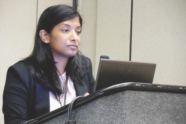
Health care reform 6 years out
Well, UnitedHealthcare has announced that it’s pulling out of the health insurance exchanges because of huge losses. This may be the mortal wound. It is apparent that health care reform is undergoing a slow-motion implosion. Most of the Affordable Care Act has been delayed or canceled, including the individual and employer mandates, the independent payment advisory board (thank goodness), Medicare Advantage payment cuts, the Cadillac health insurance tax, and auto enrollment. Half the insurance co-ops have failed, and the remainder are running at a deficit. The exchange-plan premiums are increasing dramatically. In fact, with 71 cancellations and delays, health care reform has already effectively been repealed.
Insurance coverage has increased by about 8% (10 million more into Medicaid, 80% of all new insureds), particularly of the poor. On the flip side, millions have lost their old insurance, now have very high deductibles, and have lost their doctors. High-deductible insurance means that patients really aren’t going to be able to use their insurance, unless they have a catastrophic event and are hospitalized.
High-deductible insurance that pays at Medicaid rates, and Medicaid, are a particularly bad mix for dermatologists. Medicaid does not even cover the cost of overhead in the office setting, and many patients cannot afford their high deductibles. Almost all of the cost-efficient in-office curative procedures we offer cost less than the deductible. We are all seeing patients delay and delay treatment until the end of the year, hoping they won’t have to meet their deductible.
Many patients, excited that they finally have health insurance, are bitterly disappointed to find that they really don’t, except for their annual physical exam and the emergency room. The doctor is put in the poisonous position of explaining insurance policy limitations, and being collection agent. Poor Medicaid patients, who get free care at the hospital, go to the emergency room for minor complaints that would be much more efficiently handled in the office setting. This clogs emergency rooms, and is ferociously expensive. This is the opposite of what health care reform was supposed to do.
Insurance premiums are rising rapidly because somebody has to pay for coverage of the millions who could not formerly qualify for health insurance because of preexisting conditions. The current exchanges allow dropping in and out of insurance coverage and, with the elimination of preexisting conditions, this allows patients to game the system and wait until they fall ill to buy insurance. Historically, almost all were in the pool of insureds and the risk was predictable.
There is no way for politicians to go back and remove millions of chronically ill from the coverage rolls. Imagine the nightly news. As a physician, it is impossible not to feel compassion for these chronically ill patients, but it would have cost a lot less to just make them eligible for Medicaid to begin with.
OK, it is easy to complain, but what are possible solutions? Patients need health care savings accounts for a sizable portion of their deductibles. Physician rosters need to be real time and accurate. Networks need to be adequate (another column, another day). Medicaid rates need to increase to Medicare levels, as they did for the initial 2-year teaser period for primary care physicians. Exchange plan payment rates need to match their commercial insurance wrappers, instead of Medicaid rates, so physicians can afford to accept them. Stricter enrollment periods are needed, so patients cannot game the system, signing up only when they get sick or want a joint replaced. If you are going to provide health insurance for all, then make sure the health insurance is real, not hollow.
Dr. Coldiron is a past president of the American Academy of Dermatology. He is currently in private practice, but maintains a clinical assistant professorship at the University of Cincinnati. He cares for patients, teaches medical students and residents, and has several active clinical research projects. Dr. Coldiron is the author of more than 80 scientific letters, papers, and several book chapters, and he speaks frequently on a variety of topics. Write to him at [email protected].
Summer colds
Most viral infections in summer months are caused by enteroviruses. We studied illnesses in about 400 kids aged 4-18 years seen in private pediatric practice and were surprised by what we found.
Our impression was that summer colds lasted for a shorter time span than winter colds. What we found was that the median duration of illness was about 8 days. Among the various syndromes, the most common was stomatitis (viral blisters in the throat), accounting for 58% of all cases seen. A flulike illness with fever, myalgias, and malaise was second most common (28% of cases), followed by hand/foot/mouth syndrome (8%), pleurodynia (3%), fever with viral rash (3%), and aseptic meningitis (1%). Most of the cases occurred among children 4-12 years old.

The most prevalent symptoms were fever, headache, sore throat, tiredness, muscle aches, and crankiness. Fever was present in about 85% of cases of children with stomatitis, in 95% of cases with myalgias and malaise, but in only 50% of cases of hand/foot/mouth. Headache was very common as well, occurring in about 40% of children with stomatitis, 70% of children with myalgias and malaise, and in 30% of children with hand/foot/mouth.
Illness within a household was quite common. About 50% of the children who came for care had a sibling or parent ill with a summer cold. However, while the symptoms of the family members often were the same as the child who presented for care, that was not always the case. As anticipated, most illness within a household occurred within a 2-week time span. Hand/foot/mouth was most easily recognized by parents to have spread among their children. When a parent became ill, it was almost always the mother because she was almost always the primary parent caretaker.
Summer colds took a toll on families in terms of loss of work by parents. Most of the children were ill enough to stay out of day care or school for about 2-4 days. Virtually all the children with hand/foot/mouth and stomatitis with classic viral blister lesions had a single visit to the pediatric practice, and very limited or no tests done or medications prescribed other than acetaminophen or ibuprofen. But for the children with higher fevers without hand/foot/mouth or stomatitis, the costs of care escalated as tests were much more often performed (CBC, chest x-ray), and medications prescribed (antibiotics for uncertain diagnosis in the context of high fever), and occasional referrals made to the emergency department for further work-up (100% of cases of aseptic meningitis and 50% of cases of pleurodynia).
Overall, summer colds are not so insignificant as presumed at first glance. What interests me now is why summer colds so infrequently are followed by an acute otitis media or sinusitis, whereas winter colds caused by respiratory syncytial virus, influenza, and rhinoviruses are followed by an acute otitis media in about one-third of cases. A new study is underway!
Dr. Pichichero, a specialist in pediatric infectious diseases, is director of the Research Institute, Rochester (N.Y.) General Hospital. He is also a pediatrician at Legacy Pediatrics in Rochester. He has no disclosures.
Most viral infections in summer months are caused by enteroviruses. We studied illnesses in about 400 kids aged 4-18 years seen in private pediatric practice and were surprised by what we found.
Our impression was that summer colds lasted for a shorter time span than winter colds. What we found was that the median duration of illness was about 8 days. Among the various syndromes, the most common was stomatitis (viral blisters in the throat), accounting for 58% of all cases seen. A flulike illness with fever, myalgias, and malaise was second most common (28% of cases), followed by hand/foot/mouth syndrome (8%), pleurodynia (3%), fever with viral rash (3%), and aseptic meningitis (1%). Most of the cases occurred among children 4-12 years old.
The most prevalent symptoms were fever, headache, sore throat, tiredness, muscle aches, and crankiness. Fever was present in about 85% of cases of children with stomatitis, in 95% of cases with myalgias and malaise, but in only 50% of cases of hand/foot/mouth. Headache was very common as well, occurring in about 40% of children with stomatitis, 70% of children with myalgias and malaise, and in 30% of children with hand/foot/mouth.
Illness within a household was quite common. About 50% of the children who came for care had a sibling or parent ill with a summer cold. However, while the symptoms of the family members often were the same as the child who presented for care, that was not always the case. As anticipated, most illness within a household occurred within a 2-week time span. Hand/foot/mouth was most easily recognized by parents to have spread among their children. When a parent became ill, it was almost always the mother because she was almost always the primary parent caretaker.
Summer colds took a toll on families in terms of loss of work by parents. Most of the children were ill enough to stay out of day care or school for about 2-4 days. Virtually all the children with hand/foot/mouth and stomatitis with classic viral blister lesions had a single visit to the pediatric practice, and very limited or no tests done or medications prescribed other than acetaminophen or ibuprofen. But for the children with higher fevers without hand/foot/mouth or stomatitis, the costs of care escalated as tests were much more often performed (CBC, chest x-ray), and medications prescribed (antibiotics for uncertain diagnosis in the context of high fever), and occasional referrals made to the emergency department for further work-up (100% of cases of aseptic meningitis and 50% of cases of pleurodynia).
Overall, summer colds are not so insignificant as presumed at first glance. What interests me now is why summer colds so infrequently are followed by an acute otitis media or sinusitis, whereas winter colds caused by respiratory syncytial virus, influenza, and rhinoviruses are followed by an acute otitis media in about one-third of cases. A new study is underway!
Dr. Pichichero, a specialist in pediatric infectious diseases, is director of the Research Institute, Rochester (N.Y.) General Hospital. He is also a pediatrician at Legacy Pediatrics in Rochester. He has no disclosures.
Sports safety
The National Athletic Trainers Association and the American Medical Society for Sports Medicine have developed a new program they call Collaborative Solutions for Safety in Sports, with the goal of establishing a suite of safety rules, policies, and possibly laws to protect high school athletes from injury (“School Athletes Often Lack Adequate Protection” by Jane Brody, New York Times, April 18, 2016).
After its second meeting, Dr. Jonathan Drezner, director of the Center for Sports Cardiology at the University of Washington, Seattle, said that the collaborative hopes that eventually every high school in the country will have an athletic trainer at every practice and game; an emergency action plan to respond appropriately to an athlete in distress; a publicly accessible automated external defibrillator (AED) and a school-based program in its use; and climatization policies to prevent heat injury and heat stroke.

I suspect that in communities in which the athletic facilities are located on a single campus that these guideline might be achievable. But here in Brunswick, Maine, and all of the other communities that I am familiar with, having a trainer at every practice and game is logistically impractical and financially unsustainable.
For example, on a given weekday afternoon in the spring here in Brunswick, there may be boys and girls varsity and junior varsity lacrosse, baseball, and softball practices or games on fields scattered around town – many of which are miles apart. The track team may be running on the roads and in the woods, who knows where. Staffing all of these events might be a financial windfall for the athletic trainers, if they could be found. But the money just isn’t there. Although the 50 high school athletes who died last year according to the National Athletic Trainers Association is 50 too many, I doubt that having a trainer at every practice and game would be a cost-effective solution.

When I opened my practice, Brunswick’s only pediatrician eagerly relinquished the job of school physician. Along with doing annual physical assessments on the bus drivers, I was expected to attend all of the home varsity football games. Trotting out on the field to evaluate the bumped and bruised players provided good visibility and helped build my practice. But I wondered who was going to attend to the junior varsity players and to the soccer players, who in my experience were more likely to be injured than the varsity football players. I certainly didn’t have the time. Nor could I be in five places at once.
After a couple of years, the athletic director and I hatched a plan to ask the school board to require that the coaches in every sport be certified in CPR. Our plan was quickly adopted, in part because a 15-year-old in a neighboring town had recently suffered a cardiac event during a track practice. Later, it was discovered that she had short QT syndrome, but there was an unfortunate delay in finding someone skilled in CPR.
In the 35 years since the CPR requirement was initially adopted, there has not been a single player who required resuscitation. However, last year one of the track coaches was running with a friend at dawn on a rural road when his friend dropped, pulseless. The coach’s school-required CPR training saved the man’s life.
While having a trainer at every high school practice and game is an unrealistic goal, educating coaches and players on how to identify and manage an athlete in distress makes a lot of sense. But the collaborative’s recommendation that is my personal favorite is having AEDs publicly accessible at games and practices. As a grandparent who spends a lot of time watching his grandchildren compete, I want the equipment available should I get a little too excited.
Dr. Wilkoff practiced primary care pediatrics in Brunswick, Maine, for nearly 40 years. He has authored several books on behavioral pediatrics, including “How to Say No to Your Toddler.”
The National Athletic Trainers Association and the American Medical Society for Sports Medicine have developed a new program they call Collaborative Solutions for Safety in Sports, with the goal of establishing a suite of safety rules, policies, and possibly laws to protect high school athletes from injury (“School Athletes Often Lack Adequate Protection” by Jane Brody, New York Times, April 18, 2016).
After its second meeting, Dr. Jonathan Drezner, director of the Center for Sports Cardiology at the University of Washington, Seattle, said that the collaborative hopes that eventually every high school in the country will have an athletic trainer at every practice and game; an emergency action plan to respond appropriately to an athlete in distress; a publicly accessible automated external defibrillator (AED) and a school-based program in its use; and climatization policies to prevent heat injury and heat stroke.
I suspect that in communities in which the athletic facilities are located on a single campus that these guideline might be achievable. But here in Brunswick, Maine, and all of the other communities that I am familiar with, having a trainer at every practice and game is logistically impractical and financially unsustainable.
For example, on a given weekday afternoon in the spring here in Brunswick, there may be boys and girls varsity and junior varsity lacrosse, baseball, and softball practices or games on fields scattered around town – many of which are miles apart. The track team may be running on the roads and in the woods, who knows where. Staffing all of these events might be a financial windfall for the athletic trainers, if they could be found. But the money just isn’t there. Although the 50 high school athletes who died last year according to the National Athletic Trainers Association is 50 too many, I doubt that having a trainer at every practice and game would be a cost-effective solution.
When I opened my practice, Brunswick’s only pediatrician eagerly relinquished the job of school physician. Along with doing annual physical assessments on the bus drivers, I was expected to attend all of the home varsity football games. Trotting out on the field to evaluate the bumped and bruised players provided good visibility and helped build my practice. But I wondered who was going to attend to the junior varsity players and to the soccer players, who in my experience were more likely to be injured than the varsity football players. I certainly didn’t have the time. Nor could I be in five places at once.
After a couple of years, the athletic director and I hatched a plan to ask the school board to require that the coaches in every sport be certified in CPR. Our plan was quickly adopted, in part because a 15-year-old in a neighboring town had recently suffered a cardiac event during a track practice. Later, it was discovered that she had short QT syndrome, but there was an unfortunate delay in finding someone skilled in CPR.
In the 35 years since the CPR requirement was initially adopted, there has not been a single player who required resuscitation. However, last year one of the track coaches was running with a friend at dawn on a rural road when his friend dropped, pulseless. The coach’s school-required CPR training saved the man’s life.
While having a trainer at every high school practice and game is an unrealistic goal, educating coaches and players on how to identify and manage an athlete in distress makes a lot of sense. But the collaborative’s recommendation that is my personal favorite is having AEDs publicly accessible at games and practices. As a grandparent who spends a lot of time watching his grandchildren compete, I want the equipment available should I get a little too excited.
Dr. Wilkoff practiced primary care pediatrics in Brunswick, Maine, for nearly 40 years. He has authored several books on behavioral pediatrics, including “How to Say No to Your Toddler.”

Let ’em cry … or not
A young couple already has decided to bring their as-yet-unborn child to your group. Now they are interviewing each member in hopes of finding a primary care physician who will best fit their expectations. Their second question for you is, “How do you feel about letting a baby cry itself to sleep?”
You sense that their question is a Rorschach test and a sneaky attempt to peer into what makes you tick. But let’s pretend for a moment that you are seized by a brain cramp and fail to do the obvious by turning the question around and asking them about how they feel about sleep training. Instead, you shoot from the hip. How would you respond?

Would you tell them that allowing a child to cry himself to sleep is neither dangerous nor cruel? Nor does it commit the child to a life of insecurity and emotional imbalance. In your opinion, if done correctly, it is usually the quickest and least painful way to help a child develop healthy sleep habits.
Or would you tell them that their child’s cry means that he needs something, and it is their responsibility to meet that need? That you believe letting a child cry himself to sleep is cruel and that it is better to let a child develop the skill of falling to sleep naturally at his own pace.
Because you neglected to first determine where these parents are coming from, regardless of which end of the spectrum you favor, your candid, nuance-free answer is likely to be a problem for somebody. If you revealed that you are a let-’em-cry proponent, the parents who were looking for a sensitive, child-centered pediatrician will quickly cross you off their list. However, if the parents choose you because you presented yourself as a let-nature-take-its-time pediatrician, they may have narrowed their options when their baby fails to settle in easily.
The challenge of how best to advise parents about infant sleep problems is a prime example of when practicing primary care medicine becomes an art. The answer to the let-’em-cry … or not dilemma is saturated with emotion and pretty much devoid of supporting scientific data. My gut, my personality, and 40 years of experience tell me that, more often than not, letting children cry themselves to sleep is the better approach. However, experience also has told me to keep my mouth shut when the topic of infant sleep is painted in the black-and-white question of let ‘em cry … or not.
The best approach is to learn as much as possible about the baby’s parents. Do they have similar or widely differing tolerances for a crying infant? I won’t really learn this until the parenting game has begun. Will I be able to convince these parents that, while it may be their responsibility to meet their crying child’s needs, one of those needs is the need to fall asleep? Or will I be wasting my time by trying to change their instincts?
Regardless of your own bias, your advice must be tailored to each individual family’s strengths and vulnerabilities, including the child’s temperament and the parents’ emotional resilience and tolerance for crying. Just as when we are counseling a mother who is nearing the end of her struggle with breastfeeding, a pediatrician must be prepared to become a chameleon and leave his or her bias behind.
One of the best strategies for avoiding that treacherous let-’em-cry … or not fork in the road is to promote good sleep habits from the beginning. When a baby is gaining weight, I encourage mothers to shorten feedings so that the baby finishes most feedings sated and drowsy but not fully asleep. I urge parents who find that a pacifier helps to use it only when the child is in his crib and to create a dim light, minimal-stimulation environment from around 7 p.m. to 7 a.m. By encouraging families to adopt these and other sleep-friendly practices early, I can often avoid revealing the ugly truth that, at my core, I am really a let-’em-cry guy.
Dr. Wilkoff practiced primary care pediatrics in Brunswick, Maine, for nearly 40 years. He has authored several books on behavioral pediatrics, including “How to Say No to Your Toddler.”
A young couple already has decided to bring their as-yet-unborn child to your group. Now they are interviewing each member in hopes of finding a primary care physician who will best fit their expectations. Their second question for you is, “How do you feel about letting a baby cry itself to sleep?”
You sense that their question is a Rorschach test and a sneaky attempt to peer into what makes you tick. But let’s pretend for a moment that you are seized by a brain cramp and fail to do the obvious by turning the question around and asking them about how they feel about sleep training. Instead, you shoot from the hip. How would you respond?
Would you tell them that allowing a child to cry himself to sleep is neither dangerous nor cruel? Nor does it commit the child to a life of insecurity and emotional imbalance. In your opinion, if done correctly, it is usually the quickest and least painful way to help a child develop healthy sleep habits.
Or would you tell them that their child’s cry means that he needs something, and it is their responsibility to meet that need? That you believe letting a child cry himself to sleep is cruel and that it is better to let a child develop the skill of falling to sleep naturally at his own pace.
Because you neglected to first determine where these parents are coming from, regardless of which end of the spectrum you favor, your candid, nuance-free answer is likely to be a problem for somebody. If you revealed that you are a let-’em-cry proponent, the parents who were looking for a sensitive, child-centered pediatrician will quickly cross you off their list. However, if the parents choose you because you presented yourself as a let-nature-take-its-time pediatrician, they may have narrowed their options when their baby fails to settle in easily.
The challenge of how best to advise parents about infant sleep problems is a prime example of when practicing primary care medicine becomes an art. The answer to the let-’em-cry … or not dilemma is saturated with emotion and pretty much devoid of supporting scientific data. My gut, my personality, and 40 years of experience tell me that, more often than not, letting children cry themselves to sleep is the better approach. However, experience also has told me to keep my mouth shut when the topic of infant sleep is painted in the black-and-white question of let ‘em cry … or not.
The best approach is to learn as much as possible about the baby’s parents. Do they have similar or widely differing tolerances for a crying infant? I won’t really learn this until the parenting game has begun. Will I be able to convince these parents that, while it may be their responsibility to meet their crying child’s needs, one of those needs is the need to fall asleep? Or will I be wasting my time by trying to change their instincts?
Regardless of your own bias, your advice must be tailored to each individual family’s strengths and vulnerabilities, including the child’s temperament and the parents’ emotional resilience and tolerance for crying. Just as when we are counseling a mother who is nearing the end of her struggle with breastfeeding, a pediatrician must be prepared to become a chameleon and leave his or her bias behind.
One of the best strategies for avoiding that treacherous let-’em-cry … or not fork in the road is to promote good sleep habits from the beginning. When a baby is gaining weight, I encourage mothers to shorten feedings so that the baby finishes most feedings sated and drowsy but not fully asleep. I urge parents who find that a pacifier helps to use it only when the child is in his crib and to create a dim light, minimal-stimulation environment from around 7 p.m. to 7 a.m. By encouraging families to adopt these and other sleep-friendly practices early, I can often avoid revealing the ugly truth that, at my core, I am really a let-’em-cry guy.
Dr. Wilkoff practiced primary care pediatrics in Brunswick, Maine, for nearly 40 years. He has authored several books on behavioral pediatrics, including “How to Say No to Your Toddler.”
