User login
Will Diabetes Drugs Advance Osteoarthritis Management?
VIENNA — With the glucagon-like peptide (GLP) 1 receptor agonist semaglutide (Wegovy) recently shown to significantly induce weight loss in people with osteoarthritis (OA) and obesity in the STEP-9 trial, could drugs traditionally used to treat type 2 diabetes be the next big thing for OA management?
“Hormone-based weight loss drugs are a game changer” for obesity management, Sébastien Czernichow, MD, PhD, said during a plenary session at the OARSI 2024 World Congress.
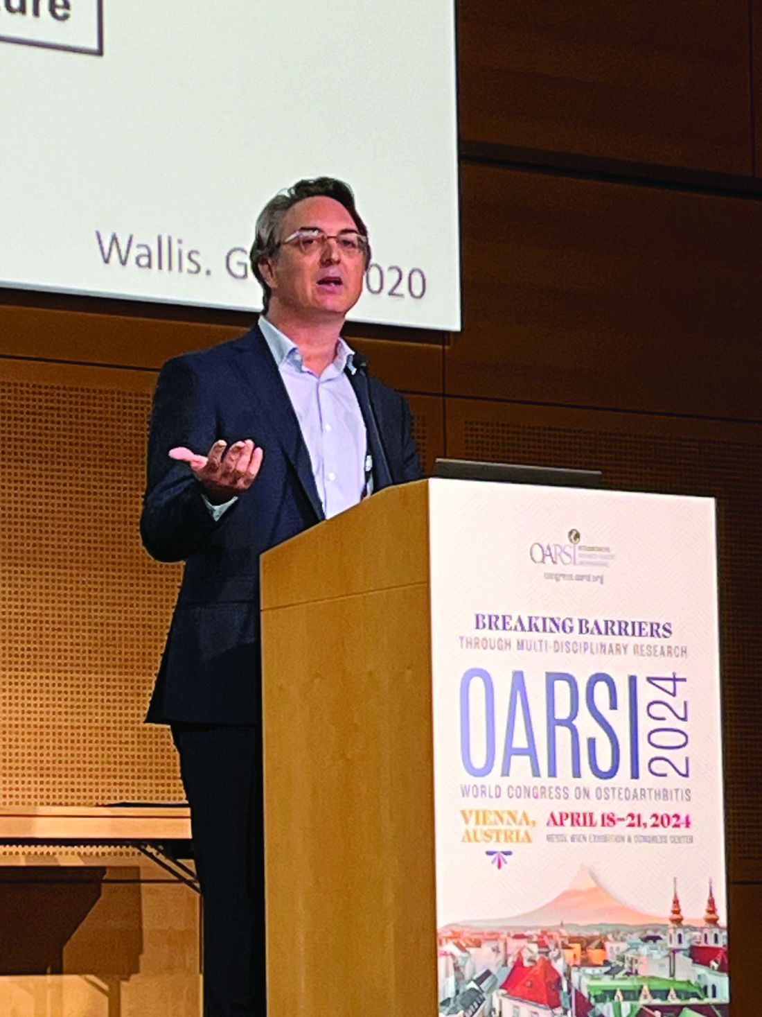
Drugs such as semaglutide may also have a cardioprotective effect, reducing the risk for major adverse cardiovascular events by as much as 20% vs placebo, added Dr. Czernichow, professor of nutrition at Paris Cité University and head of the Department of Nutrition at the George Pompidou European Hospital in Paris, France.
“You have to keep in mind that the short-term side effects are mainly gastrointestinal and [are] manageable. The mid-term side effects are an increased gallbladder [disease] risk, and the long-term benefits and risks are not really well known yet,” Dr. Czernichow said. With regard to that, the effects of these drugs on lean body mass, bone health, and nutritional deficiencies need to be further evaluated and monitored.
Weight Loss Benefits
Weight loss is one of the cornerstones of OA management, and in addition to the weight loss seen with the GLP-1 receptor agonists, there have also been changes in body composition, Dr. Czernichow said.
In SURMOUNT-1, for example, the dual glucose-dependent insulinotropic polypeptide and GLP-1 receptor agonist tirzepatide (Zepbound) was shown to significantly reduce total fat mass with a smaller decrease in total lean mass in a subanalysis.
It has been argued that effects on body composition need to be considered when evaluating new weight loss drugs, and that focusing only on the degree of weight reduction is “encouraging inaccurate measures of medication efficacy for both patients and clinicians,” Dr. Czernichow said, citing a viewpoint published in JAMA Internal Medicine.
“The real question is: Are we able to fund these drugs for everyone? Or will only the richest patients be allocated to these drugs?” Dr. Czernichow said.
Weight Rebound
Tonia Vincent, MBBS, PhD, professor of musculoskeletal biology and an honorary rheumatologist at The Kennedy Institute of Rheumatology at University of Oxford in England, was concerned about rebound weight gain.
“We hear a lot about this, that people stopping drugs actually get worse weight gain than before they started, and that’s a concern about a drug that is going to have a huge pressure for supply,” Dr. Vincent said following Dr. Czernichow’s presentation.
Another delegate said that calling GLP-1 receptor agonists a “game changer” for weight loss in OA was premature because long-term results are needed.
“You mentioned that the double-digit weight loss is getting very close to the results from bariatric surgery, but bariatric surgery you do once, and for these drugs, to maintain the weight loss, you need to take them continuously,” she said.
Weight Loss Affects Bone
Yet another delegate cautioned on the potential effects of significant weight loss on bone and cartilage. There is evidence, he said, that weight loss of 5-10 kg can significantly affect bone turnover, increasing bone resorption and thus putting patients at a risk of becoming osteopenic. “Are we looking at a new population of osteoporosis patients who may then also be at risk for fractures?” he asked.
Separately at OARSI 2024, Anne C. Bay-Jensen, PhD, chief technology officer at Nordic Bioscience in Herlev, Denmark, and colleagues reported data showing that weight loss was associated with an increase in bone and cartilage degradation.
Although Dr. Bay-Jensen and colleagues found that losing weight was associated with improved patient outcomes, there was a 1.58-fold increase in the bone resorption marker CTX-I in people who had lost weight vs a 1.37-fold gain in those whose weight remained stable and 1.11-fold increase in those who gained weight.
Moreover, there was a 1.15-fold increase in the cartilage degradation marker C2M in the weight loss group and 0.84-fold decrease in the interstitial matrix degradation marker C3M.
GLP-1 and Bone Effects
Another question is whether GLP-1 receptor agonists might be having direct effects on the bone that may be beneficial in OA. They might, postdoctoral researcher Eda Çiftci, PhD, of AO Research Institute Davos in Switzerland, and collaborators, said during the poster sessions at OARSI 2024.
Dr. Çiftci and researchers reported the findings of an in vitro study that looked at whether liraglutide might have anti-inflammatory and anabolic effects on a human chondrocytes model that had been treated with interleukin (IL)-1-beta to “mimic an inflammatory OA condition.”
The release of the proinflammatory cytokines IL-6 and IL-8 was reduced by treatment with liraglutide when compared with control chondrocytes. Furthermore, the expression of the proteoglycan aggrecan — important for articular cartilage function — also was preserved.
These results suggest that liraglutide does indeed have anabolic and anti-inflammatory effects, Dr. Çiftci and fellow researchers concluded.
New Role for Dipeptidyl Transferase Inhibitors?
Researchers are also looking at the potential role for other diabetes medications in OA management, including the dipeptidyl peptidase (DPP) 4 inhibitors.
Although these drugs are considered “weight neutral,” in vitro studies have suggested that the DPP4 enzyme may have a role to play in chondrocyte survival and inflammation, Yu-Hsiu Chen, MD, of the Tri-Service General Hospital and the National Defense Medical Center in Taipei, Taiwan, told this news organization. The DPP4 enzyme inactivates GLP-1, so there is rationale there.
“Last year, we published a paper where we found the concentration of DPP4 in the synovial fluid was correlated with radiographic change in knee OA,” Dr. Chen said. This time, “we’re trying to see if a DPP4 inhibitor can be used as a treatment.”
For their analysis, they used data on people newly diagnosed with type 2 diabetes who were and were not using DPP4 inhibitors obtained from Taiwan’s National Health Insurance Research Database. This database contains information on 99% of the Taiwanese population, Dr. Chen said.
Matching 165,333 DPP4 inhibitor users with an equal number of nonusers showed that there was a significant 58% risk reduction for developing OA with DPP4 inhibitor use (hazard ratio, 0.42; 95% CI, 0.41-0.44).
DPP4 inhibitor use was also associated with a 58% risk lower risk for total knee replacement (TKR) and a 62% lower risk for total hip replacement.
Dr. Chen and colleagues concluded: “These results strongly indicate that DPP4 inhibitors could be considered as a viable treatment approach for individuals with type 2 [diabetes mellitus] who are at risk for developing OA or [who] already have OA.”
Could Sodium-Glucose Cotransporter 2 (SGLT2) Inhibitors Be Beneficial?
So, what about SGLT2 inhibitors? Do they also have a potential role to play in managing people with OA, regardless of whether there is diabetes present? Perhaps, and their effect may be even greater than what’s been observed for GLP-1 receptor agonists, as data presented by epidemiologist S. Reza Jafarzadeh, DVM, PhD, suggested.
“While GLP-1 receptor agonist drugs have been reported to reduce OA risk, largely attributed to their weight loss effect, SGLT2 inhibitors may provide a greater protective effect on OA outcomes,” said Dr. Jafarzadeh, assistant professor at Boston University.

He presented data from a large analysis of new users of SGLT2 inhibitors and GLP-1 receptor agonists within two claims databases — Merative (n = 603,471) and TriNetX (n = 1,202,972) — showing that SGLT2 inhibitors were associated with significantly lower risks for OA and the need for TKR.
Comparing new users of SGLT2 inhibitors and GLP-1 receptor agonists in the Merative dataset, the relative risks and odds ratios for OA were a respective 0.96 and 0.80, and having a TKR, 0.88 and 0.76.
Similar results were seen using the TriNetX dataset, with respective relative risks and hazard ratios of 0.90 and 0.85 for OA, and 0.81 and 0.78 for TKR.
In an interview, Dr. Jafarzadeh said that the initial hypothesis was that because SGLT2 inhibitors have only a modest effect on weight loss, there would be no effect on OA outcomes.
“But we were surprised that it actually looked like they reduced the risk of OA outcomes even more than GLP-1 receptor agonists,” Dr. Jafarzadeh said.
Further work is needed to understand these data, but they could mean that SLGT2 inhibitors, like GLP-1 receptor agonists, may have a role to play outside their current use in type 2 diabetes.
The congress was sponsored by the Osteoarthritis Research Society International.
Dr. Czernichow disclosed ties with BariaTek Medical, Boehringer Ingelheim, Bristol Myers Squibb, Fresenius, Janssen, Jellynov, Lilly, Novo Nordisk, Novartis, and ViiV Healthcare. Dr. Vincent had no relevant disclosures. Dr. Bay-Jensen is the chief technology officer and director of immunoscience at Nordic Bioscience, which funded the work in the poster she presented at OARSI 2024. The work presented by Dr. Çiftci and colleagues was funded by the Eurostars-2 joint program with co-funding from the European Horizon 2020 research and innovation program. Dr. Çiftci had no personal disclosures to report. Dr. Chen’s work was supported by the government of Taiwan, and she had no financial conflicts of interest to disclose. Dr. Jafarzadeh had no conflicts of interest to disclose.
A version of this article appeared on Medscape.com .
VIENNA — With the glucagon-like peptide (GLP) 1 receptor agonist semaglutide (Wegovy) recently shown to significantly induce weight loss in people with osteoarthritis (OA) and obesity in the STEP-9 trial, could drugs traditionally used to treat type 2 diabetes be the next big thing for OA management?
“Hormone-based weight loss drugs are a game changer” for obesity management, Sébastien Czernichow, MD, PhD, said during a plenary session at the OARSI 2024 World Congress.
Drugs such as semaglutide may also have a cardioprotective effect, reducing the risk for major adverse cardiovascular events by as much as 20% vs placebo, added Dr. Czernichow, professor of nutrition at Paris Cité University and head of the Department of Nutrition at the George Pompidou European Hospital in Paris, France.
“You have to keep in mind that the short-term side effects are mainly gastrointestinal and [are] manageable. The mid-term side effects are an increased gallbladder [disease] risk, and the long-term benefits and risks are not really well known yet,” Dr. Czernichow said. With regard to that, the effects of these drugs on lean body mass, bone health, and nutritional deficiencies need to be further evaluated and monitored.
Weight Loss Benefits
Weight loss is one of the cornerstones of OA management, and in addition to the weight loss seen with the GLP-1 receptor agonists, there have also been changes in body composition, Dr. Czernichow said.
In SURMOUNT-1, for example, the dual glucose-dependent insulinotropic polypeptide and GLP-1 receptor agonist tirzepatide (Zepbound) was shown to significantly reduce total fat mass with a smaller decrease in total lean mass in a subanalysis.
It has been argued that effects on body composition need to be considered when evaluating new weight loss drugs, and that focusing only on the degree of weight reduction is “encouraging inaccurate measures of medication efficacy for both patients and clinicians,” Dr. Czernichow said, citing a viewpoint published in JAMA Internal Medicine.
“The real question is: Are we able to fund these drugs for everyone? Or will only the richest patients be allocated to these drugs?” Dr. Czernichow said.
Weight Rebound
Tonia Vincent, MBBS, PhD, professor of musculoskeletal biology and an honorary rheumatologist at The Kennedy Institute of Rheumatology at University of Oxford in England, was concerned about rebound weight gain.
“We hear a lot about this, that people stopping drugs actually get worse weight gain than before they started, and that’s a concern about a drug that is going to have a huge pressure for supply,” Dr. Vincent said following Dr. Czernichow’s presentation.
Another delegate said that calling GLP-1 receptor agonists a “game changer” for weight loss in OA was premature because long-term results are needed.
“You mentioned that the double-digit weight loss is getting very close to the results from bariatric surgery, but bariatric surgery you do once, and for these drugs, to maintain the weight loss, you need to take them continuously,” she said.
Weight Loss Affects Bone
Yet another delegate cautioned on the potential effects of significant weight loss on bone and cartilage. There is evidence, he said, that weight loss of 5-10 kg can significantly affect bone turnover, increasing bone resorption and thus putting patients at a risk of becoming osteopenic. “Are we looking at a new population of osteoporosis patients who may then also be at risk for fractures?” he asked.
Separately at OARSI 2024, Anne C. Bay-Jensen, PhD, chief technology officer at Nordic Bioscience in Herlev, Denmark, and colleagues reported data showing that weight loss was associated with an increase in bone and cartilage degradation.
Although Dr. Bay-Jensen and colleagues found that losing weight was associated with improved patient outcomes, there was a 1.58-fold increase in the bone resorption marker CTX-I in people who had lost weight vs a 1.37-fold gain in those whose weight remained stable and 1.11-fold increase in those who gained weight.
Moreover, there was a 1.15-fold increase in the cartilage degradation marker C2M in the weight loss group and 0.84-fold decrease in the interstitial matrix degradation marker C3M.
GLP-1 and Bone Effects
Another question is whether GLP-1 receptor agonists might be having direct effects on the bone that may be beneficial in OA. They might, postdoctoral researcher Eda Çiftci, PhD, of AO Research Institute Davos in Switzerland, and collaborators, said during the poster sessions at OARSI 2024.
Dr. Çiftci and researchers reported the findings of an in vitro study that looked at whether liraglutide might have anti-inflammatory and anabolic effects on a human chondrocytes model that had been treated with interleukin (IL)-1-beta to “mimic an inflammatory OA condition.”
The release of the proinflammatory cytokines IL-6 and IL-8 was reduced by treatment with liraglutide when compared with control chondrocytes. Furthermore, the expression of the proteoglycan aggrecan — important for articular cartilage function — also was preserved.
These results suggest that liraglutide does indeed have anabolic and anti-inflammatory effects, Dr. Çiftci and fellow researchers concluded.
New Role for Dipeptidyl Transferase Inhibitors?
Researchers are also looking at the potential role for other diabetes medications in OA management, including the dipeptidyl peptidase (DPP) 4 inhibitors.
Although these drugs are considered “weight neutral,” in vitro studies have suggested that the DPP4 enzyme may have a role to play in chondrocyte survival and inflammation, Yu-Hsiu Chen, MD, of the Tri-Service General Hospital and the National Defense Medical Center in Taipei, Taiwan, told this news organization. The DPP4 enzyme inactivates GLP-1, so there is rationale there.
“Last year, we published a paper where we found the concentration of DPP4 in the synovial fluid was correlated with radiographic change in knee OA,” Dr. Chen said. This time, “we’re trying to see if a DPP4 inhibitor can be used as a treatment.”
For their analysis, they used data on people newly diagnosed with type 2 diabetes who were and were not using DPP4 inhibitors obtained from Taiwan’s National Health Insurance Research Database. This database contains information on 99% of the Taiwanese population, Dr. Chen said.
Matching 165,333 DPP4 inhibitor users with an equal number of nonusers showed that there was a significant 58% risk reduction for developing OA with DPP4 inhibitor use (hazard ratio, 0.42; 95% CI, 0.41-0.44).
DPP4 inhibitor use was also associated with a 58% risk lower risk for total knee replacement (TKR) and a 62% lower risk for total hip replacement.
Dr. Chen and colleagues concluded: “These results strongly indicate that DPP4 inhibitors could be considered as a viable treatment approach for individuals with type 2 [diabetes mellitus] who are at risk for developing OA or [who] already have OA.”
Could Sodium-Glucose Cotransporter 2 (SGLT2) Inhibitors Be Beneficial?
So, what about SGLT2 inhibitors? Do they also have a potential role to play in managing people with OA, regardless of whether there is diabetes present? Perhaps, and their effect may be even greater than what’s been observed for GLP-1 receptor agonists, as data presented by epidemiologist S. Reza Jafarzadeh, DVM, PhD, suggested.
“While GLP-1 receptor agonist drugs have been reported to reduce OA risk, largely attributed to their weight loss effect, SGLT2 inhibitors may provide a greater protective effect on OA outcomes,” said Dr. Jafarzadeh, assistant professor at Boston University.
He presented data from a large analysis of new users of SGLT2 inhibitors and GLP-1 receptor agonists within two claims databases — Merative (n = 603,471) and TriNetX (n = 1,202,972) — showing that SGLT2 inhibitors were associated with significantly lower risks for OA and the need for TKR.
Comparing new users of SGLT2 inhibitors and GLP-1 receptor agonists in the Merative dataset, the relative risks and odds ratios for OA were a respective 0.96 and 0.80, and having a TKR, 0.88 and 0.76.
Similar results were seen using the TriNetX dataset, with respective relative risks and hazard ratios of 0.90 and 0.85 for OA, and 0.81 and 0.78 for TKR.
In an interview, Dr. Jafarzadeh said that the initial hypothesis was that because SGLT2 inhibitors have only a modest effect on weight loss, there would be no effect on OA outcomes.
“But we were surprised that it actually looked like they reduced the risk of OA outcomes even more than GLP-1 receptor agonists,” Dr. Jafarzadeh said.
Further work is needed to understand these data, but they could mean that SLGT2 inhibitors, like GLP-1 receptor agonists, may have a role to play outside their current use in type 2 diabetes.
The congress was sponsored by the Osteoarthritis Research Society International.
Dr. Czernichow disclosed ties with BariaTek Medical, Boehringer Ingelheim, Bristol Myers Squibb, Fresenius, Janssen, Jellynov, Lilly, Novo Nordisk, Novartis, and ViiV Healthcare. Dr. Vincent had no relevant disclosures. Dr. Bay-Jensen is the chief technology officer and director of immunoscience at Nordic Bioscience, which funded the work in the poster she presented at OARSI 2024. The work presented by Dr. Çiftci and colleagues was funded by the Eurostars-2 joint program with co-funding from the European Horizon 2020 research and innovation program. Dr. Çiftci had no personal disclosures to report. Dr. Chen’s work was supported by the government of Taiwan, and she had no financial conflicts of interest to disclose. Dr. Jafarzadeh had no conflicts of interest to disclose.
A version of this article appeared on Medscape.com .
FROM OARSI 2024
Vacationing Doctors Fight to Revive a Drowned Child
Emergencies happen anywhere, anytime, and sometimes, medical professionals find themselves in situations where they are the only ones who can help. Is There a Doctor in the House? is a series telling these stories.
Jennifer Suders, DO: We were in Florida with our 1-year-old daughter visiting my parents. They moved to an area called Hallandale Beach and live in a high-rise community with a few different pools and spas.
Dan and I were in the spa area at the gym. He was getting me to hurry up because we were supposed to meet my parents who were with our daughter. I was sort of moseying and taking my time.
We were walking by one of the pool decks to get into the building when I heard what sounded like a slap. My first thought was that maybe somebody was choking and someone was hitting their back. Choking has always been my biggest fear with our daughter.
I turned and saw some people who seemed frantic. I looked at Dan and started to ask, “Do you think they need help?” I don’t even think I got the whole sentence out before this mom whipped her head around. I’ll never forget her dark brown hair flying. She screamed, “HELP!”
Dan and I just ran. I let go of my backpack and iPad and water bottle. They scattered across the pool deck. I instantly had my phone in my hand dialing 911.
Daniel Suders, DO: That’s what they teach us, to call 911 first. I didn’t think of it in the moment, but Jenny did.
Jennifer Suders:
Dan and I got down on either side of the boy and checked for a pulse. We couldn’t feel anything. Dan started chest compressions. I was talking to the 911 operator, and then I gave two rescue breaths. We did a sternal rub.
I was kind of yelling in the boy’s face, trying to get him to respond. I tried English and Russian because there’s a big Russian community there, and my family speaks Russian. The grandma asked us if we knew what we were doing.
Daniel Suders: I think she asked if Jenny was a nurse.
Jennifer Suders: Common misconception. Suddenly, the boy started vomiting, and so much water poured out. We turned him on his side, and he had two or three more episodes of spitting up the water. After that, we could see the color start to come back into his face. His eyes started fluttering.
We thought he was probably coming back. But we were too scared to say that in case we were wrong, and he went back under. So, we just held him steady. We didn’t know what had happened, if he might have hit his head, so we needed to keep him still.
Daniel Suders: It was amazing when those eyes opened, and he started to wake up.
Jennifer Suders: It felt like my heart had stopped while I was waiting for his to start.
Daniel Suders: He was clutching his chest like it hurt and started calling for his mom. He was crying and wanting to get in his mom’s arms. We had to keep him from standing up and walking.
Jennifer Suders: He was clearly scared. There were all these strange faces around him. I kept looking at my phone, anxiously waiting for EMS to come. They got there about 8 or 9 minutes later.
At some point, the father walked in with their daughter, a baby under a year old. He was in shock, not knowing what was going on. The grandma explained that the boy had been jumping into the pool over and over with his brother. All of a sudden, they looked over, and he was just lying there, floating, face down. They were right there; they were watching him. It was just that quick.
Daniel Suders: They pulled him out right away, and that was a big thing on his side that it was caught so quickly. He didn’t have to wait long to start resuscitation.
Jennifer Suders: Once EMS got there and assessed him, they put him and his mom on the stretcher. I remember watching them wheel it through the double doors to get to the elevator. As soon as they were gone, I just turned around and broke down. I had been in doctor mode if you will. Straight to the point. No nonsense. Suddenly, I went back into civilian mode, and my emotions just bubbled up.
After we left, we went to meet my parents who had our kid. Dan just beelined toward her and scooped her up and wouldn’t let her go.
For the rest of the day, it was all I could think about. It took me a while to fall asleep that night, and it was the first thing I thought when I woke up the next morning. We were hopeful that the boy was going to be okay, but you never know. We didn’t call the hospital because with HIPAA, I didn’t know if they could tell us anything.
And then the next day — there they were. The family was back at the pool. The little boy was running around like nothing had happened. We were a little surprised. But I would hate for him to be scared of the pool for the rest of his life. His family was watching him like a hawk.
They told us that the boy and his mom had stayed overnight in the ER, but only as a precaution. He didn’t have any more vomiting. He was absolutely fine. They were incredibly grateful.
We got their names and exchanged numbers and took a picture. That’s all I wanted — a photo to remember them.
A day or so later, we saw them again at a nearby park. The boy was climbing trees and seemed completely normal. It was the best outcome we could have hoped for.
Daniel Suders: My biggest worry was any harm to his chest from the resuscitation, or of course how long he was without oxygen. But everyone says that kids are really resilient. I work with adults, so I don’t have a lot of experience.
As a hospitalist, we don’t always see a lot of success with CPR. It’s often an elderly person who just doesn’t have much of a chance. That same week before our vacation, I had lost a 90-year-old in the hospital. It was such a juxtaposition — a 3-year-old with their whole life in front of them. We were able to preserve that, and it was incredible.
Jennifer Suders: I’m a nephrologist, so my field is pretty calm. No big emergencies. We have patients on the floor, but if a code gets called, there’s a team that comes in from the intensive care unit. I always kind of wondered what I would do if I was presented with a scenario like this.
Daniel Suders: We have a lot of friends that do ER medicine, and I felt like those were the guys that really understood when we told them the story. One friend said to me, “By the time they get to us, they’re either in bad shape or they’re better already.” A lot depends on what happens in the field.
Jennifer Suders: I’m even more vigilant about pool safety now. I want to make sure parents know that drowning doesn›t look like flailing theatrics. It can be soundless. Three adults were right next to this little boy and didn›t realize until they looked down and saw him.
If we hadn’t been there, I don’t know if anyone would’ve been able to step in. No one else was medically trained. But I think the message is — you don’t have to be. Anyone can take a CPR class.
When I told my parents, my dad said, “Oh my gosh, I would’ve laid right down there next to that kid and passed out.” Without any training, it’s petrifying to see something like that.
I think about how we could have stayed in the gym longer and been too late. Or we could have gotten on the elevator earlier and been gone. Two minutes, and it would’ve been a story we heard later, not one we were a part of. It feels like we were at a true crossroads in that moment where that boy could have lived or died. And the stars aligned perfectly.
We had no medicine, no monitors, nothing but our hands and our breaths. And we helped a family continue their vacation rather than plan a funeral.
Jennifer Suders, DO, is a nephrologist at West Virginia University Medicine Wheeling Clinic. Daniel Suders, DO, is a hospitalist at West Virginia University Medicine Reynolds Memorial Hospital.
A version of this article appeared on Medscape.com .
Tackling Lean Mass Loss When Weight Loss is Successful
DENVER — In addition to the established gastrointestinal side effects common with the highly effective anti-obesity drugs, there is growing discussion around their potential to contribute to the loss of lean mass, necessary to keep the metabolic engine running full-steam.
And although measures should be recommended to prevent those effects, experts also want to remind clinicians that the loss of lean mass is indeed expected with most weight loss interventions — when they’re successful.
“The bottom line is if you’re successful with weight loss, it’s a normal process that you’re going to lose some lean mass,” Angela Fitch, MD, associate director of the Massachusetts General Hospital Weight Center in Boston, said during a presentation on the issue at Obesity Medicine 2024.
“It’s what we would expect to see if you successfully lost weight with bariatric surgery or with an intense lifestyle intervention,” said Dr. Fitch, past president of the Obesity Medicine Association.
“The difference is, there haven’t been nearly as many people being successful with weight loss with those other interventions,” she noted. “But with the popularity of the glucagon-like peptide 1 (GLP-1) medications, people are hearing this for the first time and saying, ‘Oh my gosh, 30% of the weight loss is muscle mass — that’s horrible.’ “
, which have been reported in clinical trials of the GLP-1s semaglutide and the dual glucose-dependent insulinotropic polypeptide tirzepatide to range from about 25% to 40%, respectively, of weight loss.
“Excess adiposity is what makes us sick — not our weight,” Dr. Fitch underscored. “The amount of fat that people are losing [with anti-obesity medications] is far more beneficial than maybe the potential that they’ve lost a little bit of lean mass,” she said.
She cited research suggesting that significant weight loss from bariatric surgery is linked to increases in life expectancy, cardiovascular risk reduction, cancer risk reduction, and a wide array of other positive effects — despite the loss of lean mass that occurs with the weight loss.
Opportunity for Awareness
The increased attention on issues of body composition accompanying weight loss importantly provides clinicians the chance to underscore to patients the importance of offsetting the loss of lean mass through strength training, nutritional choices, and other measures.
However, patients should be prepared that achieving these goals can be more challenging than expected, said Dr. Fitch.
“It can be very hard to be in an energy deficit (due to a weight loss regimen) and gain muscle mass,” she said. “When athletes are trying to gain muscle mass, they’re increasing their intake to do so. It doesn’t come naturally in today’s world.”
Nevertheless, patients can be reassured that the losses can be reversed with some effort, Dr. Fitch noted.
She cautioned that for those who succeed in building or rebuilding lean mass, the evidence may be reflected on the scale, with numbers going up, not down — something they may not wish to see.
“Patients tend to freak out when they see the scale going up after losing all of that weight, but you can reassure them that it’s okay — this is healthier weight gain.”
Special Considerations in Older Patients
Efforts at staving off lean mass loss are particularly important in older patients, who are already most vulnerable to experiencing it naturally with age, even if not on a weight loss regimen.
But Dr. Fitch offered that age does not necessarily have to be a barrier in tackling those effects.
She described two cases of treating patients in their mid-70s, a male and female, with GLP-1s for obesity. Not only were they able to achieve substantial reductions in body mass index over nearly a year on treatment, but they were also able to avoid skeletal muscle mass loss during a period when it would have likely naturally occurred.
She noted the need to augment strength training with protein intake to help build muscle, citing recommendations including consumption of 1.4-2.0 g of protein per kg of body weight for building muscle and maintaining muscle mass.
Importantly, “make sure patients aren’t too appetite suppressed so they can keep up with their nutrition,” Dr. Fitch said.
A key condition to watch for in these patients is sarcopenia. Definitions of sarcopenia vary, but it is distinguished by low skeletal muscle mass and either low muscle strength — measured, for instance, with hand grip — or low muscle performance, such as reduced walking speed or muscle power, Dr. Fitch said.
In such cases, patients may need special considerations, including avoiding significant caloric deficits and whether the risks of medication outweigh the benefits.
‘Super-Responders’ and Other Lean Mass Loss Scenarios
Further addressing the issues of body composition and weight loss at the meeting, Robert F. Kushner, MD, professor of medicine and medicine education at Northwestern University in Chicago, noted that one area of concern regarding lean mass loss is “super-responders” — patients who have exceptionally high weight loss on GLP-1s.
“We are concerned about individuals who experience very high weight loss responses to medication, [specifically] 25% or more weight loss, as well as individuals at higher risk of losing lean body mass [muscle mass], specifically people in their 50s, 60s, and 70s,” Dr. Kushner told this news organization.
“Lifestyle counseling, particularly regarding safety and body composition, is recommended in these patients,” he said, adding that in managing these patients, “the approach is to use close patient monitoring, dose reduction if needed, and emphasizing a high-protein diet accompanied by aerobic and resistance physical activity.”
Potentially dramatic lean mass loss can occur in obesity whether or not patients are on obesity medications. As evidence of this, Dr. Kushner cited a subanalysis of the Look AHEAD trial of 1019 overweight or obese patients who had a mean age of 58 years at baseline. Patients were randomized to either a physical activity and reduced calorie intervention group or simply education.
Although the results showed that fat losses in the intervention group were generally regained over 8 years, a striking, steady decline was observed in lean mass in both the intervention and control groups, including men and women.
Dr. Fitch disclosed ties to Eli Lilly, Novo Nordisk, Currax, Vivus, SideKick Health, Jenny Craig, Carmot, and Seca. Dr. Kushner is on the advisory boards of Novo Nordisk, Weight Watchers, Lilly, Boehringer Ingelheim, and Altimmune.
A version of this article appeared on Medscape.com.
Internet Use Good for Mental Well-Being?
Contrary to previous research that suggests internet use can have a deleterious effect on mental health, a new study of more than 2 million individuals suggested it can actually enhance well-being.
Between 2006 and 2021, investigators studied more than 2 million people between the ages of 15 and 99 years in 168 countries, focusing on their psychological well-being and their use of the internet. Many of the included countries have rarely or never been studied in this connection.
“We were surprised to find a positive correlation between well-being and internet use across the majority of the thousands of models we used for our analysis,” lead author Matti Vuorre, PhD, of Tilburg University, Tilburg, the Netherlands, and a research associate at Oxford Internet Institute in England, said in a news release.
The study was published online on May 13 in Technology, Mind, and Behavior.
A Global Phenomenon
Coauthor Andrew K. Przybylski, PhD, professor of human behavior and technology at Oxford Internet Institute, explained the motive for conducting the study.
“Whilst internet technologies and their platforms and their potential psychological consequences remain debated, research to date has been inconclusive and of limited geographic and demographic scope,” he said.
He noted that the “overwhelming majority” of studies have focused on the Global North and on younger people and “ignoring the fact that the penetration of the internet has been, and continues to be, a global phenomenon.”
The researchers set out to address this gap by analyzing “how internet access, mobility internet access, and active internet use might predict psychological well-being on a global level across the life stages,” Dr. Przybylski continued. “To our knowledge, no other research has directly grappled with these issues and addressed the worldwide scope of the debate.”
To study internet use, the investigators analyzed data from the 2022 Gallup World Poll, a nationally representative survey of each country’s civilian, non-institutionalized adult population (ie, aged ≥ 15 years), conducted between 2002 and 2022. The poll assessed well-being using face-to-face, as well as phone interviews, conducted by local interviewers in the respondents’ native languages.
The total sample size included 2,414,295 adults drawn from 186 countries (53.1% women), drawn from countries that included those located in Latin America, Asia, and Africa.
The researchers examined eight indicators of well-being: life satisfaction, daily negative and positive experiences, two indices of social well-being, physical well-being, community well-being, and experiences of purpose.
Covariates included respondents’ income, education, work, relationship status, the ability to meet basic needs (food and shelter), and whether they reported having health problems.
Greater Life Satisfaction
The researchers conducted a “multiverse” of 33,792 types of analyses, researching the average differences in well-being between individuals who had access to mobile internet or had used the internet in the past 7 days.
They found that for the average country, those who had access to the internet reported approximately 0.08 units greater life satisfaction, positive experiences, and social life satisfaction and 0.06 units lower negative experience than those without access.
They also reported approximately 0.08 units greater experiences of purpose and 0.1 unit greater physical, 0.02 units greater community, and 0.08 units greater social well-being than individuals without access.
Being an active internet user was associated with a 0.03- to 0.08-unit increase in life satisfaction, positive experiences, social well-being, and physical well-being and a 0.04-unit decrease in negative experiences. Access to a smartphone predicted increases of 0.06 and 0.07 units.
Although the standard deviations (SDs) of well-being outcomes were small (eg, the median life satisfaction difference was 0.36 SDs between individuals who did and did not have access to the internet), they were “not negligible.”
In fact, when the researchers examined the associations’ robustness across all analyses, they found that 84.9% resulted in positive and statistically significant associations between internet connectivity and well-being.
Of the 4.9% of associations between internet use and community well-being that were negative, most were observed among young women between the ages of 15 and 24 years.
While the researchers did not identify this as a causal relationship, they noted that this finding is consistent with previous reports of increased cyberbullying and negative associations between social media use and depressive symptoms in young women.
“Overall, we found that average associations were consistent across internet adoption predictors and well-being outcomes, with those who had access to or actively used the internet reporting meaningfully greater well-being than those who did not,” Dr. Przybylski said.
The study’s limitations included comparing individuals with each other, given that there “are likely myriad other feature of the human condition that are associated with both uptake of internet technologies and well-being in such a manner that they might case spurious associations or mask true associations,” the authors noted.
Moreover, longitudinal studies tracking participants over time can provide more information about the “contexts of how and why an individual might be affected by internet technologies and platforms.” In addition, the self-reported measures of technology might be “lacking.”
Dr. Przybylski hopes that the findings will “bring some greater context to the screen time debate; however, further work is still needed in this important area.”
He urged platform providers “to share their detailed data on user behavior with social scientists working in this field for transparent and independent scientific enquiry, to enable a more comprehensive understanding of internet technologies in our daily lives.”
A Starting Point
In a separate news release, Kevin McConway, PhD, MBA, emeritus professor of applied statistics, The Open University, Milton Keynes, England, noted that there has been “endless debate and considerable speculation on the possible effects of internet use on well-being, in general across all ages, but more specifically in relation to children and young people.”
The current study “certainly extends the available information beyond simple speculation and beyond previous studies that used participants mostly in relatively rich Northern countries,” noted Dr. McConway, who was not involved in the study.
However, he cautioned, the study is only “a starting point, and if nothing else, it casts very serious doubt on the view, held by some people, that the internet is bad for us all.”
In particular, the observational nature of the study meant that the positive associations between internet use and measure of well-being could have been caused by other factors and are not causative.
“It’s important to understand that none of the well-being measures used in this research has been properly validated by experts in psychological measurement,” said Dr. McConway.
No source of study funding was listed. Dr. Przybylski’s research is supported by the Huo Family Foundation and the Economic and Social Research Council. In the preceding 5 years, Dr. Przybylski has worked on research grants provided by the John Fell Fund, The Diana Award, and the children’s charity Barnardo’s. These research grants were paid to Dr. Przybylski’s employer, the Oxford Internet Institute. During this period, Dr. Przybylski has engaged unpaid consultations with several organizations including UNICEF, the Organization for Economic Co-operation and Development, Meta Inc., UKIE, UK Research and Innovation, The UK’s DCMS, The Office of the UK’s Chief Medical Officer, the Office of the US Surgeon General, The UK’s Academy of Medical Sciences, and the UK Parliament. There were no financial products or benefits resulting from these consultations. Dr. Vuorre reported no relevant financial relationships. Neither author reported any conflicts of interest. Dr. McConway is a trustee of the Science Media Center. However, his remarks are in the capacity of an independent professional statistician.
A version of this article appeared on Medscape.com.
Does Racism in Black Americans Boost Alzheimer’s Risk?
Racial discrimination in Black Americans is associated with an increased risk of developing Alzheimer’s disease (AD) in later life, new findings showed.
Researchers found that Black Americans who experience racism in their 40s and 50s are more likely to have increased serum levels of AD biomarkers p-tau181 and neurofilament light (NfL) more than a decade later.
“We know that Black Americans are at an elevated risk of Alzheimer’s disease and other dementias compared to non-Hispanic White Americans, but we don’t fully understand all the factors that contribute to this disproportionate risk,” Michelle Mielke, PhD, co-author and professor of epidemiology and prevention at Wake Forest University School of Medicine, Winston-Salem, North Carolina, said in a press release.
Recent data show AD is twice as prevalent in Black Americans as in Whites, at 18.6% and 10%, respectively. Dr. Mielke said this level of disparity cannot be attributed solely to genetic differences, and evidence suggests that racism and its related stress may play a role.
The findings were published online in Alzheimer’s and Dementia.
AD Biomarker Testing
To further explore a possible link between exposure to racism and AD risk, investigators analyzed data from the Family and Community Health Study, a multisite, longitudinal investigation that included more than 800 families in the United States.
Blood samples and information on racial discrimination were collected from 255 middle-aged Black Americans between 2002 and 2005.
Blood samples were tested for serum phosphorylated tau181 (p-Tau181), a marker of AD pathology; NfL, a nonspecific marker of neurodegeneration; and glial fibrillary acidic protein (GFAP), a marker of brain inflammation.
Participants answered questions about racial discrimination, which included whether they have been subjected to disrespectful treatment including racial slurs, harassment from law enforcement, or if they had ever been excluded from social activities because of their race.
The sample included 212 females and 43 males with a mean age of 46. Most participants (70%) lived in urban areas.
Stress-Related?
Investigators found no correlation between racial discrimination and increased levels of AD blood biomarkers in 2008 when participants were a mean age of 46 years. However, 11 years later, when participants were roughly 57 years old, investigators found experiencing racism in middle age was significantly correlated with higher levels of both p-Tau181 (r = 0.158; P ≤ .012) and NfL (r = 0.143; P ≤ .023). There was no significant association between reported discrimination and GFAP.
“These findings support the hypothesis that unique life stressors encountered by Black Americans in midlife become biologically embedded and contribute to AD pathology and neurodegeneration later in life,” the authors wrote.
Investigators speculated based on previous research that the stress related to discrimination may be associated with reductions in hippocampal and prefrontal cortex volumes and neurodegeneration in general.
Dr. Mielke also said it’s clear that future studies should focus on racism experienced by Black Americans to further understand their risk for dementia.
“This research can help inform policies and interventions to reduce racial disparities and reduce dementia risk,” she said.
Study limitations include the absence of amyloid biomarkers. Investigators noted that participants had non-detectable levels of amyloid, likely due to the use of serum vs cerebrospinal fluid.
The study was funded by the National Institute on Aging and the National Heart, Lung, and Blood Institute. Mielke reported serving on scientific advisory boards and/or having consulted for Acadia, Biogen, Eisai, LabCorp, Lilly, Merck, PeerView Institute, Roche, Siemens Healthineers, and Sunbird Bio.
A version of this article appeared on Medscape.com.
Lecanemab’s Promise and Peril: Alzheimer’s Treatment Dilemma
Clinicians interested in treating patients with symptoms of mild cognitive impairment or mild dementia should carefully analyze the potential benefits and harms of monoclonal amyloid beta therapy, including likelihood of side effects and overall burden on the patient, according to researchers at the annual meeting of the American Geriatrics Society (AGS).
Lecanemab (Leqembi) may help some patients by lowering the level of beta-amyloid protein in the brain. Results from a phase 3 trial presented at the conference showed participants with Alzheimer’s disease had a 27% slower progression of the disease compared with placebo.
But clinicians must weigh that advantage against risks and contraindications, according to Esther Oh, MD, PhD, an associate professor in the Division of Geriatric Medicine and Gerontology and co-director of the Johns Hopkins Memory and Alzheimer’s Treatment Center, Johns Hopkins University, Baltimore, Maryland, who spoke during a plenary session. Lecanemab gained accelerated approval by the US Food and Drug Administration in January 2023 and full approval in July 2023.
The results from CLARITY, an 18-month, multicenter, double-blind trial involving 1795 participants aged 50-90 years, showed that the variation between treatment and placebo did not meet the criteria for a minimum clinically important difference for mild cognitive impairment or mild Alzheimer’s disease.
Even more concerning to Dr. Oh was the rate of amyloid-related abnormalities on brain imaging, which can cause brain edema and hemorrhage (12.6% and 17.3%, respectively). Almost 85% of cases were asymptomatic.
The risk for abnormalities indicates that thrombolytics are contraindicated for patients taking the drug, according to Dr. Oh.
“Appropriate use recommendations exclude vitamin K antagonists such as warfarin, direct oral anticoagulants and heparin, although aspirin and other antiplatelet agents are allowed,” Dr. Oh said during the presentation.
Blood biomarkers, PET imaging, and levels of amyloid-beta proteins in cerebrospinal fluid are used to determine eligibility for lecanemab. However, tau biomarkers may indicate signs of cognitive impairment decades prior to symptoms. Some evidence indicates that the drug may be more effective in individuals with low tau levels that are evident in earlier stages of disease. Tau can also be determined from cerebrospinal fluid, however, “we do not factor in tau protein as a biomarker for treatment eligibility, but this may become an important biomarker in the future,” Dr. Oh said.
Lecanemab is cost-prohibitive for many patients, with an annual price tag of $26,000. Treatment also requires monthly infusions, a PET, intravenous administration, lab work, multiple MRIs, and potentially an APOE4 serum test.
Medicare covers the majority of services, but patients are responsible for deductibles and copays, an estimated $7000 annually, according to Shari Ling, MD, deputy chief medical officer with the US Centers for Medicare & Medicaid Services, who also spoke during the session. Supplemental or other insurance such as Medicaid are also not included in this estimate.
The Medicare population is growing more complex over time, Dr. Ling said. In 2021, 54% of beneficiaries had five or more comorbidities, which can affect eligibility for lecanemab.
“Across the healthcare system, we are learning what is necessary for coordination of delivery, for evaluation of people who receive these treatments, and for the care that is not anticipated,” Dr. Ling noted.
Neither speaker reported any financial conflicts of interest.
A version of this article first appeared on Medscape.com.
FROM AGS 2024
CBT Tops Mindfulness Therapy for Prolonged Grief Disorder
Cognitive-behavioral therapy (CBT) is superior to mindfulness-based cognitive therapy (MT) for reducing symptom severity in patients with prolonged grief disorder, results from a randomized trial showed.
While patients receiving grief-focused CBT had a superior response compared with those receiving MT, participants in both groups experienced a significant reduction in symptoms 6 months after treatment.
“We emphasize that these results do not suggest that mindfulness-based therapy was not effective in treating grief-focused CBT, but rather that grief-focused CBT was relatively more effective in reducing prolonged grief disorder, depression, and grief-related cognitions than mindfulness-based cognitive therapy,” investigators, led by Richard Bryant, PhD, of the University of New South Wales, Sydney, Australia, wrote.
The findings were published online in JAMA Psychiatry.
Barrier to Treatment
Prolonged grief disorder can affect up to 10% of bereaved individuals and is associated with increased suicide risk, cancer, immunological dysfunction, cardiac events, and functional impairment.
One barrier to treatment for individuals with prolonged grief disorder is that the treatment process can be emotionally painful. Between 15% and 25% of patients with prolonged grief offered grief-focused CBT decline to participate because they are reluctant to focus on painful emotions surrounding the death of their loved one.
To compare grief-focused CBT with mindfulness-based CT, another psychotherapeutic treatment, investigators recruited 100 adults aged 18-70 years between 2012 and 2022.
Participants who met the criteria for prolonged grief disorder were randomized on a 1:1 basis to receive either grief-focused CBT (n = 50) or CT (n = 50). All assessors were blinded to the treatment condition.
Therapy in both groups included 11 weekly 90-minute individual sessions.
Participants were assessed posttreatment at the 6-month mark for prolonged grief disorder symptom severity with the Prolonged Grief (PG)-13 Scale. They were also assessed for symptoms of depression, anxiety, and self-reported quality of life.
Grief-focused CBT entailed education on prolonged grief disorder, monitoring of daily thoughts, revisiting the death memory for several sessions, reframing maladaptive grief-related thoughts, writing a letter to the deceased loved one, relapse prevention strategies, and goal setting.
Mindfulness-based CT was adapted to problematic grief and began with psychoeducation about prolonged grief disorder. The additional sessions entailed mindfulness-orienting exercises, meditation, body scans, and how mindfulness practices can be used to tolerate aversive emotions and thoughts or to manage grief reactions.
Participants were assessed at the end of their treatment and had a mean age of 47 years, and 87% were female. The majority (71%) were White, and 21% were African, Indigenous Australian, and Pacific Islander.
While participants in both groups had similar outcomes posttreatment, at the 6-month follow-up, grief-focused CBT led to more significant reductions in scores on the PG-13 scale compared with mindfulness-based CT (mean difference, 7.1 points; 95% CI, 1.6-12.5; P = .01) with a large between-group effect size (0.8; 95% CI, 0.2-1.3).
PG-13 scores range from 11 to 55, with higher scores indicating greater prolonged grief disorder severity.
Of note, both treatment groups had a significant reduction in prolonged grief disorder symptoms (mean difference, 11.3; 95% CI, 8.6-14.1; P < .001), with a large effect size (1.2; 95% CI, 0.9-1.5).
Grief-focused CBT also led to greater reductions in depression at 6 months as measured by the Beck Depression Inventory (mean difference, 6.6; 95% CI, 0.5-12.9; P = .04). Investigators noted that this finding was unexpected and “suggests that the greater reduction of depression in participants receiving grief-focused cognitive behavior therapy may be attributed to the superior reductions in prolonged grief disorder severity, thereby leading to downstream decreases in depression.”
The investigators noted several study limitations. Most participants were White, which limits the generalizability of the results to other races and ethnicities. Therapists were not blinded to treatment conditions, and investigators did not monitor participants’ therapeutic exercises that were to be practiced posttreatment until the 6-month mark.
The study was funded by the National Health and Medical Research Council. Dr. Bryant served on the ICD Eleventh Revision Working Group on the Classification of Stress-Related Disorders. No other disclosures were reported.
A version of this article appeared on Medscape.com .
FDA OKs First Multitarget Stool RNA Test for CRC Screening
ColoSense, which had breakthrough device designation by the FDA, detects colorectal neoplasia–associated RNA markers and the presence of occult hemoglobin in human stool.
A positive ColoSense test result may indicate the presence of CRC, advanced adenomas, or serrated precancerous lesions and should be followed by a colonoscopy, the company said in a news release.
The FDA approval was based on results of the CRC-PREVENT trial, which evaluated the ColoSense mt-sRNA test in a diverse group of adults undergoing colonoscopy. 
The mt-sRNA test results were compared with the colonoscopy results.
Among all average-risk individuals, the sensitivity of the mt-sRNA test was 93% for CRC, 100% for early (stage I) CRC, and 45% for advanced adenomas. In a subgroup of those aged 45-49 years, the sensitivity was 100% for CRC and 44% for advanced adenomas.
The trial results were presented last year at the American College of Gastroenterology annual meeting and simultaneously published in JAMA .
CRC is the second deadliest cancer in the United States, and adherence rates to recommended colonoscopies as a screening modality have remained consistently low at roughly 60%.
Cases of CRC are also rising among people younger than age 50 years, leading the United States Preventive Services Task Force to recommend initiation of CRC screening at age 45 years.
“The growing number of adults diagnosed with colorectal cancer underscores the urgent need for innovative approaches in screening. It’s essential to eliminate obstacles and broaden the availability of screening methods for healthcare providers and patients,” Anjee Davis, president of Fight CRC, said in the news release.
“We hope that introducing new FDA-approved diagnostic tools, including stool-based tests like ColoSense, will help to advance access and increase screening rates, ultimately reducing the impact of late-stage colorectal cancer diagnoses,” Ms. Davis said.
The company plans to make ColoSense available in the United States later this year or early in 2025.
A version of this article appeared on Medscape.com.
ColoSense, which had breakthrough device designation by the FDA, detects colorectal neoplasia–associated RNA markers and the presence of occult hemoglobin in human stool.
A positive ColoSense test result may indicate the presence of CRC, advanced adenomas, or serrated precancerous lesions and should be followed by a colonoscopy, the company said in a news release.
The FDA approval was based on results of the CRC-PREVENT trial, which evaluated the ColoSense mt-sRNA test in a diverse group of adults undergoing colonoscopy.
The mt-sRNA test results were compared with the colonoscopy results.
Among all average-risk individuals, the sensitivity of the mt-sRNA test was 93% for CRC, 100% for early (stage I) CRC, and 45% for advanced adenomas. In a subgroup of those aged 45-49 years, the sensitivity was 100% for CRC and 44% for advanced adenomas.
The trial results were presented last year at the American College of Gastroenterology annual meeting and simultaneously published in JAMA .
CRC is the second deadliest cancer in the United States, and adherence rates to recommended colonoscopies as a screening modality have remained consistently low at roughly 60%.
Cases of CRC are also rising among people younger than age 50 years, leading the United States Preventive Services Task Force to recommend initiation of CRC screening at age 45 years.
“The growing number of adults diagnosed with colorectal cancer underscores the urgent need for innovative approaches in screening. It’s essential to eliminate obstacles and broaden the availability of screening methods for healthcare providers and patients,” Anjee Davis, president of Fight CRC, said in the news release.
“We hope that introducing new FDA-approved diagnostic tools, including stool-based tests like ColoSense, will help to advance access and increase screening rates, ultimately reducing the impact of late-stage colorectal cancer diagnoses,” Ms. Davis said.
The company plans to make ColoSense available in the United States later this year or early in 2025.
A version of this article appeared on Medscape.com.
Nocturnal Hot Flashes and Alzheimer’s Risk
In a recent article in the American Journal of Obstetrics & Gynecology, Rebecca C. Thurston, PhD, and Pauline Maki, PhD, leading scientists in the area of menopause’s impact on brain function, presented data from their assessment of 248 late perimenopausal and postmenopausal women who reported hot flashes, also known as vasomotor symptoms (VMS).
Hot flashes are known to be associated with changes in brain white matter, carotid atherosclerosis, brain function, and memory. Dr. Thurston and colleagues objectively measured VMS over 24 hours, using skin conductance monitoring. Plasma concentrations of Alzheimer’s disease biomarkers, including the amyloid beta 42–to–amyloid beta 40 ratio, were assessed. The mean age of study participants was 59 years, and they experienced a mean of five objective VMS daily.
A key finding was that VMS, particularly those occurring during sleep, were associated with a significantly lower amyloid beta 42–to–beta 40 ratio. This finding suggests that nighttime VMS may be a marker of risk for Alzheimer’s disease.
Previous research has found that menopausal hormone therapy is associated with favorable changes in Alzheimer’s disease biomarkers. Likewise, large observational studies have shown a lower incidence of Alzheimer’s disease among women who initiate hormone therapy in their late perimenopausal or early postmenopausal years and continue such therapy long term.
The findings of this important study by Thurston and colleagues provide further evidence to support the tantalizing possibility that agents that reduce nighttime hot flashes (including hormone therapy) may lower the subsequent incidence of Alzheimer’s disease in high-risk women.
Dr. Kaunitz is a tenured professor and associate chair in the department of obstetrics and gynecology at the University of Florida College of Medicine–Jacksonville, and medical director and director of menopause and gynecologic ultrasound services at the University of Florida Southside Women’s Health, Jacksonville. He disclosed ties to Sumitomo Pharma America, Mithra, Viatris, Bayer, Merck, Mylan (Viatris), and UpToDate.
A version of this article appeared on Medscape.com.
Pediatricians Face Competing Goals in Well Visits for LGBTQ+ Adolescents
TORONTO — , and there are different preferences for those with a gender identity different from their birth assignment or non-heterosexuals relative to those in neither of these categories.
In a study that surveyed more than 60,000 adolescents, one of the messages was that there is a “balancing act” that involves affirming the child’s self-identity while recognizing the substantial vulnerability at this step in development, reported Scott Jelinek, MD, a third-year pediatrics resident in the Children’s Hospital of Philadelphia, University of Pennsylvania.
Based on his work, there are two aims.
“The first is to determine the comfort level of the adolescent in discussing sensitive health information,” said Dr. Jelinek, referring to the discussion of SOGI irrespective of how the adolescent responds. “To understand this is crucial because this first encounter with healthcare can be formative.”
Yet, for those who identify as lesbian, gay, bisexual, transsexual, queer, or with another sexual or gender orientation (LBGTQ+), the encounter can be more delicate, according to Dr. Jelinek. One reason is that there is greater uncertainty about acceptance of these identities from peers, parents, and others, Dr. Jelinek said.
This was reinforced by results of a cross-sectional study of 62,695 adolescents in 31 pediatric clinics in the Philadelphia area. Of these, 10,381 (16.6%) identified as LGBTQ+. The adolescents aged in range from 13 to 21 years with a mean age of 15.3.
These data were presented at the Pediatric Academic Societies annual meeting. Dr. Jelinek received this year’s Society of Pediatric Research Richard D. Rowe Award for clinical research by a fellow.
Revealing Sensitive Information
With the intention of comparing responses from LBGTQ+ youth to those of cisgender heterosexuals, the first of two primary questions elicited information about comfort level discussing SOGI in the presence of parents or caregivers during a primary care visit. The second asked for a preference regarding electronic or oral capture of the information. “Almost half [49.4%] of the LGBTQ+ adolescents expressed discomfort discussing this information with the caregiver present,” reported Dr. Jelinek. This proportion, which was close to double the 25.5% rate among the cisgender heterosexual respondents, reached significance (P < .01). After adjustment for covariates, there was a 60% greater odds ratio (OR) among LBGTQ+ adolescents for expressing reluctance to share this information in front of a caregiver (adjusted odds ratio [aOR] 0.37; 95% CI: 0.35-0.39).
The greater preference among LBGTQ+ adolescents for electronic capture of SOGI-relevant information also reached statistical significance. Even though the proportional difference was modest (74.2% vs 72.7%; P < .01), it corresponded to about a 10% greater preference for electronic data collection after adjustment (aOR 1.08; 95% CI: 1.03-1.14), Dr. Jelinek reported.
These results were generally consistent across clinics, which were located in urban, suburban, and rural areas. Responses among Black adolescents, which represented 29.7% of the study population, were similar to those provided by White adolescents, which represented 46.1%, and Hispanics, which represented about 10% of the sample.
The results are not entirely surprising in the context of the potential for LBGTQ+ stigma, but Dr. Jelinek emphasized the need for being aware that this discussion is delicate and might have ramifications after the visit for children trying to accept and affirm their self-identification.
“Let us remember that the healthcare system has the potential to be a powerful ally in the lives of LBGTQ+ youth and to meet their unique needs,” he said.
The interaction is also delicate because parents might not yet be aware of their child’s sexual orientation. Indeed, Dr. Jelinek said that completion of the Attitudes Toward Homosexuality Questionnaire (AHQ) might be the first time that these individuals have revealed this aspect of their identity to anyone.
For confirming a non-heterosexual orientation, “pediatricians are on the front line and often the first point of contact for adolescents seeking health support and affirmation,” he said.
For this reason, it is also essential to maintain confidentiality to the degree that the patient specifies. Dr. Jelinek recognizes tension when balancing visibility and affirmation against the need for privacy, but he said both are important. Even if pediatricians should provide a positive experience for adolescents revealing their sexual orientation, there might be personal, family, and social adjustments to navigate over time.
As a result, Dr. Jelinek warned that there are issues for protecting information that an adolescent is not ready to reveal.
In this regard. “there is an urgent need for innovative solutions to balance visibility with privacy in primary care,” he said, reporting that electronic medical records (EMR) do not necessarily guarantee confidentiality, particularly from family members.
When adolescents arrive at the office to complete an AHQ, front desk staff at Dr. Jelinek’s center are instructed to hand the tablet to the child, not the caregiver. However, he recognizes that this does not prevent the caregiver from reviewing the answers or in some cases taking the tablet to complete the answers.
“If I enter the exam room and see the tablet in a parent’s lap, I am going to want to have a conversation with the patient to verify the answers,” he said.
Protecting Patients
The data from this study provoke important questions about how to achieve the goals that Dr. Jelinek described, according to Ashley M. Lekach, MSN, RN, a family nurse practitioner working in pediatric endocrinology at NewYork-Presbyterian’s Methodist Hospital in Brooklyn, New York. Ms. Lekach was not involved with the study.
“My concern is that once we are given this sensitive information, how do we make sure we are going to protect the patient from unwanted disclosure?” Ms. Lekach said. She agreed that there is a risk that EMRs can be accessed by individuals to which the patient would not want SOGI information revealed.
“It is a vote of confidence for the patient to reveal this information to me, and it is clearly our job to make sure the patient feels safe,” she said.
She also expressed concern that adolescents who reveal this information might need resources to cope with issues raised by non-heterosexual identification. She agreed that discussing sexual orientation and gender identity in the clinical setting is often a major step for adolescents, particularly young adolescents, but she believes follow-up and next steps are in the interest of the patient.
Although the need for affirmation and confidentiality are not new ideas, Ms. Lekach said that the talk provided some useful context for thinking about these issues.
Dr. Jelinek and Ms. Lekach report no potential conflicts of interest.
FROM PAS 2024