User login
Help for Guard and Reserve Members at Risk for Suicide
Every day in 2005, 18.5 service members and veterans committed suicide; of those, 2.7 were active-duty and non-activated Guard or Reserve. In 2015, those numbers had risen to 20.6 deaths per day, of which 3.8 were among active-duty or non-activated Guard and Reserve members. According to the VA’s most recent analysis, 7,298 current and former service members committed suicide in 2016. Of those, 902 were former Guard and Reserve members.
National Guard and Reserve members may not have veteran legal status due to their type of service, which can limit their access to VA benefits and services under current laws and regulations. In partnership with the DoD, VA now operates a mobile Vet Center to increase Guard and Reserve members’ access to mental health care.
To further help them, their families, and their health care providers, the VA also has developed a tool kit with links to mental health and suicide prevention resources that are available through the VA and their communities. “Extending support to former Guard and Reserve members at the community level is an important aspect of VA’s public health approach to preventing suicide,” said Dr. Keita Franklin, executive director for suicide prevention in the VA Office of Mental Health and Suicide Prevention.
The resources include online suicide prevention training, mobile apps that help with managing daily stressors, and supportive services for family members who are seeking care for former service members.
InTransition, for instance, is a free confidential program that offers coaching and specialized assistance over the phone for service members who need access to mental health care. Military OneSource provides military personnel and their families with round-the-clock support for a wide range of civilian necessities, such as tax preparation and spouse employment. PsychArmor Institute provides free online education to anyone who works with, lives with, or cares for service members, veterans, and their families. The MY3-Support Network app allows users to add the contact information of 3 people they would like to talk to when they are having thoughts of suicide.
The tool kit also offers links to programs for community members who want to learn how to help prevent suicides and support families who have gone through the trauma. The #BeThere Campaign teaches how simple acts can help save the life of a veteran in crisis. The S.A.V.E. Training video, designed in collaboration with PsychArmor Institute, teaches how to demonstrate support and compassion when talking with a veteran who may be at risk. Other links lead users to ways to help those whose loved one has committed suicide, such as the Tragedy Assistance Program for Survivors (TAPS).
Further expansion of suicide prevention activities for the former Guard and Reserve population is planned for fiscal year 2019.
The Veterans Crisis Line is available with free confidential support and crisis intervention 24 hours a day, 7 days a week, 365 days a year: Call 800.273.8255 (press 1), text to 838255, or chat online at VeteransCrisisLine.net/Chat. The tool kit is available at https://www.mentalhealth.va.gov/suicide_prevention/docs/toolkit_National_Guard_and_Reserve_members_cleared_2-21-19.pdf.
‘Fibro-fog’ confirmed with objective ambulatory testing
MILWAUKEE – Individuals with fibromyalgia had worse cognitive functioning than did a control group without fibromyalgia, according to both subjective and objective ambulatory measures.
For study participants with fibromyalgia, aggregate self-reported cognitive function over an 8-day period was poorer than for their matched controls without fibromyalgia. Objective measures of working memory, including mean and maximum error scores on a dot memory test, also were worse for the fibromyalgia group (P less than .001 for all).
Objective measures of processing speed also were slower for those with fibromyalgia, but the difference did not reach statistical significance.
These findings are “generally consistent with findings from lab-based studies of people living with [fibromyalgia], Anna Kratz, PhD, and her coauthors wrote in a poster at the scientific meeting of the American Pain Society. by using smartphone-based capture of momentary subjective and objective cognitive functioning.
In a study of 50 adults with fibromyalgia and 50 matched controls, Dr. Kratz and her colleagues at the University of Michigan, Ann Arbor, had participants complete baseline self-report and objective measures of cognitive functioning in an in-person laboratory session. Then, participants were sent home with a wrist accelerometer and a smartphone; apps on the smartphone administered objective cognitive tests as well as subjective questions about cognitive function.
Both the subjective and objective portions of the ambulatory study were completed five times daily (on waking, and on a “quasi-random” schedule throughout the day), for at least 8 days. Day 1 was considered a “training day,” and data from that day were excluded from analysis.
To assess subjective cognitive function, patients were asked to give a momentary assessment of how slow, and how foggy, their thinking was, using a 0-100 scale. These two questions were drawn from the PROMIS Applied Cognition – General Concerns item bank. Objective measures included processing speed, captured by a 16-trial exercise of matching symbol pairs. Also, working memory was tested by completing four trials of remembering the placement of three dots in a 5x5 dot matrix.
Among the participants, 88% were female. The mean age was 45 years, and about 80% of the subjects were white. Fibromyalgia patients had more pain than did their matched controls and had poorer baseline performance on four neurocognitive tasks drawn from the National Institutes of Health Toolbox. For a flanker test, a list sorting task, a dimensional change card sort test, and a pattern comparison task, mean scores for participants with fibromyalgia ranged from 39.08 to 49.76; for the control group, mean scores ranged from 43.78 to 57.36 (P less than .05 for all).
Some people with fibromyalgia report subjective diurnal variation in cognitive function, so Dr. Kratz and her coauthors were interested in tracking performance on the ambulatory cognitive tasks over the course of the day. “Diurnal patterns and associations between objective/subjective functioning were similar across the groups,” said the authors, with no hallmark diurnal pattern for the participants with fibromyalgia. Generally, participants in both groups had the highest subjective and objective levels of performance in the morning, a dip at the first reporting time, and a gradual recovery to a level somewhat below the first morning test point by the end of the day.
Dr. Kratz and her colleagues found that in both groups, “significant associations were observed between within-person momentary changes in subjective cognitive functioning and processing speed.” This association did not hold true for working memory, however.
The findings were overall generally consistent with lab-based testing of cognitive function in individuals living with fibromyalgia, the authors said.
Dr. Kratz and her colleagues reported no outside sources of funding, and reported no conflicts of interest.
SOURCE: Kratz A et al. APS 2019, Poster 117.
REPORTING FROM APS 2019
FDA to expand opioid labeling with instructions on proper tapering
The Food and Drug Administration is making changes to opioid analgesic labeling to give better information to clinicians on how to properly taper patients dependent on opioid use, according to Douglas Throckmorton, MD, deputy director for regulatory programs in the FDA’s Center for Drug Evaluation and Research.
, such as withdrawal symptoms, uncontrolled pain, and suicide. Both the FDA and the Centers for Disease Control and Prevention offer guidelines on how to properly taper opioids, Dr. Throckmorton said, but more needs to be done to ensure that patients are being provided with the correct advice and care.
The changes to the labels will include expanded information to health care clinicians and are intended to be used when both the clinician and patient have agreed to reduce the opioid dosage. When this is discussed, factors that should be considered include the dose of the drug, the duration of treatment, the type of pain being treated, and the physical and psychological attributes of the patient.
Other actions the FDA is pursuing to combat opioid use disorder include working with the National Academies of Sciences, Engineering, and Medicine on guidelines for the proper opioid analgesic prescribing for acute pain resulting from specific conditions or procedures, and advancing policies that make immediate-release opioid formulations available in fixed-quantity packaging for 1 or 2 days.
“The FDA remains committed to addressing the opioid crisis on all fronts, with a significant focus on decreasing unnecessary exposure to opioids and preventing new addiction; supporting the treatment of those with opioid use disorder; fostering the development of novel pain treatment therapies and opioids more resistant to abuse and misuse; and taking action against those involved in the illegal importation and sale of opioids,” Dr. Throckmorton said.
Find the full statement by Dr. Throckmorton on the FDA website.
Study finds higher than expected rates of hemophilia in Indiana
The state of Indiana had higher hemophilia incidence and prevalence rates, compared with national estimates, according to results from a statewide epidemiologic analysis.
“[The study] aimed to identify all persons with hemophilia, including those not served by an hemophilia treatment center, who resided in Indiana,” Amanda I. Okolo, MPH, of the Indiana Hemophilia and Thrombosis Center, Indianapolis, and her colleagues wrote in Haemophilia.
The researchers retrospectively reviewed medical chart data from federally identified hemophilia treatment centers in Indiana during 2011-2013. Various data sources were used to find hemophilia cases, including administrative claims, clinic reports, and hospital records. The team estimated incidence and prevalence rates using both confirmed and possible cases of hemophilia. With respect to incidence, the calculation was based on the 10 years leading up to the study surveillance period.
“During the study period, 599, 623, and 634 male cases of hemophilia were identified in 2011, 2012 and 2013, respectively, with a total of 704 unique male cases,” the researchers wrote. Among these cases, 35.2% had factor IX deficiency and 64.8% had factor VIII deficiency.
Health care utilization was high among this group of patients, with more than 80% of cases seen at a hemophilia treatment center at least once during the 3-year study period.
The age-adjusted prevalence rate for hemophilia in 2013 was 19.4 cases per 100,000 males. The mean incidence rate over the 10 years leading up to the study period was 30.1 per 100,000, or 1 per 3,688 live male births, noticeably higher than the generally accepted national frequency of hemophilia at about 1 per 5,000 live male births.
“The estimated hemophilia prevalence in Indiana was 45% higher than previously reported in the United States,” the researchers added.
The higher prevalence could be partly caused by improvements in hemophilia care, namely increased adoption of prophylaxis. But a more important factor may be the reduction in HIV infections and the decreasing mortality from HIV among hemophilia patients.
The researchers acknowledged a key limitation of the study was the lack of data on patients assessed in other clinical settings. As a result, the reported rates could be an underestimation.
“Our results may be relevant to other countries where the hemophilia treatment center model is utilized,” they wrote.
No funding sources were reported. The authors reported having no conflicts of interest.
SOURCE: Okolo AI et al. Haemophilia. 2019 Mar 29. doi: 10.1111/hae.13734.
FROM HAEMOPHILIA
Is Routine 39-Week Induction of Labor in Healthy Pregnancy a Reasonable Course?
In this supplement to OBG Management a panel of experts discuss the risks and benefits of routine induction of labor (IOL) at 39 weeks. The panelists examine the findings from the ARRIVE trial and the potential impact on real-world practice. The experts also describe their own approach to IOL at 39 weeks and what they see for the future.
Panelists
- Errol R. Norwitz, MD, PhD, MBA (Moderator)
- Aaron B. Caughey, MD, PhD
- John T. Repke, MD
- Sindhu K. Srinivas, MD, MSCE
Mucus buildup precedes lung damage in children with CF
according to a cross-sectional cohort study.
It has been difficult for researchers to pinpoint the mechanisms that initiate lung disease in people with CF, because it is challenging to study young people with the disease and “CF animal models often fail to recapitulate aspects of human CF disease and yield disparate findings,” wrote Charles R. Esther Jr., MD, of the division of pediatric pulmonology at the University of North Carolina at Chapel Hill and his colleagues in Science Translational Medicine.
The researchers studied 46 clinically stable young children (aged 3.3 years, plus or minus 1.7 years) with CF and 16 age-matched controls who did not have CF, but had respiratory symptoms (aged 3.2 years, plus or minus 2.0 years) using chest CT imaging and bronchoalveolar lavage fluid. BALF samples in CF patients were collected over 62 study visits and subsequently cultured for detection and quantification of pathogens. The children with CF were enrolled in the Australian Respiratory Early Surveillance Team for Cystic Fibrosis (AREST CF) program.
“We analyzed the relationships between airway mucus, inflammation, and bacterial culture/microbiome,” the researchers wrote.
BALF total mucin levels were higher in CF samples versus non-CF controls. In addition, Dr. Esther and his colleagues found that these results were the same regardless of infection status and that increased densities of mucus flakes were also seen in samples from the CF patients. “Elevated total mucin concentrations and inflammatory markers were observed in children with CF despite a low incidence of pathogens identified by culture or molecular microbiology. This muco-inflammatory state also characterized our CF population with the earliest lung disease [without substantial CT-defined structural changes] in the setting of little or no pathogen infection,” they wrote.
Based on the findings, the investigators postulated that the airways of children with CF may show distinct defects in the clearance of recently created mucins, which could contribute to early CF lung disease.
A key limitation of the study was the prophylactic use of intermittent antibiotics. As a result, bacterial infection could have contributed to the development of early CF lung disease.
“Agents designed to remove permanent mucus covering airway surfaces of young children with CF appear to be rational strategies to prevent bacterial infection and disease progression,” they concluded.
The study was supported by the National Heart, Lung, and Blood Institute; the North Carolina Translational and Clinical Sciences Institute; the National Health and Medical Research Council; and the Cystic Fibrosis Foundation. Two coauthors reported financial affiliations with Parion Sciences.
SOURCE: Esther CR et al. Sci Transl Med. 2019 Apr 3. doi: 10.1126/scitranslmed.aav3488.
FROM SCIENCE TRANSLATIONAL MEDICINE
Sodium bicarbonate decreases death and organ failure in patients with severe AKI
Clinical question: Does sodium bicarbonate treatment improve clinical outcomes in critically ill patients with severe metabolic acidosis?
Background: Severe acidemia is associated with impaired cardiac function, decreased perfusion, and increased mortality. Many physicians use sodium bicarbonate to improve hemodynamic stability in critically ill patients with acidemia. However, the use of sodium bicarbonate in this role remains controversial because the evidence to support it is limited.
Study design: Multicenter, open-label, randomized, controlled trial.
Setting: Twenty-six ICUs in France.
Synopsis: Investigators randomized 389 adult patients with severe acidemia and Sequential Organ Failure Assessment (SOFA) scores of 4 or greater or serum lactate level of 2 mmol/L or greater to receive either no sodium bicarbonate or 4.2% intravenous sodium bicarbonate. The primary composite outcome was at least organ failure at day 7 or mortality by day 28.
When compared as a whole, the treatment group did not demonstrate improvement in the primary outcome. However, patients with Acute Kidney Injury Network scores of 2 or 3 at enrollment who received bicarbonate had lower rates of the composite primary outcome (70% vs. 82%; P = .462). Additionally, 35% of the treatment group utilized a renal replacement therapy (RRT) during their ICU stay versus 52% of the control group (P = .0009).
Limitations of the study included unblinding of the ICU physicians and the lack of a control intravenous solution. Notably, 47 of the 194 patients in the control group received sodium bicarbonate as salvage therapy.
Bottom line: Sodium bicarbonate treatment may decrease the need for RRT in patients with significant metabolic acidemia and may decrease the likelihood of death or organ failure in those with severe acute kidney injury.
Citation: Jaber S et al. Sodium bicarbonate therapy for patients with severe metabolic acidaemia in the intensive care unit (BICAR-ICU): A multicentre, open-label, randomised controlled, phase 3 trial. Lancet. 2018;392(10141):31-40.
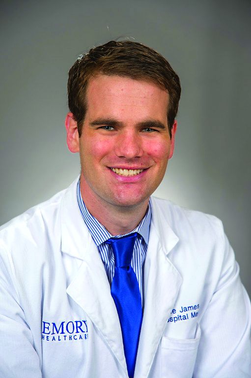
Dr. James is a hospitalist at Emory University Hospital Midtown and an assistant professor at Emory University, both in Atlanta
Geroscience brings bench science to the real-world problems of aging
NEW ORLEANS – Patients ask their doctors whether dietary manipulation can extend lifespan and promote healthy aging. Right now, basic scientists and clinicians from many disciplines are teaming up under the broad umbrella of the field of geroscience to try to answer these and other concerns relevant to an aging population.
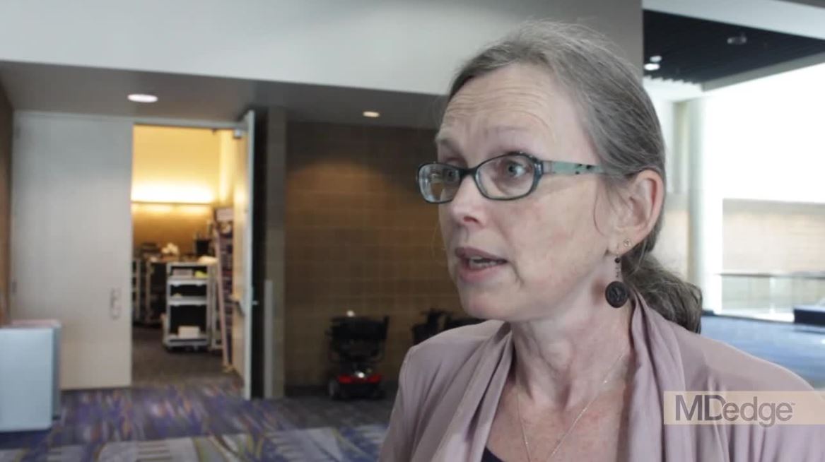
“The idea here is that, instead of going after each disease one at a time, as we do ... [we] instead go after disease vulnerability – and this is something that is shared, as a function of age,” Rozalyn Anderson, PhD, said of this new discipline. The work touches on disparate diseases such as cancer, dementia, and diabetes, she pointed out during a video interview at the annual meeting of the Endocrine Society.
“I separate these things out into ‘front-end’ and ‘back-end,’ work,” said Dr. Anderson of the University of Wisconsin-Madison’s aging and caloric restriction program. She explained that the caloric restriction she researches is back-end work to support the rapidly evolving field of nutritional modulation of aging.
When the basic science builds the framework, physicians and scientists can turn to front-end research, looking at humans to see which dietary manipulations are effective – and which are achievable.
“Take a paradigm that works, and then try to understand how it works,” said Dr. Anderson. “So [for example], we have this paradigm, and it’s tremendously effective in rodents. It’s effective in flies, in worms, in yeast, in spiders, in dogs – and in nonhuman primates.” Then, she and her team try to pull out clues “about the biology of aging itself, and what creates disease vulnerability as a function of age,” she said.
“The most important thing of all is that we can modify aging. This is not a foregone conclusion – no one would have believed it. But even in a primate species, we can change how they age. And the way in which we change is through nutrition.”
Dr Anderson added that “the paradigm of caloric restriction is tremendously effective, but [in reality], people are not going to do it.” It’s simply not practical to ask individuals to restrict calories by 30% or more over a lifespan, so “.”
Dr. Anderson reported no relevant conflicts of interest or disclosures.
REPORTING FROM ENDO 2019
Using humor in clinical practice
A patient in Seattle reported drinking alcohol on only two occasions during the year: when it rains – and when it doesn’t.
Various benefits of humor have been studied as part of the treatment modality. Humor can be a powerful resource, but it remains a complex process, and its proper use in clinical practice requires careful consideration. Despite having demonstrated the ability to relieve stress in patients and among medical professionals,1 humor has not gained widespread acceptance.
Humor has been shown to help build relationships, and establish trust and support for favorable health outcomes. It increases patients’ satisfaction, decreases medical malpractice claims, and has the potential to reduce cultural differences and hierarchy between patients and health care practitioners.2 The Accreditation Council of Graduate Medical Education values interpersonal and communication skills as being among the core competencies to be imparted to physicians in training.
Currently, there is no standard methodology for using humor in practice, as each clinical setting and circumstance can vary widely. Whichever setting you find yourself in, however, you might 1-3
Explore the benefits of humor in your clinical practice
Consider humor an integral part of your professionalism. Initiate it where you have assessed it is appropriate.
Understand your audience
Assess your patients’ capability of understanding or appreciating your humor. Do not force it on patients. Be respectful of their perspectives and mindful of cultural differences.
Reciprocate humor
If patients take the humor route to lighten what might be a tense encounter, respond to their attempt and join them in bringing levity into the mix.
Use humor to support patients
Humor can take many forms. It can be subtle and does not always require a punchline. Patients may use it to express concerns or even fear. Health care providers can use it as support and to demonstrate caring, reflecting anxieties likely displayed or revealed by patients.
Avoid certain forms of humor
Avoid using self-disparaging or gallows humor. Humor between health care providers and patients should never be sarcastic, ethnic, or sexist.
Pay attention to how your patients use humor
Explore the possible meanings of your patients’ attempts at humor and what concerns they might be seeking to express. Use your findings to discuss deeper issues.
Incorporate humor into your teaching
Students, too, can benefit from the therapeutic potential of humor. Use humor to dispel or lessen your students’ fears or anxieties. It can help in the learning process and memory. Creating a cheery ambience can help lessen nervousness, ease coping, and reduce burnout.
References
1. South Med J. 2003 Dec;96(12):1257-61.
2. J Am Board Fam Med. 2018 Mar-Apr;31(2):270-8.
3. Health Expect. 2014 Jun;17(3):332-44.
Dr. Lamba is a psychiatrist and medical director at Bayridge Hospital in Lynn, Mass. Dr. Rana is assistant professor of pediatrics at Boston University.
Stress incontinence surgery improves sexual dysfunction
TUCSON, ARIZ. –
The finding comes from a secondary analysis of two randomized, controlled trials comparing Burch colposuspension, autologous fascial slings, retropubic midurethral polypropylene slings, and transobturator midurethral polypropylene slings. The analysis looked at outcomes at 24 months after surgery. Stephanie Glass Clark, MD, a resident at Virginia Commonwealth University, Richmond, presented the results at the annual scientific meeting of the Society of Gynecologic Surgeons.
In the secondary analysis of the Stress Incontinence Surgical Treatment Efficacy Trial (SISTEr) and the Trial of Midurethral Slings (TOMUS) trials, Dr. Clark and her fellow researchers looked at the effect of surgical failure on sexual dysfunction outcomes. Subjective failure was defined as self-reported SUI symptoms or self-reported leakage by 3-day voiding diary beyond 3 months after the surgery. Objective failure was defined as any treatment for SUI after the surgery or a positive stress test or pad test beyond 3 months after the surgery.
Participants were excluded from the two studies if they were sexually inactive in the previous 6 months at baseline, at 12 months post baseline, or at 24 months. The studies employed the short form of the Pelvic Organ Prolapse/Urinary Incontinence Sexual Questionnaire (PISQ-12), which had 12 questions with scores ranging from 0 to 4. The secondary analysis sample included 488 women from SISTEr and 436 women from TOMUS.
There were some baseline differences among groups between the two trials, including vaginal deliveries, race/ethnicity, stage of prolapse, and concomitant surgeries performed at time of the anti-incontinence procedure.
All four surgeries were associated with improvements in sexual function, with no statistically significant between-group differences. Mean PISQ-12 scores improved from a range of 31-33 to a range of 36-38 at 24 months. Although there is no published minimum important difference for PISQ-12 scores, an improvement of at least one-half of a standard deviation is generally accepted as clinically meaningful. “In this case, the standard deviation at baseline was just under 3 and so the improvement of each treatment group by more than 1.5 is a clinically meaningful improvement in their sexual function,” Dr. Clark said.
“Sexual dysfunction is a much more common problem than we previously thought, so we’ve been trying to figure out if patients with pelvic floor disorders like stress incontinence are going to have any improvement in sexual dysfunction by surgically treating their stress incontinence. Previously published data had been pretty conflicting,” Dr. Clark added in an interview.
That previous research was mostly retrospective and could have been impacted by patient selection bias. By analyzing clinical trials, the researchers hoped to test their idea that the pelvic floor symptoms themselves may be key to sexual dysfunction and that treating it surgically would improve matters.
The positive result is encouraging, but it still leaves unanswered questions about the mechanism behind the relationship. Dr. Clark wondered whether leaking urine leakage during sex might be the culprit, or whether it is fear or shame associated with the condition.
The answer may come from further analysis of women who were sexually inactive at baseline, but became sexually active over the course of the studies. “I think looking at that patient population in particular is going to be an interesting area of research. Is it that it was completely related to their pelvic floor disorder, and then we fixed it [so] they could have a more fulfilling sexual life?” speculated Dr. Clark.
The study received some funding from the National Institutes of Health. Dr. Clark reported no relevant financial disclosures.
SOURCE: Clark SG et al. SGS 2019, Oral Presentation 11.
REPORTING FROM SGS 2019
