User login
Part 2: Whose Bill Is It, Anyway?
In an attempt to understand the data presented by Bai et al regarding independent billing by NPs and PAs in the emergency department (ED), I reached out to several colleagues to get their take on the study.1 Four of them are ED providers (2 MDs, 1 NP, 1 PA), and another is an experienced data analyst. In short, the analysis was analyzed!
Each member of my “expert panel” had similar comments and concerns, particularly regarding billing versus providing care. These are two different animals, or—as I often say—Bai and colleagues were comparing “oranges and shoes.” Several colleagues questioned the purpose of the article: What were the authors really trying to say?
Both MDs noted the absence of comments related to any consultation between the NP/PA and the ED physician during the patient encounter. They also pointed out the sheer volume of patients in EDs, resulting in the increasing popularity and utilization of NP/PAs to provide timely care to patients in need.
More than one of us initially wondered whether there could be potential “overbilling.” With the implementation of electronic health records (EHRs), the average charge per patient has increased. Maybe the EHR, now so common in EDs, more accurately captures the amount of time the provider spends caring for the patient and allows for more detailed documentation of the visit. This might result in a prima facie higher level of billing without necessarily representing the acuity of the presenting complaint.
One fact not presented in the Bai article is that, in many instances, providers (whether MDs, NPs, or PAs) do not complete the bills submitted to the insurance companies. In many EDs, medical coders extract the patient encounter data from the medical record. The reality is, the acuity of the patient is being decided by the person who is coding the visit—not the provider. And thus, it is potentially flawed thinking to rely on billing data alone to assess an increase in the acuity of patients seen by NPs and PAs in the ED.
Since we know (through data!) that there are more NPs and PAs providing care in EDs across the country, it follows logically that there will be more bills submitted in our names. This leads me to wonder: What was the motivation for Bai and colleagues to perform this study? What point are they actually trying to make?
Stay tuned ... There’s more to say next week! (In the meantime, you can share your thoughts by writing to me at [email protected].)
1. Bai G, Kelen GD, Frick KD, Anderson GF. Nurse practitioners and physician assistants in emergency medical services who billed independently, 2012-2016. Am J Emerg Med. https://doi.org/10.1016/j.ajem.2019.01.052. Accessed April 1, 2019.
In an attempt to understand the data presented by Bai et al regarding independent billing by NPs and PAs in the emergency department (ED), I reached out to several colleagues to get their take on the study.1 Four of them are ED providers (2 MDs, 1 NP, 1 PA), and another is an experienced data analyst. In short, the analysis was analyzed!
Each member of my “expert panel” had similar comments and concerns, particularly regarding billing versus providing care. These are two different animals, or—as I often say—Bai and colleagues were comparing “oranges and shoes.” Several colleagues questioned the purpose of the article: What were the authors really trying to say?
Both MDs noted the absence of comments related to any consultation between the NP/PA and the ED physician during the patient encounter. They also pointed out the sheer volume of patients in EDs, resulting in the increasing popularity and utilization of NP/PAs to provide timely care to patients in need.
More than one of us initially wondered whether there could be potential “overbilling.” With the implementation of electronic health records (EHRs), the average charge per patient has increased. Maybe the EHR, now so common in EDs, more accurately captures the amount of time the provider spends caring for the patient and allows for more detailed documentation of the visit. This might result in a prima facie higher level of billing without necessarily representing the acuity of the presenting complaint.
One fact not presented in the Bai article is that, in many instances, providers (whether MDs, NPs, or PAs) do not complete the bills submitted to the insurance companies. In many EDs, medical coders extract the patient encounter data from the medical record. The reality is, the acuity of the patient is being decided by the person who is coding the visit—not the provider. And thus, it is potentially flawed thinking to rely on billing data alone to assess an increase in the acuity of patients seen by NPs and PAs in the ED.
Since we know (through data!) that there are more NPs and PAs providing care in EDs across the country, it follows logically that there will be more bills submitted in our names. This leads me to wonder: What was the motivation for Bai and colleagues to perform this study? What point are they actually trying to make?
Stay tuned ... There’s more to say next week! (In the meantime, you can share your thoughts by writing to me at [email protected].)
1. Bai G, Kelen GD, Frick KD, Anderson GF. Nurse practitioners and physician assistants in emergency medical services who billed independently, 2012-2016. Am J Emerg Med. https://doi.org/10.1016/j.ajem.2019.01.052. Accessed April 1, 2019.
1. Bai G, Kelen GD, Frick KD, Anderson GF. Nurse practitioners and physician assistants in emergency medical services who billed independently, 2012-2016. Am J Emerg Med. https://doi.org/10.1016/j.ajem.2019.01.052. Accessed April 1, 2019.

Intermittent, but prolonged, calorie restriction may improve metabolic markers
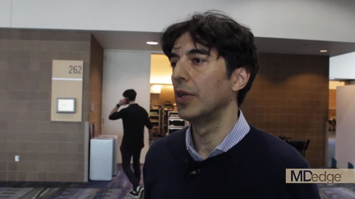
NEW ORLEANS – Can a physician-supervised, intermittent fasting strategy improve metabolic risk? Yes, according to Valter Longo, PhD.
Dr. Longo is a proponent of 5 days of reduced calories, performed once monthly or even less frequently for at-risk individuals. He calls this the “fasting-mimicking diet.”
“If somebody is obese or overweight, and has high cholesterol, high fasting glucose, and is perhaps prediabetic, then a doctor may decide to do the diet once a month for 5 days, and for the rest of the month, the person can go back to whatever it is that they do,” he said in a video interview at the annual meeting of the Endocrine Society.
“We think we are going to see more and more of this approach in the future,” said Dr. Longo, the Edna M. Jones Professor of Gerontology at the University of Southern California, Los Angeles.
Dr. Longo sees two chief practical benefits from the diet. First, patients “don’t feel they are being pushed to revolutionize their lives” because they aren’t asked to make radical lifestyle changes that have to be adhered to on a daily basis, and second, “we are starting to see that the patient slowly moves in the direction of a better diet without being asked to do it.”
which includes some healthy fats from olive oil and nuts. Fasting blood glucose, blood pressure, and insulinlike growth factor 1 levels and other metabolic markers were all reduced in the randomized crossover trial after 3 months of the diet plan.
Dr. Longo noted that in the clinical trial, effects were more pronounced for individuals with a higher risk for disease.
Dr. Longo has a majority stake in L-Nutra, which markets a commercially available fasting-mimicking diet package. He donates his proceeds to a nonprofit corporation he founded.
REPORTING FROM ENDO 2019
HM19: Pediatric medical and surgical co-management
Anticipatory and prevention-heavy approach
Presenter
Erin Shaughnessy, MD, MSHCM
Session title
Reaching Across the Aisle: Pediatric Co-Management with Surgery and Subspecialists

Session summary
Dr. Shaughnessy articulated a balanced approach to the importance of careful selection of patients needing to be co-managed by pediatric hospitalists. She compared two personal and very different experiences.
She initially managed a well-developed surgical co-management service at a quaternary, academic, free-standing children’s hospital, in which surgeons and subspecialists also admitted and managed patients to their own services. Currently, Dr. Shaughnessy is a division chief at Phoenix Children’s Hospital, a free-standing children’s hospital with a community hospital background, in which hospitalists admit most, if not all the patients, while subspecialty services have been transitioning only recently to having their own admitting services and employing the ideas of limited co-management.
She reminded the HM19 audience of the essential principles of co-management: shared responsibility, authority and accountability for the care of a hospitalized patient, discussing the scenarios, both from literature and real life, in which the line could become blurry at times.
Many pediatric programs are moving away from a traditional consultation model, Dr. Shaughnessy said, in which a consult is called for a new or a persistent problem with a patient, and where a consulting team signs off upon the resolved issue.
The more modern co-management model infuses a need for anticipatory and prevention-heavy approach, intertwined with fiscally responsible ideas that must be palatable for all: administration, hospitalists, and patients.
Dr. Shaughnessy reviewed a number of articles from both adult and pediatric literature with varied results, some that have shown decreased length of stay, decreased number of medical complications, decreased readmissions, decreased number of tests, but some that have also shown an increase in median hospital costs, emphasizing perhaps the importance of context in which one practices.
Finally, she identified patient selection, collaborative relationships, clear roles delineation, and excellence in communication as four main factors deciding the faith of a co-management model.
Key takeaways for HM
1. Careful selection of patients to be co-managed is essential and can prevent potential increase in costs and negative outcomes.
2. Success in medical and surgical co-management relies on well-delineated roles, collaborative culture, and immaculate communication.
Dr. Giordano is a pediatric neurosurgery hospitalist and assistant professor in pediatrics at Columbia University Irving Medical Center in New York.
Anticipatory and prevention-heavy approach
Anticipatory and prevention-heavy approach
Presenter
Erin Shaughnessy, MD, MSHCM
Session title
Reaching Across the Aisle: Pediatric Co-Management with Surgery and Subspecialists
Session summary
Dr. Shaughnessy articulated a balanced approach to the importance of careful selection of patients needing to be co-managed by pediatric hospitalists. She compared two personal and very different experiences.
She initially managed a well-developed surgical co-management service at a quaternary, academic, free-standing children’s hospital, in which surgeons and subspecialists also admitted and managed patients to their own services. Currently, Dr. Shaughnessy is a division chief at Phoenix Children’s Hospital, a free-standing children’s hospital with a community hospital background, in which hospitalists admit most, if not all the patients, while subspecialty services have been transitioning only recently to having their own admitting services and employing the ideas of limited co-management.
She reminded the HM19 audience of the essential principles of co-management: shared responsibility, authority and accountability for the care of a hospitalized patient, discussing the scenarios, both from literature and real life, in which the line could become blurry at times.
Many pediatric programs are moving away from a traditional consultation model, Dr. Shaughnessy said, in which a consult is called for a new or a persistent problem with a patient, and where a consulting team signs off upon the resolved issue.
The more modern co-management model infuses a need for anticipatory and prevention-heavy approach, intertwined with fiscally responsible ideas that must be palatable for all: administration, hospitalists, and patients.
Dr. Shaughnessy reviewed a number of articles from both adult and pediatric literature with varied results, some that have shown decreased length of stay, decreased number of medical complications, decreased readmissions, decreased number of tests, but some that have also shown an increase in median hospital costs, emphasizing perhaps the importance of context in which one practices.
Finally, she identified patient selection, collaborative relationships, clear roles delineation, and excellence in communication as four main factors deciding the faith of a co-management model.
Key takeaways for HM
1. Careful selection of patients to be co-managed is essential and can prevent potential increase in costs and negative outcomes.
2. Success in medical and surgical co-management relies on well-delineated roles, collaborative culture, and immaculate communication.
Dr. Giordano is a pediatric neurosurgery hospitalist and assistant professor in pediatrics at Columbia University Irving Medical Center in New York.
Romosozumab gets FDA approval for treating osteoporosis

“These are women who have a history of osteoporotic fracture or multiple risk factors or have failed other treatments for osteoporosis,” according to a news release from the agency.
The monthly treatment of two injections (given one after the other at one visit) mainly works by increasing new bone formation, but these effects wane after 12 doses. If patients still need osteoporosis therapy after that maximum of 12 doses, it’s recommended they are put on treatments that reduce bone breakdown. Romosozumab-aqqg is “a monoclonal antibody that blocks the effects of the protein sclerostin,” according to the news release.
The treatment’s efficacy and safety was evaluated in two clinical trials of more than 11,000 women with postmenopausal osteoporosis. In one trial, women received 12 months of either romosozumab-aqqg or placebo. The treatment arm had a 73% lower risk of vertebral fracture than did the placebo arm, and this benefit was maintained over a second year when both groups were switched to denosumab, another osteoporosis therapy. In the second trial, one group received romosozumab-aqqg for 1 year and then a year of alendronate, and the other group received 2 years of alendronate, another osteoporosis therapy, according to the news release. In this trial, the romosozumab-aqqg arm had 50% less risk of vertebral fractures than did the alendronate-only arm, as well as reduced risk of nonvertebral fractures.
Romosozumab-aqqg was associated with higher risks of cardiovascular death, heart attack, and stroke in the alendronate trial, so the treatment comes with a boxed warning regarding those risks and recommends that the drug not be used in patients who have had a heart attack or stroke within the previous year, according to the news release. Common side effects include joint pain and headache, as well as injection-site reactions.
Expert gives tips on timing, managing lupus pregnancies
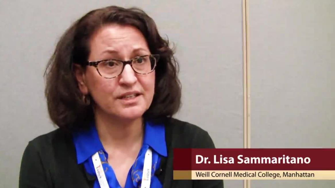
SAN FRANCISCO – Not that many years ago, women with systemic lupus erythematosus were told not to get pregnant. It was just one more lupus heartbreak.
Times have changed, according to Lisa Sammaritano, MD, a lupus specialist and associate professor of clinical medicine at Weill Cornell Medical College, New York.
While lupus certainly complicates pregnancy, it by no means rules it out these days. With careful management, the dream of motherhood can become a reality for many women. Dr. Sammaritano shared her insights about timing and treatment at an international congress on systemic lupus erythematosus.
It’s important that the disease is under control as much as possible; that means that timing – and contraception – are key. Antiphospholipid antibodies, common in lupus, complicate matters, but there are workarounds, she said.
AT LUPUS 2019
Oxytocin dampens the brain’s food-related reward circuitry
NEW ORLEANS – Oxytocin shows promise as a weight-loss medication, with encouraging results in animal models and small human studies. Now, in a calorie-rich environment.
“It is clear by now that obesity is a very serious health concern,” Liya Kerem, MD, said in a video interview at the annual meeting of the Endocrine Society. “The most adopted strategy, which is lifestyle modification, does not help [with losing or maintaining] weight in many cases, so we really need to find new treatments for obesity.”
Functional magnetic resonance imaging (fMRI) is a good tool for investigating the neurobiologic basis of overeating, said Dr. Kerem, a pediatric endocrinology fellow at Massachusetts General Hospital, Boston. In previous studies, fMRI has shown that “individuals with obesity have hyperactivation of the reward circuitry in the brain.”
Oxytocin is produced in the hypothalamus and is active in many brain areas associated with reward processing, said Dr. Kerem. Animal studies have shown a decrease in food intake and weight gain with oxytocin administration.
The hormone, which is generally seen as very safe, has had limited study in humans as a weight-loss strategy. Findings from one small study have shown that in men, a single intranasal dose of 24 IU of oxytocin resulted in less hunger-driven eating as well as lower consumption of a postmeal palatable snack, with the latter representing hedonic eating, said Dr. Kerem. A second small pilot study showed that significant weight loss occurred in obese humans after 8 weeks of daily oxytocin administration.
Findings from another study showed that participants who were overweight or obese, unlike their normal-weight counterparts, had reduced activation in the ventral tegmental area (VTA) after oxytocin administration. The VTA is an important region in the brain’s reward network, explained Dr. Kerem.
She and her colleagues used fMRI to probe dynamic changes in brain reward circuitry under the effect of oxytocin. They wanted to understand how oxytocin would “change the dialog between the VTA and the key brain areas involved in processing visual food stimuli.”
The hypothesis was that oxytocin would reduce functional connectivity between the VTA and other brain areas that are important for food reward and sensory processing when the participants were exposed to pictures of high-calorie food.
To test that hypothesis, the researchers showed the participants 100 each of four different kinds of images: high-calorie foods, low-calorie foods, nonfood images, and “fixation” images, used for calibration. The 10 participants had a mean body mass index of 29 kg/m2, and the mean age was 31 years.
Oxytocin did indeed attenuate functional connectivity between the VTA and several brain regions that are “key food motivation areas,” said Dr. Kerem. In particular, connections between the VTA and the insula were reduced with oxytocin. The insula is the “gustatory hub of the brain, key to subjective perception of food stimuli,” she explained.
Other attenuated associations included the oral area of the somatosensory cortex; the operculum, which shows fMRI activation to taste; the temporal gyrus, which is important for sensory processing; and, importantly, both the amygdala and hippocampus, known to be important for stimulus-reward learning, said Dr. Kerem. “We found that oxytocin targets exactly that hyperactivation in an overweight and obese population.”
It “reduced the functional connectivity between the VTA, a key hedonic brain region that drives efforts to obtain desired foods, and multiple brain areas involved in the cognitive, sensory, and emotional processing of food cues in men with overweight and obesity,” she said at a press conference highlighting the research. She emphasized that the effect was seen only with exposure to high-calorie food images. “Targeting hyperactivation of reward areas with oxytocin may inhibit overeating behavior,” she added.
Dr. Kerem and her colleagues are currently enrolling men and women for a larger clinical trial of oxytocin for weight loss.
Dr. Kerem reported no conflicts of interest. One of the study’s coauthors is a consultant for OXT Therapeutics, which is investigating obesity-related uses for oxytocin.
REPORTING FROM ENDO 2019
Older women have good functional recovery after POP surgery
TUCSON, ARIZ. – according to a new study.

Functional status was defined as the ability to perform activities essential to self-care, independent living, and recreation.
Patients with the highest level of function at baseline actually had a slight decrease in functionality scores, although this difference was not clinically meaningful.
“We can tell patients: You’re high functioning, your prolapse is bothering you in other ways, but we’re going to keep you as high functioning as you are now. I think that’s really important for older women who are retired and want to stay active. They want to make sure they don’t have a surgery that’s going to make them less so,” Daniel Lee, MD, of the University of Pennsylvania, Philadelphia, said in an interview. Dr. Lee presented the study at the annual scientific meeting of the Society of Gynecologic Surgeons.
The study used data from multiple centers across the United States and a range of patient ethnicities. “That helped strengthen the conclusions,” Dr. Lee said. Most previous studies used generalized questionnaires rather than functional outcome questionnaires to determine patient outcomes.
One confounder is the potential presence of cognitive dysfunction, which can occur sometimes in older women following surgery. The study relied on surveys and excluded patients who weren’t able to understand them. Two patients were much worse after the surgery, and Dr. Lee speculated that cognitive dysfunction could have been the cause, although the team could not confirm that. “There’s a possibility that preexisting dementia or cognitive dysfunction could be unmasked by the surgery,” he said. The team is working to incorporate accelerometers to more objectively measure outcomes and will soon publish a feasibility study of their use in older women with cognitive dysfunction.
One limitation of the study was that it excluded women who were considered poor candidates for surgery. But that also suggests that patient selection is working as intended. “I think it just shows that surgeons do a very good job of figuring out who is a good surgical candidate, that they turn out to be better off [functionally] or that their prolapse gets improved,” Dr. Lee said.
The researchers analyzed questionnaire data from 176 women. The mean age was 72 years, and mean body mass index was 27 kg/m2; 87% of the women were Caucasian and 10% were black. Using the Activities Assessment Scale (AAS), which covers sedentary, ambulatory, and work/exercise domains, as well as the Patient Health Questionnaire to measure depression, the researchers found that, by 3 months, 59% of patients had an improved functional status, 35% had returned to within one standard deviation of baseline, and 6% had worsened, compared with their baseline scores.
Patients in the improved group started at a mean baseline total AAS score of 85 and improved to 100 at 3 months. Those who returned to baseline started at 100 on average and returned to 100 at 3 months, while those in the worsened group had a mean baseline score of 100 and a mean score of 93 at 3 months (P less than .001 for all comparisons).
The study was funded by the Fellows Pelvic Research Network. The investigators reported no relevant financial disclosures.
SOURCE: Lee D et al. SGS 2019, Abstract 09.
TUCSON, ARIZ. – according to a new study.
Functional status was defined as the ability to perform activities essential to self-care, independent living, and recreation.
Patients with the highest level of function at baseline actually had a slight decrease in functionality scores, although this difference was not clinically meaningful.
“We can tell patients: You’re high functioning, your prolapse is bothering you in other ways, but we’re going to keep you as high functioning as you are now. I think that’s really important for older women who are retired and want to stay active. They want to make sure they don’t have a surgery that’s going to make them less so,” Daniel Lee, MD, of the University of Pennsylvania, Philadelphia, said in an interview. Dr. Lee presented the study at the annual scientific meeting of the Society of Gynecologic Surgeons.
The study used data from multiple centers across the United States and a range of patient ethnicities. “That helped strengthen the conclusions,” Dr. Lee said. Most previous studies used generalized questionnaires rather than functional outcome questionnaires to determine patient outcomes.
One confounder is the potential presence of cognitive dysfunction, which can occur sometimes in older women following surgery. The study relied on surveys and excluded patients who weren’t able to understand them. Two patients were much worse after the surgery, and Dr. Lee speculated that cognitive dysfunction could have been the cause, although the team could not confirm that. “There’s a possibility that preexisting dementia or cognitive dysfunction could be unmasked by the surgery,” he said. The team is working to incorporate accelerometers to more objectively measure outcomes and will soon publish a feasibility study of their use in older women with cognitive dysfunction.
One limitation of the study was that it excluded women who were considered poor candidates for surgery. But that also suggests that patient selection is working as intended. “I think it just shows that surgeons do a very good job of figuring out who is a good surgical candidate, that they turn out to be better off [functionally] or that their prolapse gets improved,” Dr. Lee said.
The researchers analyzed questionnaire data from 176 women. The mean age was 72 years, and mean body mass index was 27 kg/m2; 87% of the women were Caucasian and 10% were black. Using the Activities Assessment Scale (AAS), which covers sedentary, ambulatory, and work/exercise domains, as well as the Patient Health Questionnaire to measure depression, the researchers found that, by 3 months, 59% of patients had an improved functional status, 35% had returned to within one standard deviation of baseline, and 6% had worsened, compared with their baseline scores.
Patients in the improved group started at a mean baseline total AAS score of 85 and improved to 100 at 3 months. Those who returned to baseline started at 100 on average and returned to 100 at 3 months, while those in the worsened group had a mean baseline score of 100 and a mean score of 93 at 3 months (P less than .001 for all comparisons).
The study was funded by the Fellows Pelvic Research Network. The investigators reported no relevant financial disclosures.
SOURCE: Lee D et al. SGS 2019, Abstract 09.
REPORTING FROM SGS 2019
Ligelizumab maintains urticaria control for up to 1 year
WASHINGTON – in an open-label extension study, Diane Baker, MD, said at the annual meeting of the American Academy of Dermatology.
About 75% of the cohort experienced complete disease control at least once during the study. Novartis is developing ligelizumab (QGE031) as a treatment option for patients with spontaneous chronic urticaria (CSU) whose symptoms are inadequately controlled by H1-antihistamines. Like omalizumab (Xolair), which is approved in the United States and Europe for treatment of CSU, ligelizumab is a humanized anti-IgE monoclonal antibody. But the investigational agent binds to IgE with greater affinity than omalizumab, said Dr. Baker, a dermatologist who practices in Portland, Ore.
The extension study was a follow-up to a 12-week, phase 2, dose-finding trial of 382 CSU patients. In the study, which was not powered for efficacy endpoints, 51% of those who received 72 mg subcutaneously every 4 weeks had a Hives Severity Score of 0 by week 12, compared with 42% of those who received 240 mg every 4 weeks and 26% of those taking the omalizumab comparator. Additionally, 47% of those in the 72-mg group and 46% of the 240-mg group achieved a score of 0 on another indicator, the Urticaria Activity Score, which measures symptoms over 7 days (UAS7).
The extension study, which evaluated the 240-mg dose, showed the durability of that response, with 52% of those in the 240-mg group maintained a UAS7 of 0 at 1 year, according to Dr. Baker. By the end of the year, most patients (75.8%) had experienced at least one period of complete symptom control, and 84.0% experienced a UAS of 6 or lower at least once.
Adverse events were common in the cohort, with 84% experiencing at least one. But most (78%) were mild or moderate, and there was no clear side effect pattern, Dr. Baker said. Eight patients discontinued treatment because of an adverse event, and another eight dropped out because of lack of efficacy. Other reasons for discontinuation were pregnancy, protocol deviation, and physician or patient decision.
Novartis has launched two 1-year, phase 3 trials randomizing patients to 72 mg or 240 mg of ligelizumab or 300 mg of omalizumab every 4 weeks in a similar patient population, Dr. Baker said. PEARL 1 and PEARL 2, the largest pivotal trials to date in CSU, will enroll more than 2,000 patients, according to a company press release.
Dr. Baker is a clinical trials investigator for Novartis.
SOURCE: Baker D et al. AAD 2019, Session S034.
REPORTING FROM AAD 2019
SB-525 gene therapy looks viable for hemophilia A
Interim data suggest SB-525, an investigational gene therapy, may be safe and effective for patients with severe hemophilia A.
In the phase 1/2 Alta trial, SB-525 produced dose-dependent increases in factor VIII activity, with two of eight patients achieving normal factor VIII levels.
In addition, SB-525 was considered well tolerated. One patient did experience treatment-related serious adverse events – hypotension and fever – but these resolved within 24 hours.
Sangamo Therapeutics and Pfizer recently released these data in a press release and conference call.
The data include eight patients with severe hemophilia A who received SB-525 at 9e11 vg/kg, 2e12 vg/kg, 1e13 vg/kg, and 3e13 vg/kg.
Patient 1 (9e11 vg/kg), Patient 2 (9e11 vg/kg), and Patient 3 (2e12 vg/kg) did not experience clinically relevant increases in factor VIII levels and still require prophylactic recombinant factor VIII therapy.
Patient 4 (2e12 vg/kg) and Patient 5 (1e13 vg/kg) discontinued factor VIII therapy but have experienced spontaneous bleeds. Patient 4 had three bleeds in 48 weeks of follow-up, and Patient 5 had two bleeds in 40 weeks of follow-up.
Patient 6 (1e13 vg/kg), Patient 7 (3e13 vg/kg), and Patient 8 (3e13 vg/kg) have stopped factor VIII therapy and remain free of spontaneous bleeds at 28 weeks, 12 weeks, and 6 weeks of follow-up, respectively.
Patients 7 and 8 have achieved normal factor VIII levels. At 6 weeks, their factor VIII levels reached 140% and 94% of normal, respectively, according to a one-stage clotting assay, and 93% and 65%, respectively, according to a chromogenic assay.
“It appears the patients achieve their peak factor level at week 5 to 7 and maintain that level,” Edward Conner, MD, chief medical officer of Sangamo, said during the conference call.
The adverse events observed in this trial include grade 1 tachycardia (n = 1), grade 1 fatigue (n = 1), grade 1 alanine aminotransferase increase (n = 3), grade 1 myalgia (n = 1), grade 2 pyrexia (n = 2), and grade 3 hypotension (n = 1).
“These interim results indicate that SB-525 has the potential to comprise a well-tolerated, reliable, and predictable treatment, features that we believe will be a hallmark of the future gene therapy treatment of hemophilia A,” Dr. Conner said during the call.
Sangamo and Pfizer are enrolling additional patients in this trial, with the goal of expanding the 3e13 vg/kg cohort by up to five patients. The companies plan to present longer-term follow-up data at an upcoming scientific meeting.
The Alta trial is sponsored by Sangamo Therapeutics in collaboration with Pfizer.
My patient will die
“You are the best doctor I ever had.” These were the words of my patient at our final session. I kissed Rosa on the cheek and embraced her as she left my office. And I said, “It is important to show the love.”
“This may be the last time I come to your office,” she said. When Rosa came through the door at the beginning of the session, I was taken aback. She had lost weight and was using a walker; her face was drawn and sallow. I knew she had a recent diagnosis of liver cancer and had been hospitalized. She told me: “I have 3 months to live.” The cancer was inoperable.
She sat in a chair close to me, and we reminisced about 20 years as doctor and patient. She also talked about the stents in her liver; when they blocked, the pain resulted in a revisit to the emergency department. She had help from home health aides for several hours a day. Rosa’s sister arrived from Puerto Rico to be here “for as long as it takes.”
When she started therapy, Rosa was a single mother who lived in the projects with her adolescent son, Wesley. Her husband had died of AIDS. Unemployed and depressed, she told me that an uncle had sexually abused her when she was a child. Over the years, she looked to me for support: When her son, Wesley, got shot in the leg on a basketball court; when Wesley married a woman who shunned her; when her nephew who stayed with her got arrested for selling drugs and she lost her apartment as a result. Rosa remained in New York, displaced and struggling to find a reasonable home. After Wesley married, he moved with his family to rural Pennsylvania.
Whenever Rosa called to set up a therapy session, we talked about her problems. I prescribed medication for her, and I directed her to proper medical care. Often, I encouraged her to improve her diet, lose weight, and exercise – but to no avail. Her health deteriorated. She had cardiac surgery, heart failure, diabetes, hypertension, and chronic obesity. All these illnesses became her concern. She attended clinics at the hospital.
Now she told me that she would miss her son and would not see her two young grandchildren grow up. Rosa took Wesley to a funeral home to select a coffin and a headstone. It was tough for both of them, but she wanted to spare Wesley the trouble of doing it alone. She reflected, “It was like hitting a concrete wall” when she discovered her terminal diagnosis.
Rosa is facing pain, saying goodbye, and death. During her meeting, her ordeal made me cry, but I tried to contain it. I have been her doctor for so long, not a member of her family, not a friend. Yet I love her.
“Just to be is a blessing. Just to live is holy.”
– Abraham Joshua Heschel
Dr. Cohen is in private practice and is a clinical assistant professor of psychiatry at Weill Cornell Medical Center of New York-Presbyterian Hospital, and psychiatric consultant at the Hospital for Special Surgery, also in New York. She made changes to the patient’s story to protect confidentiality.