User login
Sociodemographic factors impact OS in MM

Photo courtesy of NCI
New research has revealed sociodemographic factors that appear to influence survival in younger patients with multiple myeloma (MM).
The study, which included more than 10,000 MM patients under the age of 65, suggested that marital status, insurance status, and county-level income all affect a patient’s chance of survival.
However, race/ethnicity and county-level educational achievement were not associated with overall survival (OS).
Luciano Costa, MD, PhD, of the University of Alabama at Birmingham, and his colleagues reported these findings in Cancer.
The researchers analyzed data on patients who were diagnosed with MM before the age of 65, between 2007 and 2012. There were 10,161 cases of MM, and the median follow-up was 27 months (range, 0-71 months).
The sociodemographic variables assessed were marital status, insurance status, median household income in the county of residence, and county-level educational achievement.
The researchers also looked at race/ethnicity, which was defined as a self-reported construct including Hispanic, non-Hispanic black, non-Hispanic white, and “other.”
The researchers found that patients had an increased risk of death if they were not married, did not have private insurance (were uninsured or on Medicaid), or lived in a low-income area (belonging to the lowest 2 income quartiles).
The 4-year estimated OS rate was 71.1% for patients who did not have any of these adverse sociodemographic factors, but it was 63.2% for patients with 1 factor, 53.4% for patients with 2 factors, and 46.5% for patients with all 3 factors (P<0.001).
The researchers did find that Hispanic and non-Hispanic black individuals had more adverse sociodemographic factors and worse OS than non-Hispanic whites and people belonging to the “other” category.
However, when the population was stratified by the cumulative number of sociodemographic factors and the researchers adjusted for confounders, there was no consistent association between race/ethnicity and OS.
The researchers said this suggests the apparent impact of race/ethnicity on OS may be due to the disparate distribution of sociodemographic factors observed among the different races/ethnicities rather than the race/ethnicity construct itself.
“This [study] strongly suggests that there is a huge disparity in outcomes that could potentially be overcome by improving access and affordability of treatments,” Dr Costa said.
“With the recent emphasis on comparative effectiveness in oncology, it also becomes crucial that all variables affecting outcomes—including sociodemographic factors—are accounted for when comparisons between different therapeutic approaches and healthcare systems are made.” ![]()
Drug granted orphan designation for MAS

Image from Flickr
The US Food and Drug Administration (FDA) has granted orphan drug designation for dusquetide as a treatment for macrophage activation syndrome (MAS).
Dusquetide is an innate defense regulator, a new class of short, synthetic peptides that accelerate bacterial clearance and resolution of tissue damage while modulating inflammation following exposure to bacterial pathogens, radiation, chemotherapy, and other agents.
According to researchers, dusquetide has demonstrated preclinical efficacy and safety in several animal models.
In a mouse model of MAS, dusquetide was shown to reduce pancytopenia, inhibit IL-12 responses, and improve body weight maintenance.
SGX942, the drug product containing dusquetide, has demonstrated safety in a phase 1 study of 84 healthy volunteers.
In addition, SGX942 has demonstrated preliminary efficacy and safety in an exploratory phase 2 study of 111 patients with oral mucositis due to chemoradiation therapy for head and neck cancer.
SGX942 is being developed by Solgenix, Inc.
About orphan designation
The FDA grants orphan designation to drugs and biologics intended to treat, diagnose, or prevent diseases/disorders that affect fewer than 200,000 people in the US.
The designation provides incentives for sponsors to develop products for rare diseases. This may include tax credits toward the cost of clinical trials, prescription drug user fee waivers, and 7 years of market exclusivity if the drug is approved.
About MAS
MAS is a life-threatening complication of rheumatic disease that, for unknown reasons, frequently occurs in individuals with systemic juvenile idiopathic arthritis. MAS also occurs in patients with systemic lupus erythematosus, Kawasaki disease, adult-onset Still’s disease, and various vasculitic syndromes.
MAS is characterized by pancytopenia, liver insufficiency, coagulopathy, and neurologic symptoms.
MAS is thought to be caused by the activation and uncontrolled proliferation of T lymphocytes and well-differentiated macrophages, leading to widespread hemophagocytosis and cytokine overproduction. ![]()
Seizure First Aid


ACS Submits Comment Letter to CMS on MIPS and APMs
The American College of Surgeons (ACS) submitted a comment letter June 27 on the Centers for Medicare & Medicaid Services (CMS) proposed rule on implementation of certain provisions of MACRA. Specifically, the CMS set forth proposals on the two pathways by which MACRA replaces the sustainable growth rate (SGR) formula: the Merit-Based Incentive Payment System (MIPS) and Alternative Payment Models (APMs). Among other issues, the ACS comments address the four components of MIPS: 1. quality, 2. resource use, 3. advancing care information, and 4. clinical practice improvement activities. The comments also address the criteria necessary for an APM model to be considered an “Advanced” APM, specifically: 1. use of certified electronic health records technology, 2. inclusion of quality measures comparable to MIPS, and 3. taking on more than nominal financial risk. Participation in an Advanced APM would exclude the clinician from participation in MIPS. For more information on the proposed rule or the comment letter, contact [email protected].
The American College of Surgeons (ACS) submitted a comment letter June 27 on the Centers for Medicare & Medicaid Services (CMS) proposed rule on implementation of certain provisions of MACRA. Specifically, the CMS set forth proposals on the two pathways by which MACRA replaces the sustainable growth rate (SGR) formula: the Merit-Based Incentive Payment System (MIPS) and Alternative Payment Models (APMs). Among other issues, the ACS comments address the four components of MIPS: 1. quality, 2. resource use, 3. advancing care information, and 4. clinical practice improvement activities. The comments also address the criteria necessary for an APM model to be considered an “Advanced” APM, specifically: 1. use of certified electronic health records technology, 2. inclusion of quality measures comparable to MIPS, and 3. taking on more than nominal financial risk. Participation in an Advanced APM would exclude the clinician from participation in MIPS. For more information on the proposed rule or the comment letter, contact [email protected].
The American College of Surgeons (ACS) submitted a comment letter June 27 on the Centers for Medicare & Medicaid Services (CMS) proposed rule on implementation of certain provisions of MACRA. Specifically, the CMS set forth proposals on the two pathways by which MACRA replaces the sustainable growth rate (SGR) formula: the Merit-Based Incentive Payment System (MIPS) and Alternative Payment Models (APMs). Among other issues, the ACS comments address the four components of MIPS: 1. quality, 2. resource use, 3. advancing care information, and 4. clinical practice improvement activities. The comments also address the criteria necessary for an APM model to be considered an “Advanced” APM, specifically: 1. use of certified electronic health records technology, 2. inclusion of quality measures comparable to MIPS, and 3. taking on more than nominal financial risk. Participation in an Advanced APM would exclude the clinician from participation in MIPS. For more information on the proposed rule or the comment letter, contact [email protected].
ACS Comments on Inpatient Prospective Payment System Proposed Rule
The American College of Surgeons (ACS) regulatory staff submitted a comment letter to the Centers for Medicare & Medicaid Services (CMS) on June 16 regarding the 2017 Inpatient Prospective Payment System (IPPS) proposed rule. Learn more about the proposed rule here.. The notice and comment period, which began with the proposed rule’s release April 27, enables individuals and organizations to provide input on the changes the CMS plans to make. The CMS takes these comments into consideration as it crafts the final rule.
The IPPS outlines coverage criteria for Medicare Part A inpatient hospital claims. Because a large portion of surgical care is provided in the inpatient setting, the rule both directly and indirectly affects surgeons. The ACS comment letter included feedback on the CMS’s proposed changes to the Hospital Value-Based Purchasing Program and the Hospital Acquired Conditions Reduction Program, both pay-for-performance programs, and the Hospital Inpatient Quality Reporting Program, a pay-for-reporting program. For more information about the IPPS proposed rule, see the CMS fact sheet.
Take Advantage of Early-Bird Registration for Clinical Congress 2016
The scientific program, online registration, travel and housing reservation links, and previews of other planned events for the American College of Surgeons (ACS) Clinical Congress 2016, October 16-20 in Washington, DC, are now available online. The Walter E. Washington Convention Center will be the host site for the scientific sessions, courses, and scientific posters and technical exhibits; the Marriott Marquis Washington, DC, will serve as the headquarters hotel for the meeting.
The theme for Clinical Congress 2016 is Challenges for the Second Century, as the ACS pursues its next 100 years of improving quality of care for surgical patients. Clinical Congress provides outstanding education and training opportunities for ACS Fellows, Associate Fellows, Resident Members, Medical Student Members, and other surgical team members. Attendees will find leading-edge surgical research presentations, a new lineup of Didactic/Experiential Postgraduate Courses, Surgical Skills Postgraduate Courses, timely discussions of relevant surgical topics, member engagement events, and unparalleled access to peers. You can earn up to 47.5 AMA PRA Category 1 Credits™ while attending premier educational sessions and courses.
Register by August 22 to take advantage of early-bird pricing. Various hotel options are available, so make your housing reservations and travel plans now to receive reduced Clinical Congress rates.
Advance care planning discussions: Talk is no longer cheap
Clinicians outside of the surgical specialties may consider surgeons primarily providers of technical services, but those of us who provide surgical care fully appreciate that communicating with patients and families is a large component of routine surgical practice. Typical communications in surgical practice include obtaining a history of present illness, which is a key element in the ultimate decision to offer a surgical intervention, or not; discussing the risks, benefits, and alternatives of any operation being considered; and the numerous discussions held following any surgical procedure. What many surgeons may not fully appreciate, however, is how these routine communication events can fall under the general category of advance care planning (ACP).
ACP is defined as a process in which physicians (and other health care providers) discuss a patient’s goals, values, and beliefs and determine how these inform a patient’s desire for current or future medical care. Hickman et al. (Hastings Center Report Special Report 35, no. 6 (2005):S26-S30) note that ACP should focus on defining “good” care for each patient. Furthermore, changes in a patient’s medical condition represent an opportune time to revisit a patient’s hopes and goals. Consideration of surgical intervention often represents a major change in a patient’s medical condition and therefore is an excellent opportunity to engage a patient in an ACP discussion.

Given that ACP discussions are likely occurring in surgical practices on a regular basis, surgeons need to be aware of a recent change in the Physician Fee Schedule that took effect Jan. 1, 2016. Effective this date, Current Procedural Terminology (CPT) codes 99497 and 99498 now allow for billing for ACP services. CPT code 99497 includes ACP “including the explanation and discussion of advance directives such as standard forms (with the completion of such forms, when performed), by the physician or other qualified health care professional; first 30 minutes, face-to-face with patient, family member(s) and/or surrogate.” CPT code 99498 is used for each additional 30 minutes spent in such face-to-face ACP counseling.
The nuts and bolts of how these ACP CPT codes work:
How many times can these code(s) be used? There are no limits on the number of times ACP can be reported for a given beneficiary in a given time period. For example, if an ACP discussion was held with a patient and/or family member and/or surrogate prior to a major elective procedure and again in the postoperative period, the above CPT codes could be used twice. In each instance, the ACP discussion must be documented, along with any relevant change in the patient’s clinical status that prompted another ACP discussion.
Can a patient or their family member/surrogate refuse ACP services? ACP services are voluntary; therefore, a patient or their family member/surrogate can refuse ACP services. These CPT codes only can be used if a patient or family member/surrogate consents for ACP services.
What must be documented in ACP services? Physicians should consult their Medicare Administrative Contractors for documentation requirements. Examples of elements to be included in the documentation are a brief description of the discussion with the patient or family/surrogate regarding the voluntary nature of ACP services, an explanation of advance directives and documentation if an advance directive is completed, who was present during the discussion, and time spent in the face-to-face encounter.
Does an advance directive have to be completed to bill the service? No. If an advance directive is completed, this should be documented (see above), but completion of the directive is not a requirement for billing the service.
Can ACP be reported in addition to an evaluation and management (E/M) service (such as an office visit)? Yes. CPT codes 99497 and 99498 may be billed on the same day or a different day as most other E/M services. They may be billed within the global surgical period.
Is a specific diagnosis required to use the ACP CPT codes? No, a specific diagnosis is not required for the ACP codes to be billed.
According to the 2016 Medicare Physician Fee Schedule, the reimbursement is $85.99 for CPT 99497 and $74.88 for CPT 99498. For comparison, the reimbursement for E/M CPT 99203 (30-minute initial evaluation) = $108.85, CPT 99204 (45-minute initial evaluation) = $166.13, and CPT 99205 (60-minute initial evaluation) = $208.38. Far more important than the financial remuneration for these discussions, however, is the critical need for surgeons to have and document their ACP discussions with their patients and/or their family member/surrogate. As surgeons, we are often called to see patients when they are facing a significant change in their health – whether that is a new diagnosis of cancer or after a traumatic injury. Understanding a patient’s values, hopes, and concerns is an essential component to ensuring that our patients receive the best care, as defined by them.
Dr. Fahy is associate professor of surgery and internal medicine at the University of New Mexico, Albuquerque. She is a surgical oncologist who is also board certified in hospice and palliative medicine. Dr. Fahy does not have any relevant conflicts of interest to disclose.
Clinicians outside of the surgical specialties may consider surgeons primarily providers of technical services, but those of us who provide surgical care fully appreciate that communicating with patients and families is a large component of routine surgical practice. Typical communications in surgical practice include obtaining a history of present illness, which is a key element in the ultimate decision to offer a surgical intervention, or not; discussing the risks, benefits, and alternatives of any operation being considered; and the numerous discussions held following any surgical procedure. What many surgeons may not fully appreciate, however, is how these routine communication events can fall under the general category of advance care planning (ACP).
ACP is defined as a process in which physicians (and other health care providers) discuss a patient’s goals, values, and beliefs and determine how these inform a patient’s desire for current or future medical care. Hickman et al. (Hastings Center Report Special Report 35, no. 6 (2005):S26-S30) note that ACP should focus on defining “good” care for each patient. Furthermore, changes in a patient’s medical condition represent an opportune time to revisit a patient’s hopes and goals. Consideration of surgical intervention often represents a major change in a patient’s medical condition and therefore is an excellent opportunity to engage a patient in an ACP discussion.
Given that ACP discussions are likely occurring in surgical practices on a regular basis, surgeons need to be aware of a recent change in the Physician Fee Schedule that took effect Jan. 1, 2016. Effective this date, Current Procedural Terminology (CPT) codes 99497 and 99498 now allow for billing for ACP services. CPT code 99497 includes ACP “including the explanation and discussion of advance directives such as standard forms (with the completion of such forms, when performed), by the physician or other qualified health care professional; first 30 minutes, face-to-face with patient, family member(s) and/or surrogate.” CPT code 99498 is used for each additional 30 minutes spent in such face-to-face ACP counseling.
The nuts and bolts of how these ACP CPT codes work:
How many times can these code(s) be used? There are no limits on the number of times ACP can be reported for a given beneficiary in a given time period. For example, if an ACP discussion was held with a patient and/or family member and/or surrogate prior to a major elective procedure and again in the postoperative period, the above CPT codes could be used twice. In each instance, the ACP discussion must be documented, along with any relevant change in the patient’s clinical status that prompted another ACP discussion.
Can a patient or their family member/surrogate refuse ACP services? ACP services are voluntary; therefore, a patient or their family member/surrogate can refuse ACP services. These CPT codes only can be used if a patient or family member/surrogate consents for ACP services.
What must be documented in ACP services? Physicians should consult their Medicare Administrative Contractors for documentation requirements. Examples of elements to be included in the documentation are a brief description of the discussion with the patient or family/surrogate regarding the voluntary nature of ACP services, an explanation of advance directives and documentation if an advance directive is completed, who was present during the discussion, and time spent in the face-to-face encounter.
Does an advance directive have to be completed to bill the service? No. If an advance directive is completed, this should be documented (see above), but completion of the directive is not a requirement for billing the service.
Can ACP be reported in addition to an evaluation and management (E/M) service (such as an office visit)? Yes. CPT codes 99497 and 99498 may be billed on the same day or a different day as most other E/M services. They may be billed within the global surgical period.
Is a specific diagnosis required to use the ACP CPT codes? No, a specific diagnosis is not required for the ACP codes to be billed.
According to the 2016 Medicare Physician Fee Schedule, the reimbursement is $85.99 for CPT 99497 and $74.88 for CPT 99498. For comparison, the reimbursement for E/M CPT 99203 (30-minute initial evaluation) = $108.85, CPT 99204 (45-minute initial evaluation) = $166.13, and CPT 99205 (60-minute initial evaluation) = $208.38. Far more important than the financial remuneration for these discussions, however, is the critical need for surgeons to have and document their ACP discussions with their patients and/or their family member/surrogate. As surgeons, we are often called to see patients when they are facing a significant change in their health – whether that is a new diagnosis of cancer or after a traumatic injury. Understanding a patient’s values, hopes, and concerns is an essential component to ensuring that our patients receive the best care, as defined by them.
Dr. Fahy is associate professor of surgery and internal medicine at the University of New Mexico, Albuquerque. She is a surgical oncologist who is also board certified in hospice and palliative medicine. Dr. Fahy does not have any relevant conflicts of interest to disclose.
Introducing Dr. Tyler G. Hughes
I feel honored to join Tyler G. Hughes as co-Editor of the ACS Surgery News and I am excited to work with its Managing Editor, Therese Borden, to bring to its readers breaking information on a broad range of subjects of interest and importance to practicing surgeons. Although we have a great challenge to fill the giant shoes of our immediate predecessor, Layton “Bing” Rikkers, we will do our best to address the vexing clinical, economic, social, and administrative challenges that continue to confront us no matter what our practice type and setting.
I could not ask for a more accomplished and versatile co-Editor than Tyler Hughes. As a general surgeon practicing in the truly rural setting of McPherson, Kan., since 1995, he became an articulate spokesman for rural surgeons across the country during his tenure on the ACS Board of Governors as Kansas’ at-large member. As the crisis in access to general surgical care for rural Americans became increasingly evident, Tyler was asked to speak to the Board of Regents in February 2012, and the first new Advisory Council in 50 years was formed: the Advisory Council for Rural Surgery (ACRS), of which Tyler was named the first Chair. In 4 short years, the ACRS has become a force to promote better communication among rural surgeons and to call attention to the needs of them and their patients.
Tyler’s communication skills have also been put to great use in his role as Editor of the ACS Web Portal and as the inaugural Editor-in-Chief of the ACS Communities, an activity that has met with incredible success in promoting communication among the far-flung individual surgeons who constitute the ACS membership. Along the way, he has also served as an Associate Editor of “Selected Readings in General Surgery” and a member of the steering committee of Evidence Based Reviews in Surgery.
He currently serves as a Director of the American Board of Surgery (ABS). He is therefore familiar with all of the issues of surgical training, certification, and re-certification. He is similarly well versed in the complexities surrounding the implementation of Maintenance of Certification, which remains a “work in progress” that his experience as a practicing, small-town general surgeon will certainly inform.
Tyler has distinguished himself in other leadership positions throughout his more than 30 years as a surgeon, including as President of his 600-member physician group when he initially practiced in Dallas. He has been a Fellow in the ACS for his entire surgical career and holds a deep respect, affection, and loyalty to the College. He possesses mainstream values, true to his upbringing and his long residence in America’s heartland; yet, he understands and respects the divergent views of surgeons across our country. He is also not afraid to tackle challenging problems, which is why I know that our tenure as co-Editors of ACS Surgery News is not likely to become boring.

Introducing Dr. Karen Deveney
As Layton “Bing” Rikkers leaves his post as Editor of ACS Surgery News, it has fallen to Karen Deveney and me to shepherd the paper forward as co-Editors. Dr. Rikkers felt that a combination approach of an academic surgeon and a community surgeon would bring balance to ACS Surgery News that would be representative of the nature of the American College of Surgeons (ACS).
In Karen Deveney we have an accomplished academic surgeon who has wide ranging interests in and out of surgery. Karen was raised in rural Oregon, went to Stanford for undergraduate education, and did her medical school and residency at University of California, San Francisco. Among her cohort in those times of training and her early academic career were Donald Trunkey, George Sheldon, and Brent Eastman, all of whom, like Karen, went on to have a major impact in the world of surgery.
After a stint in the military serving in Germany with her surgeon husband Cliff, Karen eventually landed at Oregon Health and Science University where she went on to serve as Program Director for 20 years at one of the best general surgery training programs in the country. She served as Second Vice-President of the ACS and is the immediate past-President of the Pacific Coast Surgical Association.
Her CV reflects varied academic interests and activities. So, Karen’s contributions to academic surgery are outstanding. But in Karen we also get a person who is alive to the needs of the population beyond the walls of her major medical center. Karen has been a leader in the march to save surgical access for rural populations. She is a founding member of the ACS Advisory Council for Rural Surgery, serving as the Education Pillar Chair of that Council. In her own institution, Karen is a pioneer in developing a model rural surgery track for general surgery residents – first in Grants Pass, Ore. and then in Coos Bay, Ore.
She has been a hardworking general and colorectal surgeon for over 30 years. And, like almost all dedicated surgical educators, she has taken call – enduring the long call schedule of her residents throughout her career.
Karen and I hope to make a good team in this new effort. We are different in many ways, but very much the same in others. We plan a synergy that will unflinchingly recognize the challenges in surgery and facilitate positive discussion and reporting of the solutions for those challenges. Among those challenges are the changing economic structure of surgery, the facilitation of useful quality efforts, and most importantly, the rapid dissemination of significant clinical and scientific information vital to surgeons everywhere.
Dr. Hughes is an ACS Fellow with the department of general surgery, McPherson Hospital, McPherson, Kan., and is the Editor in Chief of ACS Communities. He is also Associate Editor for ACS Surgery News.

Pulmonary Perspectives® New Technology Enhances Electromagnetic Navigation Bronchoscopy
Following the National Lung Screening Trial (NLST), which showed at-risk patients screened with CT scans had reduced lung cancer-specific mortality, many institutions have incorporated lung cancer screening protocols into clinical practice (Aberle et al. N Engl J Med. 2011;365[5]:395). These protocols, along with new generation, high resolution multidetector CT scans, have increased the number of detected peripheral lung nodules, many smaller in size. It is estimated that over 150,000 solitary nodules are diagnosed each year in the United States (Herth et al. Expert Rev Respir Med. 2016; 0[8]:901) and, in keeping with the NLST, greater than 25% of subjects screened have lung nodules suspicious for lung cancer. As a result, many leading health practices have created specific lung nodule programs to handle the volume in an effort to deliver timely care in the evaluation of lung cancer.
Pulmonary specialists managing patients with lung nodules are faced with the difficult challenge of deciding if a patient with a nodule is a candidate for serial surveillance, tissue biopsy (transthoracic needle aspiration [TTNA] vs. bronchoscopic biopsy [TBX]), or surgical resection. Calculation of the probability of a nodule being malignant is most helpful in making these decisions for patients with low and high malignancy risk factors, as surveillance and resection are appropriate steps, respectively. However, for those with an intermediate (5%-65%) probability of having a malignant nodule, the diagnostic procedure risks, yields, and timing have to be considered because delayed sampling or false-negative results may negatively impact survival. Kanashiki et al (Oncol Rep. 2003;10[3]:649) showed that worse survival is associated with patients with imaging-to-diagnosis times of greater than 4 months. Over the past decade, image-guided bronchoscopy has been used to improve the yield for tissue sampling of smaller peripheral nodules in a timely fashion. The most common method of image-guided bronchoscopy today is electromagnetic navigation bronchoscopy (ENB).

Electromagnetic navigation bronchoscopy has shown promise for increasing diagnostic yields for peripheral nodules (PN) over conventional bronchoscopy. Over time, the improved yields have plateaued as ENB use in clinical practice increased and limitations of the early generation technology became apparent. Earlier ENB technology uses a single inspiratory CT scan of the chest to reconstruct a 3D virtual model of the airways and parenchyma. A tracked sensor is then used to navigate through the imaging reconstructed airways toward the targeted lesion, the sensor is then removed, and through a dedicated catheter instruments are used to obtain samples from the lesion. In a meta-analysis using this technology, lesions greater than 2 cm had a diagnostic yield ranging from 66.7% to 94.7%. However, as the PN size decreased to less than or equal to 2 cm, the diagnostic yield range dropped significantly with some yields reported as low as 18.2% (van ‘t Westeinde et al. Chest. 2012;142(2):377). More recently, Ost and colleagues performed a multicenter study of consecutive patients undergoing bronchoscopic sampling of PN (Ost et al. Am J Respir Crit Care Med. 2016;193[1]:68). Although it was not a randomized trial and each bronchoscopist influenced the selection of the sampling technique, the authors reported that the diagnostic yields for navigation-guided bronchoscopy were lower than conventional bronchoscopy, 38.5% and 63.7%, respectively. Taken on face value alone, one might conclude that ENB not be used to biopsy PNs. However, deeper analysis of the data showed that 97% of the ENB procedures were performed using the earlier technology described above, suggesting that the single inspiratory imaging CT scan and navigation procedure technique, which differs significantly from conventional bronchoscopy, may have some influence on the lower than expected yields.
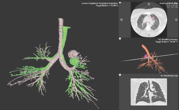
Despite increasing use and experience with ENB, diagnostic yields remain static. Chen and colleagues hypothesized that using a single inspiratory CT scan may not allow the endoscopist to make adjustments for PN movement as the lung moves during the respiratory cycle. Using different imaging protocol, the investigators assessed movement of 85 lung nodules during the respiratory cycle with paired-full inspiration and tidal-volume expiration, thin sliced (0.5-1.0 mm) CT scans. They found that the average motion of all lesions during respiration was 17.6 mm, 12.2 mm in the right-upper lobe, 10.6 mm in the left-upper lobe, and 25.3 mm and 23.8 mm in the right- and left- lower lobes, respectively (Chen et al. Chest. 2015;147[5];1275) (Fig. 1). They concluded that the location of targeted lesions on a single inspiration planning CT scan alone does not accurately represent the position of the lesion during bronchoscopy.
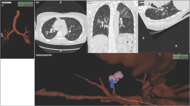
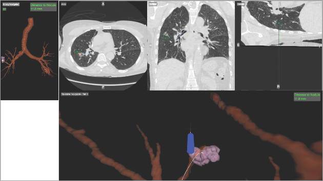
Although being able to correct for nodule movement throughout the respiratory cycle during the procedure is a significant improvement, it doesn’t guarantee that the tissue sample is obtained from the targeted lesion. To accomplish that, the system would have to be able to determine when the instrument being used to sample is in the target. The earlier ENB systems allowed for navigation to the target with a separate sensor through a steerable catheter. However, when the target was reached, the sensor had to be removed so that sampling instruments could be introduced into the catheter. Since the instruments are not tracked and the movement of the nodule is occurring, there is no guarantee that the instrument is in the target at the time of sampling. Advanced technology now allows for the tracking sensor to be placed in the tip of standard bronchoscopy instruments, making them “tip-tracked” and able to be used with standard bronchoscopes and equipment; thus, making the new ENB procedure similar to conventional bronchoscopy that was shown to have higher diagnostic yields (Figs. 2 and 3).
Our institution incorporated this technology (Veran Medical, St. Louis) into our advanced diagnostic and interventional pulmonary program for lung nodules and published our initial experience and results. During the initial 8 months of screening for lung cancer, we performed procedures on 44 patients with PNs suspicious for lung cancer. The rate for successful target sampling was 90.2% with a cancer diagnosis rate of 39%, which is similar to that found in the NLST. Those patients who had nonmalignant but abnormal pathologic findings (inflammation, granuloma, fibrosis, and so on) were monitored for a minimum of 12 months. Most of the lesions either remained stable or disappeared on follow-up imaging (Flenaugh et al. The Internet Journal of Pulmonary Medicine. 2016;18[1]). We concluded that (1) the combination of paired inspiratory and expiratory CT scan imaging accounts for nodule movement and (2) using tip-tracked conventional instruments to enter into the lesion at the time of biopsy contributes to improved yields.
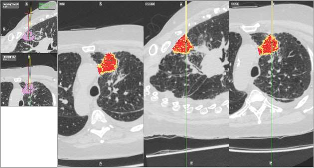
Newer ENB technology is not limited to transbronchial sampling. For PNs less than 2 cm and deep in the lung periphery, current recommendations prefer TTNA over bronchoscopic biopsy because of yield rates of 90% (Chest. 2007; 132(suppl 3):131S). Using the same paired CT scanning and tip-tracking method on transthoracic needles, the new systems allow pulmonologists to perform electromagnetic transthoracic needle aspiration (ETTNA) of PNs using the same basic equipment and during the same procedure visit (Fig. 4). This “one stop shopping” approach of bronchoscopy with the option of converting to ETTNA if the PN is not reachable endoscopically has proven to be cost efficient and allows for timely diagnosis and focused care (Yarmis et al. J Thorac Dis. 2016;8(1):186). In a prospective study designed specifically to assess feasibility, safety, and diagnostic yield of ETTNA in a single procedure, Yarmis and colleagues enrolled 24 patients to undergo endobronchial ultrasound for lung cancer staging followed by ENB and ETTNA. Ninety-six percent of the patients were candidates for ETTNA. The authors reported the yield for ETTNA was 83%, ETTNA plus ENB 87%, and ETTNA plus ENB plus endobronchial ultrasound for complete staging was 92%. Five pneumothoraces were reported; however, only two (8%) required a drainage intervention. This protocol is unique because it makes use of several advanced diagnostic procedures, including tip-tracked navigation technology, to localize, sample, diagnose, and stage during one patient procedure visit.
As lung cancer screening becomes commonplace in clinical practice and imaging technology improves, pulmonary specialists can expect to encounter and manage a greater number of pulmonary nodules. Advancements in technology now offer options for improving diagnostic accuracy while providing timely, safe, and cost effective care. While not all new technology will prove beneficial in disease management, those that improve the deficiencies of earlier technology offer us the best chance to improve practice. This perspective highlights such technology.
Dr. Flenaugh is Associate Professor, Director of Advanced Diagnostic & Interventional Pulmonary Service,Morehouse School of Medicine and Grady Hospital, Atlanta; Dr. Foreman is Professor of Medicine, Associate Chair for Research, Pulmonary & Critical Care Medicine, Morehouse School of Medicine.
Following the National Lung Screening Trial (NLST), which showed at-risk patients screened with CT scans had reduced lung cancer-specific mortality, many institutions have incorporated lung cancer screening protocols into clinical practice (Aberle et al. N Engl J Med. 2011;365[5]:395). These protocols, along with new generation, high resolution multidetector CT scans, have increased the number of detected peripheral lung nodules, many smaller in size. It is estimated that over 150,000 solitary nodules are diagnosed each year in the United States (Herth et al. Expert Rev Respir Med. 2016; 0[8]:901) and, in keeping with the NLST, greater than 25% of subjects screened have lung nodules suspicious for lung cancer. As a result, many leading health practices have created specific lung nodule programs to handle the volume in an effort to deliver timely care in the evaluation of lung cancer.
Pulmonary specialists managing patients with lung nodules are faced with the difficult challenge of deciding if a patient with a nodule is a candidate for serial surveillance, tissue biopsy (transthoracic needle aspiration [TTNA] vs. bronchoscopic biopsy [TBX]), or surgical resection. Calculation of the probability of a nodule being malignant is most helpful in making these decisions for patients with low and high malignancy risk factors, as surveillance and resection are appropriate steps, respectively. However, for those with an intermediate (5%-65%) probability of having a malignant nodule, the diagnostic procedure risks, yields, and timing have to be considered because delayed sampling or false-negative results may negatively impact survival. Kanashiki et al (Oncol Rep. 2003;10[3]:649) showed that worse survival is associated with patients with imaging-to-diagnosis times of greater than 4 months. Over the past decade, image-guided bronchoscopy has been used to improve the yield for tissue sampling of smaller peripheral nodules in a timely fashion. The most common method of image-guided bronchoscopy today is electromagnetic navigation bronchoscopy (ENB).
Electromagnetic navigation bronchoscopy has shown promise for increasing diagnostic yields for peripheral nodules (PN) over conventional bronchoscopy. Over time, the improved yields have plateaued as ENB use in clinical practice increased and limitations of the early generation technology became apparent. Earlier ENB technology uses a single inspiratory CT scan of the chest to reconstruct a 3D virtual model of the airways and parenchyma. A tracked sensor is then used to navigate through the imaging reconstructed airways toward the targeted lesion, the sensor is then removed, and through a dedicated catheter instruments are used to obtain samples from the lesion. In a meta-analysis using this technology, lesions greater than 2 cm had a diagnostic yield ranging from 66.7% to 94.7%. However, as the PN size decreased to less than or equal to 2 cm, the diagnostic yield range dropped significantly with some yields reported as low as 18.2% (van ‘t Westeinde et al. Chest. 2012;142(2):377). More recently, Ost and colleagues performed a multicenter study of consecutive patients undergoing bronchoscopic sampling of PN (Ost et al. Am J Respir Crit Care Med. 2016;193[1]:68). Although it was not a randomized trial and each bronchoscopist influenced the selection of the sampling technique, the authors reported that the diagnostic yields for navigation-guided bronchoscopy were lower than conventional bronchoscopy, 38.5% and 63.7%, respectively. Taken on face value alone, one might conclude that ENB not be used to biopsy PNs. However, deeper analysis of the data showed that 97% of the ENB procedures were performed using the earlier technology described above, suggesting that the single inspiratory imaging CT scan and navigation procedure technique, which differs significantly from conventional bronchoscopy, may have some influence on the lower than expected yields.
Despite increasing use and experience with ENB, diagnostic yields remain static. Chen and colleagues hypothesized that using a single inspiratory CT scan may not allow the endoscopist to make adjustments for PN movement as the lung moves during the respiratory cycle. Using different imaging protocol, the investigators assessed movement of 85 lung nodules during the respiratory cycle with paired-full inspiration and tidal-volume expiration, thin sliced (0.5-1.0 mm) CT scans. They found that the average motion of all lesions during respiration was 17.6 mm, 12.2 mm in the right-upper lobe, 10.6 mm in the left-upper lobe, and 25.3 mm and 23.8 mm in the right- and left- lower lobes, respectively (Chen et al. Chest. 2015;147[5];1275) (Fig. 1). They concluded that the location of targeted lesions on a single inspiration planning CT scan alone does not accurately represent the position of the lesion during bronchoscopy.
Although being able to correct for nodule movement throughout the respiratory cycle during the procedure is a significant improvement, it doesn’t guarantee that the tissue sample is obtained from the targeted lesion. To accomplish that, the system would have to be able to determine when the instrument being used to sample is in the target. The earlier ENB systems allowed for navigation to the target with a separate sensor through a steerable catheter. However, when the target was reached, the sensor had to be removed so that sampling instruments could be introduced into the catheter. Since the instruments are not tracked and the movement of the nodule is occurring, there is no guarantee that the instrument is in the target at the time of sampling. Advanced technology now allows for the tracking sensor to be placed in the tip of standard bronchoscopy instruments, making them “tip-tracked” and able to be used with standard bronchoscopes and equipment; thus, making the new ENB procedure similar to conventional bronchoscopy that was shown to have higher diagnostic yields (Figs. 2 and 3).
Our institution incorporated this technology (Veran Medical, St. Louis) into our advanced diagnostic and interventional pulmonary program for lung nodules and published our initial experience and results. During the initial 8 months of screening for lung cancer, we performed procedures on 44 patients with PNs suspicious for lung cancer. The rate for successful target sampling was 90.2% with a cancer diagnosis rate of 39%, which is similar to that found in the NLST. Those patients who had nonmalignant but abnormal pathologic findings (inflammation, granuloma, fibrosis, and so on) were monitored for a minimum of 12 months. Most of the lesions either remained stable or disappeared on follow-up imaging (Flenaugh et al. The Internet Journal of Pulmonary Medicine. 2016;18[1]). We concluded that (1) the combination of paired inspiratory and expiratory CT scan imaging accounts for nodule movement and (2) using tip-tracked conventional instruments to enter into the lesion at the time of biopsy contributes to improved yields.
Newer ENB technology is not limited to transbronchial sampling. For PNs less than 2 cm and deep in the lung periphery, current recommendations prefer TTNA over bronchoscopic biopsy because of yield rates of 90% (Chest. 2007; 132(suppl 3):131S). Using the same paired CT scanning and tip-tracking method on transthoracic needles, the new systems allow pulmonologists to perform electromagnetic transthoracic needle aspiration (ETTNA) of PNs using the same basic equipment and during the same procedure visit (Fig. 4). This “one stop shopping” approach of bronchoscopy with the option of converting to ETTNA if the PN is not reachable endoscopically has proven to be cost efficient and allows for timely diagnosis and focused care (Yarmis et al. J Thorac Dis. 2016;8(1):186). In a prospective study designed specifically to assess feasibility, safety, and diagnostic yield of ETTNA in a single procedure, Yarmis and colleagues enrolled 24 patients to undergo endobronchial ultrasound for lung cancer staging followed by ENB and ETTNA. Ninety-six percent of the patients were candidates for ETTNA. The authors reported the yield for ETTNA was 83%, ETTNA plus ENB 87%, and ETTNA plus ENB plus endobronchial ultrasound for complete staging was 92%. Five pneumothoraces were reported; however, only two (8%) required a drainage intervention. This protocol is unique because it makes use of several advanced diagnostic procedures, including tip-tracked navigation technology, to localize, sample, diagnose, and stage during one patient procedure visit.
As lung cancer screening becomes commonplace in clinical practice and imaging technology improves, pulmonary specialists can expect to encounter and manage a greater number of pulmonary nodules. Advancements in technology now offer options for improving diagnostic accuracy while providing timely, safe, and cost effective care. While not all new technology will prove beneficial in disease management, those that improve the deficiencies of earlier technology offer us the best chance to improve practice. This perspective highlights such technology.
Dr. Flenaugh is Associate Professor, Director of Advanced Diagnostic & Interventional Pulmonary Service,Morehouse School of Medicine and Grady Hospital, Atlanta; Dr. Foreman is Professor of Medicine, Associate Chair for Research, Pulmonary & Critical Care Medicine, Morehouse School of Medicine.