User login
Vascular Live
The Vascular Live stage is located in Exhibit Halls A-C, on Level 1. On the Vascular Live stage, exhibitors will present new ideas, showcase breakthrough technologies and discuss the latest trends in vascular surgery.
Thursday, June 9
12:15 – 1:15 p.m.
Sponsored by Bard Peripheral Vascular, Inc.
“Latest Outcomes from Lutonix® Global SFA Registry and Head to Head Comparison between IN.PACT and LUTONIX® DCB”
Speakers: Dr. Yvonne Bausback, Dr. Chad Laurich, and Dr. Renu Virmani
3:30 – 4 p.m.
Sponsored by Abbott
“Treatment Strategies for Complex Femoropopliteal Disease”
Speaker: Dr. Niten Singh
5:30 – 6 p.m.
Sponsored by Philips
“Physician Perspective on Outpatient Based Labs (OBLs)”
6 – 6:30 p.m.
Sponsored by GE Healthcare
“Integrated Workflow for Endograft Sizing, 3D Fusion, and Assessment, Using New EVAR ASSIST”
Speaker: Dr. Stephan Haulon
Friday, June 10
9:30 – 10 a.m.
Sponsored by Silk Road Medical
“TransCarotid Artery Revascularization (TCAR): The Way Forward in Treating Carotid Artery Disease and Stroke Prevention”
Moderator: Dr. Sumaira Macdonald
Speakers: Dr. Vikram S. Kashyap, Dr. Peter A. Schneider, and Dr. Raghu L. Motaganahalli
12:30 – 1 p.m.
Sponsored by Gore
“Five-year Patency Rates for GORE® PROPATEN® Vascular Grafts vs. ePTFE Grafts Without Heparin. See the Results”
Speaker: Dr. Russell H. Samson
1 – 1:30 p.m.
Sponsored by Gore
“Understanding How Device Design Can Impact Clinical Outcomes in Type B Dissection”
Speaker: Dr. Alan B. Lumsden
“Durable TEVAR – Results from 100,000 Implants”
Speaker: Dr. Mark A. Farber
3 – 3:30 p.m.
Sponsored by Hansen Medical
“Flexible Robotics with Electromagnetic Tracking and the Impacts on Safety and Efficiency”
Speaker: Dr. Alan B. Lumsden
Vascular Live presentations are not eligible for CME credit.
Homans Lecture: A Look Ahead in Vascular Surgery
Vascular surgery has a rich, varied and enormously interesting history, filled with innovators and creators whose inventions changed the course of patient care. This history has been well-documented innumerable times.
Dr. Frank J. Veith is a part of that storied history. However, during the John Homans Distinguished Lecture Thursday morning, he will look ahead, and not back.
During “A Look at the Future of Vascular Surgery,” he plans to discuss the implications of future changes in the field, not to mention the world of medical care, and “what vascular surgery has to do to survive,” he said.
In mid-April he was working on the VEITHsymposium he created more than 40 years ago and so had not yet finalized his thoughts on his lecture.
But at that time he believed he would revisit his long-ago Presidential Address, “Charles Darwin and Vascular Surgery,” delivered in June 1996 and which also discussed the future of vascular surgery.
He noted then that it was a “time of enormous upheaval in American medicine, and holding this office has also forced me to focus on the future of Vascular Surgery and how well it will survive these turbulent times.” As Darwin studied extinct and contemporary species, his thoughts on survival could be correlated to that of the subspecialty itself, Dr. Veith said.
The endovascular revolution had begun and he was convinced – and stated – that vascular surgeons would need to learn endovascular skills. “People thought I was crazy when I said that endovascular was going to replace most of what we did,” he said. “I turned out to be very right.”
Some of his other pronouncements that day turned out to be incorrect, he said. But because change is once again part of the field of medicine, he wants to address issues he sees and possible solutions to those issues.
“I want it to be interesting and provocative,” he said. “I don’t want the listeners to fall asleep.”
Dr. Veith is professor of surgery at New York University Medical Center and at the Cleveland Clinic Lerner College of Medicine of Case Western Reserve University. He holds the William J. von Liebig chair in Vascular Surgery at the Cleveland Clinic Foundation and has lectured throughout the world.
He is a pioneer in the area of limb-salvage surgery and endovascular grafting for traumatic, aneurysmal, and occlusive arterial disease. Each year he hosts the VEITHsymposium on what is new and important in the treatment of vascular disease.

Schedule-At-A-Glance
Wednesday, June 8
6:00 a.m. – 6:30 p.m.
Registration
7:00 – 10:00 a.m.
Postgraduate Courses
P1: Controversies and Management of Lower Extremity Arterial Occlusive Disease
P2: Complete Management of Venous Disease: From Superficial to Deep
8:00 – 11:30 a.m.
V1: VESS Paper Session 1
8:00 a.m. – 5:00 p.m.
VQI Annual Meeting
8:30 – 10:00 a.m.
Hands-on Workshops
W2: Tricks for Pedal, Tibial, Radial and Brachial Access
W3: Techniques for Challenging Embolization
W5: Atherectomy Techniques
10:15 a.m. – 1:15 p.m.
Postgraduate Courses
P3: SVS/APMA Joint Session: Management of the Diabetic Foot
P4: Hemodialysis: Challenges, Controversies and New Techniques
10:30 a.m. – 12:00 noon
Hands-on Workshops
W7: Technical Tips for Crossing CTO
W10: IVUS: An Essential Tool for the Endovascular Specialist
12:30 – 2:45 p.m.
FT: International Fast Talk
12:30 - 4:00 p.m.
V2: VESS Paper Session 2
1:30 – 3:00 p.m.
Hands-on Workshops
W14: IVC Filters: Techniques in Deployment and Recovery
W15: Non-Thermal Techniques for Venous Ablation
W16: Percutaneous Mechanical Thrombectomy: Challenges, Advances and New Techniques
W17: How to Make Carotid Stenting Competitive with CEA
W18: FEVAR Tips & Tricks: From Sizing to Implantation
2:00 – 5:00 p.m.
Postgraduate Courses
P5: Endovascular Strategies for Complex Aortic Scenarios
P6: Standing on the Shoulders of Giants: Open Operative Techniques by the Masters
2:00 – 6:00 p.m.
General Surgery Resident/Medical Student Scholarship Program – Open and Endovascular Simulation Training
3:00 – 6:00 p.m.
C1: International Forum (included among Concurrent Sessions)
3:30 – 5:00 p.m.
Hands-on Workshops
W19: DEB/DES Techniques & Applications
W22: Percutaneous Mechanical Thrombectomy: Challenges, Advances and New Techniques
W23: How to Make Carotid Stenting Competitive with CEA
W24: FEVAR Tips & Tricks: From Sizing to Implantation
4:00 – 5:00 p.m.
C2: SVS New Member Session (included among Concurrent Sessions)
5:00 – 5:30 p.m.
SVS New Member Reception
5:00 – 6:30 p.m.
Concurrent Sessions
C3: Tools to Navigate the Changing Vascular Landscape
C4: Connections and Conversations for Young to Mid-Level Vascular Surgeon Researchers
5:30 – 7:00 p.m.
SVS PAC Reception
6:15 – 7:15 p.m.
International Guest Reception
6:45 – 7:45 p.m.
General Surgery Resident/Medical Student
Program – Welcome Reception
Thursday, June 9
6:00 a.m. – 6:00 p.m.
Registration
6:30 – 8:00 a.m.
Satellite Symposia Breakfast Sessions
B1: Complex SFA Disease: Maximizing Outcomes While Minimizing Costs;* sponsored by Abbott
B2: EndoAnchors in Practice: Treatment Algorithms from the Experts;* sponsored by Medtronic
B3: New Techniques in Advanced Imaging and 3D Navigation for Complex Endovascular Procedures; sponsored by Philips;*
*not eligible for CME credit
6:30 – 8:00 a.m.
General Surgery Resident Program Breakfast
6:30 – 8:00 a.m.
Medical Student Program Breakfasts
MS1/MS2
MS3/MS4
8:00 – 8:30 a.m.
Opening Ceremony
8:30 – 10:00 a.m.
S1: William J. von Liebig Forum
10:00 – 10:30 a.m.
E1: John Homans Lecture
10:30 a.m. – 12:00 noon
F1: E. Stanley Crawford Critical Issues Forum
12:00 noon – 1:30 p.m.
Lunch and Vascular Live Presentations in Exhibit Hall
12:00 noon – 1:30 p.m.
Meet the Leaders Luncheon
General Surgery Resident/Medical Student Program – Diversity Medical Student Scholarship Luncheon
12:00 noon - 6:30 p.m.
Exhibition
1:30 – 2:50 p.m.
S2: Plenary Session 2
2:50 – 3:00 p.m.
SVS Awards Ceremony
3:00 – 3:30 p.m.
E2: Roy Greenberg Distinguished Lecture
3:30 – 4:00 p.m.
Coffee Break and Vascular Live Presentation
4:00 – 5:30 p.m.
S3: Plenary Session 3
5:30 – 6:30 p.m.
Opening Reception and Vascular Live Presentations
5:30 – 6:30 p.m.
IP: Interactive Poster Session
6:30 – 7:30 p.m.
Networking Reception for Women, Diversity and Young Surgeons7:30 p.m.
7:30 – 9:30 p.m.
Satellite Symposia
The Battle of the Bulge: Debating the Future of Aortic Aneurysm Repair;* sponsored by Endologix, Inc.
Friday, June 10
6:00 a.m. – 5:30 p.m.
Registration
6:30 – 8:00 a.m.
Breakfast Sessions
B4: Radiation Safety for the Vascular Surgeon
B5: Pediatric Vascular Surgery
B6: Treatment of Complex Iliocaval-Iliofemoral Deep Venous Thrombosis and Obstruction
6:30 – 8:00 a.m.
General Surgery Resident/Medical Student Program – Mock Interviews Practice Session
8:00 – 9:30 a.m.
S4: Plenary Session 4
9:30 – 10:00 a.m.
Coffee Break and Vascular Live Presentation
9:30 a.m. – 4:30 p.m.
Exhibition
10:00 – 11:00 a.m.
S5: Plenary Session 5
11:00 a.m. – 12:15 p.m.
Presidential Address: “We Care” (includes 15-min introduction by President-Elect)
12:15 – 1:30 p.m.
Vascular Surgery Trainee Luncheon
12:15 – 1:30 p.m.
VA Surgeons Meeting
12:15 – 1:30 p.m.
Lunch and Vascular Live Presentations in Exhibit Hall
1:30 – 3:00 p.m.
S6: Plenary Session 6
3:00 – 3:30 p.m.
Coffee Break and Vascular Live Presentation
3:30 – 5:00 p.m.
Concurrent Sessions
C5: SVS/ESVS Joint Debate Session
C6: SVS/STS Joint Session: Aortic Controversies
C7: SVS/SVM Joint Session: Medical Management of Vascular Disease
C8: Poster Competition
5:00 – 6:30 p.m.
General Surgery Resident/Medical Student Program – Residency Fair
6:30 – 8:30 p.m.
Satellite Symposia
Internal Iliac Artery Preservation Expert Panel;* sponsored by Gore
8:30 – 9:30 p.m.
Special Event: Capitol Steps Performance
Saturday, June 11
6:00 a.m. – 5:00 p.m.
Registration
6:30 – 8:00 a.m.
Breakfast Sessions
B7: Controversies in Thoracic Outlet Syndrome
B8: Hemodialysis Access: Issues and Challenges
B9: Quality Improvement and Reporting: A Survival Guide for Your Practice
8:00 – 10:00 a.m.
S7: Plenary Session 7/Late-Breaking
9:00 a.m. – 1:00 p.m.
Exhibition
9:30 – 10:00 a.m.
Coffee Break
10:30 a.m. – 12:00 noon
F2: Beyond the Journal of Vascular Surgery: “Top Ten” Papers Relevant to Vascular Surgery
10:30 a.m. – 12:00 noon
Concurrent Sessions
C9: Device Development: From Idea to IDE
C10: Essential Tools for Young Academic and Private Practice Surgeons
C11: Politics in Vascular Surgery: What Practicing Surgeons Need to Know about Power and Influence in Washington, DC
12:00 noon – 1:00 p.m.
Non-Member Lunch in Exhibit Hall
12:00 noon – 1:30 p.m.
SVS Member Business Luncheon
1:30 – 3:30 p.m.
VH: “How I Do It” Video Session
3:30 – 4:30 p.m.
F3: Poster Runoff: Championship Round
1:30 – 5:00 p.m.
Ultrasound Physics and Vascular Test Interpretation – Physician Vascular Interpretation Examination Review
*not eligible for CME credit
Concurrent Sessions Provide Younger Surgeons Information
From learning how to make research a career focus to learning what tools are needed to help adapt to the changing world of vascular surgery, the Vascular Annual Meeting has concurrent sessions designed to offer invaluable information for younger surgeons.
“C4: Connections and Conversations for Young to Mid-Level Vascular Surgeon Researchers” will be held from 5 to 6:30 p.m. Wednesday. “C10: Essential Tools for Young Academic and Private Practice Surgeons” will be held at the other end of VAM, from 10:30 a.m. to 12 p.m. Saturday.
Younger surgeons comprise a large percentage of the SVS membership as well as VAM attendees, said Dr. Grace Wang, chair of the Young Surgeons Committee. “We wanted sessions that are of interest to both private practice and academic surgeons.”
C4: Connections and Conversations
This session is cosponsored by the SVS Research Council, SVS Research and Education Committee, SVS Young Surgeons Committee, and the SVS Clinical and Comparative Effectiveness Research Committee.
“Research” now includes several disparate arenas, each with its own sets of challenges, said Dr. Wang. Accordingly, planners divided the session to include four main research areas. Experts in those fields will provide brief overviews and then facilitate small-group discussions with time allowed for both general and specific questions.
Speakers include Dr. Alan Dardik, “Basic Science and Translational Research;” Dr. Michael S. Conte, “Clinical Research” (including clinical trials); Dr. Philip P. Goodney, “Patient-Centered Research;” and Dr. Marc L. Schermerhorn, for “Database Research.” Dr. Wang will co-moderate the session with Dr. Misty D. Humphries.
Approximately 15 additional SVS member-researchers well-known for one or more of the particular topic areas will serve as small group facilitators. “I believe that this session is broadly applicable for all surgeons,” Dr. Wang said. Ensuring quality outcomes, for example, is a concern for providers at both community and academic hospitals Industry-sponsored trials also span community practice and academic hospitals. For physician-researchers who have not yet decided what specific kind of research appeals to them or what will be the main focus of their career, “this session can help refine their thinking, and allow them to connect with established mentors in that field” she added.
C10: Essential Tools
This session offers a combination of various broad topics that would be of use to both private practice and academic surgeons. Dr. Wang will co-moderate with Dr. Bradley G. Thomas, who is in private practice.
Dr. Russell Samson will advise participants about “Developing a niche and brand to build a loyal referral system,” Dr. Gilbert R. Upchurch will offer suggestions on “Developing a Robust Aortic Aneurysm Practice” and Dr. Julie A. Freischlag will discuss utilizing time management to make the most of each day.
Because a number of vascular procedures can be performed by cardiologists and/or interventional radiologists as well as vascular surgeons, Dr. Frank J. Veith will enlighten the audience on the important topic of ”Managing and Resolving Turf Battles.”
And given the changing health care system and economy, planners turned to Dr. Bhagwan Satiani, who also holds an MBA, to cover the “Top Ten Things to Understand about the Business Side of Medicine.”
An additional topic is returning to school for an advanced degree. Dr. Ruth L. Bush, one of the panelists, received not only her master’s in public health but also a law degree after getting her medical degree, and Dr. Louis Nguyen already had his MBA before he also earned his MPH. “Both obtained degrees after coming on staff, and we thought their viewpoints on advanced training would be valuable,” Dr. Wang said. “It suggests that training does not necessarily end with fellowship.”
Dr. Wang is enthused about these and other VAM sessions – plus other programming, grants, and opportunities for younger members, students, residents, and fellows that the SVS offers throughout the year.
“Our trainees and young surgeons are the future of vascular surgery, and it is exciting to see programming at the VAM that is geared toward them” she said.
The Vascular and Endovascular Surgery Society Welcomes You
I would like to invite all Vascular Annual Meeting attendees to join us for the Spring Meeting of the Vascular and Endovascular Surgery Society (VESS; formerly Peripheral Vascular Surgery Society). This year’s program will be held on Wednesday, June 8 at 8:00 a.m. and conclude at 4:00 p.m. Dr. Bernadette Aulivola and her Program Committee have selected over 25 outstanding research projects for presentation.
The format of the meeting allows great discussion between the presenters and attendees. As in previous years, the VESS and SVS Program Committees combined to select the best scientific papers for the Vascular Annual Meeting. I am sure you will be delighted with this year’s Vascular Annual Meeting.
Founded in 1976 as a club for young and recently graduated Vascular Surgeons, the society has grown to a national organization and international society boasting well over 1,000 members. Focusing on young vascular surgeons in both academic and community practice, our organization offers excellent educational opportunities and networking for career growth.
This year we are excited to introduce the VESS Women and Diversity Meet the Leaders Program, which matches young surgeons with a leader based on the needs of the young surgeon and the experience of the leader to create a mentoring relationship.
VESS has two important scientific meetings yearly. The Spring Meeting is held in conjunction with the Vascular Annual Meeting. The Annual Meeting is held in the winter and is a highlight for both new and old members.
These meetings are a special time to share research, contribute ideas, learn new technologies, and make lifelong friends. The next Annual Meeting will take place February 2-5, 2017 at the Steamboat Grand in Steamboat, Colo. The Annual Meeting will feature interesting interactive programs, engaging speakers, and exciting social events. With nearly 3,000 skiable acres, and featuring night skiing too, it will be an outstanding venue for both education and relaxation. I encourage you to take advantage of the unique setting the Annual Meeting offers. Don’t miss this great vascular meeting in a great setting.
Our members strive to promote basic and clinical research that directly improves outcomes for our vascular and endovascular surgery patients. If you are not a member, please consider joining. You won’t be disappointed. Our new website is www.vesurgery.org. I look forward to seeing you at one of our meetings and encourage you to get involved in this terrific Society!
Thomas Maldonado, MD
VESS President

Patients Who Don't Speak English are Likely to Return to the Emergency Room
(Reuters Health) - Patients in the emergency room who don't speak English well are slightly more likely to return within days, suggesting their care the first time was not as good as it could have been, researchers say.
In a study in one New York hospital, about 4 percent of English speakers made an unplanned return to the ER within three days, compared to 5 percent of people with limited English.
Low use of professional translators may partly explain the disparity in care, the researchers report in the Annals of Emergency Medicine.
"There's a necessary but not sufficient step to providing care for people with low English proficiency . . . having a good interpreter or healthcare provider who can speak to them in their language," said Dr. Elizabeth Jacobs of the University of Wisconsin-Madison, who was not part of the new study.
The study team, led by Dr. Ka Ming Ngai of the Icahn School of Medicine at Mount Sinai in New York, analyzed 2012 data from the Mount Sinai emergency department. More than 32,000 adult patients and 45,000 ER visits were included. The study did not include patients with psychiatric or substance-related
complaints, those who were nonverbal or had altered mental status, and those with a history of frequent ER visits.
Almost 3,000 patients had limited English proficiency, and in about half of cases someone served as an interpreter. Usually, this was a family member or an ER staff member. Only 527 visits in this group, 24 percent, involved a professional interpreter.
More than a quarter of patients were admitted to the hospital and 1,380 patients had an unplanned return to the ER within three days.
After accounting for age, sex, insurance, race, ethnicity, triage category and other health problems, having limited English proficiency was not tied to greater risk of being admitted to the hospital.
But those with limited English proficiency were about 24 percent more likely to return to the ER unexpectedly.
Ngai told Reuters Health by email that he has been studying the problem of language barriers for the past six years and over time has seen some improvements.
"New medical students are now routinely educated to use interpreter phones during their clinical simulation . . .however, there are still many barriers including access to interpreters and interpreter phones, time constraints, and (doctors) trying to 'get by' with their own language skills," he said.
Ngai said regulatory bodies require hospitals to make language services available. In New York State, for example, upon a request to the hospital administration by the patient, the patient's family or representative, or the provider of medical care, hospitals must provide translation services in inpatient and outpatient settings within 20 minutes and in emergency settings with 10 minutes.
Most New York Hospitals use an interpreter phone service, he said.
Patients who struggle to speak the local language are "a really important population to study and think about how we can improve their care," Jacobs said.
A 5 percent rather than 4 percent rate of return to the ER is not a large difference, but that could be due to the large number of patients excluded from the study, and because there was no validated measure of English proficiency, Jacobs said.
"That might be why we didn't see large differences, if some people considered low English proficiency actually spoke English well, or were getting good interpretive services," she said. "If you took them out, the difference might be larger."
Patients who do not speak English may struggle in other areas of the health system more than at the ER, she added.
But even having an interpreter at the hospital won't help patients deal with insurance providers, she noted.
"There are contextual issues that you may not be able to fully adequately address unless you can understand the nuances," Jacobs said.
"When dealing with immigrant population, it is almost always more than 'just' language," Ngai agreed. There can be cultural issues, too.
In addition, people with low English proficiency may also be less able to take days off of work, and to agree to be admitted to the hospital when necessary, than others, Jacobs said.
We've made tremendous progress in assuring interpreters are more available," in person, over the phone or by video, Jacobs said.
But, she said, "we are very imperfect at getting patients the services they need. It's important for providers to be educated on these issues and to understand how to access these services."
It would be ideal to try to match patients with providers by language and culture, but in the meantime, "language is a good start," Ngai said.
Test can monitor blood coagulation at home, team says
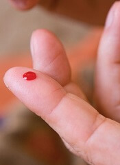
CINCINNATI—Researchers say they have developed a lateral flow assay device that patients can use at home to monitor their blood coagulation.
The device consists of nanofiber membranes inside a paper-based, porous test strip that is housed in a plastic cassette.
The researchers said this device can quickly reveal the level of the blood’s ability to clot using just a drop of blood from a finger prick.
“We have developed a blood-screening device for patients on medications like Coumadin (warfarin) or other blood thinners who need to monitor their blood-clotting levels on a regular basis,” said Andrew Steckl, PhD, of the University of Cincinnati in Ohio.
“Patients can soon monitor their blood coagulation characteristics from home quickly and painlessly before making needless trips to the lab or hospital.”
Hua Li, a student researcher in Dr Steckl’s lab, presented details on this device at the 8th International Conference on Porous Media and Annual Meeting of the International Society for Porous Media (abstract 1371).
Dr Steckl noted that slight changes in the level of coagulation properties will occur normally, but a major change in levels immediately shows up with his team’s device.
The researchers found the device was able to detect coagulation ability in rabbit blood and in blood from patients receiving warfarin.
“This simple test is not intended to replace the very careful and accurate measurements that get accomplished in a laboratory facility, but, at a relatively minimal cost, a patient can do this on their own between scheduled visits or when in doubt,” Dr Steckl said. “And it shouldn’t require a caregiver, as most patients can perform this test quickly on their own.”
Furthermore, the researchers said this technology can be calibrated to a specific patient’s condition. For example, a patient whose normal blood coagulation rate is significantly different from the general population because of a genetic disorder can use a tailor-made test kit that includes a different porous membrane.
The technology may also help patients who have a known inherited blood clotting disorder detect concerning levels early.
“By identifying potential blood-clotting problems early enough, we hope to prevent potential injury or death and the exorbitant associated costs,” Dr Steckl said. “By shifting from what were once mandatory expensive laboratory tests to using more in-home screening tests, patients can take more control of their lives, reduce healthcare costs, and, ultimately, save more lives.” ![]()
CDC issues interim guidance for Zika testing

The US Centers for Disease Control and Prevention (CDC) has released an interim guidance for testing urine for the Zika virus.
The agency noted that, in most patients, Zika virus RNA is unlikely to be detected in serum after the first week of illness.
However, recent reports have suggested that Zika virus RNA can be detected in urine for at least 2 weeks after the onset of symptoms.
Therefore, the CDC recommends that real-time reverse transcription–polymerase chain reaction (rRT-PCR) be performed on urine collected less than 14 days after the onset of symptoms in patients with suspected Zika virus.
The agency said further investigation is needed to determine the utility of rRT-PCR on urine specimens collected at 14 days or beyond.
The CDC also recommends that urine testing be performed in conjunction with serum testing for specimens collected less than 7 days after symptom onset. The agency said its recommendations for testing serum and other clinical specimens for Zika virus have not changed.
At present, the CDC’s Trioplex rRT-PCR assay is the only diagnostic tool authorized by the US Food and Drug Administration to test urine for Zika virus. ![]()
Health Canada approves ibrutinib for WM

Photo from Janssen Biotech
Health Canada has approved the BTK inhibitor ibrutinib (Imbruvica) as a treatment for patients with Waldenström’s macroglobulinemia (WM).
Ibrutinib was first approved in Canada in November 2014 for the treatment of patients with chronic lymphocytic leukemia (CLL), including those with 17p deletion, who have received at least one prior therapy, or for the frontline treatment of patients with CLL and 17p deletion.
In July 2015, ibrutinib was granted conditional approval for the treatment of patients with relapsed or refractory mantle cell lymphoma.
Health Canada’s approval of ibrutinib for WM was based on results of a multicenter, phase 2 study in which researchers tested the drug (given at 420 mg once daily) in 63 patients with previously treated WM.
The patients’ median age was 63 (range, 44-86), and their median number of prior therapies was 2 (range, 1-11).
Initial data showed an overall response rate of 87.3% in patients who received ibrutinib for a median of 11.7 months.
Updated results from the study were published in NEJM in April 2015. After a median treatment duration of 19.1 months, the overall response rate was 91%.
At 24 months, the estimated rate of progression-free survival was 69%, and the estimated rate of overall survival was 95%.
The most common grade 2-4 adverse events were neutropenia (22%) and thrombocytopenia (14%). Ibrutinib-related neutropenia and thrombocytopenia were reversible but required a dose reduction in 3 patients and treatment discontinuation in 4 patients.
Grade 2 or higher bleeding events occurred in 4 patients, and there were 15 infections considered possibly related to ibrutinib.
Treatment-related atrial fibrillation (AFib) occurred in 3 patients, all of whom had a prior history of paroxysmal AFib. AFib resolved when treatment was withheld, and all 3 patients were able to continue on therapy per protocol without an additional event.
Ibrutinib is co-developed by Cilag GmbH International (a member of the Janssen Pharmaceutical Companies) and Pharmacyclics LLC, an AbbVie company. Janssen Inc. markets ibrutinib as Imbruvica in Canada. ![]()
Drug may reduce severity of AEs from dexamethasone

Photo by Bill Branson
Adding a physiologic dose of hydrocortisone to treatment with dexamethasone can reduce the severity of certain adverse effects (AEs) in pediatric patients with acute lymphoblastic leukemia (ALL), according to researchers.
Hydrocortisone did not decrease the incidence of psychosocial problems or sleep-related issues in these patients, but the drug did reduce the severity of these problems among patients who experienced them.
Lidewij T. Warris, MD, of Erasmus MC-Sophia Children’s Hospital in Rotterdam, the Netherlands, and her colleagues reported these results in the Journal of Clinical Oncology.
The team conducted this study to determine whether a physiologic dose of hydrocortisone could reduce neuropsychologic and metabolic AEs in children with ALL who were receiving dexamethasone.
The study enrolled 50 patients (ages 3 to 16) who were set to receive 2 consecutive courses of dexamethasone in accordance with Dutch Childhood Oncology Group ALL protocols.
The patients were randomized to receive either hydrocortisone or placebo in a circadian rhythm (10 mg/m2/d) during their first dexamethasone course. During their second course, the patients were assigned to the opposite arm.
The treatment groups were similar with regard to age, type of leukemia, treatment protocol, and CNS status at diagnosis.
Psychosocial problems
The researchers assessed psychosocial problems by having parents complete the Strength and Difficulties Questionnaire (SDQ). Forty-six parents completed the questionnaire at all 4 time points tested.
The results showed that 4 days of dexamethasone treatment significantly increased patient problems, as reported by all SDQ scales and subscales. However, one-third of the population did not have any increase in SDQ total difficulties with dexamethasone.
The addition of hydrocortisone did not affect patients’ total difficulties score (mean difference, -0.8 ± 5.5; P=0.33), emotional symptoms (mean difference, -0.6 ± 2.3; P=0.08), conduct problems (mean difference, 0.0 ± 1.5; P=1.00), or other SDQ subscales.
However, hydrocortisone did have a clinically significant effect in the subset of 16 patients who had clinically relevant dexamethasone-related AEs. This was defined as an increase of ≥5 in their SDQ total difficulties score.
In these patients, hydrocortisone improved the total difficulties delta-score (median difference, -5.0; IQR, -7.8 to -3.0), emotional symptoms score (median difference, -1.5; IQR, -4.0 to -1.0), conduct problems score (median difference, -1.0; IQR, -2.0 to 0.0), and impact of stress score (median difference, -1.0; IQR, -2.0 to 0.0).
Sleep issues
The researchers used the Sleep Disturbance Scale for Children (SDSC) to assess sleep quality and sleep disturbances. Forty-seven parents completed the questionnaire at all 4 time points tested.
Results showed that dexamethasone significantly increased disorders of arousal (P=0.04), sleep-wake transition disorders (P=0.01), and disorders of excessive somnolence (P=0.01).
The addition of hydrocortisone had no significant effect on patients’ total SDSC score (P=0.84), disorders of initiating and maintaining sleep (P=0.74), disorders of excessive somnolence (P=0.29), or sleep-wake transition disorder (P=0.29).
However, hydrocortisone did have a clinically significant effect in the subset of 9 children who had clinically relevant dexamethasone-induced sleeping problems, which were defined as a change of ≥7 in SDSC total score.
Hydrocortisone reduced SDSC total scores (median difference, -11.0; IQR, -16.0 to 0.0) and disorders of initiating and maintaining sleep scores (median difference, -3.0; IQR, -7.0 to –0.5).
Other outcomes
The researchers also found that dexamethasone treatment alone did not affect patients’ attention, visual-spatial functions, memory, or processing speed.
However, the addition of hydrocortisone significantly improved patients’ long-term visual memory (P=0.01).
Hydrocortisone did not have any effect on other neuropsychological tests or on metabolic parameters. ![]()