User login
Experts urge review of global sepsis guidelines

Investigators are calling for a global review of guidelines used to diagnose sepsis, after a study showed that 1 in 8 patients with infections severe enough to necessitate admission to an intensive care unit did not meet current diagnostic criteria.
The researchers identified 109,663 patients with possible sepsis who had infection and organ failure. However, more than 13,000 patients from this group did not meet the classic criteria used to diagnose sepsis.
“To be diagnosed with sepsis, a patient must be thought to have an infection and exhibit at least 2 of the following criteria: abnormal body temperature or white blood cell count, high heart rate, high respiratory rate, or low carbon dioxide level in the blood,” said Maija Kaukonen, MD, PhD, of Monash University in Melbourne, Victoria, Australia.
“But our study found that many patients—for example, the elderly or those on medications that affect heart rate or the immune system—may not meet the classic criteria to diagnose sepsis, despite having severe infections and organ failure. If we continue to use these criteria, we may miss the opportunity to identify many critically ill patients with sepsis.”
The study was published in NEJM.
The investigators studied 1,171,797 patients from 172 intensive care units in New Zealand and Australia.
The team identified patients with infection and organ failure and categorized them according to whether they had signs meeting 2 or more systemic inflammatory response syndrome (SIRS) criteria (SIRS-positive severe sepsis) or less than 2 SIRS criteria (SIRS-negative severe sepsis).
Of the 109,663 patients who had infection and organ failure, 96,385 (87.9%) had SIRS-positive severe sepsis and 13,278 (12.1%) had SIRS-negative severe sepsis.
Over 14 years, the 2 patient groups had similar characteristics and changes in mortality. Mortality decreased from 36.1% (829/2296) to 18.3% (2037/11,119) in the SIRS-positive group (P<0.001) and from 27.7% (100/361) to 9.3% (122/1315) in the SIRS-negative group (P<0.001).
This similarity between the groups remained after the researchers adjusted their analysis for baseline characteristics. In both groups, the odds ratio was 0.96 (P=0.12).
The investigators also noted that, in the adjusted analysis, mortality increased linearly with each additional SIRS criterion (P<0.001), without any transitional increase in risk at a threshold of 2 SIRS criteria.
Rinaldo Bellomo, MD, PhD, also of Monash University, conceived this study. He said that although the classic definition of sepsis has been widely used throughout the world, he believed that, after 20 years, it was time for it to be reviewed.
“There are clear signs from this study that if we continue to use these criteria, we may fail to identify septic patients and, therefore, potentially delay their treatment,” he said. ![]()
Transradial PCI outperforms transfemoral for acute coronary syndromes
SAN DIEGO – The unshakable grip that transfemoral access has held on coronary artery catheterization for the U.S. practice of interventional cardiology may finally loosen with results from an 8,000-patient, multinational controlled trial.
MATRIX showed that transradial access for coronary catheterization of patients with acute coronary syndrome (ACS) produced significantly fewer access-site bleeding events and significantly improved patient survival, compared with transfemoral access.
“Our results, in conjunction with the updated meta-analysis, suggest that the radial approach should become the default access for patients with acute coronary syndrome undergoing invasive management,” Dr. Marco Valgimigli said at the annual meeting of the American College of Cardiology. “Access site does matter, and a reduction in access-site bleeding complications seems to translate into a mortality benefit,” said Dr. Valgimigli, an interventional cardiologist at Erasmus University Medical Center in Rotterdam, the Netherlands.
The MATRIX (Minimizing Adverse Haemorrhagic Events by Transradial Access Site and Systemic Implementation of Angiox) trial enrolled 8,404 ACS patients at 78 sites in four European countries: Italy, Spain, Sweden, and the Netherlands. The study randomized patients undergoing percutaneous coronary intervention (PCI) to catheterization via either the patient’s radial or femoral artery. After 30 days, the combined rate of death, myocardial infarction, stroke, and major bleeding was reduced by an absolute rate of 1.9% among the patients treated with transradial access, a 17% relative risk reduction that was statistically significant for one of the study’s two primary endpoints.
This outcome difference seemed driven primarily by significant reductions in major bleeds and specifically major access-site bleeds, and this led to a statistically significant reduction in all-cause death by 0.6%, a 28% relative risk reduction in 30-day mortality tied to transradial access, Dr. Valgimigli said.
“I think this will be the study that helps change guidelines, to make radial artery access the default approach,” commented Dr. Sanjit S. Jolly, an interventional cardiologist at McMaster University in Hamilton, Ont.
“The United States is very behind in the use of transradial access; it’s used in about 20% of coronary PCIs,” noted Dr. Cindy L. Grines, an interventional cardiologist at the Detroit Medical Center. “We need to make a concerted effort in the United States to retrain practitioners to do transradial procedures. This approach is initially more time consuming, involves more radiation exposure, and can be frustrating, so we probably need to incentivize physicians by increasing their reimbursement for transradial PCIs and by making it part of quality assurance programs. Unless we do something like that, transradial use may not change,” Dr. Grines said in an interview.

The significant superiority of transradial over transfemoral access for both patient survival and for one of the study’s primary endpoints contrasted with the neutral result seen in an earlier major study that compared the two access approaches, RIVAL (Radial Versus Femoral Access for Coronary Angiography and Intervention in Patients With Acute Coronary Syndromes; Lancet 2011;377:1409-20).
Dr. Valgimigli also reported results from a meta-analysis that combined the MATRIX and RIVAL results as well as data from a few additional much smaller trials. This combined analysis, which involved a total of more than 19,000 ACS patients randomized to PCI via one of the two access sites, further confirmed that transradial catheterization linked with statistically significant reductions in death, in major bleeds not associated with coronary artery surgery, and in the combined endpoint of death, myocardial infarction, and stroke, he said. Concurrent with his report at the meeting, the MATRIX results as well as the updated meta-analysis results, appeared in an article published online (Lancet 2015 [doi:10.1016/S0140-6736(15)60292-6]).
The MATRIX study used only highly experienced interventionalists who had extensive familiarity with performing PCI using both types of access. They successfully used transradial access in 94% of patients randomized to that approach, but in the other 6% technical difficulties resulted in a crossover to the transfemoral route. Among patients randomized to transfemoral access, 2% required crossover to a transradial procedure.
MATRIX was an investigator-initiated study that received grant support from Terumo and the Medicines Co. Dr. Valgimigli had no relevant financial disclosures. Dr. Jolly has been a consultant to AstraZeneca, has been a speaker on behalf of St. Jude, and has received research grants from Medtronic. Dr. Grines has been a consultant to and received honoraria from Abbott Vascular, the Medicines Co., Merck, and the Volcano Group.
[email protected]
On Twitter @mitchelzoler
U.S. interventionalists have lagged in adopting transradial access for percutaneous coronary interventions. I reviewed the status within the past year and found that about 17% of all U.S. percutaneous coronary interventions were done by transradial access, and this level jibes with recent results from a survey of U.S. interventionalists.
|
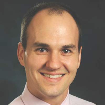
Dr. David E. Kandzari |
American use of transradial access grew markedly over the last decade. Ten years ago, the rate stood at about 3%. But it remains well behind most other countries. Results reported at the ACC meeting from another large international study of coronary interventions in ST-elevation myocardial infarction patients showed a 68% worldwide rate of transradial access (N. Engl. J. Med. 2015 [doi:10.1056/NEJMoa1415098])
I believe that the MATRIX results will help further fuel change in U.S. practice. Soon, quality assurance programs at many U.S. hospitals may incorporate transradial access as a performance measure.
The accumulated evidence, now including the MATRIX results, supports transradial access as the default approach for vascular access during coronary procedures. However, in some patients transradial access is impossible, especially in some women, in the elderly, and in patients with a high body mass index.
Dr. David E. Kandzari, director of interventional cardiology at the Piedmont Heart Institute in Atlanta, made these comments in an interview. He has been a consultant to Medtronic and Boston Scientific and has received research support from Abbott Vascular, Biotronik, Boston Scientific, and Medtronic.
U.S. interventionalists have lagged in adopting transradial access for percutaneous coronary interventions. I reviewed the status within the past year and found that about 17% of all U.S. percutaneous coronary interventions were done by transradial access, and this level jibes with recent results from a survey of U.S. interventionalists.
|
|
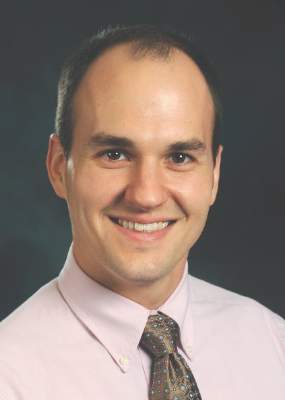
Dr. David E. Kandzari |
American use of transradial access grew markedly over the last decade. Ten years ago, the rate stood at about 3%. But it remains well behind most other countries. Results reported at the ACC meeting from another large international study of coronary interventions in ST-elevation myocardial infarction patients showed a 68% worldwide rate of transradial access (N. Engl. J. Med. 2015 [doi:10.1056/NEJMoa1415098])
I believe that the MATRIX results will help further fuel change in U.S. practice. Soon, quality assurance programs at many U.S. hospitals may incorporate transradial access as a performance measure.
The accumulated evidence, now including the MATRIX results, supports transradial access as the default approach for vascular access during coronary procedures. However, in some patients transradial access is impossible, especially in some women, in the elderly, and in patients with a high body mass index.
Dr. David E. Kandzari, director of interventional cardiology at the Piedmont Heart Institute in Atlanta, made these comments in an interview. He has been a consultant to Medtronic and Boston Scientific and has received research support from Abbott Vascular, Biotronik, Boston Scientific, and Medtronic.
SAN DIEGO – The unshakable grip that transfemoral access has held on coronary artery catheterization for the U.S. practice of interventional cardiology may finally loosen with results from an 8,000-patient, multinational controlled trial.
MATRIX showed that transradial access for coronary catheterization of patients with acute coronary syndrome (ACS) produced significantly fewer access-site bleeding events and significantly improved patient survival, compared with transfemoral access.
“Our results, in conjunction with the updated meta-analysis, suggest that the radial approach should become the default access for patients with acute coronary syndrome undergoing invasive management,” Dr. Marco Valgimigli said at the annual meeting of the American College of Cardiology. “Access site does matter, and a reduction in access-site bleeding complications seems to translate into a mortality benefit,” said Dr. Valgimigli, an interventional cardiologist at Erasmus University Medical Center in Rotterdam, the Netherlands.
The MATRIX (Minimizing Adverse Haemorrhagic Events by Transradial Access Site and Systemic Implementation of Angiox) trial enrolled 8,404 ACS patients at 78 sites in four European countries: Italy, Spain, Sweden, and the Netherlands. The study randomized patients undergoing percutaneous coronary intervention (PCI) to catheterization via either the patient’s radial or femoral artery. After 30 days, the combined rate of death, myocardial infarction, stroke, and major bleeding was reduced by an absolute rate of 1.9% among the patients treated with transradial access, a 17% relative risk reduction that was statistically significant for one of the study’s two primary endpoints.
This outcome difference seemed driven primarily by significant reductions in major bleeds and specifically major access-site bleeds, and this led to a statistically significant reduction in all-cause death by 0.6%, a 28% relative risk reduction in 30-day mortality tied to transradial access, Dr. Valgimigli said.
“I think this will be the study that helps change guidelines, to make radial artery access the default approach,” commented Dr. Sanjit S. Jolly, an interventional cardiologist at McMaster University in Hamilton, Ont.
“The United States is very behind in the use of transradial access; it’s used in about 20% of coronary PCIs,” noted Dr. Cindy L. Grines, an interventional cardiologist at the Detroit Medical Center. “We need to make a concerted effort in the United States to retrain practitioners to do transradial procedures. This approach is initially more time consuming, involves more radiation exposure, and can be frustrating, so we probably need to incentivize physicians by increasing their reimbursement for transradial PCIs and by making it part of quality assurance programs. Unless we do something like that, transradial use may not change,” Dr. Grines said in an interview.
The significant superiority of transradial over transfemoral access for both patient survival and for one of the study’s primary endpoints contrasted with the neutral result seen in an earlier major study that compared the two access approaches, RIVAL (Radial Versus Femoral Access for Coronary Angiography and Intervention in Patients With Acute Coronary Syndromes; Lancet 2011;377:1409-20).
Dr. Valgimigli also reported results from a meta-analysis that combined the MATRIX and RIVAL results as well as data from a few additional much smaller trials. This combined analysis, which involved a total of more than 19,000 ACS patients randomized to PCI via one of the two access sites, further confirmed that transradial catheterization linked with statistically significant reductions in death, in major bleeds not associated with coronary artery surgery, and in the combined endpoint of death, myocardial infarction, and stroke, he said. Concurrent with his report at the meeting, the MATRIX results as well as the updated meta-analysis results, appeared in an article published online (Lancet 2015 [doi:10.1016/S0140-6736(15)60292-6]).
The MATRIX study used only highly experienced interventionalists who had extensive familiarity with performing PCI using both types of access. They successfully used transradial access in 94% of patients randomized to that approach, but in the other 6% technical difficulties resulted in a crossover to the transfemoral route. Among patients randomized to transfemoral access, 2% required crossover to a transradial procedure.
MATRIX was an investigator-initiated study that received grant support from Terumo and the Medicines Co. Dr. Valgimigli had no relevant financial disclosures. Dr. Jolly has been a consultant to AstraZeneca, has been a speaker on behalf of St. Jude, and has received research grants from Medtronic. Dr. Grines has been a consultant to and received honoraria from Abbott Vascular, the Medicines Co., Merck, and the Volcano Group.
[email protected]
On Twitter @mitchelzoler
AT ACC 15
Key clinical point: ACS patients randomized to transradial access for PCI had significantly better survival at 30 days, compared with those treated via the transfemoral route.
Major finding: Thirty-day all-cause death occurred in 0.6% fewer patients using transradial access, a 28% relative risk reduction.
Data source: MATRIX, a multicenter, randomized controlled trial with 8,404 patients.
Disclosures: MATRIX was an investigator-initiated study that received grant support from Terumo and the Medicines Co. Dr. Valgimigli had no relevant financial disclosures. Dr. Jolly has been a consultant to AstraZeneca, has been a speaker on behalf of St. Jude, and has received research grants from Medtronic. Dr. Grines has been a consultant to and received honoraria from Abbott Vascular, the Medicines Co., Merck, and the Volcano Group.

Time to listen
Doctors talk more than they listen, especially when discussing end-of-life care with families, a recent study in Pediatrics suggests. Researchers from the University of Amsterdam followed 27 physicians and 37 parents as they navigated the difficult waters of end-of-life decision making for their children. By analyzing recorded conversations, they found that physicians spoke 67% of the time, while parents spoke only 30% of the time and nurses 3%. Additionally, they found that physicians “focused primarily on providing medical information, explaining the preferred course of action, and informing parents about the decision being reached by the team”(Pediatrics 2015;135:e465-76). Although parents were present during discussions, they were not routinely part of the decision-making process.
While this study was performed in Amsterdam and may not perfectly reflect the cultural norms of the United States, the results still should give us pause and raise important questions. Do we spend too much time talking, and too little listening? What role do parents have in decision making in our own country? Although we all participate in family-centered rounds, how often are the parents present but not involved? Do we pause often enough to explain in plain English what we had just rattled off in medicalese?
The challenge of listening is that it takes time and energy. With many other patients to care for and a long list of notes and orders to be entered, spending more time listening to families can seem exhausting and less important. However, this is the crux of the physician-patient relationship, and this is what makes the role of physician so important. When sick children and their families are at the most vulnerable point in their lives, it is our presence as empathizing, listening humans that matters most. Treating the disease with the correct medications is important but insufficient. And the sicker the patient, the higher the stakes.
As medical trainees, we often can feel powerless in these high-intensity situations. Yet, we can play a key role by advocating on behalf of our patients and their families, by giving them a voice. We can do this only by taking time to ask questions and to listen. After spending a few more minutes with these families in need, we can better understand their hopes and values, and we can identify the ways in which our goals align. As our medical teams are zipping through family-centered-rounds, we can advocate for families by raising their questions and concerns, ensuring that their voices are heard. By taking time to listen, we can provide that pivotal bridge of understanding between the medical team and the family.
Dr. Sisk is a pediatrics resident at St. Louis Children’s Hospital. E-mail him at [email protected].

Procedure type found to drive readmissions
The type of surgical procedure performed is a significant predictor of hospital readmission, reported Dr. Kevin R. Kasten and his associates at East Carolina University, Greenville, N.C.
In an analysis of 217,389 surgery patients, postoperative adverse events, specifically unplanned operating room return (odds ratio, 8.5; CI, 8.0-9.0), pulmonary embolism (OR, 8.2; CI, 7.1-9.6), deep incisional infection (OR, 7.5; CI, 6.7-8.5), and organ space infection (OR, 5.8; CI, 5.3-6.3), were significantly associated with an increased readmission risk. In addition, specific procedures associated with a higher risk for readmission included cystectomy, proctectomy, pancreatectomy, and lower-extremity vascular interventions.
The findings of this study suggest that “adverse events are a better predictor of 30[-day] readmission than patient comorbidity,” the authors said in the report. “As such, efforts to prevent adverse events such as return to the operating room, pulmonary embolism, surgical site infections, and myocardial infarction are crucial to prevention of readmission.”
Read the full article in the Journal of Surgical Research.
Some VTE patients may receive anticoagulants for too long
Even after excluding patients with atrial fibrillation, the duration of anticoagulant treatment for patients with unprovoked venous thromboembolism (VTE) and patients with transient risk factors often exceeded 1 year, according to a study published in Thrombosis Research.
In a large prospective cohort study, lead author Dr. Walter Ageno of the University of Insubria, Varese, Italy, and his associates examined 6,944 patients with a first episode of VTE. In this sample, 55% of patients with unprovoked events, 42% of patients with a transient risk factor, and 43% of patients with cancer received anticoagulant treatment for more than 12 months. The American College of Chest Physicians guideline recommends a 3-month treatment duration for patients with VTE secondary to transient risk factors. Pulmonary embolism at presentation, VTE recurrence while on treatment, chronic heart failure, and advanced age were independently associated with treatment for more than 12 months. Patients who died during the first year of treatment were excluded from the results.
Although clinicians tend to base their VTE treatment decisions on individual risk stratification, “this approach may expose a substantial proportion of patients, in particular those with VTE secondary to transient risk factors, to a possibly unnecessary risk of bleeding,” the investigators concluded.
Read the full article here: Thrombosis Research 2015.
Most probable postsurgical VTEs diagnosed after hospital discharge
Among surgical patients treated in and discharged from VA hospitals, 49.4% of possible venous thromboembolism s and 47.8% of probable VTEs were diagnosed within 30 days after surgery, and 63.1% of possible VTEs and 62.9% of probable VTEs were diagnosed within 90 days after surgery, according to Dr. Richard E. Nelson of the University of Utah, Salt Lake City.
In a retrospective cohort study, researchers examined medical records from 468,515 operations on 383,551 patients who underwent surgery without prior VTE between Jan. 1, 2005 and Dec. 31, 2010. In total, VTE occurred in 1.3% of surgical admissions for VA patients in the 90 days after surgery. However, the researchers argued that presumed VTE diagnoses should not be monitored based on inpatient records alone.
“Reliable monitoring of postoperative VTE events should incorporate two aspects. First, it should use data sources that allow for extraction of information from unstructured medical records. Second, it should use data sources that have the ability to follow patients after discharge from the hospital,” they said.
Read the full article in Thrombosis Research.
Can we do less?
It is a busy night in the emergency department, and patients are lining up in the waiting room. The next patient is a 2-year-old boy with a cough, runny nose, and increased work of breathing. My stethoscope picks up a chorus of noises in his lungs, but no wheezes. The attending physician walks into the room with me, a pediatrics resident, and the mother looks on expectantly, hoping I will make her baby better.
The attending agrees with me, this child is doing poorly and needs to be admitted. Then the question comes: “What do you want to do for him?” A few minutes later, the patient is receiving an albuterol treatment. Unsurprisingly, he does not improve, but soon he disappears off to the floor and I move onto the next patient.
In medicine, the urge to help patients drives physicians every day. The true challenge comes when the only way to help patients is by doing less. In 2014, the American Academy of Pediatrics released new bronchiolitis treatment guidelines. In this document, they cited numerous studies showing lack of benefit from albuterol or racemic epinephrine treatments, and they recommended against treatment trials in children with bronchiolitis. Additionally, they recommended against X-rays and steroids. This leaves pediatricians with the unsatisfying options of suctioning, watching, and waiting.
Physicians tend to be “fixers” by nature. Patients come to us to feel better, and we feel driven (internally and externally) to provide these cures. This desire can drive us to prescribe antibiotics for presumed viral infections, order imaging for minor head injuries, or offer trial bronchodilators in the setting of bronchiolitis. As medical trainees, we have the additional onus of answering to our attending physicians. Perhaps we are willing to watch a patient with bronchiolitis slowly evolve, but maybe some of our supervisors are not. How firmly do we stand our ground? What authority do we have?
Perhaps we have more to offer than we think. As trainees, we are exposed to education and updates from diverse fields of pediatrics, and this developing knowledge base can benefit our medical teams. We can utilize our knowledge of neurology to abort a seizure on the oncology floor. We can guide the evaluation for anemia while at an outpatient clinic. And we can apply our awareness of bronchiolitis guidelines to patients in the ED. By continuing to develop and apply an evidence base for our medical practice, we can provide meaningful insights about which interventions should (or should not) be done for our patients. Although uncomfortable at times, such situations provide us with the opportunity to improve medical practice while protecting our patients from unintended harms, gently remind our attending physicians which interventions should (or should not) be done for our patients. With education and a bit of spine, we can help our medical teams to follow that foremost of imperatives for the medical profession: Primum non nocere – First do no harm.
Dr. Sisk is a pediatrics resident at St. Louis Children’s Hospital. E-mail him at [email protected].
Drug produces ‘dramatic’ results in HL

The anti-CD30 antibody-drug conjugate brentuximab vedotin can prolong progression-free survival (PFS) in Hodgkin lymphoma (HL) patients who have undergone autologous stem cell transplant (ASCT), results of the phase 3 AETHERA trial have shown.
The median PFS for patients who received brentuximab vedotin immediately after ASCT was nearly twice that of patients who received placebo—42.9 months and 24.1 months, respectively.
“No medication available today has had such dramatic results in patients with hard-to-treat Hodgkin lymphoma,” said Craig Moskowitz, MD, of Memorial Sloan Kettering Cancer Center in New York, New York.
Dr Moskowitz and his colleagues detailed these results in The Lancet. The results were previously presented at the 2014 ASH Annual Meeting. The research was funded by Seattle Genetics, Inc., and Takeda Pharmaceutical Company Limited, the companies developing brentuximab vedotin.
The AETHERA study included 329 HL patients age 18 or older who were thought to be at high risk of relapse or progression after ASCT. Patients were randomized to receive placebo or 16 cycles of brentuximab vedotin once every 3 weeks.
After a median observation time of 30 months (range, 0-50 months), the rate of PFS was significantly higher in the brentuximab vedotin arm than the placebo arm. The hazard ratio was 0.57 (P=0.0013), according to an independent review group.
The estimated 2-year PFS was 63% in the brentuximab vedotin arm and 51% in the placebo arm, according to the independent review group. But according to investigators, the estimated 2-year PFS was 65% in the brentuximab vedotin arm and 45% in the placebo arm.
“Nearly all of these patients who are progression-free at 2 years are likely to be cured, since relapse 2 years after a transplant is unlikely,” Dr Moskowitz noted.
An interim analysis revealed no significant difference between the treatment arms with regard to overall survival.
The researchers said brentuximab vedotin was generally well-tolerated. The most common adverse events were peripheral neuropathy—occurring in 67% of brentuximab vedotin-treated patients and 13% of placebo-treated patients—and neutropenia—occurring in 35% and 12%, respectively.
In all, 53 patients died, 17% of those in the brentuximab vedotin arm and 16% of those in the placebo arm. The proportion of patients who died from disease-related illness was the same in both arms—11%.
“The bottom line is that brentuximab vedotin is a very effective drug in poor-risk Hodgkin lymphoma, and it spares patients from the harmful effects of further traditional chemotherapy by breaking down inside the cell, resulting in less toxicity,” Dr Moskowitz said.
Writing in a linked comment article, Andreas Engert, MD, of the University Hospital of Cologne in Germany, discussed how best to define which patients are at high risk of relapse and should receive brentuximab vedotin.
“AETHERA is a positive study establishing a promising new treatment approach for patients with Hodgkin’s lymphoma at high risk for relapse,” he wrote. “However, with a progression-free survival of about 50% at 24 months in the placebo group, whether this patient population is indeed high-risk could be debated.”
“An international consortium is currently reassessing the effect of risk factors in patients with relapsed Hodgkin’s lymphoma to define a high-risk patient population in need of consolidation treatment. We look forward to a better definition of patients with relapsed Hodgkin’s lymphoma who should receive consolidation treatment with brentuximab vedotin.” ![]()
Antidote can reverse effects of anticoagulant

Image by Andre E.X. Brown
SAN DIEGO—A factor Xa inhibitor antidote can reverse the anticoagulation activity of rivaroxaban (Xarelto) in healthy subjects, results of a phase 3 study suggest.
The antidote, andexanet alfa, reversed the anti-factor Xa activity of rivaroxaban, reduced the level of free rivaroxaban in the plasma, and restored thrombin generation to within the normal baseline range.
Subjects did not experience any severe or serious adverse events, and they did not develop antibodies to factor X or Xa.
Alexander Michael Gold, MD, of Portola Pharmaceuticals, and his colleagues presented these results at the American College of Cardiology’s 64th Annual Scientific Session (abstract 912-08*).
The study, ANNEXA-R, is sponsored by Portola Pharmaceuticals, the company developing andexanet alfa.
ANNEXA-R is a randomized, double-blind, phase 3 study in which researchers are evaluating the safety and efficacy of andexanet alfa for reversing rivaroxaban-induced anticoagulation in healthy volunteers ages 50 to 75.
In the first part of the study, 41 subjects received rivaroxaban at 20 mg once daily for 4 days. They were then randomized in a 2:1 ratio to receive either an 800 mg IV bolus of andexanet alfa (n=27) or placebo (n=14).
Results showed that andexanet alfa reduced the anti-factor Xa activity of rivaroxaban from baseline to nadir by more than 90%, a statistically significant difference from placebo (P<0.0001).
Significantly more andexanet alfa-treated subjects (96%) than placebo-treated subjects (0%) had a 90% or greater reduction in anti-factor Xa activity from baseline to nadir (P<0.0001).
Andexanet alfa also reduced the free (unbound) rivaroxaban concentration significantly from baseline to nadir, compared with placebo (P<0.0001).
Endogenous thrombin potential significantly increased from baseline to peak in andexanet alfa-treated subjects compared with placebo-treated subjects (P<0.0001).
And 96% of andexanet alfa-treated subjects saw their thrombin generation return to the normal range within 10 minutes of completing treatment.
The researchers said andexanet alfa was well-tolerated. There were no serious or severe adverse events, no thrombotic events, and no antibodies to factor X or Xa.
For the second part of the ANNEXA-R study, researchers plan to treat 40 healthy volunteers with rivaroxaban at 20 mg once daily for 4 days.
Then, subjects will be randomized in a 2:1 ratio to receive either andexanet alfa administered as an 800 mg IV bolus followed by a continuous infusion of 8 mg/min for 120 minutes or to placebo. Data from this part of the study are expected in mid-2015. ![]()
*Information in the abstract differs from that presented at the meeting.
Extended DAPT may not benefit patients with BMS

Photo by Frank C. Müller
Extended-duration dual antiplatelet therapy (DAPT) does not appear to confer any benefits for patients implanted with a bare-metal stent (BMS), according to a study published in JAMA.
An additional 18 months of DAPT among patients with a BMS did not result in significant differences in rates of stent thrombosis, major adverse cardiac and cerebrovascular events, or moderate or severe bleeding, compared to patients who received placebo.
Study authors noted, however, that the sample of BMS patients studied was small, which makes it difficult to draw definitive conclusions.
Dean J. Kereiakes, MD, of the Christ Hospital Heart and Vascular Center in Cincinnati, Ohio, and his colleagues conducted this research.
They analyzed 11,648 patients who received a BMS (n=1687) or drug-eluting stent (n=9961) and had completed 12 months of DAPT without bleeding or ischemic events.
The patients were randomized to continue DAPT—thienopyridine and aspirin—or receive placebo and aspirin for months 12 through 30.
Among the BMS patients, rates of stent thrombosis were 0.5% in the DAPT arm and 1.11% in the placebo arm (P=0.24).
Rates of major adverse cardiac and cerebrovascular events (a composite of death, heart attack, and stroke) were 4.04% and 4.69%, respectively (P=0.72). And rates of moderate/severe bleeding were 2.03% and 0.90%, respectively (P=0.07).
Among all the patients analyzed (both types of stent), the rates of stent thrombosis were 0.41% for patients who received DAPT and 1.32% for those who received placebo (P<0.001).
Rates of major adverse cardiac and cerebrovascular events were 4.29% and 5.74%, respectively (P<0.001). And rates of moderate/severe bleeding were 2.45% and 1.47%, respectively (P<0.001).
Dr Kereiakes and his colleagues noted that fewer BMS patients were enrolled in this trial because of the prevailing use of drug-eluting stents in clinical practice.
So the study may have been underpowered to identify differences in adverse events among BMS patients, and additional trials are needed to confirm the results of this research. ![]()