User login
In early days, bioabsorbable stent rivals nonabsorbable devices
At 6 months follow-up, a new-generation resorbable stent with a magnesium scaffold appears to perform at a level comparable to nonabsorbable drug-eluting stents (DES), according to first-in-man results presented as a late-breaker at the Cardiovascular Research Technologies conference, sponsored by MedStar Heart & Vascular Institute.
“IVUS [intravascular ultrasound] assessment demonstrated preservation of the scaffold area from post procedure up to 6 months with a low mean neointimal area,” reported Michael Haude, MD, PhD, director of the Heart & Vascular Center, Neuss, Germany.
Neointimal formation and late lumen loss (LLL) have been the Achilles’ heel of previous efforts to develop a viable fully absorbable stent, making these 6-month data highly encouraging.
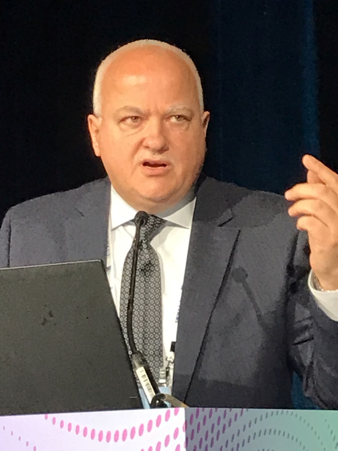
The tested device is the most recent iteration of the DREAMS (drug-eluting resorbable magnesium scaffold) technology. Relative to DREAMS 2G, the DREAMS 3G device has several design changes, including a higher radial force and reduced strut thickness.
The goal was to build on the promise of DREAMS 2G while avoiding its limitations.
“The problem with DREAMS 2G was that it showed low–target lesion failure and scaffold thrombosis rates in multiple trials, but in-scaffold LLL was not comparable to LLL values observed with historical PLLA [poly-L-lactic acid]–based scaffolds or contemporary DES,” Dr. Haude said.
The 6-month data with DREAMS 3G were drawn from the BIOMAG-I study. Patients with stable or unstable angina were enrolled if they had no angiographic evidence of thrombus at the target lesion. Patients were also required to have no more than two single de novo lesions requiring revascularization.
Of 116 patients enrolled, 115 were available for evaluation at 6 months. The study was not controlled, but outcomes were compared at 6 months to those observed with the DREAMS 2G device in the BIOSOLVE-II trial, published several years ago in the Lancet.
For the primary outcome of in-scaffold LLL at 6 months, the mean LLL from baseline at 6 months was more than 50% lower with the DREAMS 3G device in BIOMAG-I than DREAMS 2G in BIOSOLVE-II (0.21 vs. 0.44 mm). In a post hoc superiority analysis employing a weighted mean, a superiority analysis supported a highly significant difference in favor of the newer device (P < .0001).
More importantly, the low LLL in BIOMAG-I was not just favorable relative to previously evaluated bioabsorbable stents, but it appears to compete with nonabsorbable options at least after this length of follow-up.
In terms of LLL at 6 months, “these data suggested that DREAMS 3G “is now on the level of contemporary DES,” Dr. Haude said.
The relative difference in favor of DREAMS 3G was even greater at 6 months for the secondary endpoint of in-segment LLL (0.05 vs. 0.27 mm) with similar significance for the superiority margin in a post hoc analysis (P < .0001).
Serial optical coherence tomography (OCT) was conducted post procedure, and indicated that the struts “were well embedded in the vessel wall,” according to Dr. Haude. Only 4.4% of struts on average were malapposed. The total incomplete strut apposition area was on average 0.08 mm. At 6 months, most struts were no long discernible on OCT, documenting device resorption.
Clinical results at 6 months were supportive. There were no cases of definite or probable scaffold thrombosis, and there were no target vessel myocardial infarctions or cardiac deaths. There was one clinically driven target lesion revascularization.
DREAMS 3G has other features designed to make it easier to deploy, Dr. Haude said. For example, radiopaque markers are now situated on both ends of the stent, making it easier to see on imaging. There are also plans to make these stents available in 15 sizes to accommodate a broad range of anatomy.
The data were impressive for many of the panelists invited to discuss the results.
“For the first time, we are seeing a bioabsorbable device showing excellent healing and very little late lumen loss,” said Michael H. Joner, MD, professor of early clinical trials at the German Center for Cardiovascular Research, Munich. “The next step is some sort of direct comparison with a drug-eluting stent.”
Describing himself as “a little more skeptical,” Aoke V Finn, MD, medical director and chief scientific officer, CVPath Institute, University of Maryland, Baltimore, said he wants to know more about the speed of device degradation and to see more long-term results in terms of clinical events. Although he considers the data promising so far, he considers it too early to embark on a randomized trial.
Longer-term data are coming, according to Dr. Haude. In addition to the 12-month follow-up that will include OCT and IVUS evaluations, there are annual clinical follow-up analyses planned to 5 years.
Dr. Haude reports financial relationships with Biotronik, Cardiac Dimensions, OrbusNeich, and Philips. Dr. Joner reports no potential conflicts of interest. Dr. Finn reports financial relationships with 19 pharmaceutical companies including those that manufacture cardiovascular stents.
At 6 months follow-up, a new-generation resorbable stent with a magnesium scaffold appears to perform at a level comparable to nonabsorbable drug-eluting stents (DES), according to first-in-man results presented as a late-breaker at the Cardiovascular Research Technologies conference, sponsored by MedStar Heart & Vascular Institute.
“IVUS [intravascular ultrasound] assessment demonstrated preservation of the scaffold area from post procedure up to 6 months with a low mean neointimal area,” reported Michael Haude, MD, PhD, director of the Heart & Vascular Center, Neuss, Germany.
Neointimal formation and late lumen loss (LLL) have been the Achilles’ heel of previous efforts to develop a viable fully absorbable stent, making these 6-month data highly encouraging.
The tested device is the most recent iteration of the DREAMS (drug-eluting resorbable magnesium scaffold) technology. Relative to DREAMS 2G, the DREAMS 3G device has several design changes, including a higher radial force and reduced strut thickness.
The goal was to build on the promise of DREAMS 2G while avoiding its limitations.
“The problem with DREAMS 2G was that it showed low–target lesion failure and scaffold thrombosis rates in multiple trials, but in-scaffold LLL was not comparable to LLL values observed with historical PLLA [poly-L-lactic acid]–based scaffolds or contemporary DES,” Dr. Haude said.
The 6-month data with DREAMS 3G were drawn from the BIOMAG-I study. Patients with stable or unstable angina were enrolled if they had no angiographic evidence of thrombus at the target lesion. Patients were also required to have no more than two single de novo lesions requiring revascularization.
Of 116 patients enrolled, 115 were available for evaluation at 6 months. The study was not controlled, but outcomes were compared at 6 months to those observed with the DREAMS 2G device in the BIOSOLVE-II trial, published several years ago in the Lancet.
For the primary outcome of in-scaffold LLL at 6 months, the mean LLL from baseline at 6 months was more than 50% lower with the DREAMS 3G device in BIOMAG-I than DREAMS 2G in BIOSOLVE-II (0.21 vs. 0.44 mm). In a post hoc superiority analysis employing a weighted mean, a superiority analysis supported a highly significant difference in favor of the newer device (P < .0001).
More importantly, the low LLL in BIOMAG-I was not just favorable relative to previously evaluated bioabsorbable stents, but it appears to compete with nonabsorbable options at least after this length of follow-up.
In terms of LLL at 6 months, “these data suggested that DREAMS 3G “is now on the level of contemporary DES,” Dr. Haude said.
The relative difference in favor of DREAMS 3G was even greater at 6 months for the secondary endpoint of in-segment LLL (0.05 vs. 0.27 mm) with similar significance for the superiority margin in a post hoc analysis (P < .0001).
Serial optical coherence tomography (OCT) was conducted post procedure, and indicated that the struts “were well embedded in the vessel wall,” according to Dr. Haude. Only 4.4% of struts on average were malapposed. The total incomplete strut apposition area was on average 0.08 mm. At 6 months, most struts were no long discernible on OCT, documenting device resorption.
Clinical results at 6 months were supportive. There were no cases of definite or probable scaffold thrombosis, and there were no target vessel myocardial infarctions or cardiac deaths. There was one clinically driven target lesion revascularization.
DREAMS 3G has other features designed to make it easier to deploy, Dr. Haude said. For example, radiopaque markers are now situated on both ends of the stent, making it easier to see on imaging. There are also plans to make these stents available in 15 sizes to accommodate a broad range of anatomy.
The data were impressive for many of the panelists invited to discuss the results.
“For the first time, we are seeing a bioabsorbable device showing excellent healing and very little late lumen loss,” said Michael H. Joner, MD, professor of early clinical trials at the German Center for Cardiovascular Research, Munich. “The next step is some sort of direct comparison with a drug-eluting stent.”
Describing himself as “a little more skeptical,” Aoke V Finn, MD, medical director and chief scientific officer, CVPath Institute, University of Maryland, Baltimore, said he wants to know more about the speed of device degradation and to see more long-term results in terms of clinical events. Although he considers the data promising so far, he considers it too early to embark on a randomized trial.
Longer-term data are coming, according to Dr. Haude. In addition to the 12-month follow-up that will include OCT and IVUS evaluations, there are annual clinical follow-up analyses planned to 5 years.
Dr. Haude reports financial relationships with Biotronik, Cardiac Dimensions, OrbusNeich, and Philips. Dr. Joner reports no potential conflicts of interest. Dr. Finn reports financial relationships with 19 pharmaceutical companies including those that manufacture cardiovascular stents.
FROM CRT 2023
Measles exposures in Kentucky have CDC on alert
The Centers for Disease Control and Prevention has issued a Health Alert Network (HAN) health advisory notifying clinicians and public health officials of a confirmed measles case in an individual who for 2 days (February 17-18) attended a large religious gathering that was attended by an estimated 20,000 people at Asbury University in Wilmore, Ky.
Given that large numbers of people might have been exposed to the attendee (who was not vaccinated) and that the individual had a history of recent international travel, the CDC has encouraged clinicians to be vigilant for patients presenting with symptoms that meet the measles case definition. A steady increase in measles cases from 49 in 2021 to 121 in 2022 in children who were not fully vaccinated – coupled with outbreaks in Ohio and Minnesota – underscores the potential gravity of the CDC advisory as well as the need to mitigate the risk of ongoing or secondary transmission.
Currently, little is known about the individual who contracted measles other than the fact that he is a resident of Jessamine County, Ky., according to a news release issued by the Kentucky Department of Public Health. It is the third confirmed case in Kentucky over the past 3 months. State and national health officials are concerned that the individual might have transmitted measles to attendees visiting from other states.
David Sugerman, MD, MPH, a medical officer in CDC’s division of viral diseases and lead for the measles, rubella, and cytomegalovirus team, noted that the timing of the alert coincides with the period in which persons who had had contact with the initial case patient might be expected to develop symptoms.
For clinicians, “It’s really about considering measles in any un- or undervaccinated patient that arrives at a clinic and recently traveled internationally,” Dr. Sugerman told this news organization. He explained that “when doctors are seeing patients, they’re not going to necessarily share that information off the bat when they present with fever or rash, or if their child has fever and rash, or that they traveled internationally. So, eliciting that history from the patient or their parents is really critical.”
The CDC recommends that measles be considered in anyone presenting with a febrile illness and symptoms that are clinically compatible with measles (that is, rash, cough, coryza, or conjunctivitis), as well as in patients who have recently traveled abroad, especially to countries with ongoing outbreaks, including India, Somalia, and Yemen.
“In general, if they’ve traveled internationally and they are undervaccinated, measles should be part of the differential diagnosis,” Sugerman said. He also emphasized the need to follow airborne isolation precautions in addition to general infection control measures.
Immediate triage is critical, especially since overcrowded waiting rooms might be filled with patients who are not yet eligible for vaccination or are not up to date or fully vaccinated.
“Measles is under airborne isolation criteria and precautions, and therefore, [patients] need to be placed as soon as possible into a negative pressure or airborne infection isolation room – and that should be a single room,” he explained. He noted, “In some settings, there may not be a negative pressure room, e.g., an outpatient pediatrics or family medicine office.”
Dr. Sugerman said that in these circumstances, patients should be placed in a room with masked health care providers who have received two doses of measles, mumps, and rubella (MMR) vaccine and that they should wear an N95 mask when entering the room and interviewing the patient.
Clinicians should follow CDC’s testing recommendations and collect a nasopharyngeal or throat swab or a urine specimen for PCR testing and a blood specimen for serology. In addition, they should immediately report cases to local and state public health authorities.
For all patients, it’s critical to be up to date on MMR vaccines, especially persons who are going to be traveling internationally. “We recommend that when they’ve got infants traveling with them who are 6-11 months of age, that they get a first dose (which we consider a zero dose), because they need a routine dose at 12-15 months, and then 4-6 years,” said Dr. Sugerman. He said that it’s safe for adults who are unsure of their status to receive an MMR dose as well.
Dr. Sugerman stressed that despite major strides, “we just don’t have enough coverage in all individuals in this country. Because people are traveling as often as they are, it can be imported. Until measles is eliminated globally, there’s going to be an ongoing risk of importation and potential spread amongst others in their household or community, especially amongst individuals who are not fully vaccinated and, in particular, amongst those who are unvaccinated,” he said.
Dr. Sugerman reports no relevant financial relationships.
A version of this article first appeared on Medscape.com.
FDA to review dupilumab for treating chronic spontaneous urticaria
The that is inadequately controlled by current standard of care.
CSU is an inflammatory skin condition that causes sudden hives and angioedema, most often on the face, hands, and feet. However, the throat and upper airways also can be affected. CSU is generally treated with H1 antihistamines, but this strategy is insufficient for approximately 50% of patients, according to a press release from the manufacturer, Regeneron, announcing the FDA acceptance of the application on March 7.
Dupilumab (Dupixent), first approved in 2017 for treating atopic dermatitis in adults, is a fully human monoclonal antibody that inhibits the signaling of the interleukin (IL)-4 and IL-13 pathways.
The application for FDA approval for CSU is based on data from a pair of phase 3 trials in two different populations, LIBERTY-CUPID A and B.
The first study (LIBERTY-CUPID A) randomized 138 CSU patients aged 6 years and older who were uncontrolled on antihistamines to additional treatment with dupilumab or placebo over 24 weeks. The dupilumab-treated patients showed a 63% reduction in itch severity compared with a 35% reduction in patients who received the placebo, measured by changes in a 0-21 itch severity scale, according to data presented at the 2022 American Academy of Allergy, Asthma and Immunology (AAAAI) meeting.
Patients in the dupilumab group also showed a 65% reduction in the severity of urticaria activity (itch and hives) compared with 37% of those on placebo. Overall rates of adverse events were similar between groups; the most common were injection site reactions, according to the company.
The second study (LIBERTY-CUPID B) assessed efficacy and safety of dupilumab in 108 patients with CSU aged 12-80 years who were symptomatic despite standard-of-care treatment and were intolerant or incomplete responders to the anti-IgE antibody omalizumab (Xolair), approved for CSU. Last year, the company announced that this study had been halted after an interim analysis found that while there were positive numerical trends in reducing itch and hives, they “did not meet statistical significance.” In the March 7 press release, the company said that results from this study provide “additional supporting data” for the approval application.
The target date for the FDA’s decision is Oct. 22, 2023, according to Regeneron. Regeneron and Sanofi also are investigating dupilumab for treating chronic inducible urticaria triggered by cold in a phase 3 study.
One in four parents lied about kids’ COVID status: Survey
More than 1 in 4 parents lied to school officials about their children’s COVID-19 status or refused to comply with public health rules during the height of the pandemic, a new study found. Researchers said they suspected the 26% of parents who misrepresented their children’s health status may have undercounted the actual figure.
“If anything, 26% is probably the minimum” of parents who misled school officials, said Angela Fagerlin, PhD, a researcher at the University of Utah Medical School, Salt Lake City.
In the survey, many parents said they considered it their right as parents to make their own decision about their children’s health status, said Dr. Fagerlin, who is also the chair of the department of population health sciences at the University of Utah School of Medicine.
“It appears that many parents were concerned about their children missing school,” she said. “At the same time, they’re potentially exposing other kids to a serious illness.”
In the survey, parents were asked whether they lied or misrepresented information about their children on seven different COVID-19 topics, including illness and vaccination status and if they followed quarantine protocols. Researchers tallied survey responses collected in December 2021 from 580 parents, whose average age was 36 and of whom 70% were women. Results were published in the journal JAMA Network Open.
Overall, 24% of parents said they lied to people that their children were with while knowing or suspecting the children had COVID. About half of parents cited at least one of the following reasons for doing so: parental freedom, child did not feel very sick, or wanted the child’s life to feel “normal.”
About 20% of parents said they avoided testing when they thought their child had COVID, and parents also reported allowing children to break quarantine rules at a similar rate. More than half of parents who avoided testing said they were worried testing would hurt or feel uncomfortable.
About 4 in 10 parents who lied about their child’s illness status or who lied about whether their child should be in quarantine said they did so because of guidance from a public figure such as a celebrity or politician. At least 3 in 10 said they lied because they could not miss work to stay home with their child.
“We need to do a better job of providing support mechanisms like paid sick leave for family illness so that parents don’t feel like their only option is to engage in misrepresentation or non-adherence to public health guidelines during a future infectious disease outbreak that matches or exceeds the magnitude of COVID-19,” says researcher Andrea Gurmankin Levy, PhD, of Middlesex (Conn.) Community College.
A version of this article first appeared on WebMD.com.
FROM JAMA NETWORK OPEN
MS looks homogeneous
SAN DIEGO – , rather than a mixture of conditions with different genetic or other causes, according to a new analysis. The work suggests that personalized therapy based on clinical characteristics is likely to be the best approach, rather than precision medicine based on molecular or other subtypes.
Think MS is heterogeneous? Think again
The work drew upon data from 22,000 individuals, 32,000 attacks, 156,000 EDSS scores, 250,000 observation years, and 110,000 treatment years recorded in the Swedish MS registry. The researchers examined distributions in age of onset, severity, and distribution of relapses. Among patients treated with one of 12 disease-modifying therapies, they examined patterns of EDSS progression, appearance of new lesions, and relapses.
“

Regardless of which clinical characteristic of the MS syndrome that I study, I find a uniform distribution with very few if any outliers. That argues that MS is likely to be a homogeneous condition with some variation, but it’s highly unlikely that MS is a mixture of different conditions masquerading as the same thing,” said Jan Hillert, MD, PhD, who presented the study during a poster session at the annual meeting held by the Americas Committee for Treatment and Research in Multiple Sclerosis.
“There are big efforts out there trying to decipher the molecular basis of the MS syndrome, thinking that it’s a mixture of different things. And I would argue that our data very strongly argue against that,” said Dr. Hillert, who is a professor of neurology at Karolinska Institutet in Solna, Sweden.
Specific subtypes should produce individual groupings rather than a broad distribution. “If you have a multitude of factors, then this (finding of a broad distribution) is what you have. If you have a small number of strongly acting factors, like if there were genetic subgroups, then you would have a different distribution. So this is in line with the polygenic, complex nature of MS that we have been thinking about for many, many years, and which the genetics also support,” said Dr. Hillert.
The findings suggest that physicians should be emphasizing personalized treatment of MS based on factors like age, weight, disease activity and severity, disability, side effects, and other factors. “Personalized medicine I embrace, but the concept of precision medicine is naive. It’s not founded on any sound scientific evidence,” said Dr. Hillert.
An evolving definition
The conclusion is compelling, according to Patricia Coyle, MD, who was asked to comment on the study. “I have not seen this sort of analysis before. I think it doesn’t absolutely prove the case in talking about very small numbers, but it does make a very logical argument. This is not showing any meaningful large subgroups that you can call out,” said Dr. Coyle, who is a professor of neurology and director of the MS Comprehensive Care Center at Stonybrook Neurosciences Institute in New York.

“I think that it’s interesting, because we have routinely said we think this is heterogeneous, because no two patients are alike. But this is speaking against meaningful heterogeneity in MS. When you look at these sorts of statistical results, this is what you’d expect in a normal population, not in a disease where you might say, genetically, or molecularly, you could define significant subsets of individuals,” said Dr. Coyle.
The study isn’t the last word. “You would probably like to see some follow-up data, perhaps in other very large databases to make it more convincing, but I think you’re not hearing as many people talk about MS heterogeneity anymore. We know there’s a focal inflammatory component, we know there’s a neurodegenerative component. Both are present in all MS. People are even arguing that maybe it’s not meaningful to call out progressive and relapsing MS. I don’t agree with that, but I think the concept that there are meaningful subsets of patients is probably incorrect. [The idea that] one set is due to perhaps an infection, another might be molecular mimicry. ... Maybe that’s not the case at all,” said Dr. Coyle.
For example, some companies are looking into whether B cell depletion treatments might be more effective for one set of patients versus another. “The issue is, can you dissect out subsets where hitting B cells is really good and others where it doesn’t seem to matter? That really hasn’t [been successful],” said Dr. Coyle.
Dr. Hillert has served on scientific advisor boards for or received speaker’s fees from Biogen, Bristol Myers Squibb/Celgene, Janssen, Novartis, Teva, Merck KGaA, Sandoz, and Sanofi Genzyme. He has received research support from Biogen, Bristol Myers Squibb/Celgene, Merck, Janssen, Novartis, Roche, and Sanofi-Genzyme. Dr. Coyle has consulted for or received speaker fees from Accordant, Biogen, Bristol Myers Squibb, GlaxoSmithKline, Horizon Therapeutics, LabCorp, Eli Lilly and Company, Mylan, Novartis, Sanofi Genzyme, TG Therapeutics. She has received research support from Actelion, Alkermes, Celgene, CorEvitas, Genentech/Roche, Janssen, MedDay, NINDS, Novartis, and Sanofi Genzyme.
SAN DIEGO – , rather than a mixture of conditions with different genetic or other causes, according to a new analysis. The work suggests that personalized therapy based on clinical characteristics is likely to be the best approach, rather than precision medicine based on molecular or other subtypes.
Think MS is heterogeneous? Think again
The work drew upon data from 22,000 individuals, 32,000 attacks, 156,000 EDSS scores, 250,000 observation years, and 110,000 treatment years recorded in the Swedish MS registry. The researchers examined distributions in age of onset, severity, and distribution of relapses. Among patients treated with one of 12 disease-modifying therapies, they examined patterns of EDSS progression, appearance of new lesions, and relapses.
“
Regardless of which clinical characteristic of the MS syndrome that I study, I find a uniform distribution with very few if any outliers. That argues that MS is likely to be a homogeneous condition with some variation, but it’s highly unlikely that MS is a mixture of different conditions masquerading as the same thing,” said Jan Hillert, MD, PhD, who presented the study during a poster session at the annual meeting held by the Americas Committee for Treatment and Research in Multiple Sclerosis.
“There are big efforts out there trying to decipher the molecular basis of the MS syndrome, thinking that it’s a mixture of different things. And I would argue that our data very strongly argue against that,” said Dr. Hillert, who is a professor of neurology at Karolinska Institutet in Solna, Sweden.
Specific subtypes should produce individual groupings rather than a broad distribution. “If you have a multitude of factors, then this (finding of a broad distribution) is what you have. If you have a small number of strongly acting factors, like if there were genetic subgroups, then you would have a different distribution. So this is in line with the polygenic, complex nature of MS that we have been thinking about for many, many years, and which the genetics also support,” said Dr. Hillert.
The findings suggest that physicians should be emphasizing personalized treatment of MS based on factors like age, weight, disease activity and severity, disability, side effects, and other factors. “Personalized medicine I embrace, but the concept of precision medicine is naive. It’s not founded on any sound scientific evidence,” said Dr. Hillert.
An evolving definition
The conclusion is compelling, according to Patricia Coyle, MD, who was asked to comment on the study. “I have not seen this sort of analysis before. I think it doesn’t absolutely prove the case in talking about very small numbers, but it does make a very logical argument. This is not showing any meaningful large subgroups that you can call out,” said Dr. Coyle, who is a professor of neurology and director of the MS Comprehensive Care Center at Stonybrook Neurosciences Institute in New York.
“I think that it’s interesting, because we have routinely said we think this is heterogeneous, because no two patients are alike. But this is speaking against meaningful heterogeneity in MS. When you look at these sorts of statistical results, this is what you’d expect in a normal population, not in a disease where you might say, genetically, or molecularly, you could define significant subsets of individuals,” said Dr. Coyle.
The study isn’t the last word. “You would probably like to see some follow-up data, perhaps in other very large databases to make it more convincing, but I think you’re not hearing as many people talk about MS heterogeneity anymore. We know there’s a focal inflammatory component, we know there’s a neurodegenerative component. Both are present in all MS. People are even arguing that maybe it’s not meaningful to call out progressive and relapsing MS. I don’t agree with that, but I think the concept that there are meaningful subsets of patients is probably incorrect. [The idea that] one set is due to perhaps an infection, another might be molecular mimicry. ... Maybe that’s not the case at all,” said Dr. Coyle.
For example, some companies are looking into whether B cell depletion treatments might be more effective for one set of patients versus another. “The issue is, can you dissect out subsets where hitting B cells is really good and others where it doesn’t seem to matter? That really hasn’t [been successful],” said Dr. Coyle.
Dr. Hillert has served on scientific advisor boards for or received speaker’s fees from Biogen, Bristol Myers Squibb/Celgene, Janssen, Novartis, Teva, Merck KGaA, Sandoz, and Sanofi Genzyme. He has received research support from Biogen, Bristol Myers Squibb/Celgene, Merck, Janssen, Novartis, Roche, and Sanofi-Genzyme. Dr. Coyle has consulted for or received speaker fees from Accordant, Biogen, Bristol Myers Squibb, GlaxoSmithKline, Horizon Therapeutics, LabCorp, Eli Lilly and Company, Mylan, Novartis, Sanofi Genzyme, TG Therapeutics. She has received research support from Actelion, Alkermes, Celgene, CorEvitas, Genentech/Roche, Janssen, MedDay, NINDS, Novartis, and Sanofi Genzyme.
At ACTRIMS FORUM 2023
Black people are less likely to receive dementia meds
, preliminary data from a retrospective study show.
“There have been disparities regarding the use of cognition-enhancing medications in the treatment of dementia described in the literature, and disparities in the use of adjunctive treatments for other neuropsychiatric symptoms of dementia described in hospital and nursing home settings,” said study investigator Alice Hawkins, MD, with the department of neurology, Icahn School of Medicine at Mount Sinai, New York. “However, less is known about use of dementia medications that people take at home. Our study found disparities in this area as well,” Dr. Hawkins said.
The findings were released ahead of the study’s scheduled presentation at the annual meeting of the American Academy of Neurology.
More research needed
The researchers analyzed data on 3,655 Black and 12,885 White patients with a diagnosis of dementia who were seen at Mount Sinai. They evaluated utilization of five medication classes:
- cholinesterase inhibitors.
- N-methyl D-aspartate (NMDA) receptor antagonists.
- selective serotonin reuptake inhibitors (SSRIs).
- antipsychotics.
- benzodiazepines.
They found that Black patients with dementia received cognitive enhancers less often than White patients with dementia (20% vs. 30% for cholinesterase inhibitors; 10% vs. 17% for NMDA antagonists).
Black patients with dementia were also less likely to receive medications for behavioral and psychological symptom management, compared with White peers (24% vs. 40% for SSRIs; 18% vs. 22% for antipsychotics; and 18% vs. 37% for benzodiazepines).
These disparities remained even after controlling for factors such as demographics and insurance coverage.
“Larger systemic forces such as systemic racism, quality of care, and provider bias are harder to pin down, particularly in the medical record, though they all may be playing a role in perpetuating these inequities. More research will be needed to pinpoint all the factors that are contributing to these disparities,” said Dr. Hawkins.
The researchers found Black patients who were referred to a neurologist received cholinesterase inhibitors and NMDA antagonists at rates comparable with White patients. “Therefore, referrals to specialists such as neurologists may decrease the disparities for these prescriptions,” Dr. Hawkins said.
Crucial research
Commenting on the findings, Carl V. Hill, PhD, MPH, Alzheimer’s Association chief diversity, equity, and inclusion officer, said the study “adds to previous research that points to inequities in the administering of medications for dementia symptoms, and highlights the inequities we know exist in dementia care.”
“Cognitive enhancers and other behavioral/psychological management drugs, while they don’t stop, slow, or cure dementia, can offer relief for some of the challenging symptoms associated with diseases caused by dementia. If people aren’t being appropriately prescribed medications that may offer symptom relief from this challenging disease, it could lead to poorer health outcomes,” said Dr. Hill.
“These data underscore the importance of health disparities research that is crucial in uncovering inequities in dementia treatment, care, and research for Black individuals, as well as all underrepresented populations.
“We must create a society in which the underserved, disproportionately affected, and underrepresented are safe, cared for, and valued. This can be done through enhancing cultural competence in health care settings, improving representation within the health care system, and engaging and building trust with diverse communities,” Dr. Hill said.
The Alzheimer’s Association has partnered with more than 500 diverse community-based groups on disease education programs to ensure families have information and resources to navigate this devastating disease.
The study was supported by the American Academy of Neurology Resident Research Scholarship. Dr. Hawkins and Dr. Hill reported no relevant financial relationships.
A version of this article first appeared on Medscape.com.
Incommunicado no more
A few weeks ago I wrote about my glasses and the discovery that they’d been made incorrectly. The headline for the story was “The Way I See It.”
That’s the opening line from Joni Mitchell’s 1974 song “Free Man in Paris.” But I grew up in a Neil Diamond household (Dad always had Neil Diamond on when he was working at home) so the first time I heard the song was in 1977, when Diamond covered it. In fact, I didn’t even realize it was originally Mitchell’s song until I was in my 50s.
It’s about a world that doesn’t exist anymore.

The song is about music promoter David Geffen and a trip he took to Paris. Back in southern California, he was always working. There were continual phone calls, deals, meetings, and people looking for favors.
But on his trip to Paris in the early 1970s, he became just another person. No one could find him to ask for help or cut a deal. He couldn’t be reached. He felt “unfettered and alive” and could go from “cafe to cabaret,” relax, and enjoy himself.
Medical practice was once that way. You’d check-out patients to your call partners, leave town, and relax for a week or two.
Try doing that today.
We even have a new psychiatric condition – nomophobia – for the fear of not having our mobile phone handy. Every time I leave my house or office I repeat a simple mantra “phone, wallet, keys” as I pat my pockets.
Unless you can part with your gadget – which ain’t easy – no one is a “free man in Paris” (or Tokyo, or Rio, or Beijing) anymore. Even ships have cell service at sea. There are still places on Earth remote enough that you can’t be reached, but they get fewer and smaller every year.
When was the last time you really went somewhere and had no communication with your office at all? Emails, texts, anything? Unless you’re in a shift-work branch of medicine, like ER or hospitalist, I’m going to guess it’s been a while. And even in those branches you probably get emails about administrative matters, scheduling questions, and pointless memos.
Being in solo practice I’ve come to accept this, but it’s a conscious decision on my part. It’s easier than finding a call partner, and if I’m handling my own stuff at least I’m not going to come home to any surprises. So I’ve covered patients from as far west as Hawaii, north as Juneau, south as Panama City, and east as Le Havre.
Granted, this is medicine, and many other jobs don’t require the degree of involvement that it does. But I suspect pretty much any professional - attorney, accountant, executive - still has to deal with work-related stuff while traveling. Back in the 1970s to 1980s my dad, a solo-practice lawyer, had a set time each vacation weekday afternoon where he’d call his secretary to go over stuff. Today it would be by email or texts.
We’ve done this to ourselves. We’ve accepted the trade-off of better connectivity with family and friends for expanding our time at work. The same technology that lets me send in prescription refills from London also lets me send family pictures back from Maui. It’s not easy to draw a solid line between them, and I’m not so sure many of us want to.
Today, 50 years after Ms. Mitchell wrote the song, the idea of being a “free man in Paris” – or anywhere – really doesn’t exist for most of us anymore. You can argue whether that’s good or bad, but it’s where we are.
Dr. Block has a solo neurology practice in Scottsdale, Ariz.
A few weeks ago I wrote about my glasses and the discovery that they’d been made incorrectly. The headline for the story was “The Way I See It.”
That’s the opening line from Joni Mitchell’s 1974 song “Free Man in Paris.” But I grew up in a Neil Diamond household (Dad always had Neil Diamond on when he was working at home) so the first time I heard the song was in 1977, when Diamond covered it. In fact, I didn’t even realize it was originally Mitchell’s song until I was in my 50s.
It’s about a world that doesn’t exist anymore.
The song is about music promoter David Geffen and a trip he took to Paris. Back in southern California, he was always working. There were continual phone calls, deals, meetings, and people looking for favors.
But on his trip to Paris in the early 1970s, he became just another person. No one could find him to ask for help or cut a deal. He couldn’t be reached. He felt “unfettered and alive” and could go from “cafe to cabaret,” relax, and enjoy himself.
Medical practice was once that way. You’d check-out patients to your call partners, leave town, and relax for a week or two.
Try doing that today.
We even have a new psychiatric condition – nomophobia – for the fear of not having our mobile phone handy. Every time I leave my house or office I repeat a simple mantra “phone, wallet, keys” as I pat my pockets.
Unless you can part with your gadget – which ain’t easy – no one is a “free man in Paris” (or Tokyo, or Rio, or Beijing) anymore. Even ships have cell service at sea. There are still places on Earth remote enough that you can’t be reached, but they get fewer and smaller every year.
When was the last time you really went somewhere and had no communication with your office at all? Emails, texts, anything? Unless you’re in a shift-work branch of medicine, like ER or hospitalist, I’m going to guess it’s been a while. And even in those branches you probably get emails about administrative matters, scheduling questions, and pointless memos.
Being in solo practice I’ve come to accept this, but it’s a conscious decision on my part. It’s easier than finding a call partner, and if I’m handling my own stuff at least I’m not going to come home to any surprises. So I’ve covered patients from as far west as Hawaii, north as Juneau, south as Panama City, and east as Le Havre.
Granted, this is medicine, and many other jobs don’t require the degree of involvement that it does. But I suspect pretty much any professional - attorney, accountant, executive - still has to deal with work-related stuff while traveling. Back in the 1970s to 1980s my dad, a solo-practice lawyer, had a set time each vacation weekday afternoon where he’d call his secretary to go over stuff. Today it would be by email or texts.
We’ve done this to ourselves. We’ve accepted the trade-off of better connectivity with family and friends for expanding our time at work. The same technology that lets me send in prescription refills from London also lets me send family pictures back from Maui. It’s not easy to draw a solid line between them, and I’m not so sure many of us want to.
Today, 50 years after Ms. Mitchell wrote the song, the idea of being a “free man in Paris” – or anywhere – really doesn’t exist for most of us anymore. You can argue whether that’s good or bad, but it’s where we are.
Dr. Block has a solo neurology practice in Scottsdale, Ariz.
Experts share real-world experience prescribing voclosporin, belimumab for lupus nephritis
Although patients with lupus nephritis recently gained two new add-on treatment options in voclosporin (Lupkynis) and belimumab (Benlysta), there have been little data published with real-world experience in using these drugs.
Voclosporin, a calcineurin inhibitor, was approved by the Food and Drug Administration in January 2021 to treat lupus nephritis in combination with immunosuppressive medication. Belimumab, a human monoclonal antibody and B-lymphocyte stimulator, was approved in December 2020 in the United States as an add-on treatment for lupus nephritis in adults and later in July 2022 for children who are already receiving standard therapy.
How the two drugs are prescribed for patients with lupus nephritis so far appears to be influenced by presence of extrarenal manifestations of lupus, proteinuria level, clinicians’ prior experience with belimumab, costs of the drugs, and patient preference, experts said.
Voclosporin’s approval was based on data from the phase 3 AURORA 1 trial and phase 2 AURA-LV trial. AURORA 1 evaluated 357 patients with systemic lupus erythematosus (SLE) and lupus nephritis who were randomized to receive voclosporin or placebo with mycophenolate mofetil and tapered low-dose oral steroids. In the voclosporin group, the results showed a significantly higher complete renal response at 52 weeks, compared with the placebo group, while having a similar adverse event profile. The AURA-LV trial, evaluating efficacy and safety of 179 patients with lupus nephritis, showed adding low-dose voclosporin to induction therapy improved renal response, compared with placebo. AURORA 2, a continuation of the AURORA trial, showed patients with lupus nephritis receiving voclosporin have a stable estimated glomerular filtration rate and reductions in proteinuria up to 3 years of follow-up.
Results from the phase 3 BLISS-LN trial of 448 patients with confirmed lupus nephritis were the basis for belimumab’s approval and showed a significantly higher proportion of patients who received belimumab had a primary efficacy renal response, complete renal response, and significantly lower risk of a renal-related adverse event or death, compared with the placebo group.
Lack of real-world data
The lack of real-world data on either of these treatments can be attributed to lupus nephritis being a rare disease, and the approvals happening fairly recently, experts said.
“This is really due to the recency of the approvals for both of these medications for lupus nephritis,” Amit Saxena, MD, a rheumatologist and assistant professor of medicine in the division of rheumatology at NYU Langone Health in New York, said in an interview.

“It’s too soon for any appreciable data to be collected.”
Ashira D. Blazer, MD, MSCI, a rheumatologist at Hospital for Special Surgery and assistant professor of medicine at Weill Cornell Medical College, both in New York, said that rheumatologists “are a little bit hesitant” to use newer agents rather than existing therapies, and have existing guidance from the American College of Rheumatology (ACR) on treating the condition.

“I think when someone has something like lupus nephritis that’s so serious, rheumatologists pull for the tried-and-true drugs that we know will affect the inflammation quickly and get that patient to remission,” she said.
Donald E. Thomas Jr., MD, of Arthritis and Pain Associates of P.G. County in Greenbelt, Md., said he was surprised there was a lack of case studies on voclosporin or belimumab for lupus nephritis, but pointed to the time and cost of publishing a case report and the rheumatologist shortage as potential reasons.

“Most community-based rheumatologists such as myself are too busy,” he said. “Why we are not getting case series from major medical centers, I am not sure.”
When this news organization asked GlaxoSmithKline (GSK) if the company tracked data on real-world use of belimumab, a spokesperson responded that the drug “has extensive clinical efficacy and safety data, and 12 years of postapproval experience, demonstrating its efficacy in SLE to reduce disease activity in multiple organ systems, reduce severe flares, and enabling some patients to taper steroid use over time.”
The spokesperson also referenced published data where belimumab “showed improvement in lupus nephritis when compared to standard therapy alone,” and that the drug “has an established safety profile that has shown to be consistent in diverse patient populations across multiple clinical trials.”
Aurinia Pharmaceuticals did not respond when sent an inquiry on whether the company tracked similar real-world data on voclosporin use.
Prescribing experience
Despite the lack of published data on real-world use, the drugs are being prescribed, Dr. Thomas said.
“I have quite a few patients on these drugs,” he said, citing one patient with severe membranoproliferative lupus nephritis not in remission who is receiving a combination of voclosporin, belimumab, and hydroxychloroquine.
“I have had absolutely no problems getting either drug. The indications for the medicines are crystal clear,” he said.
Irene Blanco, MD, MS, professor in the department of medicine-rheumatology at Northwestern University, Chicago, said that in her experience, both voclosporin and belimumab have been easy to get for patients.

However, she noted she was seeing mostly patients with government-based insurance in the Bronx, N.Y., prior to moving to Northwestern in September 2022. Belimumab had been available from the New York State Medicaid program for indications other than lupus nephritis for some time, and the program was quick to add voclosporin once it became available. “It wasn’t hard to get at all,” she said.
Dr. Saxena noted the respective pharmaceutical companies have provided help in prescribing voclosporin and belimumab through offering patient assistance programs and navigating insurers’ prior authorization hurdles. As belimumab has been available for many years, its availability hasn’t changed, he noted. “Voclosporin has seen more formulary restrictions, but in my experience, I have been able to get the drug utilizing authorization procedures,” he said.
One issue Dr. Blazer said that she encounters is cost. According to prices obtained from drugs.com in March 2023, belimumab has an estimated annual price of $58.389.96 per patient, and voclosporin has an estimated annual price of $86,506.20 per patient.
“I tend to treat patients who can have some socioeconomic challenges, and so I think very long and hard before prescribing either of them,” she explained. “[C]ertainly in the case of voclosporin, when there are older, cheaper calcineurin inhibitors and I think I need one, I’m more likely to reach for one of the others.”
While GSK offers a patient assistance program for belimumab, which Dr. Blazer said she has used, physicians may not be aware of the program or have the resources in their offices to provide social work support for their patients.
“I have had patients who started it and ... continued to have a flare and needed to go on disability or leave their jobs, and they were just too concerned with the ongoing cost burden, and so I ended up taking them off the medication for that reason at their request,” she said.
The fact that Black patients have lupus nephritis more often than White patients do, as well as greater socioeconomic barriers, points to access to care and cost as major factors in why new drugs are not being used, Dr. Blazer said. “I think that understanding how we can improve access is going to be extremely important in getting more real-world data and getting more patients treated,” she said.
Treatment preference
A chart audit recently released by market research firm Spherix Global Insights highlighted a potential treatment preference for lupus nephritis. Use of voclosporin increased among rheumatologists and nephrologists, but patients with lupus nephritis under the care of rheumatologists were more likely to be treated with belimumab than voclosporin.
Dr. Saxena said he has experience with both and doesn’t have a preference, instead using factors other than experience when deciding the best treatment for patients. “For example, if there are nonrenal manifestations such as arthritis or rashes, I may lean towards belimumab, but if a more rapid reduction in proteinuria is important, I may lean towards voclosporin,” he said.
Dr. Thomas weighs the pros and cons of voclosporin and belimumab with the patient. “With many lupus nephritis scenarios, either drug may be a good choice and it comes down to patient preference. The main scenario where I would choose [voclosporin] over [belimumab] is in patients with [proteinuria of] 3 g protein/day or more,” he said, while belimumab would be the choice for a patient with “nonrenal manifestations of SLE in addition to their nephritis.”
For other rheumatologists, comfort level with belimumab may play a role. “We always had [belimumab] and we were always using [belimumab], and so it would make sense that like we would go for a med, again, that we’re really familiar with and we use,” Dr. Blanco said.
Dr. Blanco has prescribed belimumab, but had been using tacrolimus until recently. “I’ve been using tacrolimus since 2016. I’m probably going to lean on the [tacrolimus] rather than going to [belimumab], which works, but maybe it’s not the end-all, be-all in terms of lupus,” she said.
Although she hasn’t yet prescribed voclosporin, Dr. Blazer said she had “much more experience with belimumab.
“I’ve prescribed other calcineurin inhibitors in the past, and usually for a patient who’s very proteinuric and as an adjunct to that standard of care to try to bring down the proteinuria,” she said.
With belimumab, she would consider adding it to a patient with severe disease who has failed treatment with mycophenolate mofetil or cyclophosphamide and has a recurrent lupus nephritis flare. “It’s something I can use as an adjunct, and I think that I can get some extra benefit from it, and it also tends to be well tolerated,” Dr. Blazer said.
How patients are responding
Dr. Thomas’ patients have been responding well on voclosporin and belimumab. “I was an early adopter of [belimumab] and had patients with lupus nephritis do great on it, way before the FDA approval,” he said.
For voclosporin, Dr. Thomas highlighted the “incredibly rapid” proteinuria response. “I had a patient have marked reduction in proteinuria in just 2 weeks. Proteinuria reduction is the number one predictor of long-term better outcomes,” he said.
Many patients receiving mycophenolate and cyclophosphamide do not go into complete remission, while the clinical trials for voclosporin and belimumab had significantly higher rates of complete response and faster response rates, compared with older therapies. “That is what we need,” he said.
“These drugs are game changers in the treatment of lupus nephritis. In my mind, belimumab and voclosporin should be considered the standard of medical care treating lupus nephritis patients,” he added.
Dr. Blanco said her patients appear to like and are tolerating voclosporin and belimumab well, but because there are no pregnancy data on voclosporin, she may choose belimumab or tacrolimus for patients of reproductive age who are considering starting a family.
Patients with extrarenal symptoms tend to do particularly well with belimumab, such as those with arthritis and skin rash, Dr. Blazer said. “In my experience, as an adjunct with those standard of care medications, I have been able to maintain remission in my patients,” she said.
Dr. Saxena said both medications are “important options” for lupus nephritis in patients who don’t respond to standard therapy. “As more doctors utilize each medication and additional data is published, I’d expect an increase uptake in both medications in the future,” he said.
Dr. Blazer reported being a contributor to GSK’s SLE Educators’ Network and has been a consultant for Aurinia. Dr. Saxena reported being a consultant for GSK and Aurinia. Dr. Thomas reported being on the speakers bureau for GSK and Aurinia. Dr. Blanco reported having no relevant financial relationships with pharmaceutical companies.
Although patients with lupus nephritis recently gained two new add-on treatment options in voclosporin (Lupkynis) and belimumab (Benlysta), there have been little data published with real-world experience in using these drugs.
Voclosporin, a calcineurin inhibitor, was approved by the Food and Drug Administration in January 2021 to treat lupus nephritis in combination with immunosuppressive medication. Belimumab, a human monoclonal antibody and B-lymphocyte stimulator, was approved in December 2020 in the United States as an add-on treatment for lupus nephritis in adults and later in July 2022 for children who are already receiving standard therapy.
How the two drugs are prescribed for patients with lupus nephritis so far appears to be influenced by presence of extrarenal manifestations of lupus, proteinuria level, clinicians’ prior experience with belimumab, costs of the drugs, and patient preference, experts said.
Voclosporin’s approval was based on data from the phase 3 AURORA 1 trial and phase 2 AURA-LV trial. AURORA 1 evaluated 357 patients with systemic lupus erythematosus (SLE) and lupus nephritis who were randomized to receive voclosporin or placebo with mycophenolate mofetil and tapered low-dose oral steroids. In the voclosporin group, the results showed a significantly higher complete renal response at 52 weeks, compared with the placebo group, while having a similar adverse event profile. The AURA-LV trial, evaluating efficacy and safety of 179 patients with lupus nephritis, showed adding low-dose voclosporin to induction therapy improved renal response, compared with placebo. AURORA 2, a continuation of the AURORA trial, showed patients with lupus nephritis receiving voclosporin have a stable estimated glomerular filtration rate and reductions in proteinuria up to 3 years of follow-up.
Results from the phase 3 BLISS-LN trial of 448 patients with confirmed lupus nephritis were the basis for belimumab’s approval and showed a significantly higher proportion of patients who received belimumab had a primary efficacy renal response, complete renal response, and significantly lower risk of a renal-related adverse event or death, compared with the placebo group.
Lack of real-world data
The lack of real-world data on either of these treatments can be attributed to lupus nephritis being a rare disease, and the approvals happening fairly recently, experts said.
“This is really due to the recency of the approvals for both of these medications for lupus nephritis,” Amit Saxena, MD, a rheumatologist and assistant professor of medicine in the division of rheumatology at NYU Langone Health in New York, said in an interview.
“It’s too soon for any appreciable data to be collected.”
Ashira D. Blazer, MD, MSCI, a rheumatologist at Hospital for Special Surgery and assistant professor of medicine at Weill Cornell Medical College, both in New York, said that rheumatologists “are a little bit hesitant” to use newer agents rather than existing therapies, and have existing guidance from the American College of Rheumatology (ACR) on treating the condition.
“I think when someone has something like lupus nephritis that’s so serious, rheumatologists pull for the tried-and-true drugs that we know will affect the inflammation quickly and get that patient to remission,” she said.
Donald E. Thomas Jr., MD, of Arthritis and Pain Associates of P.G. County in Greenbelt, Md., said he was surprised there was a lack of case studies on voclosporin or belimumab for lupus nephritis, but pointed to the time and cost of publishing a case report and the rheumatologist shortage as potential reasons.
“Most community-based rheumatologists such as myself are too busy,” he said. “Why we are not getting case series from major medical centers, I am not sure.”
When this news organization asked GlaxoSmithKline (GSK) if the company tracked data on real-world use of belimumab, a spokesperson responded that the drug “has extensive clinical efficacy and safety data, and 12 years of postapproval experience, demonstrating its efficacy in SLE to reduce disease activity in multiple organ systems, reduce severe flares, and enabling some patients to taper steroid use over time.”
The spokesperson also referenced published data where belimumab “showed improvement in lupus nephritis when compared to standard therapy alone,” and that the drug “has an established safety profile that has shown to be consistent in diverse patient populations across multiple clinical trials.”
Aurinia Pharmaceuticals did not respond when sent an inquiry on whether the company tracked similar real-world data on voclosporin use.
Prescribing experience
Despite the lack of published data on real-world use, the drugs are being prescribed, Dr. Thomas said.
“I have quite a few patients on these drugs,” he said, citing one patient with severe membranoproliferative lupus nephritis not in remission who is receiving a combination of voclosporin, belimumab, and hydroxychloroquine.
“I have had absolutely no problems getting either drug. The indications for the medicines are crystal clear,” he said.
Irene Blanco, MD, MS, professor in the department of medicine-rheumatology at Northwestern University, Chicago, said that in her experience, both voclosporin and belimumab have been easy to get for patients.
However, she noted she was seeing mostly patients with government-based insurance in the Bronx, N.Y., prior to moving to Northwestern in September 2022. Belimumab had been available from the New York State Medicaid program for indications other than lupus nephritis for some time, and the program was quick to add voclosporin once it became available. “It wasn’t hard to get at all,” she said.
Dr. Saxena noted the respective pharmaceutical companies have provided help in prescribing voclosporin and belimumab through offering patient assistance programs and navigating insurers’ prior authorization hurdles. As belimumab has been available for many years, its availability hasn’t changed, he noted. “Voclosporin has seen more formulary restrictions, but in my experience, I have been able to get the drug utilizing authorization procedures,” he said.
One issue Dr. Blazer said that she encounters is cost. According to prices obtained from drugs.com in March 2023, belimumab has an estimated annual price of $58.389.96 per patient, and voclosporin has an estimated annual price of $86,506.20 per patient.
“I tend to treat patients who can have some socioeconomic challenges, and so I think very long and hard before prescribing either of them,” she explained. “[C]ertainly in the case of voclosporin, when there are older, cheaper calcineurin inhibitors and I think I need one, I’m more likely to reach for one of the others.”
While GSK offers a patient assistance program for belimumab, which Dr. Blazer said she has used, physicians may not be aware of the program or have the resources in their offices to provide social work support for their patients.
“I have had patients who started it and ... continued to have a flare and needed to go on disability or leave their jobs, and they were just too concerned with the ongoing cost burden, and so I ended up taking them off the medication for that reason at their request,” she said.
The fact that Black patients have lupus nephritis more often than White patients do, as well as greater socioeconomic barriers, points to access to care and cost as major factors in why new drugs are not being used, Dr. Blazer said. “I think that understanding how we can improve access is going to be extremely important in getting more real-world data and getting more patients treated,” she said.
Treatment preference
A chart audit recently released by market research firm Spherix Global Insights highlighted a potential treatment preference for lupus nephritis. Use of voclosporin increased among rheumatologists and nephrologists, but patients with lupus nephritis under the care of rheumatologists were more likely to be treated with belimumab than voclosporin.
Dr. Saxena said he has experience with both and doesn’t have a preference, instead using factors other than experience when deciding the best treatment for patients. “For example, if there are nonrenal manifestations such as arthritis or rashes, I may lean towards belimumab, but if a more rapid reduction in proteinuria is important, I may lean towards voclosporin,” he said.
Dr. Thomas weighs the pros and cons of voclosporin and belimumab with the patient. “With many lupus nephritis scenarios, either drug may be a good choice and it comes down to patient preference. The main scenario where I would choose [voclosporin] over [belimumab] is in patients with [proteinuria of] 3 g protein/day or more,” he said, while belimumab would be the choice for a patient with “nonrenal manifestations of SLE in addition to their nephritis.”
For other rheumatologists, comfort level with belimumab may play a role. “We always had [belimumab] and we were always using [belimumab], and so it would make sense that like we would go for a med, again, that we’re really familiar with and we use,” Dr. Blanco said.
Dr. Blanco has prescribed belimumab, but had been using tacrolimus until recently. “I’ve been using tacrolimus since 2016. I’m probably going to lean on the [tacrolimus] rather than going to [belimumab], which works, but maybe it’s not the end-all, be-all in terms of lupus,” she said.
Although she hasn’t yet prescribed voclosporin, Dr. Blazer said she had “much more experience with belimumab.
“I’ve prescribed other calcineurin inhibitors in the past, and usually for a patient who’s very proteinuric and as an adjunct to that standard of care to try to bring down the proteinuria,” she said.
With belimumab, she would consider adding it to a patient with severe disease who has failed treatment with mycophenolate mofetil or cyclophosphamide and has a recurrent lupus nephritis flare. “It’s something I can use as an adjunct, and I think that I can get some extra benefit from it, and it also tends to be well tolerated,” Dr. Blazer said.
How patients are responding
Dr. Thomas’ patients have been responding well on voclosporin and belimumab. “I was an early adopter of [belimumab] and had patients with lupus nephritis do great on it, way before the FDA approval,” he said.
For voclosporin, Dr. Thomas highlighted the “incredibly rapid” proteinuria response. “I had a patient have marked reduction in proteinuria in just 2 weeks. Proteinuria reduction is the number one predictor of long-term better outcomes,” he said.
Many patients receiving mycophenolate and cyclophosphamide do not go into complete remission, while the clinical trials for voclosporin and belimumab had significantly higher rates of complete response and faster response rates, compared with older therapies. “That is what we need,” he said.
“These drugs are game changers in the treatment of lupus nephritis. In my mind, belimumab and voclosporin should be considered the standard of medical care treating lupus nephritis patients,” he added.
Dr. Blanco said her patients appear to like and are tolerating voclosporin and belimumab well, but because there are no pregnancy data on voclosporin, she may choose belimumab or tacrolimus for patients of reproductive age who are considering starting a family.
Patients with extrarenal symptoms tend to do particularly well with belimumab, such as those with arthritis and skin rash, Dr. Blazer said. “In my experience, as an adjunct with those standard of care medications, I have been able to maintain remission in my patients,” she said.
Dr. Saxena said both medications are “important options” for lupus nephritis in patients who don’t respond to standard therapy. “As more doctors utilize each medication and additional data is published, I’d expect an increase uptake in both medications in the future,” he said.
Dr. Blazer reported being a contributor to GSK’s SLE Educators’ Network and has been a consultant for Aurinia. Dr. Saxena reported being a consultant for GSK and Aurinia. Dr. Thomas reported being on the speakers bureau for GSK and Aurinia. Dr. Blanco reported having no relevant financial relationships with pharmaceutical companies.
20 years of clinical research in cardiology
In February 2003, when Cardiology News published its first edition, there were a handful of articles reporting results from randomized clinical trials. These included a trial of bivalirudin for percutaneous coronary intervention (PCI) anticoagulation (REPLACE-2) and a small controlled pilot study of soy nuts for blood pressure reduction in postmenopausal women. Also included was a considered discussion of the ALLHAT findings.
These trials and the incremental gain they offered belie the enormous global impact the cardiology community has had in clinical research over the last several decades. In fact, more than any other medical specialty, cardiology has led the way in evidence-based practice.

“When you step back and take a look at the compendium of cardiology advances, it’s unbelievable how much we’ve accomplished in the last 20 years,” said Steven E. Nissen, MD.
Dr. Nissen, a prodigious researcher, is the chief academic officer at the Sydell and Arnold Miller Family Heart, Vascular and Thoracic Institute, and holds the Lewis and Patricia Dickey Chair in Cardiovascular Medicine at the Cleveland Clinic.
The needle mover: LDL lowering
“From a population health perspective, LDL cholesterol lowering is clearly the big winner,” said Christopher Cannon, MD, from Harvard Medical School and Brigham and Women’s Hospital, both in Boston, said in an interview.

“We’ve been at it with LDL cholesterol for about 50 years now, but I think things really accelerated over the last 20 years when the conversation shifted from just lowering LDL-C to recognizing that lower is better. This pushed us toward high-intensity statin treatment and add-on drugs to push LDL down further,” he said.
“Concurrent with this increase in the use of statins and other LDL-lowering drugs, cardiovascular death has fallen significantly, which in my mind is likely a result of better LDL lowering and getting people to stop smoking, which we’ve also done a better job of in the last 20 years,” said Dr. Cannon.
Indeed, until cardiovascular mortality started rising in 2020, the first year of the COVID-19 pandemic, mortality rates had been dropping steadily for several decades. The progress in the past 2 decades has been so fast, noted Dr. Cannon, that the American Heart Association’s stated goal in 1998 of reducing coronary heart disease, stroke, and risk by 25% by the year 2008 was accomplished about 4 years ahead of schedule.
Coincidentally, Dr. Cannon and Dr. Nissen were both important players in this advance. Dr. Cannon led the PROVE-IT trial, which showed in 2004 that an intensive lipid-lowering statin regimen offers greater protection against death or major cardiovascular events than does a standard regimen in patients with recent acute coronary syndrome.
That trial was published just months after REVERSAL, Dr. Nissen’s trial that showed for the first time that intensive lipid-lowering treatment reduced progression of coronary atherosclerosis, compared with a moderate lipid-lowering approach.
“Added to this, we have drugs like ezetimibe and the PCSK9 [proprotein convertase subtilisin/kexin type 9] inhibitor, and now they’re even using CRISPR gene editing to permanently switch off the gene that codes for PCSK9, testing this in people with familial hypercholesterolemia,” said Dr. Cannon. “In the preclinical study, they showed that with one treatment they lowered blood PCSK9 protein levels by 83% and LDL-C by 69%..”
At the same time as we’ve seen what works, we’ve also seen what doesn’t work, added Dr. Nissen. “Shortly after we saw the power of LDL lowering, everyone wanted to target HDL and we had epidemiological evidence suggesting this was a good idea, but several landmark trials testing the HDL hypothesis were complete failures.” Debate continues as to whether HDL cholesterol is a suitable target for prevention.
Not only has the recent past in lipidology been needle-moving, but the hits keep coming. Inclisiran, a first-in-class LDL cholesterol–lowering drug that shows potent lipid-lowering efficacy and excellent safety and tolerability in phase 3 study, received Food and Drug Administration approval in December 2021. The drugs twice-a-year dosing has been called a game changer for adherence.
And at the 2023 annual scientific sessions of the American College of Cardiology in March, Dr. Nissen presented results of the CLEAR Outcomes trial on bempedoic acid (Nexletol), a 14,000-patient, placebo-controlled trial of bempedoic acid in statin intolerant patients at high cardiovascular risk. Bempedoic acid is a novel compound that inhibits ATP citrate lyase, which catalyzes a step in the biosynthesis of cholesterol upstream of HMG-CoA reductase, the target of statins.
Findings revealed a significant reduction in risk for a composite 4-point major adverse cardiovascular events endpoint of time to first cardiovascular death, nonfatal MI, nonfatal stroke, or coronary revascularization. The trial marks the first time an oral nonstatin drug has met the MACE-4 primary endpoint, Dr. Nissen reported.
“We also have new therapies for lowering lipoprotein(a) and outcome trials underway for antisense and short interfering RNA targeting of Lp(a), which I frankly think herald a new era in which we can have these longer-acting directly targeted drugs that work at the translation level to prevent a protein that is not desirable,” added Dr. Nissen. “These drugs will undoubtedly change the face of atherosclerotic cardiovascular disease in the next 2 decades.”
Other important successes and equally important failures
Perhaps consideration of some of the treatments we didn’t have 20 years ago is more revealing than a list of advances. Two decades ago, there were no direct direct-acting anticoagulants on the market, “so no alternative to warfarin, which is difficult to use and associated with excess bleeding,” said Dr. Cannon. These days, warfarin is little used, mostly after valve replacement, Dr. Nissen added.
There were also no percutaneous options for the treatment of valvular heart disease and no catheter ablation of atrial fibrillation, “huge developments that are now being done everywhere,” Dr. Nissen said.
Also in the catheterization laboratory, there was also a far less sophisticated understanding of the optimal role of PCI in treating coronary artery disease.
“We’ve moved from what we called the ‘oculostenotic reflex’– if you see an obstruction, you treat it – to a far more nuanced understanding of who should and shouldn’t have PCI, such that now PCI has contracted to the point where most of the time it’s being done for urgent indications like ST-segment elevation MI or an unstable non-STEMI. And this is based on a solid evidence base, which is terribly important,” said Dr. Nissen.
The rise and fall of CVOTs
Certainly, the heart failure world has seen important advances in recent years, including the first mineralocorticoid receptor antagonist, spironolactone, shown in the 1999 RALES trial to be life prolonging in patients with heart failure with reduced ejection fraction and a first in class angiotensin neprilysin inhibitor, sacubitril/valsartan. But it’s a fair guess that heart failure has never seen anything like the sodium-glucose cotransporter 2 (SGLT2) inhibitors.
Likely very few in the cardiology world had ever heard of SGLT2 inhibition 20 years ago, even though the idea of SGLT2 inhibition dates back more than 150 years, to when a French chemist isolated a substance known as phlorizin from the bark of the apple tree and subsequent investigations found that ingestion of it caused glucosuria. The SGLT2 story is one of great serendipity and one in which Dr. Nissen played a prominent role. It also hints to something that has both come and gone in the last 20 years: the FDA-mandated cardiovascular outcome trial (CVOT).
It was Dr. Nissen’s meta-analysis published in 2007 that started the ball rolling for what has been dubbed the CVOT or cardiovascular outcomes trials.
His analysis suggested increased cardiovascular risk associated with the thiazolidinedione rosiglitazone (Avandia), then a best-selling diabetes drug.
“At the time, Avandia was the top selling diabetes drug in the world, and our meta-analysis was terribly controversial,” said Dr. Nissen. In 2008, he gave a presentation to the FDA where he suggested they should require properly powered trials to rule out excess cardiovascular risk for any new diabetes drugs.
Others also recognized that the findings of his meta-analysis hinted to a failure of the approval process and the postapproval monitoring process, something which had been seen previously, with cardiac safety concerns emerging over other antihyperglycemic medications. The FDA was also responding to concerns that, given the high prevalence of cardiovascular disease in diabetes, approving a drug with cardiovascular risk could be disastrous.
In 2008 they mandated the CVOT, one of which, the EMPA-REG OUTCOME trial, showed that the SGLT2 inhibitor empagliflozin significantly reduced the risk of a composite of cardiovascular death, nonfatal myocardial infarction, or nonfatal stroke by 14% (P = .04), driven by a 38% relative risk reduction in cardiovascular death (P < .001).Treatment with empagliflozin was also associated with a 35% reduction in heart failure hospitalization and a 32% reduction in all-cause death in that trial.
Additional groundbreaking CVOTs of empagliflozin and other SGLT2 inhibitors went on to show significant cardiorenal benefits and risk reduction in patients across the spectrum of heart failure, including those with preserved ejection fraction and in those with kidney disease.
“I think it’s fair to say that, had the FDA not mandated CVOTs for all new diabetes drugs, then the SGLT2 inhibitors and the GLP-1 [glucagonlike peptide–1] receptor agonists would have been approved on the basis of trials involving a few thousand patients showing that they lowered blood sugar, and we might never have found out what we know now about their benefits in individuals with established cardiovascular disease, in heart failure, and their ability to help people lose weight,” said Dr. Nissen. “And, of course, Avandia is long gone, which is a good thing.”
Interestingly, the FDA no longer requires extensive cardiovascular testing for new glucose-lowering agents in the absence of specific safety signals, replacing the CVOT mandate with one requiring broader inclusion of patients with underlying CV disease, chronic kidney disease, and older patients in stage 3 clinical trials of new agents.
“The SGLT2 inhibitors are already hugely important and with the growing prevalence of diabetes, their role is just going to get bigger. And it looks like the same thing will happen with the GLP-1 receptor agonists and obesity. We don’t have the outcomes trials for semaglutide and tirzepatide yet in patients with obesity, but given every other trial of this class in patients with diabetes has shown cardiovascular benefit, assuming those trials do too, those drugs are going to be very important,” added Dr. Cannon.
“The truth is, everywhere you look in cardiology, there have been major advances,” Dr. Cannon said. “It’s a wonderful time to work in this field because we’re making important progress across the board and it doesn’t appear to be slowing down at all.”
Clinical research for the next 20 years
Twenty years ago, clinical research was relatively simple, or at least it seemed so. All that was needed was a basic understanding of the scientific method and randomized controlled trials (RCTs), a solid research question, a target sample of sufficient size to ensure statistical power, and some basic statistical analysis, et violà, evidence generation.
Turns out, that might have been in large part true because medicine was in a more simplistic age. While RCTs remain the cornerstone of determining the safety and efficacy of new therapeutic strategies, they traditionally have severely lacked in age, gender, ethnic, and racial diversity. These issues limit their clinical relevance, to the chagrin of the large proportion of the population (women, minorities, children, and anyone with comorbidities) not included in most studies.
RCTs have also grown exceedingly time consuming and expensive. “We really saw the limitations of our clinical trial system during the pandemic when so many of the randomized COVID-19 trials done in the United States had complex protocols with a focus on surrogate outcomes such that, with only the 500 patients they enrolled, they ended up showing nothing,” Dr. Cannon said in an interview.
“And then we looked at the RECOVERY trial program that Martin Landray, MBChB, PhD, and the folks at Oxford [England] University pioneered. They ran multiple trials for relatively little costs, used a pragmatic design, and asked simple straightforward questions, and included 10,000-15,000 patients in each trial and gave us answers quickly,” he said.
RECOVERY is an ongoing adaptive multicenter randomized controlled trial evaluating several potential treatments for COVID-19. The RECOVERY Collaborative are credited with running multiple streamlined and easy to administer trials that included more than 47,000 participants spread across almost 200 hospital sites in six countries. The trials resulted in finding four effective COVID-19 treatments and proving that five others clearly were not effective.
Importantly, only essential data were collected and, wherever possible, much of the follow-up information was derived from national electronic health records.
“Now the question is, Can the U.S. move to doing more of these pragmatic trials?” asked Dr. Cannon.
Time to be inclusive
Where the rules of generating evidence have changed and will continue to change over the next many years is inclusivity. Gone are the days when researchers can get away with running a randomized trial with, say, few minority patients, 20% representation of women, and no elderly patients with comorbidities.
“I’m proud of the fact that 48% of more than 14,000 participants in the CLEAR outcomes trial that I presented at the ACC meeting are women,” Dr. Nissen said in an interview.
“Should it have been like that 20 years ago? Yes, probably. But we weren’t as conscious of these things. Now we’re working very hard to enroll more women and more underrepresented groups into trials, and this is a good thing.”
In a joint statement entitled “Randomized trials fit for the 21st century,” the leadership of the European Society of Cardiology, American Heart Association, American College of Cardiology, and the World Heart Federation urge investigators and professional societies to “promote trials that are relevant to a broad and varied population; assuring diversity of participants and funded researchers (e.g., with appropriate sex, age, racial, ethnic, and socioeconomic diversity).”
The statement also recognizes that the present clinical research model is “unsustainable” and encourages wider adoption of “highly streamlined” conduct like that taken by the RECOVERY investigators during the pandemic.
Stick with randomization
Some have suggested that loosening the standards for evidence generation in medicine to include observational data, big data, artificial intelligence, and alternative trial strategies, such as Mendelian randomization and causal inference of nonrandomized data, might help drive new treatments to the clinic faster. To this, Dr. Nissen and Dr. Cannon offer an emphatic no.
“The idea that you can use big data or any kind of nonrandomized data to replace randomized control trials is a bad idea, and the reason is that nonrandomized data is often bad data,” Dr. Nissen said in an interview.
“I can’t count how many bad studies we’ve seen that were enormous in size, and where they tried to control the variables to balance it out, and they still get the wrong answer,” he added. “The bottom line is that observational data has failed us over and over again.”
Not to say that observational studies have no value, it’s just not for determining which treatments are most efficacious or safe, said Dr. Cannon. “If you want to identify markers of disease or risk factors, you can use observational data like data collected from wearables and screen for patients who, say, might be at high risk of dying of COVID-19. Or even more directly, you can use a heart rate and temperature monitor to identify people who are about to test positive for COVID-19.
“But the findings of observational analyses, no matter how much you try to control for confounding, are only ever going to be hypothesis generating. They can’t be used to say this biomarker causes death from COVID or this blood thinner is better than that blood thinner.”
Concurring with this, the ESC, AHA, ACC, and WHF statement authors acknowledged the value of nonrandomized evidence in today’s big data, electronic world, but advocated for the “appropriate use of routine EHRs (i.e. ‘real-world’ data) within randomized trials, recognizing the huge potential of centrally or regionally held electronic health data for trial recruitment and follow-up, as well as to highlight the severe limitations of using observational analyses when the purpose is to draw causal inference about the risks and benefits of an intervention.”
In February 2003, when Cardiology News published its first edition, there were a handful of articles reporting results from randomized clinical trials. These included a trial of bivalirudin for percutaneous coronary intervention (PCI) anticoagulation (REPLACE-2) and a small controlled pilot study of soy nuts for blood pressure reduction in postmenopausal women. Also included was a considered discussion of the ALLHAT findings.
These trials and the incremental gain they offered belie the enormous global impact the cardiology community has had in clinical research over the last several decades. In fact, more than any other medical specialty, cardiology has led the way in evidence-based practice.
“When you step back and take a look at the compendium of cardiology advances, it’s unbelievable how much we’ve accomplished in the last 20 years,” said Steven E. Nissen, MD.
Dr. Nissen, a prodigious researcher, is the chief academic officer at the Sydell and Arnold Miller Family Heart, Vascular and Thoracic Institute, and holds the Lewis and Patricia Dickey Chair in Cardiovascular Medicine at the Cleveland Clinic.
The needle mover: LDL lowering
“From a population health perspective, LDL cholesterol lowering is clearly the big winner,” said Christopher Cannon, MD, from Harvard Medical School and Brigham and Women’s Hospital, both in Boston, said in an interview.
“We’ve been at it with LDL cholesterol for about 50 years now, but I think things really accelerated over the last 20 years when the conversation shifted from just lowering LDL-C to recognizing that lower is better. This pushed us toward high-intensity statin treatment and add-on drugs to push LDL down further,” he said.
“Concurrent with this increase in the use of statins and other LDL-lowering drugs, cardiovascular death has fallen significantly, which in my mind is likely a result of better LDL lowering and getting people to stop smoking, which we’ve also done a better job of in the last 20 years,” said Dr. Cannon.
Indeed, until cardiovascular mortality started rising in 2020, the first year of the COVID-19 pandemic, mortality rates had been dropping steadily for several decades. The progress in the past 2 decades has been so fast, noted Dr. Cannon, that the American Heart Association’s stated goal in 1998 of reducing coronary heart disease, stroke, and risk by 25% by the year 2008 was accomplished about 4 years ahead of schedule.
Coincidentally, Dr. Cannon and Dr. Nissen were both important players in this advance. Dr. Cannon led the PROVE-IT trial, which showed in 2004 that an intensive lipid-lowering statin regimen offers greater protection against death or major cardiovascular events than does a standard regimen in patients with recent acute coronary syndrome.
That trial was published just months after REVERSAL, Dr. Nissen’s trial that showed for the first time that intensive lipid-lowering treatment reduced progression of coronary atherosclerosis, compared with a moderate lipid-lowering approach.
“Added to this, we have drugs like ezetimibe and the PCSK9 [proprotein convertase subtilisin/kexin type 9] inhibitor, and now they’re even using CRISPR gene editing to permanently switch off the gene that codes for PCSK9, testing this in people with familial hypercholesterolemia,” said Dr. Cannon. “In the preclinical study, they showed that with one treatment they lowered blood PCSK9 protein levels by 83% and LDL-C by 69%..”
At the same time as we’ve seen what works, we’ve also seen what doesn’t work, added Dr. Nissen. “Shortly after we saw the power of LDL lowering, everyone wanted to target HDL and we had epidemiological evidence suggesting this was a good idea, but several landmark trials testing the HDL hypothesis were complete failures.” Debate continues as to whether HDL cholesterol is a suitable target for prevention.
Not only has the recent past in lipidology been needle-moving, but the hits keep coming. Inclisiran, a first-in-class LDL cholesterol–lowering drug that shows potent lipid-lowering efficacy and excellent safety and tolerability in phase 3 study, received Food and Drug Administration approval in December 2021. The drugs twice-a-year dosing has been called a game changer for adherence.
And at the 2023 annual scientific sessions of the American College of Cardiology in March, Dr. Nissen presented results of the CLEAR Outcomes trial on bempedoic acid (Nexletol), a 14,000-patient, placebo-controlled trial of bempedoic acid in statin intolerant patients at high cardiovascular risk. Bempedoic acid is a novel compound that inhibits ATP citrate lyase, which catalyzes a step in the biosynthesis of cholesterol upstream of HMG-CoA reductase, the target of statins.
Findings revealed a significant reduction in risk for a composite 4-point major adverse cardiovascular events endpoint of time to first cardiovascular death, nonfatal MI, nonfatal stroke, or coronary revascularization. The trial marks the first time an oral nonstatin drug has met the MACE-4 primary endpoint, Dr. Nissen reported.
“We also have new therapies for lowering lipoprotein(a) and outcome trials underway for antisense and short interfering RNA targeting of Lp(a), which I frankly think herald a new era in which we can have these longer-acting directly targeted drugs that work at the translation level to prevent a protein that is not desirable,” added Dr. Nissen. “These drugs will undoubtedly change the face of atherosclerotic cardiovascular disease in the next 2 decades.”
Other important successes and equally important failures
Perhaps consideration of some of the treatments we didn’t have 20 years ago is more revealing than a list of advances. Two decades ago, there were no direct direct-acting anticoagulants on the market, “so no alternative to warfarin, which is difficult to use and associated with excess bleeding,” said Dr. Cannon. These days, warfarin is little used, mostly after valve replacement, Dr. Nissen added.
There were also no percutaneous options for the treatment of valvular heart disease and no catheter ablation of atrial fibrillation, “huge developments that are now being done everywhere,” Dr. Nissen said.
Also in the catheterization laboratory, there was also a far less sophisticated understanding of the optimal role of PCI in treating coronary artery disease.
“We’ve moved from what we called the ‘oculostenotic reflex’– if you see an obstruction, you treat it – to a far more nuanced understanding of who should and shouldn’t have PCI, such that now PCI has contracted to the point where most of the time it’s being done for urgent indications like ST-segment elevation MI or an unstable non-STEMI. And this is based on a solid evidence base, which is terribly important,” said Dr. Nissen.
The rise and fall of CVOTs
Certainly, the heart failure world has seen important advances in recent years, including the first mineralocorticoid receptor antagonist, spironolactone, shown in the 1999 RALES trial to be life prolonging in patients with heart failure with reduced ejection fraction and a first in class angiotensin neprilysin inhibitor, sacubitril/valsartan. But it’s a fair guess that heart failure has never seen anything like the sodium-glucose cotransporter 2 (SGLT2) inhibitors.
Likely very few in the cardiology world had ever heard of SGLT2 inhibition 20 years ago, even though the idea of SGLT2 inhibition dates back more than 150 years, to when a French chemist isolated a substance known as phlorizin from the bark of the apple tree and subsequent investigations found that ingestion of it caused glucosuria. The SGLT2 story is one of great serendipity and one in which Dr. Nissen played a prominent role. It also hints to something that has both come and gone in the last 20 years: the FDA-mandated cardiovascular outcome trial (CVOT).
It was Dr. Nissen’s meta-analysis published in 2007 that started the ball rolling for what has been dubbed the CVOT or cardiovascular outcomes trials.
His analysis suggested increased cardiovascular risk associated with the thiazolidinedione rosiglitazone (Avandia), then a best-selling diabetes drug.
“At the time, Avandia was the top selling diabetes drug in the world, and our meta-analysis was terribly controversial,” said Dr. Nissen. In 2008, he gave a presentation to the FDA where he suggested they should require properly powered trials to rule out excess cardiovascular risk for any new diabetes drugs.
Others also recognized that the findings of his meta-analysis hinted to a failure of the approval process and the postapproval monitoring process, something which had been seen previously, with cardiac safety concerns emerging over other antihyperglycemic medications. The FDA was also responding to concerns that, given the high prevalence of cardiovascular disease in diabetes, approving a drug with cardiovascular risk could be disastrous.
In 2008 they mandated the CVOT, one of which, the EMPA-REG OUTCOME trial, showed that the SGLT2 inhibitor empagliflozin significantly reduced the risk of a composite of cardiovascular death, nonfatal myocardial infarction, or nonfatal stroke by 14% (P = .04), driven by a 38% relative risk reduction in cardiovascular death (P < .001).Treatment with empagliflozin was also associated with a 35% reduction in heart failure hospitalization and a 32% reduction in all-cause death in that trial.
Additional groundbreaking CVOTs of empagliflozin and other SGLT2 inhibitors went on to show significant cardiorenal benefits and risk reduction in patients across the spectrum of heart failure, including those with preserved ejection fraction and in those with kidney disease.
“I think it’s fair to say that, had the FDA not mandated CVOTs for all new diabetes drugs, then the SGLT2 inhibitors and the GLP-1 [glucagonlike peptide–1] receptor agonists would have been approved on the basis of trials involving a few thousand patients showing that they lowered blood sugar, and we might never have found out what we know now about their benefits in individuals with established cardiovascular disease, in heart failure, and their ability to help people lose weight,” said Dr. Nissen. “And, of course, Avandia is long gone, which is a good thing.”
Interestingly, the FDA no longer requires extensive cardiovascular testing for new glucose-lowering agents in the absence of specific safety signals, replacing the CVOT mandate with one requiring broader inclusion of patients with underlying CV disease, chronic kidney disease, and older patients in stage 3 clinical trials of new agents.
“The SGLT2 inhibitors are already hugely important and with the growing prevalence of diabetes, their role is just going to get bigger. And it looks like the same thing will happen with the GLP-1 receptor agonists and obesity. We don’t have the outcomes trials for semaglutide and tirzepatide yet in patients with obesity, but given every other trial of this class in patients with diabetes has shown cardiovascular benefit, assuming those trials do too, those drugs are going to be very important,” added Dr. Cannon.
“The truth is, everywhere you look in cardiology, there have been major advances,” Dr. Cannon said. “It’s a wonderful time to work in this field because we’re making important progress across the board and it doesn’t appear to be slowing down at all.”
Clinical research for the next 20 years
Twenty years ago, clinical research was relatively simple, or at least it seemed so. All that was needed was a basic understanding of the scientific method and randomized controlled trials (RCTs), a solid research question, a target sample of sufficient size to ensure statistical power, and some basic statistical analysis, et violà, evidence generation.
Turns out, that might have been in large part true because medicine was in a more simplistic age. While RCTs remain the cornerstone of determining the safety and efficacy of new therapeutic strategies, they traditionally have severely lacked in age, gender, ethnic, and racial diversity. These issues limit their clinical relevance, to the chagrin of the large proportion of the population (women, minorities, children, and anyone with comorbidities) not included in most studies.
RCTs have also grown exceedingly time consuming and expensive. “We really saw the limitations of our clinical trial system during the pandemic when so many of the randomized COVID-19 trials done in the United States had complex protocols with a focus on surrogate outcomes such that, with only the 500 patients they enrolled, they ended up showing nothing,” Dr. Cannon said in an interview.
“And then we looked at the RECOVERY trial program that Martin Landray, MBChB, PhD, and the folks at Oxford [England] University pioneered. They ran multiple trials for relatively little costs, used a pragmatic design, and asked simple straightforward questions, and included 10,000-15,000 patients in each trial and gave us answers quickly,” he said.
RECOVERY is an ongoing adaptive multicenter randomized controlled trial evaluating several potential treatments for COVID-19. The RECOVERY Collaborative are credited with running multiple streamlined and easy to administer trials that included more than 47,000 participants spread across almost 200 hospital sites in six countries. The trials resulted in finding four effective COVID-19 treatments and proving that five others clearly were not effective.
Importantly, only essential data were collected and, wherever possible, much of the follow-up information was derived from national electronic health records.
“Now the question is, Can the U.S. move to doing more of these pragmatic trials?” asked Dr. Cannon.
Time to be inclusive
Where the rules of generating evidence have changed and will continue to change over the next many years is inclusivity. Gone are the days when researchers can get away with running a randomized trial with, say, few minority patients, 20% representation of women, and no elderly patients with comorbidities.
“I’m proud of the fact that 48% of more than 14,000 participants in the CLEAR outcomes trial that I presented at the ACC meeting are women,” Dr. Nissen said in an interview.
“Should it have been like that 20 years ago? Yes, probably. But we weren’t as conscious of these things. Now we’re working very hard to enroll more women and more underrepresented groups into trials, and this is a good thing.”
In a joint statement entitled “Randomized trials fit for the 21st century,” the leadership of the European Society of Cardiology, American Heart Association, American College of Cardiology, and the World Heart Federation urge investigators and professional societies to “promote trials that are relevant to a broad and varied population; assuring diversity of participants and funded researchers (e.g., with appropriate sex, age, racial, ethnic, and socioeconomic diversity).”
The statement also recognizes that the present clinical research model is “unsustainable” and encourages wider adoption of “highly streamlined” conduct like that taken by the RECOVERY investigators during the pandemic.
Stick with randomization
Some have suggested that loosening the standards for evidence generation in medicine to include observational data, big data, artificial intelligence, and alternative trial strategies, such as Mendelian randomization and causal inference of nonrandomized data, might help drive new treatments to the clinic faster. To this, Dr. Nissen and Dr. Cannon offer an emphatic no.
“The idea that you can use big data or any kind of nonrandomized data to replace randomized control trials is a bad idea, and the reason is that nonrandomized data is often bad data,” Dr. Nissen said in an interview.
“I can’t count how many bad studies we’ve seen that were enormous in size, and where they tried to control the variables to balance it out, and they still get the wrong answer,” he added. “The bottom line is that observational data has failed us over and over again.”
Not to say that observational studies have no value, it’s just not for determining which treatments are most efficacious or safe, said Dr. Cannon. “If you want to identify markers of disease or risk factors, you can use observational data like data collected from wearables and screen for patients who, say, might be at high risk of dying of COVID-19. Or even more directly, you can use a heart rate and temperature monitor to identify people who are about to test positive for COVID-19.
“But the findings of observational analyses, no matter how much you try to control for confounding, are only ever going to be hypothesis generating. They can’t be used to say this biomarker causes death from COVID or this blood thinner is better than that blood thinner.”
Concurring with this, the ESC, AHA, ACC, and WHF statement authors acknowledged the value of nonrandomized evidence in today’s big data, electronic world, but advocated for the “appropriate use of routine EHRs (i.e. ‘real-world’ data) within randomized trials, recognizing the huge potential of centrally or regionally held electronic health data for trial recruitment and follow-up, as well as to highlight the severe limitations of using observational analyses when the purpose is to draw causal inference about the risks and benefits of an intervention.”
Learning with virtual reality helps medical students retain more information
Virtual Reality Learning Environments (VRLEs) can help medical students understand stages of fetal development and better retain the information, at least in the short term, results from a randomized, controlled trial suggest.
However, the authors note a major limitation in using these devices is the potential for cybersickness and in this study almost half of the participants using the VR headsets experienced dizziness (43%) and disorientation (48%); 38% reported impaired balance.
Findings of a research team led by Grace Ryan, a medical student at the University College Dublin Perinatal Research Centre, were published online in the International Journal of Gynecology and Obstetrics.
Forty-one University College Dublin students completed the study after randomization either to a group that had a 15-minute VRLE learning experience on the stages of fetal development (n = 21) or to a traditional PowerPoint tutorial via Zoom on the same topic (n = 20).
Knowledge gaged with multiple-choice questions
Students’ knowledge was then assessed with multiple-choice questionnaires at three time points: before the presentations, immediately after the presentations, and a week after the intervention.
The secondary outcome was level of satisfaction and self-confidence.
Within-group differences in knowledge scores were significant among all three time points for both the intervention (P < .01; 95% confidence interval, 5.33-6.19) and control group (P = .02; 95% CI, 5.74-6.49).
But students retained the information level only in the VR group 1 week after the training.
In the VR group, knowledge scores were significantly higher after the intervention, compared with baseline. In the control group, knowledge scores were significantly higher immediately after the presentation, compared with baseline, but scores decreased nonsignificantly between the time point after the presentation, compared with 1 week after.
Mean levels of satisfaction and self-confidence in learning were higher in the VRLE group, compared with the control group: 54.2 (standard deviation, 7.5) and 50.5 (SD, 7.2), respectively, but were not significant (P = .21).
VR group went on a ‘treasure hunt’
The VR intervention involved an immersive experience that explored the stages of fetal development. The experience was designed in collaboration with the University College Dublin School of Computer Science using online tutorials and documentation from the literature.
Obstetrics and gynecology clinical professionals provided expert validation.
The VR program took the students on a “treasure hunt” for information, linking images to key learning points on fetal development. Students used controllers to select organs on the fetal images they visualized relevant to a stage of development, unlocking key information that appeared on the assessment.
Zoom vs. VR
The traditional control group had a 15-minute face-to-face teaching tutorial (via Zoom because of COVID-19 restrictions) on the information and used a Microsoft PowerPoint presentation. The researchers took 15 minutes before the tutorial to explain the study and requirements for participation.
The factual content was the same for both groups and was taught by the same clinical tutor in both groups for consistency.
Aparna Srindhar, MD, of the department of obstetrics and gynecology at the University of California, Los Angeles, said in an interview that, from the images in the VR system in this study, “It is unclear what the advantage of the VR over Zoom was. If the VR is showing texts and images very similar to the Zoom in-person teaching, then it may not produce drastically different results in short-term knowledge retention.”
At UCLA, she says, clinicians have used virtual reality in the patient care setting but not in the teaching setting; they have used smart glasses and other modalities in teaching real-time procedures.
Mastering information on fetal development can be difficult because the material is complex and includes details not visible to the naked eye, the authors note.
Regarding the cybersickness side effects, the authors write, “With increased use and the advancement of VR technology, side effect profiles are expected to decrease. Future studies should include a larger cohort to explore the use of VR further as a learning tool for medical students.”
The study authors and Dr. Sridhar report no relevant financial relationships.
INTERNATIONAL JOURNAL OF GYNECOLOGY AND OBSTETRICS