User login
Bringing you the latest news, research and reviews, exclusive interviews, podcasts, quizzes, and more.
div[contains(@class, 'header__large-screen')]
div[contains(@class, 'read-next-article')]
div[contains(@class, 'nav-primary')]
nav[contains(@class, 'nav-primary')]
section[contains(@class, 'footer-nav-section-wrapper')]
footer[@id='footer']
div[contains(@class, 'main-prefix')]
section[contains(@class, 'nav-hidden')]
div[contains(@class, 'ce-card-content')]
nav[contains(@class, 'nav-ce-stack')]
Early Treatment of Lyme Disease Prompted by Histopathologic Analysis of the Abdomen of an Engorged Tick
To the Editor:
Lyme disease is caused by spirochetes of the Borrelia burgdorferi sensu lato species complex and transmitted to humans by the bite of the Ixodes scapularis tick. It was first classified as a nationally notifiable disease in 1991, and the incidence has risen remarkably since then.1 More than 63,000 cases are reported annually to the Centers for Disease Control and Prevention; however, this number reflects severe underreporting, as the true incidence of the disease is projected to be closer to 476,000 cases per year.2 Additionally, 95% of US cases occur in the Northeast and upper Midwest.3 Given the pervasiveness of Lyme disease, early and reliable diagnostic methodology is critical, especially in cases in which the timeline of inoculation is unclear. We present a case of Lyme disease that was discovered during a routine dermatologic visit.
A 77-year-old White man with no relevant medical history presented to a dermatology clinic in west-central Virginia for a routine skin check. Physical examination revealed a well-appearing patient without overt skin abnormalities. However, on closer evaluation, a 0.2×0.1-cm engorged black I scapularis tick was visualized on the left lateral upper back. There was a surrounding zone of erythema that measured less than the 5-cm-diameter criterion for erythema migrans.1
Upon questioning, the patient reported that he was unaware of the tick and could not provide a timeline for inoculation. To ensure proper treatment, the tick was removed in the office and a specimen was sent for histopathology. The arthropod was formalin fixed and paraffin embedded, and it was examined using hematoxylin and eosin and Warthin-Starry stains. Histopathology of the specimen revealed a blood-engorged arthropod. Warthin-Starry stain of the abdomen of the tick highlighted tiny strandlike spirochetes within the gut that were compatible with B burgdorferi (Figure). This finding prompted treatment with a 3-week course of doxycycline. Following treatment, erythema resolved. The patient experienced no sequelae.

Lyme disease can cause a range of serious complications if left untreated, including arthritis, neurologic deficits, and heart block. During the early stages of disease, the sensitivity and specificity of diagnostic methods such as serologic testing are limited.4 The gold standard for the diagnosis of Lyme disease comprises culture and subsequent confirmation by polymerase chain reaction.1 However, cultivation of B burgdorferi is challenging.5 The Centers for Disease Control and Prevention recommends 2-tiered serologic antibody analysis, which has 27% sensitivity during the first week of cutaneous symptoms, and involves an enzyme-linked immunoassay followed by reflexive immunoblotting for positive or indeterminate cases.2,6 The precision of this method is limited by several variables; for example, seroconversion fails to occur in approximately 40% of cases, even after proven exposure to the spirochete.7 Furthermore, the sensitivity of the test is particularly low during the first 4 to 6 weeks of infection—before the body mounts a proper immune response; fewer than 50% of patients exhibit a positive response to the test at initial presentation.3
Clinical diagnosis of Lyme disease is possible, though the pathognomonic erythema migrans rash can be delayed for as long as 30 days and remains absent in 20% to 30% of patients.1 Prophylactic treatment can be offered to individuals who reside in a hyperendemic area and have a rash or have had an engorged Ixodes tick attached for longer than 36 hours.8
More definitive techniques for early diagnosis are needed to enable selective and accurate treatment. The standard of care for Lyme disease includes a 10-day course of doxycycline or a 14-day course of cefuroxime axetil or amoxicillin.9 Many patients tolerate treatment and achieve resolution of disease, but antibiotics are not benign, as some patients experience drug-related adverse effects such as photosensitivity, urticaria, diarrhea, nausea, vomiting, esophagitis, hepatotoxicity, and the Jarisch-Herxheimer reaction (fever, chills, rigors, nausea and vomiting, headache, tachycardia, hypotension, hyperventilation, flushing, myalgia, and exacerbation of lesions).10,11 In a group of 123 patients with Lyme disease, 30% treated with cefuroxime axetil and 32% treated with doxycycline had 1 or more drug-related adverse events.10 Additionally, avoidable antibiotic use is associated with increasing antibiotic resistance.12 Improved diagnostic accuracy would prevent unnecessary treatment. Galan and colleagues7 reported that Warthin-Starry staining of prepared sections of the abdomen of a tick allowed for detection of B burgdorferi with a sensitivity of 71% and specificity of 83%. This technique did not delay the final biopsy report and may be a promising adjunct to the diagnosis of early Lyme disease.7
Anecdotally, many patients who present with an attached and engorged tick are unaware of the timeline of their exposure. Histologic analysis of a removed tick could aid in early clinical decision-making—ie, when the diagnosis is unclear and treatment guidelines vary by region and circumstance. Improved sensitivity and specificity of diagnosis can prevent unnecessary antibiotic treatment, which is associated with adverse effects and escalation of antibiotic resistance.
- Borchers AT, Keen CL, Huntley AC, et al. Lyme disease: a rigorous review of diagnostic criteria and treatment. J Autoimmun. 2015;57:82-115. doi:10.1016/j.jaut.2014.09.004
- Centers for Disease Control and Prevention. Lyme disease: data and surveillance. February 14, 2024. Accessed March 5, 2024. https://www.cdc.gov/lyme/datasurveillance/index.html
- Marques AR. Laboratory diagnosis of Lyme disease. Infect Dis Clin North Am. 2015;29:295-307. doi:10.1016/j.idc.2015.02.005
- Bratton RL, Whiteside JW, Hovan MJ, et al. Diagnosis and treatment of Lyme disease. Mayo Clin Proc. 2008;83:566-571. doi:10.4065/83.5.566
- Berger B, Johnson R, Kodner C. Cultivation of Borrelia burgdorferi from human tick bite sites: a guide to the risk of infection. J Am Acad Dermatol. 1995;32(2 pt 1):184-187. doi:10.1016/0190-9622(95)90123-x
- Branda JA, Linskey K, Kim YA, et al. Two-tiered antibody testing for Lyme disease with use of 2 enzyme immunoassays, a whole-cell sonicate enzyme immunoassay followed by a VlsE C6 peptide enzyme immunoassay. Clin Infect Dis. 2011;53:541-547. doi:10.1093/cid/cir464
- Galan A, Kupernik P, Cowper SE. Detection of Borrelia in Ixodes scapularis ticks by silver stain, immunohistochemical and direct immunofluorescent methods. J Cutan Pathol. 2018;45:473-477. doi:10.1111/cup.13143
- Nadelman RB, Nowakowski J, Fish D, et al; Prophylaxis with single-dose doxycycline for the prevention of Lyme disease after an Ixodes scapularis tick bite. N Engl J Med. 2001;345:79-84. doi:10.1056/NEJM200107123450201
- Lantos PM, Rumbaugh J, Bockenstedt LK, et al. Clinical practice guidelines by the Infectious Diseases Society of America (IDSA), American Academy of Neurology (AAN), and American College of Rheumatology (ACR): 2020 guidelines for the prevention, diagnosis, and treatment of Lyme disease. Arthritis Rheumatol. 2021;73:12-20. doi:10.1002/art.41562
- Nadelman RB, Luger SW, Frank E, et al. Comparison of cefuroxime axetil and doxycycline in the treatment of early Lyme disease. Ann Intern Med. 1992;117:273-280. doi:10.7326/0003-4819-117-4-273
- Gresser U. Amoxicillin–clavulanic acid therapy may be associated with severe side effects—review of the literature. Eur J Med Res. 2001;6:139-149.
- Nathan C, Cars O. Antibiotic resistance—problems, progress, and prospects. N Engl J Med. 2014;371:1761-1763. doi:10.1056/NEJMp1408040
To the Editor:
Lyme disease is caused by spirochetes of the Borrelia burgdorferi sensu lato species complex and transmitted to humans by the bite of the Ixodes scapularis tick. It was first classified as a nationally notifiable disease in 1991, and the incidence has risen remarkably since then.1 More than 63,000 cases are reported annually to the Centers for Disease Control and Prevention; however, this number reflects severe underreporting, as the true incidence of the disease is projected to be closer to 476,000 cases per year.2 Additionally, 95% of US cases occur in the Northeast and upper Midwest.3 Given the pervasiveness of Lyme disease, early and reliable diagnostic methodology is critical, especially in cases in which the timeline of inoculation is unclear. We present a case of Lyme disease that was discovered during a routine dermatologic visit.
A 77-year-old White man with no relevant medical history presented to a dermatology clinic in west-central Virginia for a routine skin check. Physical examination revealed a well-appearing patient without overt skin abnormalities. However, on closer evaluation, a 0.2×0.1-cm engorged black I scapularis tick was visualized on the left lateral upper back. There was a surrounding zone of erythema that measured less than the 5-cm-diameter criterion for erythema migrans.1
Upon questioning, the patient reported that he was unaware of the tick and could not provide a timeline for inoculation. To ensure proper treatment, the tick was removed in the office and a specimen was sent for histopathology. The arthropod was formalin fixed and paraffin embedded, and it was examined using hematoxylin and eosin and Warthin-Starry stains. Histopathology of the specimen revealed a blood-engorged arthropod. Warthin-Starry stain of the abdomen of the tick highlighted tiny strandlike spirochetes within the gut that were compatible with B burgdorferi (Figure). This finding prompted treatment with a 3-week course of doxycycline. Following treatment, erythema resolved. The patient experienced no sequelae.
Lyme disease can cause a range of serious complications if left untreated, including arthritis, neurologic deficits, and heart block. During the early stages of disease, the sensitivity and specificity of diagnostic methods such as serologic testing are limited.4 The gold standard for the diagnosis of Lyme disease comprises culture and subsequent confirmation by polymerase chain reaction.1 However, cultivation of B burgdorferi is challenging.5 The Centers for Disease Control and Prevention recommends 2-tiered serologic antibody analysis, which has 27% sensitivity during the first week of cutaneous symptoms, and involves an enzyme-linked immunoassay followed by reflexive immunoblotting for positive or indeterminate cases.2,6 The precision of this method is limited by several variables; for example, seroconversion fails to occur in approximately 40% of cases, even after proven exposure to the spirochete.7 Furthermore, the sensitivity of the test is particularly low during the first 4 to 6 weeks of infection—before the body mounts a proper immune response; fewer than 50% of patients exhibit a positive response to the test at initial presentation.3
Clinical diagnosis of Lyme disease is possible, though the pathognomonic erythema migrans rash can be delayed for as long as 30 days and remains absent in 20% to 30% of patients.1 Prophylactic treatment can be offered to individuals who reside in a hyperendemic area and have a rash or have had an engorged Ixodes tick attached for longer than 36 hours.8
More definitive techniques for early diagnosis are needed to enable selective and accurate treatment. The standard of care for Lyme disease includes a 10-day course of doxycycline or a 14-day course of cefuroxime axetil or amoxicillin.9 Many patients tolerate treatment and achieve resolution of disease, but antibiotics are not benign, as some patients experience drug-related adverse effects such as photosensitivity, urticaria, diarrhea, nausea, vomiting, esophagitis, hepatotoxicity, and the Jarisch-Herxheimer reaction (fever, chills, rigors, nausea and vomiting, headache, tachycardia, hypotension, hyperventilation, flushing, myalgia, and exacerbation of lesions).10,11 In a group of 123 patients with Lyme disease, 30% treated with cefuroxime axetil and 32% treated with doxycycline had 1 or more drug-related adverse events.10 Additionally, avoidable antibiotic use is associated with increasing antibiotic resistance.12 Improved diagnostic accuracy would prevent unnecessary treatment. Galan and colleagues7 reported that Warthin-Starry staining of prepared sections of the abdomen of a tick allowed for detection of B burgdorferi with a sensitivity of 71% and specificity of 83%. This technique did not delay the final biopsy report and may be a promising adjunct to the diagnosis of early Lyme disease.7
Anecdotally, many patients who present with an attached and engorged tick are unaware of the timeline of their exposure. Histologic analysis of a removed tick could aid in early clinical decision-making—ie, when the diagnosis is unclear and treatment guidelines vary by region and circumstance. Improved sensitivity and specificity of diagnosis can prevent unnecessary antibiotic treatment, which is associated with adverse effects and escalation of antibiotic resistance.
- Borchers AT, Keen CL, Huntley AC, et al. Lyme disease: a rigorous review of diagnostic criteria and treatment. J Autoimmun. 2015;57:82-115. doi:10.1016/j.jaut.2014.09.004
- Centers for Disease Control and Prevention. Lyme disease: data and surveillance. February 14, 2024. Accessed March 5, 2024. https://www.cdc.gov/lyme/datasurveillance/index.html
- Marques AR. Laboratory diagnosis of Lyme disease. Infect Dis Clin North Am. 2015;29:295-307. doi:10.1016/j.idc.2015.02.005
- Bratton RL, Whiteside JW, Hovan MJ, et al. Diagnosis and treatment of Lyme disease. Mayo Clin Proc. 2008;83:566-571. doi:10.4065/83.5.566
- Berger B, Johnson R, Kodner C. Cultivation of Borrelia burgdorferi from human tick bite sites: a guide to the risk of infection. J Am Acad Dermatol. 1995;32(2 pt 1):184-187. doi:10.1016/0190-9622(95)90123-x
- Branda JA, Linskey K, Kim YA, et al. Two-tiered antibody testing for Lyme disease with use of 2 enzyme immunoassays, a whole-cell sonicate enzyme immunoassay followed by a VlsE C6 peptide enzyme immunoassay. Clin Infect Dis. 2011;53:541-547. doi:10.1093/cid/cir464
- Galan A, Kupernik P, Cowper SE. Detection of Borrelia in Ixodes scapularis ticks by silver stain, immunohistochemical and direct immunofluorescent methods. J Cutan Pathol. 2018;45:473-477. doi:10.1111/cup.13143
- Nadelman RB, Nowakowski J, Fish D, et al; Prophylaxis with single-dose doxycycline for the prevention of Lyme disease after an Ixodes scapularis tick bite. N Engl J Med. 2001;345:79-84. doi:10.1056/NEJM200107123450201
- Lantos PM, Rumbaugh J, Bockenstedt LK, et al. Clinical practice guidelines by the Infectious Diseases Society of America (IDSA), American Academy of Neurology (AAN), and American College of Rheumatology (ACR): 2020 guidelines for the prevention, diagnosis, and treatment of Lyme disease. Arthritis Rheumatol. 2021;73:12-20. doi:10.1002/art.41562
- Nadelman RB, Luger SW, Frank E, et al. Comparison of cefuroxime axetil and doxycycline in the treatment of early Lyme disease. Ann Intern Med. 1992;117:273-280. doi:10.7326/0003-4819-117-4-273
- Gresser U. Amoxicillin–clavulanic acid therapy may be associated with severe side effects—review of the literature. Eur J Med Res. 2001;6:139-149.
- Nathan C, Cars O. Antibiotic resistance—problems, progress, and prospects. N Engl J Med. 2014;371:1761-1763. doi:10.1056/NEJMp1408040
PRACTICE POINTS
- Lyme disease is increasingly common in the United States.
- Lyme disease can cause debilitating sequelae if left untreated, including arthritis, neurologic deficits, and heart block.
- Diagnostic methods for identifying early Lyme disease have limited sensitivity and specificity, necessitating alternative strategies for making an accurate diagnosis and initiating treatment.

Purpuric Eruption in a Patient With Hairy Cell Leukemia
The Diagnosis: Purpuric Drug Eruption
Histopathology revealed interface dermatitis, spongiosis, and a perivascular lymphocytic infiltrate with extravasated red blood cells consistent with a purpuric drug eruption. Our patient achieved remission of hairy cell leukemia after receiving only 2 of 5 expected doses of cladribine. The rash resolved completely in 3 weeks following a prednisone taper (Figure).
Hairy cell leukemia is a rare indolent lymphoproliferative disorder of B cells that accounts for approximately 2% of adult leukemias in the United States. Cladribine, a purine nucleoside analog that impairs DNA synthesis and repair, has become the mainstay of therapy, demonstrating a 95% complete response rate.1 Although few reports have addressed the cutaneous reactions seen with cladribine therapy, they can occur in more than 50% of patients.1,2 The most common skin manifestation associated with cladribine therapy is a morbilliform rash, but Stevens-Johnson syndrome and toxic epidermal necrolysis (TEN) have been reported.1
Few cases of purpuric eruption secondary to cladribine treatment have been described, and nearly all reports involve concomitant medications such as allopurinol, which our patient was taking, and antibiotics including trimethoprim-sulfamethoxazole and penicillins.1,3,4 In a cohort of 35 patients receiving cladribine,1 only concomitant treatment with cladribine and allopurinol caused cutaneous reactions, further supporting the hypothesis of cladribine-induced drug sensitivity. Allopurinol often is prescribed during induction therapy for prophylaxis against tumor lysis syndrome; similarly, antibiotics frequently are given prophylactically and therapeutically for neutropenic fever. It is believed that T-cell imbalance and profound lymphopenia induced by cladribine increase susceptibility to drug hypersensitivity reactions.1,3
The typical purpuric eruption develops within 2 days of starting cladribine therapy. Diascopy will reveal petechiae, and biopsy should be performed to rule out other serious drug-induced reactions, such as erythema multiforme, Stevens-Johnson syndrome, and TEN. A cladribine-induced purpuric eruption typically is self-resolving and carries a favorable prognosis, though high-dose corticosteroids often are prescribed to hasten recovery. The rare reports of serious cutaneous reactions secondary to cladribine therapy have been with maculopapular, not purpuric eruptions.2 Based on limited available data, cladribine-induced purpura should not be a limitation to continued treatment in patients who need it.1 Careful consideration of concomitant drug use is necessary, as the current literature demonstrates resolution of rash with withdrawal of other therapies, namely allopurinol.2-4 Future studies are needed to examine the safety of withholding offending medications and to further elucidate the mechanisms contributing to drug hypersensitivity due to cladribine.
Widespread purpura and petechiae can pose a wide differential; the patient’s recent history of cladribine administration pointed to a classic purpuric eruption. Other diagnoses such as toxic erythema of chemotherapy (TEC) and TEN are not purpuric, though plaques can be violaceous. Lack of bullae, blisters, and facial or mucosal surface involvement suggest TEN.5 Thrombotic thrombocytopenic purpura and disseminated intravascular coagulation do manifest with petechiae and purpura, though such a robust eruption in the context of recent cladribine therapy is less likely. The classic retiform purpura and necrosis were not present to suggest purpura fulminans from disseminated intravascular coagulation.
Several of the proposed diagnoses as well as a purpuric drug eruption would demonstrate extravasated red blood cells on histopathology, but the presence of interface dermatitis narrows the differential to a purpuric drug eruption. Necrotic keratinocytes and full-thickness necrosis were not present on biopsy to support a diagnosis of TEN in our patient. Characteristic features of TEC—including eccrine squamous syringometaplasia, dermal edema, and keratinocyte atypia—were not present on biopsy.6 Finally, although TEN should resolve with steroid treatment, TEC is self-limited and thrombotic thrombocytopenic purpura and disseminated intravascular coagulation would not resolve with use of steroids alone.
- Ganzel C, Gatt ME, Maly A, et al. High incidence of skin rash in patients with hairy cell leukemia treated with cladribine. Leuk Lymphoma. 2012;53:1169-1173. doi:10.3109/10428194.2011.635864
- Chubar Y, Bennett M. Cutaneous reactions in hairy cell leukaemia treated with 2-chlorodeoxyadenosine and allopurinol. Br J Haematol. 2003;122:768-770. doi:10.1046/j.1365-2141.2003.04506.x
- Espinosa Lara P, Quirós Redondo V, Aguado Lobo M, et al. Purpuric exanthema in a patient with hairy cell leukemia treated with cladribine and allopurinol. Ann Hematol. 2017;96:1209-1210. doi:10.1007 /s00277-017-2992-z
- Hendrick A. Purpuric rash following treatment with 2-chlorodeoxyadenosine. Clin Lab Haematol. 2001;23:67-68. doi:10.1046 /j.1365-2257.2001.0346b.x
- Kang S, Amagai M, Bruckner AL, et al, eds. Fitzpatrick’s Dermatology. 9th ed. McGraw-Hill Education; 2019.
- Bolognia JL, Cooper DL, Glusac EJ. Toxic erythema of chemotherapy: a useful clinical term. J Am Acad Dermatol. 2008;59:524-529.
The Diagnosis: Purpuric Drug Eruption
Histopathology revealed interface dermatitis, spongiosis, and a perivascular lymphocytic infiltrate with extravasated red blood cells consistent with a purpuric drug eruption. Our patient achieved remission of hairy cell leukemia after receiving only 2 of 5 expected doses of cladribine. The rash resolved completely in 3 weeks following a prednisone taper (Figure).
Hairy cell leukemia is a rare indolent lymphoproliferative disorder of B cells that accounts for approximately 2% of adult leukemias in the United States. Cladribine, a purine nucleoside analog that impairs DNA synthesis and repair, has become the mainstay of therapy, demonstrating a 95% complete response rate.1 Although few reports have addressed the cutaneous reactions seen with cladribine therapy, they can occur in more than 50% of patients.1,2 The most common skin manifestation associated with cladribine therapy is a morbilliform rash, but Stevens-Johnson syndrome and toxic epidermal necrolysis (TEN) have been reported.1
Few cases of purpuric eruption secondary to cladribine treatment have been described, and nearly all reports involve concomitant medications such as allopurinol, which our patient was taking, and antibiotics including trimethoprim-sulfamethoxazole and penicillins.1,3,4 In a cohort of 35 patients receiving cladribine,1 only concomitant treatment with cladribine and allopurinol caused cutaneous reactions, further supporting the hypothesis of cladribine-induced drug sensitivity. Allopurinol often is prescribed during induction therapy for prophylaxis against tumor lysis syndrome; similarly, antibiotics frequently are given prophylactically and therapeutically for neutropenic fever. It is believed that T-cell imbalance and profound lymphopenia induced by cladribine increase susceptibility to drug hypersensitivity reactions.1,3
The typical purpuric eruption develops within 2 days of starting cladribine therapy. Diascopy will reveal petechiae, and biopsy should be performed to rule out other serious drug-induced reactions, such as erythema multiforme, Stevens-Johnson syndrome, and TEN. A cladribine-induced purpuric eruption typically is self-resolving and carries a favorable prognosis, though high-dose corticosteroids often are prescribed to hasten recovery. The rare reports of serious cutaneous reactions secondary to cladribine therapy have been with maculopapular, not purpuric eruptions.2 Based on limited available data, cladribine-induced purpura should not be a limitation to continued treatment in patients who need it.1 Careful consideration of concomitant drug use is necessary, as the current literature demonstrates resolution of rash with withdrawal of other therapies, namely allopurinol.2-4 Future studies are needed to examine the safety of withholding offending medications and to further elucidate the mechanisms contributing to drug hypersensitivity due to cladribine.
Widespread purpura and petechiae can pose a wide differential; the patient’s recent history of cladribine administration pointed to a classic purpuric eruption. Other diagnoses such as toxic erythema of chemotherapy (TEC) and TEN are not purpuric, though plaques can be violaceous. Lack of bullae, blisters, and facial or mucosal surface involvement suggest TEN.5 Thrombotic thrombocytopenic purpura and disseminated intravascular coagulation do manifest with petechiae and purpura, though such a robust eruption in the context of recent cladribine therapy is less likely. The classic retiform purpura and necrosis were not present to suggest purpura fulminans from disseminated intravascular coagulation.
Several of the proposed diagnoses as well as a purpuric drug eruption would demonstrate extravasated red blood cells on histopathology, but the presence of interface dermatitis narrows the differential to a purpuric drug eruption. Necrotic keratinocytes and full-thickness necrosis were not present on biopsy to support a diagnosis of TEN in our patient. Characteristic features of TEC—including eccrine squamous syringometaplasia, dermal edema, and keratinocyte atypia—were not present on biopsy.6 Finally, although TEN should resolve with steroid treatment, TEC is self-limited and thrombotic thrombocytopenic purpura and disseminated intravascular coagulation would not resolve with use of steroids alone.
- Ganzel C, Gatt ME, Maly A, et al. High incidence of skin rash in patients with hairy cell leukemia treated with cladribine. Leuk Lymphoma. 2012;53:1169-1173. doi:10.3109/10428194.2011.635864
- Chubar Y, Bennett M. Cutaneous reactions in hairy cell leukaemia treated with 2-chlorodeoxyadenosine and allopurinol. Br J Haematol. 2003;122:768-770. doi:10.1046/j.1365-2141.2003.04506.x
- Espinosa Lara P, Quirós Redondo V, Aguado Lobo M, et al. Purpuric exanthema in a patient with hairy cell leukemia treated with cladribine and allopurinol. Ann Hematol. 2017;96:1209-1210. doi:10.1007 /s00277-017-2992-z
- Hendrick A. Purpuric rash following treatment with 2-chlorodeoxyadenosine. Clin Lab Haematol. 2001;23:67-68. doi:10.1046 /j.1365-2257.2001.0346b.x
- Kang S, Amagai M, Bruckner AL, et al, eds. Fitzpatrick’s Dermatology. 9th ed. McGraw-Hill Education; 2019.
- Bolognia JL, Cooper DL, Glusac EJ. Toxic erythema of chemotherapy: a useful clinical term. J Am Acad Dermatol. 2008;59:524-529.
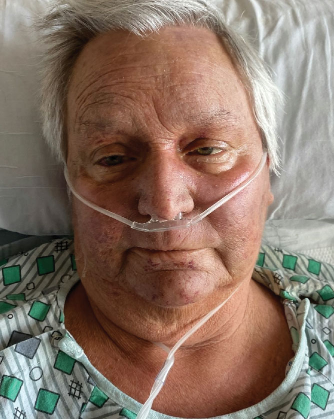
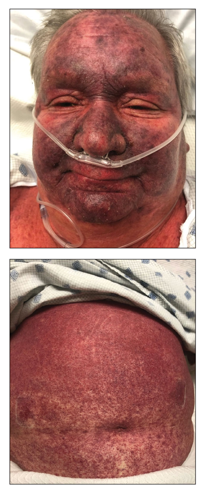
A 68-year-old woman presented to the emergency department with neutropenic fever and a rash over the body after receiving 2 doses of cladribine therapy for hairy cell leukemia. Physical examination demonstrated marked facial (top), lip, and tongue swelling, as well as a diffuse dusky nonpalpable purpuric rash on the abdomen (bottom) and back involving 90% of the body surface area. Bilateral ear edema was appreciated with accentuation of the earlobe crease. The patient exhibited subconjunctival hemorrhage, ectropion, and scleral injection. A punch biopsy of the thigh was performed.

Plastic Surgeon Illegally Restricted Negative Reviews, Judge Rules
A plastic surgeon broke federal law when he restricted patients from posting negative reviews by requiring them to sign nondisclosure agreements before they received care, a district judge has ruled.
Seattle-based surgeon Javad Sajan, MD, ran afoul of the Consumer Review Fairness Act (CRFA) by requiring more than 10,000 patients to sign the agreements, according to a recent decision by US District Judge Ricardo S. Martinez. The law protects consumers’ rights to post truthful reviews about businesses.
Judge Martinez wrote that the terms of Dr. Sajan’s nondisclosure agreements “clearly include language prohibiting or restricting patients from posting negative reviews,” in violation of CRFA. Penalties for the offense will be determined at a September trial.
This news organization contacted Dr. Sajan’s office and his attorney for comment but did not get a response.
The decision is the latest development in an ongoing legal dispute between Dr. Sajan and the State of Washington over whether the surgeon’s efforts to limit negative online reviews were illegal.
Beginning in 2017, Dr. Sajan and his practice, Allure Esthetic, introduced agreements that “forced” patients to contact the business directly if they had concerns rather than post a negative review, according to a 2022 lawsuit against Dr. Sajan filed by Washington Attorney General Robert Ferguson.
“Online reviews are often the first stop when consumers are determining who to trust,” Mr. Ferguson said in a statement. “That’s especially critical when those services deal with a patient’s health and safety. We will take action against those who illegally stop Washingtonians from sharing reviews with the public.”
If patients posted negative reviews, the clinic, in some cases, threatened litigation, according to the complaint. In other cases, patients were allegedly offered money and free services in exchange for taking the reviews down. Patients who accepted cash or services were required to sign a second agreement forbidding them from posting future negative reviews and imposing a $250,000 penalty for failure to comply, according to court documents.
In court documents, Dr. Sajan’s attorneys argued the agreements did not violate CRFA because patients had the opportunity to modify the language or decline signing them, which hundreds did. The CRFA requires Mr. Ferguson to prove that consumers lacked a meaningful opportunity to negotiate the terms, attorneys for Dr. Sajan argued in court records.
But Judge Martinez wrote that the patients who declined to sign the agreements or changed the terms represented only a “tiny fraction” of the affected patients.
The agreement language restricts patients from speaking out by forcing dissatisfied patients to work with Allure until a resolution is reached, Judge Martinez noted in his decision. “At the very least, this would delay patients from posting such reviews and force patients to interact in some way with Allure, and it certainly appears to prohibit posting reviews until Allure agrees to some kind of favorable resolution.”
Surgeon Posted Fake Positive Reviews to Counteract Bad Reviews, AG Says
Employee accounts in court documents describe a physician fixated on reviews who went to great lengths to ensure positive reviews about his work outweighed the negative.
Former employees said they were instructed to track down patients who left negative reviews and either “threaten” them to take the posts down or offer them “money” or other things, according to Mr. Ferguson’s lawsuit. If patients could not be identified, the practice would file a defamation lawsuit against the anonymous person who posted the review and use litigation to subpoena the website for the reviewer’s IP address in order to identify them, according to court documents.
Employees testified they had regular meetings to review current negative reviews and discuss what steps they were taking to get them removed. At team meetings, in-house counsel would regularly present an Excel spreadsheet with updates on progress in getting patients to remove negative reviews, according to court documents.
In addition to restricting negative reviews, Mr. Ferguson accuses Dr. Sajan of posting fake positive reviews and “buying” thousands of fake followers on social media.
At Dr. Sajan’s direction, employees created Gmail accounts using stock photos for their profile pictures and used the accounts to post fake reviews of Allure Esthetic and Dr. Sajan, according to the complaint. The practice also used members of an online forum called BlackHatWorld.com to create fake email accounts and to post fake reviews, the attorney general alleges. Many of the fake positive reviews, including the fake Google reviews, still appear on online review sites today, the attorney general contends.
Dr. Sajan and his practice also allegedly manipulated social media to appear more popular. Mr. Ferguson claims that Dr. Sajan instructed his former web designer to purchase 60,000 followers through a vendor on BlackHatWorld.com. Most of Dr. Sajan’s current Instagram followers are not real, according to Mr. Ferguson.
The practice also used a social media bot tool to buy thousands of fake likes on Instagram, YouTube, and other social media, according to court documents.
In addition, Dr. Sajan and his practice are accused of significantly altering “before and after” photos of patients and using fake email accounts to allow the clinic to take skincare rebates intended for patients.
All of these practices violated HIPAA, the state Consumer Protection Act (CPA) and the federal CRFA, according to Mr. Ferguson.
Surgeon Claims Competitor Behind Allegations
Attorneys for Dr. Sajan argue a competitor is behind the accusations and that other regulatory entities determined the practice did nothing wrong.
The competitor, a Seattle-based plastic surgeon, filed numerous complaints about Dr. Sajan to the Washington Medical Commission (WMC), according to court documents. The medical commission reviewed the third agreement and closed its investigation, finding that if the allegations were true, “no violation of law occurred,” court records show.
“Defendants relied upon this closing code from the WMC that the (non-disclosure) forms were lawful,” Dr. Sajan’s attorneys wrote in court documents.
The US Department of Health & Human Services Office for Civil Rights (OCR) also reviewed and audited Dr. Sajan’s use of the agreements, his attorneys noted. In a notice from OCR included in court exhibits, the agency wrote that all matters at issue have now been resolved through the practice’s voluntary compliance actions and that it was closing its investigation.
Attorneys for Dr. Sajan accuse Mr. Ferguson and state investigators of withholding the full extent of the competitor’s involvement in their investigation and failing to identify the competitor in written discovery or any of its initial disclosures. Dr. Sajan and his team discovered that the competitor was a source of key information through public records requests, according to court documents.
The remaining claims against Dr. Sajan will be addressed at trial, set for September 9, 2024.
A version of this article appeared on Medscape.com.
Study Evaluates CVD, Mortality Risks In Patients With Prurigo Nodularis
TOPLINE:
, particularly among women and White patients.
METHODOLOGY:
- Studies have shown increased risks for cardiovascular diseases in patients with PN, but limited sample sizes have hindered further subgroup analysis. Given PN’s pronounced sex and ethnicity skew, it is important to examine underrepresented groups to accurately assess their cardiovascular risk.
- In this propensity-score matched analysis, researchers identified 64,801 patients (59.44% women) with PN using electronic health reports from the Global Collaborative Network of TriNetX and matched to individuals without PN.
- Researchers calculated risks for 15 cardiovascular endpoints and all-cause mortality within 10 years of diagnosis. Major adverse cardiovascular events (MACE) included acute cerebral and myocardial infarction (MI), heart failure, ventricular arrhythmia, and sudden cardiac death.
TAKEAWAY:
- Patients with PN showed a higher risk for death (hazard ratio [HR], 1.1243) and MACE (HR, 1.117) (P < .0001 for both).
- PN was also associated with a higher risk for heart failure (HR, 1.062), thrombotic venous disease (HR, 1.26), angina pectoris (HR, 1.096), and peripheral arterial diseases (HR, 1.082) (P < .0001 for all) and for acute MI (HR, 1.11; P = .0015) and valve disorders (HR, 1.08; P = .0018).
- White patients with PN had a significantly increased risk for MACE, death, heart failure, cardiac arrest, vascular diseases, and acute MI, but this was not observed in people of color.
- Women exhibited a higher risk for MACE, heart failure, peripheral artery disease, acute MI, conduction disease, and valve disorders, while men did not have an increased risk for major or acute cardiovascular events. Both men and women had a higher risk for death, chronic ischemic heart disease, and venous disease.
IN PRACTICE:
“Although no novel PN-specific treatment rationale can be derived from the presented data, the potential risk of subsequent cardiovascular disease should be considered in the care of patients with PN, which includes screening and optimal management of other additional cardiovascular risk factors,” the authors wrote.
LIMITATIONS:
Retrospective observational design introduced inherent biases. Misdiagnosis or false coding in electronic health records could affect the data accuracy and ethnicity-specific analyses.
SOURCE:
This work, led by Henning Olbrich, from the Department of Dermatology, University of Lübeck, Germany, was published online in eBioMedicine.
DISCLOSURES:
The study was supported by the University of Lübeck, the Deutsche Forschungsgemeinschaft, and the State of Schleswig-Holstein. One author declared financial ties outside this work, and one author is an employee of TriNetX.
A version of this article appeared on Medscape.com.
Docs Vent As Feds Investigate Private Equity, Consolidation in Medicine
As three federal agencies investigate how private equity ownership and consolidation of healthcare organizations affects patient care and costs, physicians are giving them an earful.
“Before I retired, I could already see the damage private equity was doing to hospitals and medical practices. Well-regarded physician groups were being bought and the respected doctors and staff forced out to squeeze out profit for the buyers. Hospital-based physicians were being hit especially hard,” wrote Rhonda Wright, MD, of Brookhaven, Georgia.
“Now, the rot is setting in for emergency rooms. One in four ERs is now (under-)staffed by private equity firms. This is leading to longer wait times, deterioration in patient care, and higher bills,” Dr. Wright continued. “Private equity takeover of medicine must be stopped. All such deals should be strictly regulated and should be heavily scrutinized, if not barred altogether. Our health depends upon it!”
The federal government is accepting public comments like Dr. Wright’s through June 5 and has even set up a website (healthycompetition.gov) to make it easier to file complaints against health organizations possibly violating antitrust laws.
The US Department of Justice’s Antitrust Division, the Federal Trade Commission (FTC), and the Department of Health and Human Services want to hear from physicians and the public about how private equity firms’ investments in healthcare entities, such as hospitals, nursing homes, or specialty service providers, affect patients and healthcare workers. The investigation will also evaluate how market pricing, competition, and referral patterns change when practices and hospitals are acquired by health systems or insurers.
Maintaining competition in the provider and payer markets benefits healthcare workers through higher pay, while patients can access quality care at lower prices, the joint request for information said. However, consolidation and mergers — potentially driven by private equity’s entry into the market — can diminish these benefits.
Investigating private equity and consolidation in medicine is part of the Biden Administration’s focus on lowering medical and prescription drug costs and strengthening competition in healthcare. The FTC’s vote last week to ban noncompete agreements, which business groups have vowed to challenge in court, falls under the same initiative.
Alexandra Nicole Thran, MD, FACEP, president of the Vermont Chapter of the American College of Emergency Physicians, said that the private equity business model is problematic because it ties physicians’ wages to patient satisfaction and the number of patients they see per hour.
A Connecticut primary care physician expressed similar sentiments. “Physicians are being forced into a system where corporations provide financial incentives and punitive policies to direct healthcare decisions towards a profitable aim,” said Eric Schwaber, MD.
While a majority of comments criticized the role of private equity and consolidation, some reflected a more positive view.
“Private equity helps make healthcare more efficient and effective. It brings needed operational and managerial expertise to allow for better patient care,” said Reenie Abraham, MD, an associate professor in the Department of Internal Medicine at University of Texas Southwestern Medical Center, Dallas. The University of Texas is facing a lawsuit involving the liability status of its physicians who work for a private equity-backed hospital partly owned by the university.
Several public comments point to the increasing market influence UnitedHealth Group (UHG) and other payers have obtained through recent acquisitions. Retired emergency room physician Scott Davis, MD, said that the “astronomical” rate of burnout among providers has been exacerbated by “the economic takeover of the healthcare system by…United Healthcare [and] private equity groups who put profits over anything else.”
The healthcare conglomerate employs approximately 10% of active US physicians, including many through its subsidiary, Optum Health, which provides primary, urgent, and surgical care. UHG has also invested heavily in acquiring physician practices to advance its value-based care model.
“If a publicly traded private insurance or private equity company is interested in their short-term quarterly profits or stock price, there is little interest in the…effective management of chronic disease, other than that which fulfills a ‘value-based’ metric,” wrote Kenneth Dolkart, MD, FACP, clinical assistant professor at the Dartmouth Geisel School of Medicine in Hanover, New Hampshire.
Sarah Ealy, a revenue cycle professional, commented that payers like UHG have outsized bargaining power when negotiating rates with providers. “In many states, United Healthcare and its subsidiaries pay a lower reimbursement rate than state Medicaid plans — these rates are nearly 50% of the breakeven per-visit rate that practices need to keep the lights on.”
Another comment ties the recent cyberattack on UHG-owned Change Healthcare to private equity ownership and “healthcare behemoths buying up practices and data.”
“The ramrodding of consolidation and private oversight with little to no barriers to foreign intrusions…is a testament to how ill prepared [the] US market is to private equity healthcare takeovers,” said SW Dermatology Practice LLC.
The agencies request comments from all health market participants, including physicians, nurses, employers, administrators, and patients.
A version of this article first appeared on Medscape.com.
Satisfactory Results, Less Pain When Surface Anesthesia Used with Thermomechanical Fractional Injury Therapy
BALTIMORE — , a small study of the recently cleared device found.
The study enrolled 12 patients who were undergoing treatment for periorbital rhytides, or wrinkles, around the eyes. Seven of them received topical anesthetic cream 20 minutes before the procedure, while five were given ice packs to self-apply for 5 minutes beforehand. Patients received four treatment sessions with a month between sessions and were then evaluated up to 3 months after their last session. Study results were presented at the annual conference of the American Society for Laser Medicine and Surgery.
“Based on the approach that we had used, which was to keep the device parameters the same for those who received topical anesthetics and those who received ice, when we looked at the pain levels that the patients had relayed to us when we were doing the procedure, we found that both of them were almost exactly the same in terms of discomfort, a level of 3-4 out of 10, with 10 being the highest discomfort level,” lead investigator Jerome M. Garden, MD, said in an interview after the conference.
“In terms of patient satisfaction using a range of 0-5, again it was fairly equivalent” between the two groups, said Dr. Garden, professor of clinical dermatology and biomedical engineering at Northwestern University, Chicago. “And the overall satisfaction rate was high.”

The same device settings were used for all procedures: A pulse duration of 10 milliseconds and a protrusion depth of 400 micrometers. Double passes were applied using the standard device tip, with the smaller tip used in tighter areas, Dr. Garden said. Three patients were Fitzpatrick skin type (FST) I, six were FST II, and three were FST III.
Study Results
The results for the different anesthetic methods were almost identical. Those using ice reported a 0-10 average pain level of 3.95 ± 1.5, while those who received the topical anesthetic reported a pain level of 3.92 ± 1.5. In terms of self-graded improvement at 3-month follow-up, using a scale of 1-4, with 1 representing up to a 25% improvement and 4 a 75%-100% improvement, the patients using ice had a 2.6 ± 0.5 improvement and those using topical cream a 2.8 ± 0.5 improvement, Dr. Garden said during his presentation.
In terms of patient satisfaction, rated on a scale of 1-5, with 5 being very satisfied, the average grade was 3.9 ± 0.9, Dr. Garden added, with 70% rating ≥ 4.
“This is a relatively new device, which uses an approach to help texture changes in wrinkling on the skin in a different fashion than any of the other devices that are currently out there,” Dr. Garden told this news organization after the conference. “I wanted to understand more in depth the different parameters that may impact the outcome” with this device, he added.
The thermomechanical fractional injury device, originally cleared by the US Food and Drug Administration (FDA) in 2021, with a second-generation device cleared in June 2023, deposits heat into the skin, producing controlled thermal injury to promote collagen and elastin production. The device uses only heat, not a laser, which penetrates the skin. A heat sensation on the skin during the procedure can affect patients differently depending on their level of tolerance, Dr. Garden said during his presentation.
Managing Patient Discomfort
Kachiu C. Lee, MD, MPH, of the Main Line Center for Laser Surgery in Ardmore, Pennsylvania, has used the device in her own practice and found that “it can definitely get a little bit uncomfortable for patients,” she said in an interview after the conference.
“I would say that as the doctor, my number one priority is to always make sure my patients are comfortable, especially when treating a sensitive area like the eyes,” added Dr. Lee, who was not involved with the study. “I don’t want them to suddenly jump or move from the discomfort when I have a device right next to their eye. I think that the patient comfort is very important to make sure that we’re managing their discomfort so that the procedure is tolerable.”

She added, “Dr. Garden’s study was effective at showing that surface anesthesia, whether it be with an ice pack 5 minutes before or a topical numbing cream, can be very effective in reducing the pain level while also not interfering with the efficacy of the treatment itself.”
Dr. Garden serves on the medical advisory board for Novoxel, maker of the device. Dr. Lee had no relevant disclosures.
A version of this article appeared on Medscape.com.
BALTIMORE — , a small study of the recently cleared device found.
The study enrolled 12 patients who were undergoing treatment for periorbital rhytides, or wrinkles, around the eyes. Seven of them received topical anesthetic cream 20 minutes before the procedure, while five were given ice packs to self-apply for 5 minutes beforehand. Patients received four treatment sessions with a month between sessions and were then evaluated up to 3 months after their last session. Study results were presented at the annual conference of the American Society for Laser Medicine and Surgery.
“Based on the approach that we had used, which was to keep the device parameters the same for those who received topical anesthetics and those who received ice, when we looked at the pain levels that the patients had relayed to us when we were doing the procedure, we found that both of them were almost exactly the same in terms of discomfort, a level of 3-4 out of 10, with 10 being the highest discomfort level,” lead investigator Jerome M. Garden, MD, said in an interview after the conference.
“In terms of patient satisfaction using a range of 0-5, again it was fairly equivalent” between the two groups, said Dr. Garden, professor of clinical dermatology and biomedical engineering at Northwestern University, Chicago. “And the overall satisfaction rate was high.”
The same device settings were used for all procedures: A pulse duration of 10 milliseconds and a protrusion depth of 400 micrometers. Double passes were applied using the standard device tip, with the smaller tip used in tighter areas, Dr. Garden said. Three patients were Fitzpatrick skin type (FST) I, six were FST II, and three were FST III.
Study Results
The results for the different anesthetic methods were almost identical. Those using ice reported a 0-10 average pain level of 3.95 ± 1.5, while those who received the topical anesthetic reported a pain level of 3.92 ± 1.5. In terms of self-graded improvement at 3-month follow-up, using a scale of 1-4, with 1 representing up to a 25% improvement and 4 a 75%-100% improvement, the patients using ice had a 2.6 ± 0.5 improvement and those using topical cream a 2.8 ± 0.5 improvement, Dr. Garden said during his presentation.
In terms of patient satisfaction, rated on a scale of 1-5, with 5 being very satisfied, the average grade was 3.9 ± 0.9, Dr. Garden added, with 70% rating ≥ 4.
“This is a relatively new device, which uses an approach to help texture changes in wrinkling on the skin in a different fashion than any of the other devices that are currently out there,” Dr. Garden told this news organization after the conference. “I wanted to understand more in depth the different parameters that may impact the outcome” with this device, he added.
The thermomechanical fractional injury device, originally cleared by the US Food and Drug Administration (FDA) in 2021, with a second-generation device cleared in June 2023, deposits heat into the skin, producing controlled thermal injury to promote collagen and elastin production. The device uses only heat, not a laser, which penetrates the skin. A heat sensation on the skin during the procedure can affect patients differently depending on their level of tolerance, Dr. Garden said during his presentation.
Managing Patient Discomfort
Kachiu C. Lee, MD, MPH, of the Main Line Center for Laser Surgery in Ardmore, Pennsylvania, has used the device in her own practice and found that “it can definitely get a little bit uncomfortable for patients,” she said in an interview after the conference.
“I would say that as the doctor, my number one priority is to always make sure my patients are comfortable, especially when treating a sensitive area like the eyes,” added Dr. Lee, who was not involved with the study. “I don’t want them to suddenly jump or move from the discomfort when I have a device right next to their eye. I think that the patient comfort is very important to make sure that we’re managing their discomfort so that the procedure is tolerable.”
She added, “Dr. Garden’s study was effective at showing that surface anesthesia, whether it be with an ice pack 5 minutes before or a topical numbing cream, can be very effective in reducing the pain level while also not interfering with the efficacy of the treatment itself.”
Dr. Garden serves on the medical advisory board for Novoxel, maker of the device. Dr. Lee had no relevant disclosures.
A version of this article appeared on Medscape.com.
FROM ASLMS 2024
Consider a Four-Step Approach to Shared Decision-Making in Pediatric Dermatology
SAN DIEGO — In the clinical experience of Kelly M. Cordoro, MD, .
“SDM is a cornerstone of person-centered care,” Dr. Cordoro, professor of dermatology and pediatrics at the University of California, San Francisco, said at the Society for Pediatric Dermatology meeting, held in advance of the annual meeting of the American Academy of Dermatology. “We do it all the time. It can be patient-led, clinician-led, or a patient/family dyad approach. If we do it well, it can improve outcomes. Patients report more satisfying interactions with their care team. It brings adolescent patients especially a sense of independence and they adapt faster to their illness.”

Conditions such as acne, psoriasis, and atopic dermatitis have multiple treatment options, often without a single best choice. The ideal treatment depends on disease characteristics (extent, sites affected, symptoms, and natural history), the patient (age, comorbidities, overall disease burden), therapies (safety, efficacy, duration, and adverse events), and preferences (logistics, time, shots vs. pills, etc.). “These factors vary between patients and within the same patient over time, and at each step along the course of the condition, SDM approaches are relevant,” she said.
AHRQ’s Five-Step Approach
The Agency for Healthcare Research and Quality developed a five-step approach to SDM known as SHARE: Seek your patient’s participation; Help your patient explore and compare treatment options; Assess your patient’s values and preferences; Reach a decision with your patient, and Evaluate your patient’s decision. “We do this all the time in practice with adult patients, but may not label it as SDM,” said Dr. Cordoro, chief and fellowship director of pediatric dermatology at UCSF.
“Where it gets a little murkier is in pediatric decision-making, which is a complex type of surrogate decision-making.” In this situation the patient — a minor — does not have full autonomy. The challenge for caregivers is that giving or withholding permission for interventions is a difficult role. “Their job is to protect the patient’s well-being while empowering them toward independence,” she said. “It can be hard for caregivers to understand complex information.” The challenge for clinicians, she continued, is to know when to invite SDM. This requires relational and sharp communication skills. “We must consider our patient’s/family’s health literacy and be sure the information we share is understood,” she said. “What are the social and structural determinants of health that are going to influence decision-making? You want to move into a relationship like this with cultural humility so you can understand what their preferences are and how they’re seeing the problem. Because there’s no universal agreement on the age at which minors should be deemed decision-making competent in health care, the approach is nuanced and depends on each individual patient and family.”
Dr. Cordoro proposed the following four-step approach to SDM to use in pediatric dermatology:
Step 1: Share relevant information about the condition and treatment options in a clear and understandable manner. The average US resident is at the seventh-to eighth-grade level, “so we have to avoid medical jargon and use plain language,” Dr. Cordoro said. Then, use the teach-back approach to assess their understanding. “Ask, ‘What is your understanding of the most important points that we talked about?’ Or, ‘Please share with me what you heard so I’m sure we all understand the plan.’ Using these techniques will reduce the barriers to care such as health literacy.”
Step 2: Solicit and understand patient/patient family perspectives, preferences and priorities. The goal here is to uncover their beliefs, concerns, and assumptions that may influence their decisions. “Be mindful of power asymmetry,” she noted. “Many families still believe the doctor is the boss and they are there to be told what to do. Be clear that the patient has a say. Talk directly to the patient about their interests if developmentally appropriate.”
Step 3: Invite patients/family into a shared decision-making conversation. Consider statements like, “There are many reasonable options here. Let’s work together to come up with the decision that’s right for you.” Or, “Let’s start by exploring your specific goals and concerns. As you think about the options I just talked to you about, what’s important to you?” Or, “Do you want to think about this decision with anyone else?”
Step 4: Check back in frequently. Pause between significant points and check in. “See how they’re doing during the conversation,” she said. “At future appointments, remember to solicit their input on additional decisions.”
In Dr. Cordoro’s opinion, one potential pitfall of SDM is an over-reliance on patient decision aids. “Very few are available in dermatology,” she said. “Some are relevant but none specifically to pediatric dermatology. They are often complex and require a high reading comprehension level. This disadvantages patients and families with low health literacy. Keep it clear and simple. Your patients will appreciate it.”
Dr. Cordoro reported having no relevant disclosures.
SAN DIEGO — In the clinical experience of Kelly M. Cordoro, MD, .
“SDM is a cornerstone of person-centered care,” Dr. Cordoro, professor of dermatology and pediatrics at the University of California, San Francisco, said at the Society for Pediatric Dermatology meeting, held in advance of the annual meeting of the American Academy of Dermatology. “We do it all the time. It can be patient-led, clinician-led, or a patient/family dyad approach. If we do it well, it can improve outcomes. Patients report more satisfying interactions with their care team. It brings adolescent patients especially a sense of independence and they adapt faster to their illness.”
Conditions such as acne, psoriasis, and atopic dermatitis have multiple treatment options, often without a single best choice. The ideal treatment depends on disease characteristics (extent, sites affected, symptoms, and natural history), the patient (age, comorbidities, overall disease burden), therapies (safety, efficacy, duration, and adverse events), and preferences (logistics, time, shots vs. pills, etc.). “These factors vary between patients and within the same patient over time, and at each step along the course of the condition, SDM approaches are relevant,” she said.
AHRQ’s Five-Step Approach
The Agency for Healthcare Research and Quality developed a five-step approach to SDM known as SHARE: Seek your patient’s participation; Help your patient explore and compare treatment options; Assess your patient’s values and preferences; Reach a decision with your patient, and Evaluate your patient’s decision. “We do this all the time in practice with adult patients, but may not label it as SDM,” said Dr. Cordoro, chief and fellowship director of pediatric dermatology at UCSF.
“Where it gets a little murkier is in pediatric decision-making, which is a complex type of surrogate decision-making.” In this situation the patient — a minor — does not have full autonomy. The challenge for caregivers is that giving or withholding permission for interventions is a difficult role. “Their job is to protect the patient’s well-being while empowering them toward independence,” she said. “It can be hard for caregivers to understand complex information.” The challenge for clinicians, she continued, is to know when to invite SDM. This requires relational and sharp communication skills. “We must consider our patient’s/family’s health literacy and be sure the information we share is understood,” she said. “What are the social and structural determinants of health that are going to influence decision-making? You want to move into a relationship like this with cultural humility so you can understand what their preferences are and how they’re seeing the problem. Because there’s no universal agreement on the age at which minors should be deemed decision-making competent in health care, the approach is nuanced and depends on each individual patient and family.”
Dr. Cordoro proposed the following four-step approach to SDM to use in pediatric dermatology:
Step 1: Share relevant information about the condition and treatment options in a clear and understandable manner. The average US resident is at the seventh-to eighth-grade level, “so we have to avoid medical jargon and use plain language,” Dr. Cordoro said. Then, use the teach-back approach to assess their understanding. “Ask, ‘What is your understanding of the most important points that we talked about?’ Or, ‘Please share with me what you heard so I’m sure we all understand the plan.’ Using these techniques will reduce the barriers to care such as health literacy.”
Step 2: Solicit and understand patient/patient family perspectives, preferences and priorities. The goal here is to uncover their beliefs, concerns, and assumptions that may influence their decisions. “Be mindful of power asymmetry,” she noted. “Many families still believe the doctor is the boss and they are there to be told what to do. Be clear that the patient has a say. Talk directly to the patient about their interests if developmentally appropriate.”
Step 3: Invite patients/family into a shared decision-making conversation. Consider statements like, “There are many reasonable options here. Let’s work together to come up with the decision that’s right for you.” Or, “Let’s start by exploring your specific goals and concerns. As you think about the options I just talked to you about, what’s important to you?” Or, “Do you want to think about this decision with anyone else?”
Step 4: Check back in frequently. Pause between significant points and check in. “See how they’re doing during the conversation,” she said. “At future appointments, remember to solicit their input on additional decisions.”
In Dr. Cordoro’s opinion, one potential pitfall of SDM is an over-reliance on patient decision aids. “Very few are available in dermatology,” she said. “Some are relevant but none specifically to pediatric dermatology. They are often complex and require a high reading comprehension level. This disadvantages patients and families with low health literacy. Keep it clear and simple. Your patients will appreciate it.”
Dr. Cordoro reported having no relevant disclosures.
FROM AAD 2024
Do Patients Benefit from Cancer Trial Participation?
TOPLINE:
METHODOLOGY:
- The view that patients with cancer benefit from access to investigational drugs in the clinical trial setting is widely held but does necessarily align with trial findings, which often show limited evidence of a clinical benefit. First, most investigational treatments assessed in clinical trials fail to gain regulatory approval, and the minority that are approved tend to offer minimal clinical benefit, experts explained.
- To estimate the survival benefit and toxicities associated with receiving experimental treatments, researchers conducted a meta-analysis of 128 trials comprising 141 comparisons of an investigational drug and a control treatment, which included immunotherapies and targeted therapies.
- The analysis included 42 trials in non–small cell lung cancer (NSCLC), 37 in breast cancer, 15 in hepatobiliary cancer, 13 in pancreatic cancer, 12 in colorectal cancer, and 10 in prostate cancer, involving a total of 47,050 patients.
- The primary outcome was PFS and secondary outcomes were overall survival and grades 3-5 serious adverse events.
TAKEAWAY:
- Overall, the experimental treatment was associated with a 20% improvement in PFS (pooled hazard ratio [HR], 0.80), corresponding to a median 1.25-month PFS advantage. The PFS benefit was seen across all cancer types, except pancreatic cancer.
- Overall survival improved by 8% with experimental agents (HR, 0.92), corresponding to 1.18 additional months. A significant overall survival benefit was seen across NSCLC, breast cancer, and hepatobiliary cancer trials but not pancreatic, prostate, colorectal cancer trials.
- Patients in the experimental intervention group, however, experienced much higher risk for grade 3-5 serious adverse events (risk ratio [RR], 1.27), corresponding to 7.40% increase in absolute risk. The greater risk for serious adverse events was significant for all indications except prostate cancer (RR, 1.13; 95% CI, 0.91-1.40).
IN PRACTICE:
“We believe our findings are best interpreted as suggesting that access to experimental interventions that have not yet received full FDA approval is associated with a marginal but nonzero clinical benefit,” the authors wrote.
“Although our findings seem to reflect poorly on trials as a vehicle for extending survival for participants, they have reassuring implications for clinical investigators, policymakers, and institutional review boards,” the researchers said, explaining that this “scenario allows clinical trials to continue to pursue promising new treatments — supporting incremental advances that sum to large gains over extended periods of research — without disadvantaging patients in comparator groups.”
SOURCE:
Renata Iskander, MSc, of McGill University, Montreal, Quebec, Canada, led this work, which was published online on April 29, 2024, in Annals of Internal Medicine.
LIMITATIONS:
There was high heterogeneity across studies due to variations in drugs tested, comparators used, and populations involved. The use of comparators below standard care could have inflated survival benefits. Additionally, data collected from ClinicalTrials.gov might be biased due to some trials not being reported.
DISCLOSURES:
Canadian Institutes of Health Research supported this work. The authors received grants for this work from McGill University, Rossy Cancer Network, and National Science Foundation. One author received consulting fees outside this work. The other authors declared no competing interests.
A version of this article appeared on Medscape.com.
Do Health-Related Social Needs Raise Mortality Risk in Cancer Survivors?
Little is known about the specific association between health-related social needs (HRSNs) and mortality risk even though HRSNs, defined as challenges in affording food, housing, and other necessities of daily living, are potential challenges for cancer survivors, wrote Zhiyuan Zheng, PhD, of the American Cancer Society, Atlanta, and colleagues.
A 2020 study by Dr. Zheng and colleagues published in the Journal of the National Comprehensive Cancer Network (NCCN) showed that food insecurity and financial worries had a negative impact on cancer survivorship. In the new study, published in Cancer, the researchers identified cancer survivors using the 2013-2018 National Health Interview Survey (NHIS) and the NHIS Mortality File through December 31, 2019. The researchers examined mortality using the data from the Centers for Disease Control and Prevention’s National Death Index (NDI) through December 31, 2019, which links to the National Health Interview Survey Data used in the study.
Individuals’ HRSNs were categorized into three groups: severe, moderate, and minor/none. HRSNs included food insecurity and nonmedical financial concerns, such as housing costs (rent, mortgage). Medical financial hardship included material, psychological, and behavioral domains and was divided into three groups: 2-3 domains, 1 domain, or 0 domains.
What Are the Potential Financial Implications of this Research?
The high costs of cancer care often cause medical financial hardships for cancer survivors, and expenses also may cause psychological distress and nonmedical financial hardship as survivors try to make ends meet while facing medical bills, wrote Dr. Zheng and colleagues.
Policy makers are increasingly interested in adding HRSNs to insurance coverage; recent guidance from the Centers for Medicare & Medicaid Services (CMS) allows individual states to apply to provide nutrition and housing supports through state Medicaid programs, according to authors of a 2023 article published in JAMA Health Forum.
The new study adds to the understanding of how HRSNs impact people with cancer by examining the association with mortality risk, Yelak Biru, MSc, president and chief executive officer of the International Myeloma Foundation, said in an interview.
“This is a key area of study for addressing the disparities in treatments and outcomes that result in inequities,” said Mr. Biru, a patient advocate and multiple myeloma survivor who was not involved in the study.
What Does the New Study Show?
The new study characterized HRSNs in 5,855 adult cancer survivors aged 18-64 years and 5,918 aged 65-79 years. In the 18- to 64-year-old group, 25.5% reported moderate levels of HRSNs, and 18.3% reported severe HRSNs. In patients aged 65-79 years, 15.6% and 6.6% reported moderate HRSNs and severe HRSNs, respectively.
Severe HRSN was significantly associated with higher mortality risk in an adjusted analysis in patients aged 18-64 years (hazard ratio 2.00, P < .001).
Among adults aged 65-79 years, severe HRSN was not associated with higher mortality risk; however, in this older age group, those with 2-3 domains of medical financial hardship had significantly increased mortality risk compared with adults aged 65-79 years with zero domains of medical financial hardship (HR 1.58, P = .007).
Although the findings that HRSNs were associated with increased mortality risk, especially in the younger group, were not surprising, they serve as a call to action to address how HRSNs are contributing to cancer mortality, Mr. Biru said in an interview. “HRSNs, like food or housing insecurity, can lead to patients being unable to undergo the best treatment approach for their cancer,” he said.
What Are the Limitations and Research Gaps?
The study findings were limited by several factors including the use of self-reports to measure medical financial hardship, food insecurity, and nonmedical financial concerns in the NHIS, the researchers wrote in their discussion. More research with longer follow-up time beyond 1-5 years is needed, wrote Dr. Zheng and colleagues.
Studies also are needed to illustrate how patient navigation can help prevent patients from falling through the cracks with regard to social needs and financial hardships, Mr. Biru told this news organization.
Other areas for research include how addressing social needs affects health outcomes and whether programs designed to address social needs are effective, he said.
“Finally, qualitative research is needed to capture the lived experiences of cancer survivors facing these challenges. This knowledge can inform the development of more patient-centered interventions and policies that effectively address the social determinants of health and improve overall outcomes for all cancer survivors,” Mr. Biru said.
What Is the Takeaway Message for Clinicians?
HRSNs and financial hardship are significantly associated with increased risk of mortality in adult cancer survivors, Dr. Zheng and colleagues concluded. Looking ahead, comprehensive assessment of HRSNs and financial hardship may help clinicians connect patients with relevant services to mitigate the social and financial impacts of cancer, they wrote.
“The takeaway message for oncologists in practice is that addressing [HRSNs] and financial hardship is crucial for providing comprehensive and equitable cancer care,” Mr. Biru said during his interview.
“The impact of social determinants of health on cancer outcomes cannot be ignored, and oncologists play a vital role in identifying and addressing these needs,” he said. Sensitive, discussion-based screenings are needed to identify core needs such as food and transportation, but clinicians also can consider broader social factors and work with a team to connect patients to appropriate resources, he added.
“By recognizing the importance of HRSN screening and taking proactive steps to address these needs, oncologists can contribute to improving health outcomes, reducing healthcare disparities, and providing more equitable cancer care for their patients,” he said.
What Other Guidance Is Available?
“High-quality cancer care requires treating the whole person; measuring and addressing anything in their life that could result in poorer health outcomes is a key component of comprehensive care,” Mr. Biru emphasized.
In September 2023, the National Comprehensive Cancer Network (NCCN) convened a working group cochaired by Mr. Biru that developed recommendations for how oncology practices should routinely measure HRSNs (NCCN.org/social-needs).
“The working group proposed that every cancer patient be assessed for food, transportation access, and financial and housing security at least once a year, and be reassessed at every care transition point as well,” Mr. Biru said. Such screenings should include follow-up to connect patients with services to address any HRSNs they are experiencing, he added.
Lead author Dr. Zheng is employed by the American Cancer Society, which as a nonprofit receives funds from the public through fundraising and contributions, as well as some support from corporations and industry to support its mission programs and services. Mr. Biru had no financial conflicts to disclose.
FROM CANCER
New HIV Infections After Vampire Facials at Unlicensed Spa
At least three clients of an unlicensed spa in New Mexico contracted HIV after receiving platelet-rich plasma (PRP) microneedling facials, according to an investigation by the US Centers for Disease Control and Prevention (CDC).
The investigation, spanning 5 years with parts of it still ongoing, has resulted in the closure of the spa and is raising questions about public safety in cosmetic clinics.
Though transmission of HIV by unsterile injection practices is a known risk, this is the first time it has been linked to cosmetic injection services, said Anna Stadelman-Behar, PhD, MPH, of the CDC’s Epidemic Intelligence Service.
Sometimes called a vampire facial, the PRP treatment involves taking a patient’s own blood and separating it in a centrifuge. The portion containing a high concentration of platelets is then reinjected with a syringe or microneedling device.
“The idea is that when you inject this concentrated amount of platelets, the growth factors that the platelets release help to stimulate the regenerative nature of that area,” said Anthony Rossi, MD, professor of dermatology at Weill Cornell Medical College in New York, and attending dermatologist at Memorial Sloan Kettering Cancer Center.
The infections under investigation first came to light when a woman was diagnosed with HIV with no known risk factors for the disease other than exposure to microneedling facials at a cosmetic spa.
The New Mexico Department of Health and the CDC launched an investigation of the spa and discovered a litany of “gross violations of infection control practices,” said Dr. Stadelman-Behar.
Infection-Control Violations
At the spa in New Mexico, investigators found:
- On a kitchen counter, a centrifuge, a heating dry bath, and a rack of unlabeled tubes containing blood
- In a refrigerator, unlabeled tubes of blood and medical injectables including botox and lidocaine stored along with food
- Unwrapped syringes in drawers, on counters, and discarded in regular trash cans
- No autoclave for steam sterilization on the premises
- Only surface cleaning for procedure equipment with ammonium chloride disinfecting spray and benzalkonium chloride disinfecting wipes after each client visit
- Disposable electric desiccator tips cleaned only by alcohol immersion to be reused
The spa’s owner operated without appropriate licenses at multiple locations and did not have an appointment scheduling system that stored client contact information.
Investigators contacted as many people as they could find and launched a large-scale community outreach effort to find more.
In total, four clients and one intimate partner of a client were diagnosed with HIV during the investigation, but one client and her partner were determined to likely have been infected before the spa visit.
It is not clear whether the infections were due to unlabeled contaminated blood products being given to the wrong client or contamination on shared needles. Investigators did not have the authority to collect specimens during their site visit that would have allowed them to study that.
“We can’t definitively say what the route of contamination was,” noted Dr. Stadelman-Behar.
Anne Chapas, MD, a board-certified dermatologist, and instructor at Mount Sinai Hospital in New York, added that just because a procedure is cosmetic, that doesn’t mean it is not medical. “Personally, I feel it should only be done by medical practitioners who understand the risks.”
A Medical Procedure
PRP microneedling has been used extensively in orthopedic surgery to promote joint regeneration. For the past 10 years, it has also been used in dermatology to treat hair loss from alopecia, to augment wound healing, and cosmetically to reduce facial wrinkles.
It is generally done in a doctor’s office or medical spa, and the procedure takes about half an hour.
Dr. Stadelman-Behar said that this ongoing investigation highlights the importance of front-line healthcare workers using their clinical expertise to help identify potential new routes of transmission for infections. “It was provider-led intuition that sparked this investigation, so it’s important to let the department of health know if there is something amiss with any of the exposures that the patient might have had,” she said.
A version of this article appeared on Medscape.com.