User login
What's the diagnosis?
At the week follow-up, the lesions were unchanged and the swelling on the left lateral eyebrow was worsening. A biopsy of the yellow lesion on the back and one of the scaly papules on the abdomen was performed. A fungal and bacterial cultures were also ordered.
He was referred to ophthalmology for evaluation of the eyelid swelling and an ultrasound was requested.
The skin biopsy showed a clonal proliferation of reniform histiocytes with eosinophils within the dermis. The cells were positive for S100, CD207 (langerin), and CD1a and negative for pancytokeratin and Melan-A, supportive of the diagnosis of Langerhans cell histiocytosis (LCH).
Diagnosis
The patient was admitted to the hospital, where a skeletal survey was performed, which showed an asymmetric lucency involving the left frontal calvarium extending to the superior lateral orbital rim. The brain MRI demonstrated a destructive avidly enhancing soft-tissue process which involved the superior left orbital rim likely with some degree of intracranial extension. This lesion exerts mass effect upon surrounding structures to the left ocular globe. With the skin and skeletal findings, the patient was diagnosed with LCH. His blood count was significant for thrombocytopenia. His liver and kidney function were normal. His electrolytes were also with in normal range. He was started on chemotherapy with vinblastine and systemic corticosteroids with resolution of the rash and decrease on the size of the lesion on the orbit within a few weeks.
Infantile LCH is a rare neoplastic disorder of hematopoietic myeloid precursor cells caused by activating mutations in the mitogen-activated protein kinase (MAPK) pathway, particularly BRAF-V600E mutation. White male children are mostly affected, with a peak incidence of 1-3 years of age. Nine out of 10 children with cutaneous involvement also have multisystemic disease, such as the case of our patient. LCH is classified as single or multisystem organ disease. Two-thirds of the cases present with single system involvement. Organs most commonly affected include the bone (the skull being the most commonly affected), skin, and high-risk organs like the liver, spleen, and bone marrow, and less commonly the lungs, lymph nodes, and central nervous system. Some patients can present with fever, lethargy, and weight loss. None were noted in our patient.
Skin findings of LCH can have multiple morphologies and presentations and often described as a big mimicker. In young infants like our patient, the seborrheic dermatitis–mimicking type is often seen. In other cases, the skin lesions can appear eczematous, petechial, with scabbing, crusting, or purpura. Xanthoma-like lesions, like that one our patient had in the back, have also been described. Resistant diaper dermatitis and cradle cap should prompt the clinician to think about LCH. Lesions can be so varied that can present with hypopigmentation (vitiligo like), hyperpigmentation, varicella-like papulo-pustules, and red blue nodules within others. Oral mucosa and nail involvement can also occur.
Bone involvement can present as soft-tissue mass with swelling and pain as it occur in our patient.
Endocrinopathies have been described in patients with LCH including diabetes insipidus, growth hormone deficiency, and less likely thyroid disease.
Multidisciplinary care
The diagnosis of LCH in infants necessitates a combination of clinical, radiological, and histopathologic findings. In infants, cutaneous involvement is a frequent initial presentation, with characteristic lesions that are often misdiagnosed as other dermatologic conditions. Timely recognition of these lesions and appropriate skin biopsies for histological examination are essential steps in achieving an accurate diagnosis.
Radiological imaging, including x-rays, CT, and MRI, plays a crucial role in assessing the extent of involvement.
The management of LCH in infants requires a well-coordinated multidisciplinary approach involving pediatric oncologists, dermatologists, radiologists, orthopedic surgeons, and other relevant specialists. Treatment strategies vary depending on the extent of disease involvement and the presence of risk factors. In localized cases, observation with close monitoring may be considered, as some cases of LCH in infants may undergo spontaneous regression. However, cases with severe symptoms, extensive organ involvement, or high-risk features may require systemic therapies.

Chemotherapy agents, including vinblastine and prednisone have been utilized in the treatment of infantile LCH with varying success. The selection of treatment regimens should be tailored to each individual case, considering disease severity, potential toxicities, and long-term effects. In cases of bone lesions causing significant deformities or functional impairment, surgical intervention may be necessary. Skin only disease can be treated with topical corticosteroids.
Prognosis
Survival rates in patients with single-organ involvement without risk-organ involvement is close to 100% and with risk-organ involvement of 98% at 5 years.
Long-term follow-up is essential for infants diagnosed with LCH, as recurrence and late effects can occur even after successful treatment. Continued monitoring allows for the timely detection of relapses or the development of secondary complications.
Infants thought to have common skin conditions like eczema, seborrheic dermatitis, or diaper dermatitis not responding to treatment should be referred to pediatric dermatology for evaluation to rule out the possibility of LCH.
Dr. Matiz is a pediatric dermatologist at Southern California Permanente Medical Group, San Diego.
References
Krooks J et al. J Am Acad Dermatol. 2018 Jun;78(6):1035-44.
Krooks J et al. J Am Acad Dermatol. 2018 Jun;78(6):1047-56.
Leung AKC et al. World J Pediatr. 2019 Dec;15(6):536-45.
At the week follow-up, the lesions were unchanged and the swelling on the left lateral eyebrow was worsening. A biopsy of the yellow lesion on the back and one of the scaly papules on the abdomen was performed. A fungal and bacterial cultures were also ordered.
He was referred to ophthalmology for evaluation of the eyelid swelling and an ultrasound was requested.
The skin biopsy showed a clonal proliferation of reniform histiocytes with eosinophils within the dermis. The cells were positive for S100, CD207 (langerin), and CD1a and negative for pancytokeratin and Melan-A, supportive of the diagnosis of Langerhans cell histiocytosis (LCH).
Diagnosis
The patient was admitted to the hospital, where a skeletal survey was performed, which showed an asymmetric lucency involving the left frontal calvarium extending to the superior lateral orbital rim. The brain MRI demonstrated a destructive avidly enhancing soft-tissue process which involved the superior left orbital rim likely with some degree of intracranial extension. This lesion exerts mass effect upon surrounding structures to the left ocular globe. With the skin and skeletal findings, the patient was diagnosed with LCH. His blood count was significant for thrombocytopenia. His liver and kidney function were normal. His electrolytes were also with in normal range. He was started on chemotherapy with vinblastine and systemic corticosteroids with resolution of the rash and decrease on the size of the lesion on the orbit within a few weeks.
Infantile LCH is a rare neoplastic disorder of hematopoietic myeloid precursor cells caused by activating mutations in the mitogen-activated protein kinase (MAPK) pathway, particularly BRAF-V600E mutation. White male children are mostly affected, with a peak incidence of 1-3 years of age. Nine out of 10 children with cutaneous involvement also have multisystemic disease, such as the case of our patient. LCH is classified as single or multisystem organ disease. Two-thirds of the cases present with single system involvement. Organs most commonly affected include the bone (the skull being the most commonly affected), skin, and high-risk organs like the liver, spleen, and bone marrow, and less commonly the lungs, lymph nodes, and central nervous system. Some patients can present with fever, lethargy, and weight loss. None were noted in our patient.
Skin findings of LCH can have multiple morphologies and presentations and often described as a big mimicker. In young infants like our patient, the seborrheic dermatitis–mimicking type is often seen. In other cases, the skin lesions can appear eczematous, petechial, with scabbing, crusting, or purpura. Xanthoma-like lesions, like that one our patient had in the back, have also been described. Resistant diaper dermatitis and cradle cap should prompt the clinician to think about LCH. Lesions can be so varied that can present with hypopigmentation (vitiligo like), hyperpigmentation, varicella-like papulo-pustules, and red blue nodules within others. Oral mucosa and nail involvement can also occur.
Bone involvement can present as soft-tissue mass with swelling and pain as it occur in our patient.
Endocrinopathies have been described in patients with LCH including diabetes insipidus, growth hormone deficiency, and less likely thyroid disease.
Multidisciplinary care
The diagnosis of LCH in infants necessitates a combination of clinical, radiological, and histopathologic findings. In infants, cutaneous involvement is a frequent initial presentation, with characteristic lesions that are often misdiagnosed as other dermatologic conditions. Timely recognition of these lesions and appropriate skin biopsies for histological examination are essential steps in achieving an accurate diagnosis.
Radiological imaging, including x-rays, CT, and MRI, plays a crucial role in assessing the extent of involvement.
The management of LCH in infants requires a well-coordinated multidisciplinary approach involving pediatric oncologists, dermatologists, radiologists, orthopedic surgeons, and other relevant specialists. Treatment strategies vary depending on the extent of disease involvement and the presence of risk factors. In localized cases, observation with close monitoring may be considered, as some cases of LCH in infants may undergo spontaneous regression. However, cases with severe symptoms, extensive organ involvement, or high-risk features may require systemic therapies.
Chemotherapy agents, including vinblastine and prednisone have been utilized in the treatment of infantile LCH with varying success. The selection of treatment regimens should be tailored to each individual case, considering disease severity, potential toxicities, and long-term effects. In cases of bone lesions causing significant deformities or functional impairment, surgical intervention may be necessary. Skin only disease can be treated with topical corticosteroids.
Prognosis
Survival rates in patients with single-organ involvement without risk-organ involvement is close to 100% and with risk-organ involvement of 98% at 5 years.
Long-term follow-up is essential for infants diagnosed with LCH, as recurrence and late effects can occur even after successful treatment. Continued monitoring allows for the timely detection of relapses or the development of secondary complications.
Infants thought to have common skin conditions like eczema, seborrheic dermatitis, or diaper dermatitis not responding to treatment should be referred to pediatric dermatology for evaluation to rule out the possibility of LCH.
Dr. Matiz is a pediatric dermatologist at Southern California Permanente Medical Group, San Diego.
References
Krooks J et al. J Am Acad Dermatol. 2018 Jun;78(6):1035-44.
Krooks J et al. J Am Acad Dermatol. 2018 Jun;78(6):1047-56.
Leung AKC et al. World J Pediatr. 2019 Dec;15(6):536-45.
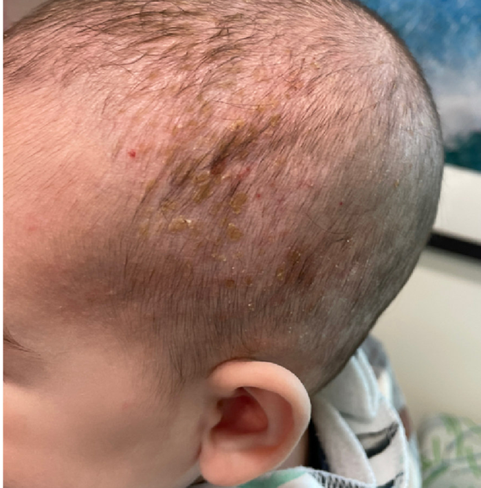
A 4-month male was referred to the pediatric dermatology clinic for a rash on the scalp, torso, and the diaper area since he was 2 months of age. He has been treated with nystatin, clotrimazole, and zinc oxide paste with partial improvement. After 2 months of partial improvement the rash worsened again, and he was referred to pediatric dermatology. The mother also reported asymptomatic left upper lateral eyebrow swelling noted a few weeks prior.

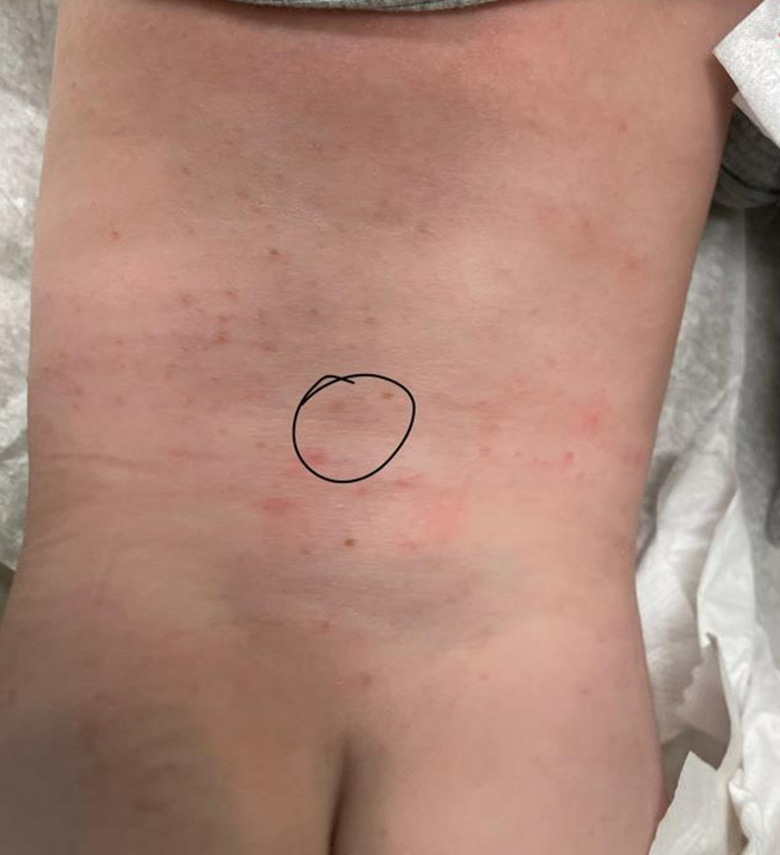
On the torso and diaper area, he had multiple scaly pink papules. On the groin he had eroded pink scaly plaques (Picture 2).
On his back he had a 3-mm yellow papule (Picture 3).
Piroxicam boosts success of levonorgestrel for emergency contraception
Adding oral piroxicam to oral levonorgestrel significantly improved the efficacy of emergency contraception, based on data from 860 women.
Oral hormonal emergency contraception (EC) is the most widely used EC method worldwide, but the two currently available drugs, levonorgestrel and ulipristal acetate (UPA), are not effective when given after ovulation, wrote Raymond Hang Wun Li, MD, of the University of Hong Kong, and colleagues. Previous studies suggest that cyclo-oxygenase (COX) inhibitors may disrupt follicular rupture and prevent ovulation, but data on their use in combination with current oral ECs are lacking, the researchers said.
In a study published in The Lancet, the researchers randomized 430 women to receive a single oral dose of 1.5 mg levonorgestrel plus 40 mg of the COX-2 inhibitor piroxicam or 1.5 mg levonorgestrel plus a placebo. The study participants were women aged 18 years and older who requested EC within 72 hours of unprotected sex and who had regular menstrual cycles between 24 and 42 days long. The median age of the participants was 30 years; 97% were Chinese. The median time from intercourse to treatment was 18 hours for both groups.
The primary outcome was the percentage of pregnancies prevented, based on pregnancy status 1-2 weeks after treatment.
One pregnancy occurred in the piroxicam group, compared with seven pregnancies in the placebo group, which translated to a significant difference in the percentage of pregnancies prevented (94.7% vs. 63.4%, P < .0001).
No trend toward increased failure rates appeared based on the time elapsed between intercourse and EC use in either group, and no differences appeared in the return or delay of subsequent menstrual periods between the groups.
The most common adverse events (reported by more than 5% of participants in both groups) included fatigue or weakness, nausea, lower abdominal pain, dizziness, and headache.
The choice of piroxicam as the COX inhibitor in conjunction with levonorgestrel for the current study had several potential advantages, the researchers wrote in their discussion. These advantages include the widespread availability and long-acting characteristics of piroxicam, which is also true of levonorgestrel, they said.
The findings were limited by several factors including the generalizability to other settings and populations, the researchers noted. The efficacy of the levonorgestrel/piroxicam combination in women with a body mass index greater than 26 kg/m2 may be lower, but the current study population did not have enough women in this category to measure the potential effect, they said. The study also did not examine the effect of piroxicam in combination with ulipristal acetate.
However, the results are the first known to demonstrate the improved effectiveness of oral piroxicam coadministered with oral levonorgestrel for EC, they said.
“The strength of this recommendation and changes in clinical guidelines may be determined upon demonstration of reproducible results in further studies,” they added.
Pill combination shows potential and practicality
Oral emergency contraception on demand is an unmet need on a global level, Erica P. Cahill, MD, of the department of obstetrics and gynecology and division of family planning services at Stanford (Calif.) University, wrote in an accompanying editorial.
Dr. Cahill noted the longer half-life of piroxicam compared with other COX-2 inhibitors, which made it a practical choice. Although the study was not powered to evaluate secondary outcomes, bleeding patterns consistent with use of EC pills were observed. Documentation of these patterns is worthwhile, Dr. Cahill said, “because people using emergency contraceptive pills might also be using fertility awareness methods and need to know when they can be certain they are not pregnant.”
Overall, the study supports the addition of 40 mg piroxicam to 1.5 mg levonorgestrel as emergency contraception, said Dr. Cahill. Future studies can build on the current findings by evaluating repeat dosing of the piroxicam/levonorgestrel combination and by evaluating the combination of COX-2 inhibitors and ulipristal acetate to prevent pregnancy, she said.
The study received no outside funding. The researchers and Dr. Cahill had no financial conflicts to disclose.
FROM THE LANCET
American Geriatrics Society 2023 updated Beers Criteria highlights
Every 4 years, an interprofessional panel of experts from the American Geriatrics Society provides updated guidelines on safe prescribing of medications in older adults, known as the Beers Criteria. A 2023 update was released in May 2023 after panel review of more 1,500 clinical trials and research studies published since the last update.
Anticoagulants
Notable changes to the 2023 guidelines include updated recommendations for anticoagulation. Warfarin should be avoided as initial therapy for venous thromboembolism or nonvalvular atrial fibrillation unless there are contraindications to direct oral anticoagulants (DOACs) or other substantial barriers to use.

Rivaroxaban should also be avoided, and dabigatran used with caution in favor of apixaban, which is felt to have a better safety profile in older adults. Rivaroxaban may be considered if once daily dosing is deemed to be more clinically appropriate. Financial barriers regarding drug coverage and formulary options were acknowledged as a significant barrier to equitable access to preferred direct oral anticoagulants in older adults.
Diabetes medication
Regarding diabetes management, short-acting sulfonylureas should be avoided in addition to long-acting sulfonylureas, because of the increased risk of hypoglycemia, and cardiovascular and all-cause mortality in older adults. Sodium-glucose cotransporter 2 inhibitors as an entire class are recommended to be used with caution, as older adults are at higher risk of euglycemic ketoacidosis and urogenital infections, particularly in women in the first month of initiating treatment.
Like DOACs, the panel acknowledged that financial considerations may lead to limited options for oral diabetic treatment. In circumstances where a sulfonylurea is used, short-acting forms are preferred over long acting to reduce the risk of prolonged hypoglycemia.
Aspirin for primary prevention
Alongside the U.S. Preventive Services Task Force guideline update in 2022 regarding aspirin for primary prevention of cardiovascular disease and stroke, the Beer’s Criteria recommend against initiation of aspirin for primary prevention in older adults. Ticagrelor and prasugrel should be used with caution because of the increased risk of major bleeding in older adults over the age of 75, compared with clopidogrel. If prasugrel is used, a lower dose of 5 mg is recommended, in line with guidelines by the American College of Cardiology and American Heart Association.
Pain medication
For pain management, the Beer’s Criteria updated recommendations to avoid NSAIDs, particularly when used in combination with steroids or anticoagulants. The panel highlights that even short-term use of NSAIDs is high risk when used in combination with steroids or anticoagulants. If no other alternatives are possible, patients should be placed on a proton pump inhibitor or misoprostol while taking NSAIDs.
Baclofen should be avoided in older adults with renal insufficiency (estimated glomerular filtration rate < 60 mL/min per 1.73 m2) because of the increased risk of encephalopathy, and when used, should be given at the lowest effective dose with close monitoring for mental status changes.
Androgen and estrogen replacement therapy
For androgen replacement therapy, the panel notes that testosterone supplementation should be avoided because of cardiovascular risks unless there is confirmed hypogonadism. The panel revised their recommendation on the basis of emerging data that a history of prostate cancer is not an absolute contraindication for exogenous testosterone. A risk versus benefit discussion about exogenous testosterone should be had with a medical oncologist or urologist in those with a history of prostate cancer.
Regarding estrogen, systemic formulations should not be initiated in women over the age of 60 because of increased risk of cardiovascular events, venous thromboembolism, and dementia. In women with a history of breast cancer, vaginal estrogens are generally felt to be safe to use at low doses, such as less than 25 mcg twice weekly.
Dr. Wang is a geriatrician and general internist at Harborview Medical Center, Seattle.
Every 4 years, an interprofessional panel of experts from the American Geriatrics Society provides updated guidelines on safe prescribing of medications in older adults, known as the Beers Criteria. A 2023 update was released in May 2023 after panel review of more 1,500 clinical trials and research studies published since the last update.
Anticoagulants
Notable changes to the 2023 guidelines include updated recommendations for anticoagulation. Warfarin should be avoided as initial therapy for venous thromboembolism or nonvalvular atrial fibrillation unless there are contraindications to direct oral anticoagulants (DOACs) or other substantial barriers to use.
Rivaroxaban should also be avoided, and dabigatran used with caution in favor of apixaban, which is felt to have a better safety profile in older adults. Rivaroxaban may be considered if once daily dosing is deemed to be more clinically appropriate. Financial barriers regarding drug coverage and formulary options were acknowledged as a significant barrier to equitable access to preferred direct oral anticoagulants in older adults.
Diabetes medication
Regarding diabetes management, short-acting sulfonylureas should be avoided in addition to long-acting sulfonylureas, because of the increased risk of hypoglycemia, and cardiovascular and all-cause mortality in older adults. Sodium-glucose cotransporter 2 inhibitors as an entire class are recommended to be used with caution, as older adults are at higher risk of euglycemic ketoacidosis and urogenital infections, particularly in women in the first month of initiating treatment.
Like DOACs, the panel acknowledged that financial considerations may lead to limited options for oral diabetic treatment. In circumstances where a sulfonylurea is used, short-acting forms are preferred over long acting to reduce the risk of prolonged hypoglycemia.
Aspirin for primary prevention
Alongside the U.S. Preventive Services Task Force guideline update in 2022 regarding aspirin for primary prevention of cardiovascular disease and stroke, the Beer’s Criteria recommend against initiation of aspirin for primary prevention in older adults. Ticagrelor and prasugrel should be used with caution because of the increased risk of major bleeding in older adults over the age of 75, compared with clopidogrel. If prasugrel is used, a lower dose of 5 mg is recommended, in line with guidelines by the American College of Cardiology and American Heart Association.
Pain medication
For pain management, the Beer’s Criteria updated recommendations to avoid NSAIDs, particularly when used in combination with steroids or anticoagulants. The panel highlights that even short-term use of NSAIDs is high risk when used in combination with steroids or anticoagulants. If no other alternatives are possible, patients should be placed on a proton pump inhibitor or misoprostol while taking NSAIDs.
Baclofen should be avoided in older adults with renal insufficiency (estimated glomerular filtration rate < 60 mL/min per 1.73 m2) because of the increased risk of encephalopathy, and when used, should be given at the lowest effective dose with close monitoring for mental status changes.
Androgen and estrogen replacement therapy
For androgen replacement therapy, the panel notes that testosterone supplementation should be avoided because of cardiovascular risks unless there is confirmed hypogonadism. The panel revised their recommendation on the basis of emerging data that a history of prostate cancer is not an absolute contraindication for exogenous testosterone. A risk versus benefit discussion about exogenous testosterone should be had with a medical oncologist or urologist in those with a history of prostate cancer.
Regarding estrogen, systemic formulations should not be initiated in women over the age of 60 because of increased risk of cardiovascular events, venous thromboembolism, and dementia. In women with a history of breast cancer, vaginal estrogens are generally felt to be safe to use at low doses, such as less than 25 mcg twice weekly.
Dr. Wang is a geriatrician and general internist at Harborview Medical Center, Seattle.
Few meet eligibility for newer Alzheimer’s drugs
, a cross sectional study has found.
Reporting in the journal Neurology, researchers from the Mayo Clinic in Rochester, Minn., and the University of Chicago found that only a small percentage of patients in the Mayo Clinic Study of Aging (MCSA) with mild cognitive impairment (MCI) or mild dementia due to Alzheimer’s disease would meet the clinical trial eligibility requirements of either agent.

“Our study results show only a small percentage of people with early Alzheimer’s disease may be eligible to receive treatment, mostly due to chronic health conditions and brain scan abnormalities common in older adults,” said lead researcher Maria Vassilaki, MD, PhD, an epidemiologist at Mayo Clinic in Rochester, Minn.
Applying clinical trial exclusion criteria to a broader population
The study included 237 people aged 50-90, 222 who had MCI and 15 with mild dementia, and whose brain scans showed increased amounts of amyloid-beta plaques. Average age of the participants was 80.9 years and 97.5% were White (99.6% not Hispanic or Latino).
The researchers then looked at the eligibility criteria for the pivotal clinical trials for lecanemab, which the U.S. Food and Drug Administration approved in January this year, and aducanumab, which the FDA cleared in 2021. Both drugs received FDA accelerated approval.
For lecanemab, clinical trial inclusion required specific scores for the Clinical Dementia Rating (CDR) (other than 0.5 or 1.0), Wechsler Memory Scale (WMS-R) Logical Memory II (which varied with age group), or Mini-Mental State Examination (MMSE) (22 to 30). A body mass index between 17 and 35 kg/m2 was also an inclusion criteria. Only 112 people, or 47%, met the inclusion criteria. Exclusion criteria included a history of cardiovascular disease or cancer, Parkinson’s disease, or brain injury, or a positive brain scan. When the exclusion criteria were applied, only 19 people, or 8%, qualified for the lecanemab trial.
When the researchers modified the exclusion criteria to include all study participants with MCI but not applying results from additional cognitive tests, 17.4% of MCSA patients would have been eligible for the lecanemab trial.
Aducanumab clinical trial inclusion criteria were a CDR global score other than 0.5 and an MMSE below 24, with an age cutoff of 85 years. Only 104 of the MCSA population, or 44%, met the clinical trial criteria. When the researchers applied the exclusion criteria for cardiovascular disease, central nervous system-related exclusions (such as brain cancer or epilepsy), a history of cancer, or brain scan abnormalities, they found that only 12 people, or 5%, would have been eligible for an aducanumab trial.
“Clinical trials often have strict eligibility criteria and could exclude those with other conditions that could be common in older adults,” Dr. Vassilaki said in emailed comments. “Thus, we wanted to examine if we apply these criteria to a study that recruits participants from the community, how many of the individuals in the early symptomatic stages, mild cognitive impairment or mild dementia due to Alzheimer’s disease, would be eligible for the treatment.”
Dr. Vassilaki said these drugs need to be studied in larger, more diverse populations, as well as in less healthy populations, before they’re more widely available to people with Alzheimer’s disease. “In addition,” she said, “we can learn more from the postmarketing surveillance of side effects and also from registries of patients receiving these treatments.”
One limitation of the study Dr. Vassilaki pointed out is the overwhelmingly White population. Evaluating the clinical trial eligibility criteria in more diverse populations is crucial, she said.
Estimating the number of patients who would qualify for treatment
In an accompanying commentary, Matthew Howes, MD, of Butler Hospital and Brown University in Providence, R.I., and colleagues wrote that the study findings provide health systems planning to offer amyloid-lowering antibodies for Alzheimer’s disease an estimate of how many patients would be eligible for the treatments. “Providers must exercise clinical judgment in selecting patients for treatment with shared decision-making with patients and families,” the commentators wrote.
The study was supported by the National Institutes of Health, the National Institute on Aging, the Alexander Family Alzheimer’s Disease Research Professorship of the Mayo Clinic, the Mayo Foundation for Medical Education and Research, the Liston Award, the GHR Foundation, and the Schuler Foundation. Dr. Vassilaki disclosed relationships with F. Hoffmann-La Roche, Abbott Laboratories, Johnson & Johnson, Medtronic, Merck, and Amgen. Dr. Howe has no relevant disclosures.
, a cross sectional study has found.
Reporting in the journal Neurology, researchers from the Mayo Clinic in Rochester, Minn., and the University of Chicago found that only a small percentage of patients in the Mayo Clinic Study of Aging (MCSA) with mild cognitive impairment (MCI) or mild dementia due to Alzheimer’s disease would meet the clinical trial eligibility requirements of either agent.
“Our study results show only a small percentage of people with early Alzheimer’s disease may be eligible to receive treatment, mostly due to chronic health conditions and brain scan abnormalities common in older adults,” said lead researcher Maria Vassilaki, MD, PhD, an epidemiologist at Mayo Clinic in Rochester, Minn.
Applying clinical trial exclusion criteria to a broader population
The study included 237 people aged 50-90, 222 who had MCI and 15 with mild dementia, and whose brain scans showed increased amounts of amyloid-beta plaques. Average age of the participants was 80.9 years and 97.5% were White (99.6% not Hispanic or Latino).
The researchers then looked at the eligibility criteria for the pivotal clinical trials for lecanemab, which the U.S. Food and Drug Administration approved in January this year, and aducanumab, which the FDA cleared in 2021. Both drugs received FDA accelerated approval.
For lecanemab, clinical trial inclusion required specific scores for the Clinical Dementia Rating (CDR) (other than 0.5 or 1.0), Wechsler Memory Scale (WMS-R) Logical Memory II (which varied with age group), or Mini-Mental State Examination (MMSE) (22 to 30). A body mass index between 17 and 35 kg/m2 was also an inclusion criteria. Only 112 people, or 47%, met the inclusion criteria. Exclusion criteria included a history of cardiovascular disease or cancer, Parkinson’s disease, or brain injury, or a positive brain scan. When the exclusion criteria were applied, only 19 people, or 8%, qualified for the lecanemab trial.
When the researchers modified the exclusion criteria to include all study participants with MCI but not applying results from additional cognitive tests, 17.4% of MCSA patients would have been eligible for the lecanemab trial.
Aducanumab clinical trial inclusion criteria were a CDR global score other than 0.5 and an MMSE below 24, with an age cutoff of 85 years. Only 104 of the MCSA population, or 44%, met the clinical trial criteria. When the researchers applied the exclusion criteria for cardiovascular disease, central nervous system-related exclusions (such as brain cancer or epilepsy), a history of cancer, or brain scan abnormalities, they found that only 12 people, or 5%, would have been eligible for an aducanumab trial.
“Clinical trials often have strict eligibility criteria and could exclude those with other conditions that could be common in older adults,” Dr. Vassilaki said in emailed comments. “Thus, we wanted to examine if we apply these criteria to a study that recruits participants from the community, how many of the individuals in the early symptomatic stages, mild cognitive impairment or mild dementia due to Alzheimer’s disease, would be eligible for the treatment.”
Dr. Vassilaki said these drugs need to be studied in larger, more diverse populations, as well as in less healthy populations, before they’re more widely available to people with Alzheimer’s disease. “In addition,” she said, “we can learn more from the postmarketing surveillance of side effects and also from registries of patients receiving these treatments.”
One limitation of the study Dr. Vassilaki pointed out is the overwhelmingly White population. Evaluating the clinical trial eligibility criteria in more diverse populations is crucial, she said.
Estimating the number of patients who would qualify for treatment
In an accompanying commentary, Matthew Howes, MD, of Butler Hospital and Brown University in Providence, R.I., and colleagues wrote that the study findings provide health systems planning to offer amyloid-lowering antibodies for Alzheimer’s disease an estimate of how many patients would be eligible for the treatments. “Providers must exercise clinical judgment in selecting patients for treatment with shared decision-making with patients and families,” the commentators wrote.
The study was supported by the National Institutes of Health, the National Institute on Aging, the Alexander Family Alzheimer’s Disease Research Professorship of the Mayo Clinic, the Mayo Foundation for Medical Education and Research, the Liston Award, the GHR Foundation, and the Schuler Foundation. Dr. Vassilaki disclosed relationships with F. Hoffmann-La Roche, Abbott Laboratories, Johnson & Johnson, Medtronic, Merck, and Amgen. Dr. Howe has no relevant disclosures.
FROM NEUROLOGY
On the trail of a new vaccine for Lyme disease
The results of their study were published in the journal Microbiome.
Ticks are vectors of many harmful pathogens that can cause life-threatening illnesses. Ixodes ricinus (in Europe) and Ixodes scapularis (in Canada and the United States) carry Borrelia, the bacteria that cause Lyme disease. At the moment, there is no vaccine for this disease. But that could all change, thanks to the findings of scientists at the National Research Institute for Agriculture, Food, and Environment (INRAE), in collaboration with the Agency for Food, Environmental, and Occupational Health and Safety and the National Veterinary School of Alfort, France.
“Ticks can transmit a broad variety of pathogens of medical importance, including Borrelia afzelii, the causative agent of Lyme borreliosis in Europe. Tick microbiota is an important factor modulating not only vector physiology, but also the vector competence,” the team reported. They focused their efforts on developing a vaccine that would disturb the tick microbiota and thus reduce Borrelia colonization.
To explore this indirect approach, they injected a harmless strain of Escherichia coli bacteria into mice, which then produced antibodies. Their reasoning was that when a tick bites one of these mice, the antibodies would pass into the arachnid’s microbiota and disturb it, thereby making the tick less harmful. And indeed, the researchers’ work showed that in the ticks that fed on vaccinated mice, levels of Borrelia levels were much lower than in than ticks that fed on unvaccinated mice (see video for an explanation). So, when given to a mouse, this vaccine “protects” the tick against colonization by Borrelia but does not protect the mouse against the disease.
The study has advanced this area of research in two significant ways: It provides new information on the importance of the microbiota when it comes to ticks that are infected with Borrelia, and it suggests an innovative vaccination strategy. Indeed, the results confirm that tick microbiota is essential for the development of Borrelia in the arachnid. As noted in an INRAE press release, “This is a key piece of data that opens the door to one day having an innovative vaccination strategy aimed at perturbing the microbiota of the vector of the Lyme disease agent.”
Dengue, Zika virus, and malaria are also transmitted by a vector – the mosquito. Innovative antimicrobiota vaccines may be able to control these diseases as well.
This article was translated from the Medscape French Edition. A version of this article appeared on Medscape.com.
FROM MICROBIOME
COVID hospitalizations climb for fourth straight week
Weekly new hospitalizations for COVID-19 have climbed for the fourth straight week.
according to newly updated Centers for Disease Control and Prevention figures. Hospitalizations reached an all-time low of about 6,300 per week in July.
The CDC stopped tracking the number of people infected by the virus earlier in 2023, and now relies on hospitalization data to gauge the current impact of COVID-19.
“We have to remember that we’re still dealing with numbers that are far less than what we’ve seen for the pandemic,” John Brownstein, PhD, a professor of biomedical informatics at Harvard Medical School, Boston, told ABC News. “We have to zoom out to look at our experience for the entire pandemic, to understand that what we’re dealing with now is far from any crisis that we’ve experienced with previous waves.”
The current predominant strain remains EG.5, and experts believe it is not more severe or more contagious than other recent variants.
Dr. Brownstein told ABC News that one reason for the concern about rising COVID metrics, despite their overall low levels, is that a surge occurred in the summer of 2021 with the dangerous Delta variant.
“But each new variant so far that has come through has subsequently had less of a population impact,” he said. “Now, is it possible we may see one in the future that is worthy, a real concern? Absolutely. But overall, we’ve seen a dampening of effect over the last several variants that have come through.”
A version of this article appeared on WebMD.com.
Cancer rates rise among people under age 50
From 2010 to 2019, the rate of cancer diagnoses rose from 100 to 103 cases per 100,000 people, according to the study, published in JAMA Network Open. The increases were driven by jumps in certain types of cancer and within specific age, racial, and ethnic groups. Researchers analyzed data for more than 560,000 people under age 50 who were diagnosed with cancer during the 9-year period.
Breast cancer remained the most common type of cancer to affect younger people, while the most striking increase was seen in gastrointestinal cancers. The rate of people with GI cancers rose 15%.
Women were more likely to be diagnosed with cancer, whereas the rate of cancer among men under age 50 declined by 5%. When the researchers analyzed the data based on a person’s race or ethnicity, they found that cancer rates were increasing among people who are Asian, Pacific Islander, Hispanic, American Indian, or Alaska Native. The rate of cancer among Black people declined and was steady among White people.
The only age group that saw cancer rates increase was 30- to 39-year-olds. One of the top concerns for younger people with cancer is that there is a greater risk for the cancer to spread.
The cancer rate has been declining among older people, the researchers noted. One doctor told The Washington Post that it’s urgent that the reasons for the increases among young people be understood.
“If we don’t understand what’s causing this risk and we can’t do something to change it, we’re afraid that as time goes on, it’s going to become a bigger and bigger challenge,” said Paul Oberstein, MD, director of the gastrointestinal medical oncology program at NYU Langone’s Perlmutter Cancer Center, New York. He was not involved in the study.
It’s unclear why cancer rates are rising among young people, but some possible reasons are obesity, alcohol use, smoking, poor sleep, sedentary lifestyle, and things in the environment like pollution and carcinogens, the Post reported.
A version of this article first appeared on WebMD.com.
FROM JAMA NETWORK OPEN
Child assault tied to triple the risk for mental illness within 1 year
The greatest risk was found in the first year following the assault, increasing to three times the risk of being diagnosed with mental illness, compared with children not assaulted. Mood and anxiety disorders were the most common diagnoses.
“From a clinical and policy perspective, our study highlights that there is a critical opportunity for health care clinicians to support children in the first year following physical assault,” Natasha Saunders, MD, MSc, of the Hospital for Sick Children, Toronto, and colleagues wrote. “There is a need to develop and implement targeted mental illness prevention, screening, and treatment programs for assaulted children.”
The findings were published online in JAMA Network Open.
While it has been well established that children exposed to assault have an increased risk for subsequent mental illness, Dr. Saunders and coinvestigators noted that using an age-matched, population-based cohort study would enable them to obtain detailed information on the patterns and timing of subsequent psychiatric diagnoses.
To that end, the researchers used several medical databases in Ontario to find 5,487 children (infants to age 13 years) who required an ED visit or hospitalization for a physical assault in Ontario between 2006 and 2014.
These children were matched on a 1:4 basis with 21,948 children not exposed to physical assault. The children were followed until their 18th birthday or until the study ended in March 2019.
The researchers found that more than a third of the children (39%) who were exposed to assault received a mental health diagnosis, according to health records, compared with 23% of unexposed children.
Mood and anxiety disorders were the most common diagnoses among children exposed to assault (16.2% vs. 10.6%, respectively); followed by select childhood behavior disorders, such as ADHD, oppositional defiant disorder, or conduct disorder (9.9% vs. 5.2%); and substance use disorders (2.4% vs. 0.4%).
Triple risk of mental illness in first year
The researchers found that the children exposed to assault were nearly twice as likely to be diagnosed with a mental illness over a median follow-up of 7 years, compared with those not exposed to assault (adjusted hazard ratio, 1.96; 95% confidence interval, 1.85,2.08).
In the year following the assault, children exposed to assault bore three times the risk of being diagnosed with a mental illness, compared with unexposed children (aHR, 3.08; 95% CI, 2.68,3.54).
In addition, the children who had been assaulted were more likely to be diagnosed in an acute care setting than those who were not assaulted (14% vs. 2.8%).
The children who had been assaulted were an average age of 7 years and were more often boys (55% vs. 45%). Children who were assaulted were also more likely to have mothers with mental illness (35% vs. 19%).
The investigators noted that the study likely underestimated the number of children exposed to assault, as many do not end up in the ED.
In addition to highlighting the need for medical personnel to support children in the first year following assault, the investigators wrote that “our results also advocate for accessible mental health care outside of the acute setting and for care that addresses the social and health needs of mothers, who themselves have high social and health risks.”
This study received funding from the National Foundation to End Child Abuse and Neglect and the Ontario Ministry of Health and the Ministry of Long-Term Care. Dr. Saunders reported receiving personal fees from The BMJ Group, Archives of Diseases in Childhood outside the submitted work.
A version of this article first appeared on Medscape.com.
FROM JAMA NETWORK OPEN
Healthy babies can still get very sick from RSV
Any parent might naturally assume that their newborn is at little risk from respiratory syncytial virus (RSV), which in healthy infants has been thought to cause mild symptoms similar to having a cold. But a new study challenges the assumption that only infirm children are at risk for the worst outcomes from RSV, finding that
The researchers, who published their study in JAMA Network Open, said the results reinforce the importance of a new preventive injection that can lower the risk for severe RSV infection in babies.
“RSV is the number one cause of hospitalizations in young infants,” said Natasha Halasa, MD, MPH, an infectious disease specialist at Vanderbilt University Medical Center in Nashville, Tenn., and the lead author of the new study. But “the vast majority of kids didn’t have underlying medical conditions” when they got sick.
Every infant in the study was in an intensive care unit for at least 24 hours, Dr. Halasa said, and most babies gave no prior indication that RSV would affect them so profoundly.
“Two to three of every 100 babies in the United States will be hospitalized for RSV in their first year of life,” added study author Angela Campbell, MD, MPH, of the Coronavirus and Other Respiratory Viruses Division of the Centers for Disease Control and Prevention in Atlanta.
Until recently, only one treatment was available for children up to age 2 at high risk for RSV, the monoclonal antibody palivizumab (Synagis). Palivizumab is reserved for children who are born prematurely, are immunocompromised, or have chronic heart or lung disease. The injection is given monthly during the 5-month peak of RSV season, from fall to spring.
In July, the Food and Drug Administration approved, and the CDC has since recommended, a new monoclonal antibody called nirsevimab (Beyfortus) to prevent the worst effects of RSV. Nirsevimab is intended for all newborns under age 8 months who were born during the RSV season, or babies who will be entering that season before reaching 8 months. The injection is given only once and can act for 150 days. The FDA and CDC actions came following a clinical trial showing that nirsevimab lowers the risk for hospitalization from RSV among infants by more than 75%.
“We’re very excited that this product exists now,” Dr. Campbell said.
Chart reviews during the ‘tripledemic’
In fall 2022 the United States experienced a “tripledemic” of elevated hospitalizations for COVID-19, influenza, and RSV. For the new study, Dr. Halasa and her colleagues examined the medical records of 600 infants (under age 1; average age, 2.6 months) admitted to U.S. ICUs for lower respiratory tract infections caused by RSV from October to December 2022, during the height of the tripledemic.
More than 60% of admissions, 361, were boys; 44% were White, 23% were Hispanic, 16% were Black, 10% were unknown race, 5% were multiple race, and 2% were Asian.
Of the 600 infants, 572 (95.3%) required oxygen at the hospital and 487 (81.2%) had no underlying medical conditions linked to higher risk from RSV. The other infants had at least one ailment, such as a cardiac or lung condition, that could result in more severe RSV outcomes.
The 169 preemies in the study population were more likely to be intubated in the ICU than were those born at term. But 90 of the 143 total recorded intubations happened among full-term infants. Two children in the study group died.
Christopher Horvat, MD, MHA, who works in the pediatric ICU at the University of Pittsburgh Medical Center, called the new study “important,” adding that it shows “the RSV burden is substantial for children who are otherwise healthy.” Dr. Horvat, who was not involved in the work, said the new data highlight the value of preventive measures to prevent any repeat of the tripledemic.
On the same day the new study was published, the American Academy of Pediatrics (AAP) released a statement calling for widespread access to nirsevimab.
“The American Academy of Pediatrics recommends that all infants – and especially those at high risk – receive the new preventive antibody, nirsevimab, to protect against severe disease caused by respiratory syncytial virus (RSV), which is common, highly contagious, and sometimes deadly,” the organization said in a statement.
The AAP called for the CDC and the Centers for Medicaid & Medicare Services to work together to ensure that any parent in America can obtain nirsevimab for their children if needed. Anyone who cannot access nirsevimab this year, the AAP said, should rely on the older treatment palivizumab instead.
The sources in this story reported no relevant financial relationships.
A version of this article first appeared on Medscape.com.
FROM JAMA NETWORK OPEN
Playing football linked to higher Parkinson’s risk
In a cross-sectional study of older men, former tackle football players had a 61% higher likelihood of reporting a diagnosis of parkinsonism or PD, compared with men who played non-football sports.
Longer duration of football participation and higher level of play (college and professional) were associated with higher risk.
Lead researcher Michael L. Alosco, PhD, director of the Boston University Alzheimer’s Disease Research Center, said it’s important to note that the findings are from a cohort of men “enriched” for having PD.
“These are people who are likely already concerned for or at risk for having this disease. We don’t yet know how our findings translate to the general population,” Dr. Alosco said in an interview.
The study was published online in JAMA Network Open.
Repetitive head impacts
Dating back to the 1920s, PD and parkinsonism an umbrella term that refers to motor symptoms associated with PD and other conditions have long been described in boxers who suffer repetitive head impacts.
Multiple studies have linked tackle football with progressive brain diseases such as chronic traumatic encephalopathy. Few studies, however, have investigated the association between participation in football and PD.
For their study, Dr. Alosco and colleagues leveraged data from Fox Insight, a longitudinal online study of some people with and some without PD that is sponsored by the Michael J. Fox Foundation for Parkinson’s Research.
They focused their analyses on 1,875 men (mean age, 67 years) who reported playing any organized sport. As noted, the cohort was enriched for parkinsonism or PD. A total of 1,602 (85%) had received a diagnosis of parkinsonism/PD, and 273 had not.
Altogether, 729 men had a history of playing tackle football, and 1,146 men played non-football sports (control group). Among the football players, 82% played at youth sports or at the high school level; 17% played at the college level; and fewer than 1% played at the pro or semi-pro level.
Among the football players, 648 (89%) reported a parkinsonism/PD diagnosis.
A history of playing football was associated with higher odds of reporting a parkinsonism/PD diagnosis (odds ratio, 1.61; 95% confidence interval, 1.19-2.17) after accounting for age, education level, history of diabetes and heart disease, body mass index (BMI), traumatic brain injury with loss of consciousness, and family history of PD.
Football players who had longer careers and who played at higher levels of competition were at increased risk of having parkinsonism or PD.
Playing one to four seasons yielded an OR of 1.39 (95% CI, 0.98-1.98). The OR was 2.18 (95% CI, 1.36-3.49) for playing five or more seasons.
Football players who competed at the college or professional level had nearly triple the odds of reporting a parkinsonism/PD diagnosis (OR, 2.93; 95% CI, 1.28-6.73), compared with athletes who played at the youth or high school level.
Age at first exposure to football was not associated with a parkinsonism/PD diagnosis.
The researchers cautioned that this was a convenience sample of mostly White people, and the sample was enriched for having PD – factors that limit the generalizability of the findings.
Also, diagnosis of PD was self-reported by participants through online assessments, and objective in-person evaluations were not conducted.
Unequivocal link?
“This is among the first and largest to look at the relationship between football and having a diagnosis of PD in a large cohort of people from the Fox Insight online study,” Dr. Alosco said.
He cautioned that “not all people who play football will develop later-life neurological problems. That being said, the study adds to the accumulating evidence that suggests playing football is one risk factor for the development of later-life brain diseases.
“This represents an opportunity to educate the communities on the potential risks of playing football (short and long term), including what we know and what we don’t know, so that people can make informed decisions on participating in tackle football and develop additional ways to mitigate risk,” Dr. Alosco said.
In a comment, Shaheen Lakhan, MD, PhD, a neurologist and researcher from Boston, said: “The emerging body of research leaves little doubt that engaging in football raises the risk of developing Parkinson’s disease and parkinsonism.
“This progressive line of investigation serves to enhance our understanding, unequivocally demonstrating that even participation in amateur football, including at the youth and high school levels, constitutes a significant risk factor for the onset of Parkinson’s disease,” said Dr. Lakhan, who was not involved in the study.
However, he said it’s “crucial to underscore that the statistics reveal a notable distinction: individuals who have a history of college or professional football play face odds nearly three times higher of receiving a diagnosis of parkinsonism or Parkinson’s disease when compared to their counterparts who engaged in football during their youth or high-school years.
“Ultimately, determinations regarding involvement in sports should be a collaborative endeavor involving parents, young athletes, and health care providers. It is incumbent upon physicians to equip parents and youth with a comprehensive comprehension of the potential risks and rewards inherent in football participation,” Dr. Lakhan said.
He added, though, that there are multifaceted advantages to playing football. “This pursuit nurtures cardiovascular well-being, fosters invaluable social interactions, cultivates teamwork, instills discipline through regimented routines, and hones a spectrum of physical proficiencies,” Dr. Lakhan said.
“It’s worth noting that a constellation of alternative sports, including track and field, swimming, soccer, baseball, and tennis, can be cogently discussed as substitutes, all while preserving the manifold benefits of athletic engagement,” Dr. Lakhan added.
The Fox Insight Study is funded by the Michael J. Fox Foundation for Parkinson’s Research. The study was conducted in collaboration with the Michael J. Fox Foundation for Parkinson’s Research, the sponsor of the Fox Insight study, which collected and aggregated data used in the study. It was also supported by the National Institute of Neurological Disorders and Stroke. Dr. Alosco received grants from the National Institutes of Health during the conduct of the study, an honorarium from the Michael J. Fox Foundation for work unrelated to the study, and royalties from Oxford University Press outside the submitted work. Dr. Lakhan disclosed no relevant financial relationships.
A version of this article first appeared on Medscape.com.
FROM JAMA NETWORK OPEN