User login
Hospitals Launch Bedside Procedure Services
A dedicated procedure team or service can give hospitals needed expertise without requiring a one-size-fits-all approach. In many cases, hospitalists run procedure services, but interventional radiologists and pulmonary critical care specialists also oversee some of them.
At The Johns Hopkins Hospital, the bedside procedure service began in the department of medicine and has since expanded throughout the hospital.
“I think a proceduralist service is as important as the hospitalist service,” says David Lichtman, PA, director of the service. He calls it “essential” for good patient care because it can allow experienced providers to be consistently involved in the process, whether proceduralists, medical students, or new interns perform the procedure.
“Patients have the benefit of expert care, and the trainees have the ability to learn and do without having to worry about working without a safety net,” he says. As a result, the service keeps patients safe while maximizing medical education.
At many institutions, a service or team can also meet a pressing need. In its seven years of existence, for example, the hospitalist-led procedures team at the University of Miami Jackson Memorial Hospital Medical Campus has been called upon to do more than 7,500 procedures.
“The idea behind procedure services is that you consolidate the expertise and training within a few people, be it a few hospitalists or a few proceduralists,” says Michelle Mourad, MD, director of quality improvement and patient safety for the division of hospital medicine at the University of California
San Francisco (UCSF). But a successful service can require significant investments in infrastructure and other resources. When they run the numbers, many hospitalist groups are forced to conclude that they simply don’t have sufficient demand to justify the expense of maintaining provider competency.
“People are really struggling with this,” she says.

The few studies conducted on procedure services, however, suggest that hospitals can benefit from improved patient satisfaction and a potential reduction in some complications.
“We were worried that that use of trainees and the teaching that went on at the bedside might be a concern for patients,” Dr. Mourad says of the UCSF procedures program. “We found that, instead, patients were reassured by having a designated expert in the room and recognized that it hadn’t always been the case in the past.” Accordingly, she says, a survey of satisfaction recorded “exceptionally high” rates.3
Initial research also suggests a reduction in such complications as thoracentesis-related pneumothorax.
“We have some inkling that perhaps the rigor with which we approach procedures, the high level of experience that we bring to procedures, and the presence of an expert in the room for every procedure may have decreased the complication rate for thoracentesis at our institution,” Dr. Mourad says.
At Boston University, the procedure service is based in the department of pulmonary critical care, and the department’s attending physicians supervise internal medicine residents. It was developed after “identifying some potential patient safety concerns with unsupervised resident procedures,” says Melissa Tukey, MD, MSc, now a pulmonology critical care physician at Lahey Clinic in Burlington, Mass. A major aim of the procedure service, she says, is to provide supervision and teaching to medical house staff performing the procedures.
To test whether the service was delivering on those goals, Dr. Tukey and colleagues studied thoracentesis, paracentesis, central line, and lumbar puncture procedures.4 The study, an 18-month comparison of the procedures performed by the dedicated procedure service versus those done by other providers, found no significant difference in what were already quite low complication rates.
Unexpectedly, the researchers didn’t see higher levels of resident engagement in procedures performed by the procedure team, but they did find improvement in “best practice safety process measures,” such as whether ultrasound use followed established recommendations.
“I think that whenever you’re looking at quality improvement initiatives, you have to have an understanding of what might be the potential benefits,”
Dr. Tukey says. Her study, at least, suggests that launching a procedure service primarily to reduce the number of severe complications may not be the most appropriate goal. On the other hand, she says, the data do support the “very realistic goals” of improving residency education and maintaining procedure quality.
A dedicated service may not be a cure-all, in other words. And it’s certainly not for everyone. But given enough resources and buy-in, experts say, it could at least help put a hospital’s ailing bedside procedure strategy on the road to recovery without overextending its providers.
A dedicated procedure team or service can give hospitals needed expertise without requiring a one-size-fits-all approach. In many cases, hospitalists run procedure services, but interventional radiologists and pulmonary critical care specialists also oversee some of them.
At The Johns Hopkins Hospital, the bedside procedure service began in the department of medicine and has since expanded throughout the hospital.
“I think a proceduralist service is as important as the hospitalist service,” says David Lichtman, PA, director of the service. He calls it “essential” for good patient care because it can allow experienced providers to be consistently involved in the process, whether proceduralists, medical students, or new interns perform the procedure.
“Patients have the benefit of expert care, and the trainees have the ability to learn and do without having to worry about working without a safety net,” he says. As a result, the service keeps patients safe while maximizing medical education.
At many institutions, a service or team can also meet a pressing need. In its seven years of existence, for example, the hospitalist-led procedures team at the University of Miami Jackson Memorial Hospital Medical Campus has been called upon to do more than 7,500 procedures.
“The idea behind procedure services is that you consolidate the expertise and training within a few people, be it a few hospitalists or a few proceduralists,” says Michelle Mourad, MD, director of quality improvement and patient safety for the division of hospital medicine at the University of California
San Francisco (UCSF). But a successful service can require significant investments in infrastructure and other resources. When they run the numbers, many hospitalist groups are forced to conclude that they simply don’t have sufficient demand to justify the expense of maintaining provider competency.
“People are really struggling with this,” she says.
The few studies conducted on procedure services, however, suggest that hospitals can benefit from improved patient satisfaction and a potential reduction in some complications.
“We were worried that that use of trainees and the teaching that went on at the bedside might be a concern for patients,” Dr. Mourad says of the UCSF procedures program. “We found that, instead, patients were reassured by having a designated expert in the room and recognized that it hadn’t always been the case in the past.” Accordingly, she says, a survey of satisfaction recorded “exceptionally high” rates.3
Initial research also suggests a reduction in such complications as thoracentesis-related pneumothorax.
“We have some inkling that perhaps the rigor with which we approach procedures, the high level of experience that we bring to procedures, and the presence of an expert in the room for every procedure may have decreased the complication rate for thoracentesis at our institution,” Dr. Mourad says.
At Boston University, the procedure service is based in the department of pulmonary critical care, and the department’s attending physicians supervise internal medicine residents. It was developed after “identifying some potential patient safety concerns with unsupervised resident procedures,” says Melissa Tukey, MD, MSc, now a pulmonology critical care physician at Lahey Clinic in Burlington, Mass. A major aim of the procedure service, she says, is to provide supervision and teaching to medical house staff performing the procedures.
To test whether the service was delivering on those goals, Dr. Tukey and colleagues studied thoracentesis, paracentesis, central line, and lumbar puncture procedures.4 The study, an 18-month comparison of the procedures performed by the dedicated procedure service versus those done by other providers, found no significant difference in what were already quite low complication rates.
Unexpectedly, the researchers didn’t see higher levels of resident engagement in procedures performed by the procedure team, but they did find improvement in “best practice safety process measures,” such as whether ultrasound use followed established recommendations.
“I think that whenever you’re looking at quality improvement initiatives, you have to have an understanding of what might be the potential benefits,”
Dr. Tukey says. Her study, at least, suggests that launching a procedure service primarily to reduce the number of severe complications may not be the most appropriate goal. On the other hand, she says, the data do support the “very realistic goals” of improving residency education and maintaining procedure quality.
A dedicated service may not be a cure-all, in other words. And it’s certainly not for everyone. But given enough resources and buy-in, experts say, it could at least help put a hospital’s ailing bedside procedure strategy on the road to recovery without overextending its providers.
Physician Residency Training Gets Boost from Quality Improvement Clinics
To address these serious gaps, SHM and the Committee of Interns and Residents Policy and Education Initiative (CIR PEI) are partnering to run Resident Quality Improvement Clinics at Harlem Hospital Center and Bronx-Lebanon Hospital Center. Resident physicians at these two hospitals have been actively engaged in patient safety and quality initiatives.

“This is a great opportunity for our doctors in training,” says Harlem Hospital Medical Director Maurice Wright, MD. “Most physicians deliver care as a part of a multidisciplinary team. They must garner the knowledge and skills necessary to provide high quality and safe healthcare within a team model. This training will give them the knowledge and skills needed to create effective systems for care delivery.”
Under the auspices of the CIR PEI QI Innovation Institute, the QI clinics will provide residents at Harlem Hospital Center and Bronx-Lebanon Hospital Center with the knowledge, skills, and professional development required to champion quality improvement and patient safety practices and apply their newly acquired knowledge to the development of a quality improvement project.
SHM will be applying many of the principles from its existing programs to the new program. The organization, which represents hospitalists, has been teaching quality improvement through its Quality and Safety Educators Academy and award-winning mentorship programs to hundreds of hospitalists and hospitals nationwide.
“We are thrilled to participate in this groundbreaking program,” SHM President Burke Kealey, MD, SFHM, says. “We fully expect that this program will build competencies in patient safety and quality improvement for residents and educators and—in the process—improve the quality of care delivered at these hospitals.”
SHM will also be offering quality improvement content to medical students, residents, and early-career hospitalists at its annual meeting in March in National Harbor, Md., just outside of Washington, D.C. (www.hospitalmedicine2015.org).
Resident physicians at Harlem are eager to begin working with the SHM mentors.
“This is a wonderful learning opportunity to enable resident doctors to enhance not only our clinical skills but organizational and administrative skills that will, overall, impact healthcare in a more meaningful way,” Harlem resident Paroma Mitra, MD, says. “And we’ll become better spokespeople on direct issues that impact patients.”
Brendon Shank is SHM’s associate vice president of communications. This article was adapted from a January 20 joint press release from SHM and the CIR Policy and Education Initiative (CIR PEI).
To address these serious gaps, SHM and the Committee of Interns and Residents Policy and Education Initiative (CIR PEI) are partnering to run Resident Quality Improvement Clinics at Harlem Hospital Center and Bronx-Lebanon Hospital Center. Resident physicians at these two hospitals have been actively engaged in patient safety and quality initiatives.
“This is a great opportunity for our doctors in training,” says Harlem Hospital Medical Director Maurice Wright, MD. “Most physicians deliver care as a part of a multidisciplinary team. They must garner the knowledge and skills necessary to provide high quality and safe healthcare within a team model. This training will give them the knowledge and skills needed to create effective systems for care delivery.”
Under the auspices of the CIR PEI QI Innovation Institute, the QI clinics will provide residents at Harlem Hospital Center and Bronx-Lebanon Hospital Center with the knowledge, skills, and professional development required to champion quality improvement and patient safety practices and apply their newly acquired knowledge to the development of a quality improvement project.
SHM will be applying many of the principles from its existing programs to the new program. The organization, which represents hospitalists, has been teaching quality improvement through its Quality and Safety Educators Academy and award-winning mentorship programs to hundreds of hospitalists and hospitals nationwide.
“We are thrilled to participate in this groundbreaking program,” SHM President Burke Kealey, MD, SFHM, says. “We fully expect that this program will build competencies in patient safety and quality improvement for residents and educators and—in the process—improve the quality of care delivered at these hospitals.”
SHM will also be offering quality improvement content to medical students, residents, and early-career hospitalists at its annual meeting in March in National Harbor, Md., just outside of Washington, D.C. (www.hospitalmedicine2015.org).
Resident physicians at Harlem are eager to begin working with the SHM mentors.
“This is a wonderful learning opportunity to enable resident doctors to enhance not only our clinical skills but organizational and administrative skills that will, overall, impact healthcare in a more meaningful way,” Harlem resident Paroma Mitra, MD, says. “And we’ll become better spokespeople on direct issues that impact patients.”
Brendon Shank is SHM’s associate vice president of communications. This article was adapted from a January 20 joint press release from SHM and the CIR Policy and Education Initiative (CIR PEI).
Hospitalists Can Help Create the Future of Hospital Medicine
Two years ago, then-SHM President Eric Howell, MD, SFHM, challenged society members to recruit 1,000 medical students and residents to the hospitalist movement. Since then, SHM’s members have not only met that goal but continue to exceed it.

Today, the number of house staff continues to grow well beyond Dr. Howell’s original goal, and SHM offers even more programming with medical students and residents in mind.
The key to this success is twofold: the allure of a career in hospital medicine and the current generation of hospitalists who serve as a critical link to future hospitalists.
Brendon Shank is SHM’s associate vice president of communications.
Two years ago, then-SHM President Eric Howell, MD, SFHM, challenged society members to recruit 1,000 medical students and residents to the hospitalist movement. Since then, SHM’s members have not only met that goal but continue to exceed it.
Today, the number of house staff continues to grow well beyond Dr. Howell’s original goal, and SHM offers even more programming with medical students and residents in mind.
The key to this success is twofold: the allure of a career in hospital medicine and the current generation of hospitalists who serve as a critical link to future hospitalists.
Brendon Shank is SHM’s associate vice president of communications.
Ways Hospitalists Can Support Advocacy for Patients, Hospital Medicine
There are so many ways to advocate for your patients, for your profession, for the future of hospital medicine. The easiest way? Getting involved.
We know how important it is to you that your patients receive the best care possible. We know that you do your absolute best as their provider but that sometimes there are hurdles that can hinder your capabilities until some kind of legislative change is enacted. SHM does its best to foresee these obstacles and works rigorously to achieve positive legislative outcomes, but often there are details we cannot fathom without your input and expertise. That’s why we need you, our hospitalist members, to fill in the gaps.
On April 1, the final day of Hospital Medicine 2015, SHM is hosting another “Hospitalists on the Hill” in Washington, D.C. We are so excited to join members on Capitol Hill again. Discussing healthcare issues that impact your patients and the specialty by meeting personally with legislators and their staff is an opportunity to share your experiences as a frontline hospitalist and directly impact key policy issues.
Want to learn more about how you can impact the process prior to heading to the Hill? Unable to attend Hill Day, but still want a better understanding of the legislative process and how SHM gets involved? Come to our “Policy Basics 101” session March 31 at HM15, where you’ll hear from SHM’s Government Relations team and from members of the Public Policy Committee. You will not only learn about the legislative and regulatory processes, but you can also discover where hospitalists can take part and exert influence along the way.
If you find that you’re unable to attend the face-to-face meetings on April 1, or even if you are, make sure that you are a member of SHM’s Grassroots Network. SHM uses this venue to keep you informed of the healthcare policy decisions on the horizon and asks you to take only a few minutes to reach out to your representatives via e-mail to take action on the issues most important to hospital medicine.
The Grassroots Network has grown substantially over the past few years, but we are always looking for more hospitalists to take up the cause. Strength in numbers is the most effective way to tell Congress where change is needed. Sign up directly.
Whether you do it in person on Capitol Hill or through periodic e-mails to legislators, advocating for patients and the specialty of hospital medicine is important work, and we hope you’ll continue to help us in even greater numbers in the future. Hospitalists have a unique voice in the healthcare system—one that needs to be shared and engaged in critical policy discussions. We hope you’ll join us in the movement to advocate for hospitalists, for your patients, and for hospital medicine.
Ellen Boyer is SHM’s government relations project coordinator.
HM15 At Hand Conference App Helps Hospitalists Plan Schedule
With the largest national conference for hospitalists just weeks away, now is the time to use the HM15 conference app to plan your schedule. HM15 At Hand—available now—can help you create a simple agenda by distilling down the dozens of sessions, exhibitors, and events.
HM15 At Hand is the ultimate companion for HM15 attendees, providing:
- Personalized agenda. Look up sessions of interest by day, track, speaker, or even how they apply to maintenance of certification.
- Presentation materials. Slide decks and other materials will be available exclusively on HM15 At Hand.
- Other attendees. HM15 makes finding and connecting with other attendees easy.
- Exhibitor information. Hundreds of exhibitors will be at HM15. HM15 At Hand makes your smartphone or tablet your guide to making the right connections with the leaders in the hospital medicine marketplace.
- Maps. HM15 At Hand can help you find your way through the Gaylord National Resort and Convention Center with digital maps.
Educational Opportunities for Hospitalists Beyond HM15
Whether you’re packing your bags for HM15 or following from afar, there are plenty of other opportunities to get the most up to date clinical, management, and quality improvement information in the specialty:
- Leadership Academy 2015
October 19-22
Austin, Texas
Get the managerial confidence you need to take your hospital medicine career to the next level. All three Leadership Academy courses will be offered in what’s now called the “Live Music Capital of the World.” www.hospitalmedicine.org/leadership
- Quality and Safety Educators Academy
May 7-9
Tempe, Ariz.
Medical students and residents are turning to hospitalists to learn about quality improvement and patient safety. The Quality and Safety Educators Academy (QSEA) is a great way to stay up to speed on the latest knowledge and tools to teach quality improvement. www.hospitalmedicine.org/qsea
- Project BOOST
Ongoing Applications
Have you thought about enrolling your hospital in SHM’s award-winning Project BOOST only to find that you missed the enrollment deadline? SHM has now made Project BOOST’s application process more flexible by accepting rolling applications throughout the year. www.hospitalmedicine.org/boost
- ABIM Maintenance of Certification
Deadline is August 1
Now is the time to start planning to enroll in the Focused Practice in Hospital Medicine Maintenance of Certification program. The enrollment deadline for the Fall 2015 exam is August 1, but don’t wait until the end of July to get started! http://www.hospitalmedicine.org/moc
Society of Hospital Medicine's Quality Improvement Module Approved for ABIM Maintenance of Certification
If you’re among the many physicians enrolled in the American Board of Internal Medicine’s (ABIM) Maintenance of Certification (MOC) program, you have to earn a combined 100 points in medical knowledge and practice improvement throughout your 10-year certificate period. SHM wants to help you with this process.
SHM is pleased to announce that the ABIM has approved SHM’s Hospital Quality Improvement and Patient Safety Medical Knowledge Module for credit in the ABIM MOC program.
Take the QI and Patient Safety Medical Knowledge Module and many other online courses—free for members—at www.shmlearningportal.org.
How Academic Hospitalists Can Balance Teaching, Nonteaching Roles
As a group director at a growing, university-based hospitalist program, I often interview aspiring academic hospitalists. Inevitably, the conversation turns to a coveted aspect of the job. I’m not talking about the salary. Applicants want to know, “How much time will I spend on teaching services?”
Because hospitalists at academic institutions typically are passionate about their work as instructors and mentors, they highly value time with trainees. Unfortunately, the 2011 Accreditation Council for Graduate Medical Education (ACGME) work hour rules triggered an expansion in non-teaching services at many teaching hospitals, forcing groups either to divide teaching service among an increasing number of attending physicians or to allocate this commodity unevenly on grounds such as seniority. For many group leaders, striking the right balance between teaching and non-teaching service can be an important contributor to recruitment and retention. During these interviews, I’ve often wondered how our group compares to others around the country.
The 2014 State of Hospital Medicine report (SOHM) shines light on this topic.
Among the 422 groups that only care for adults, 52 self-reported as academic groups. The groups were then asked to describe how they distribute work duties. In these academic practices, about half (52.5%) of the group’s full-time equivalents (FTEs) were devoted to clinical work in which the attending supervises learners delivering care. The remaining FTEs were devoted to a mix of clinical work on non-teaching services, administration, and protected time for research.
Interestingly, the portion devoted to clinical teaching differs substantially between university-based and affiliated community teaching hospitals (36.2% vs. 79.1%), suggesting that hospitalists face tough competition for teaching time at the main campuses of academic systems but might have more opportunities to teach at the bedside in faculty jobs at affiliated hospitals.
The above FTE figures represent averages, which don’t tell the whole story. Groups might not distribute teaching time evenly; the approach to allocation ranges from a completely egalitarian approach to a system with two tiers that separate teaching and non-teaching hospitalists.

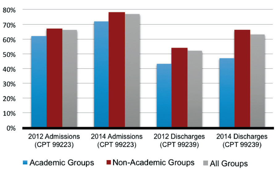
To address the ranges, the State of Hospital Medicine survey asked respondents to divide their faculty into five categories of individual job types, ranging from “No clinical activity with trainees” to “>75% of clinical activity with trainees” (see Figure 1). The results show a broad array of teaching responsibilities, with 20% of academic hospitalists spending no time teaching and another 21% spending almost all of their time teaching.
I suspect this distribution partially reflects the underlying interests of the individual hospitalists, but it is also a product of the available opportunities. A few factors might influence those opportunities, such as decisions by the hospital to hire hospitalists rather than nurse practitioners and physicians assistants to cover new services, or the presence of specialists and general internists who share teaching service slots with hospitalists.
One of the great things about SHM’s State of Hospital Medicine report is how it depicts the wide variety of careers available to hospitalists; the teaching environment is no exception. Although I strive to help my colleagues tailor positions to suit their interests, we never have quite enough resident service time to meet the demands of our enthusiastic teachers. Fortunately, this report allows me to discuss our job openings with candidates knowing how we stack up against academic programs around the country.
Dr. White is assistant professor of medicine at the University of Washington and hospitalist group director at the University of Washington Medical Center in Seattle.
As a group director at a growing, university-based hospitalist program, I often interview aspiring academic hospitalists. Inevitably, the conversation turns to a coveted aspect of the job. I’m not talking about the salary. Applicants want to know, “How much time will I spend on teaching services?”
Because hospitalists at academic institutions typically are passionate about their work as instructors and mentors, they highly value time with trainees. Unfortunately, the 2011 Accreditation Council for Graduate Medical Education (ACGME) work hour rules triggered an expansion in non-teaching services at many teaching hospitals, forcing groups either to divide teaching service among an increasing number of attending physicians or to allocate this commodity unevenly on grounds such as seniority. For many group leaders, striking the right balance between teaching and non-teaching service can be an important contributor to recruitment and retention. During these interviews, I’ve often wondered how our group compares to others around the country.
The 2014 State of Hospital Medicine report (SOHM) shines light on this topic.
Among the 422 groups that only care for adults, 52 self-reported as academic groups. The groups were then asked to describe how they distribute work duties. In these academic practices, about half (52.5%) of the group’s full-time equivalents (FTEs) were devoted to clinical work in which the attending supervises learners delivering care. The remaining FTEs were devoted to a mix of clinical work on non-teaching services, administration, and protected time for research.
Interestingly, the portion devoted to clinical teaching differs substantially between university-based and affiliated community teaching hospitals (36.2% vs. 79.1%), suggesting that hospitalists face tough competition for teaching time at the main campuses of academic systems but might have more opportunities to teach at the bedside in faculty jobs at affiliated hospitals.
The above FTE figures represent averages, which don’t tell the whole story. Groups might not distribute teaching time evenly; the approach to allocation ranges from a completely egalitarian approach to a system with two tiers that separate teaching and non-teaching hospitalists.
To address the ranges, the State of Hospital Medicine survey asked respondents to divide their faculty into five categories of individual job types, ranging from “No clinical activity with trainees” to “>75% of clinical activity with trainees” (see Figure 1). The results show a broad array of teaching responsibilities, with 20% of academic hospitalists spending no time teaching and another 21% spending almost all of their time teaching.
I suspect this distribution partially reflects the underlying interests of the individual hospitalists, but it is also a product of the available opportunities. A few factors might influence those opportunities, such as decisions by the hospital to hire hospitalists rather than nurse practitioners and physicians assistants to cover new services, or the presence of specialists and general internists who share teaching service slots with hospitalists.
One of the great things about SHM’s State of Hospital Medicine report is how it depicts the wide variety of careers available to hospitalists; the teaching environment is no exception. Although I strive to help my colleagues tailor positions to suit their interests, we never have quite enough resident service time to meet the demands of our enthusiastic teachers. Fortunately, this report allows me to discuss our job openings with candidates knowing how we stack up against academic programs around the country.
Dr. White is assistant professor of medicine at the University of Washington and hospitalist group director at the University of Washington Medical Center in Seattle.
Society of Hospital Medicine’s Quality Improvement Toolkits Bring Best Practices to Hospitals
SHM’s free quality improvement toolkits bring the very best practices in the most pressing hospital issues right to your hospital.
With topics like discharging to post-acute care facilities, pain management, and diabetes, SHM helps thousands of hospitals tackle these issues—all with the confidence and expertise of the leaders in the field.
For the latest toolkits, including those designed for post-acute care and glycemic control, visit www.hospitalmedicine.org/qi.
Greater Transparency for Financial Information in Healthcare Will Prompt Questions from Patients
The movement toward greater transparency of financial information in healthcare is providing patients with access to data that might affect their healthcare decisions. Not all of this information is provided in ways that give patients the full picture, and they may turn to you for some added clarity.

Financial Relationships
The Physician Payments Sunshine Act (“Sunshine Act’) was passed as a part of the Affordable Care Act and requires the public disclosure of financial relationships between physicians and the manufacturers of pharmaceuticals, devices, and supplies, as well as group purchasing organizations. The first wave of financial information was publicly disclosed in 2014 on the federal Open Payments website. When it went live, the website disclosed approximately $3.5 billion in payments made by manufacturers to physicians and teaching hospitals during the last five months of 2013. These payments include research grants, consulting fees, speaking fees, travel, and other expenses. In the future, the payments reported will span an entire year, further increasing the total dollar amount paid by industry.
The Sunshine Act is intended to expose potential conflicts of interest in healthcare so that patients are more informed consumers of healthcare services. The relationships between healthcare providers and industry have been scrutinized much more heavily over the past decade. The concern is that physicians with a financial interest, whether through a consultancy relationship with industry or through the development of new technology, might be biased in treating patients because of these relationships. On the other hand, the majority of relationships between healthcare providers and industry can be beneficial. The relationships provide education to other providers, encourage the development of new treatment options, and improve the effectiveness of existing treatments.
The Centers for Medicare and Medicaid Services (CMS) explains on its website that the disclosed financial ties are not necessarily indicators of any wrongdoing, and that the intent of publishing the information is to promote transparency and discourage inappropriate relationships. Without the proper context, these relationships could be viewed as improper by patients and the general public. Therefore, providers should be prepared to answer patients’ questions and possibly even proactively provide details, such as the scope of any relationship with industry. Many providers begin to consult with a pharmaceutical or device manufacturer because of their experience using a particular product, rather than using a particular product after forming that financial relationship. This context could shift patients’ views of what it means for their providers to have this type of connection with industry.
Providers also need to be aware that government agencies, insurers, and attorneys can track this data. Although it is still too early to know the full scope of the potential uses of this information in government investigations, insurance carrier decisions, malpractice, or other legal actions, it does provide further reason to ensure that the information posted is accurate.
During the initial launch of the Open Payments website, some data was temporarily removed due to inaccuracies, including payments linked incorrectly to physicians with the same first and last names. While these issues are being reviewed by CMS, their existence proves how important it is for all physicians (even those not affiliated with the industry) to review the data reported in order to ensure the accuracy of their information.
Procedure Costs
Another transparency requirement in the Affordable Care Act was implemented on Oct. 1, 2014, as part of the inpatient prospective payment system final rule. Hospitals are now required to make their prices for procedures public and update the list annually. The final rule is not explicit with respect to the manner of the disclosure, except that either a price list or the policy for obtaining access must be made public. Some complain that the rule is difficult to comply with because it is vague, while others point out that this fact gives hospitals necessary flexibility in the method of reporting. It is at the hospital’s discretion whether to post the information online or in a physical location.
It is important to note that patients with private payer insurance coverage have distinct rates that are set through agreements between their health plans and the hospitals, so information on the public list very likely will not be applicable to those patients and could be a source of confusion.
As patients have more access to information about the costs for procedures, providers need to be aware of where within the facility they should refer patients with questions or concerns, including information on a hospital’s financial assistance programs.
There are so many sources of information that patients and their families can obtain before ever setting foot in the hospital. An open dialogue with patients that emphasizes the context of any financial relationships with industry, including the benefits, can help to minimize the potential that the information will be treated as suspect by your patients.
Further, as patients bear more of the costs of healthcare, questions surrounding the costs of procedures relative to published data may be encountered more frequently at the bedside and in office visits. This information may have an impact on patients’ decisions about their care.

The movement toward greater transparency of financial information in healthcare is providing patients with access to data that might affect their healthcare decisions. Not all of this information is provided in ways that give patients the full picture, and they may turn to you for some added clarity.
Financial Relationships
The Physician Payments Sunshine Act (“Sunshine Act’) was passed as a part of the Affordable Care Act and requires the public disclosure of financial relationships between physicians and the manufacturers of pharmaceuticals, devices, and supplies, as well as group purchasing organizations. The first wave of financial information was publicly disclosed in 2014 on the federal Open Payments website. When it went live, the website disclosed approximately $3.5 billion in payments made by manufacturers to physicians and teaching hospitals during the last five months of 2013. These payments include research grants, consulting fees, speaking fees, travel, and other expenses. In the future, the payments reported will span an entire year, further increasing the total dollar amount paid by industry.
The Sunshine Act is intended to expose potential conflicts of interest in healthcare so that patients are more informed consumers of healthcare services. The relationships between healthcare providers and industry have been scrutinized much more heavily over the past decade. The concern is that physicians with a financial interest, whether through a consultancy relationship with industry or through the development of new technology, might be biased in treating patients because of these relationships. On the other hand, the majority of relationships between healthcare providers and industry can be beneficial. The relationships provide education to other providers, encourage the development of new treatment options, and improve the effectiveness of existing treatments.
The Centers for Medicare and Medicaid Services (CMS) explains on its website that the disclosed financial ties are not necessarily indicators of any wrongdoing, and that the intent of publishing the information is to promote transparency and discourage inappropriate relationships. Without the proper context, these relationships could be viewed as improper by patients and the general public. Therefore, providers should be prepared to answer patients’ questions and possibly even proactively provide details, such as the scope of any relationship with industry. Many providers begin to consult with a pharmaceutical or device manufacturer because of their experience using a particular product, rather than using a particular product after forming that financial relationship. This context could shift patients’ views of what it means for their providers to have this type of connection with industry.
Providers also need to be aware that government agencies, insurers, and attorneys can track this data. Although it is still too early to know the full scope of the potential uses of this information in government investigations, insurance carrier decisions, malpractice, or other legal actions, it does provide further reason to ensure that the information posted is accurate.
During the initial launch of the Open Payments website, some data was temporarily removed due to inaccuracies, including payments linked incorrectly to physicians with the same first and last names. While these issues are being reviewed by CMS, their existence proves how important it is for all physicians (even those not affiliated with the industry) to review the data reported in order to ensure the accuracy of their information.
Procedure Costs
Another transparency requirement in the Affordable Care Act was implemented on Oct. 1, 2014, as part of the inpatient prospective payment system final rule. Hospitals are now required to make their prices for procedures public and update the list annually. The final rule is not explicit with respect to the manner of the disclosure, except that either a price list or the policy for obtaining access must be made public. Some complain that the rule is difficult to comply with because it is vague, while others point out that this fact gives hospitals necessary flexibility in the method of reporting. It is at the hospital’s discretion whether to post the information online or in a physical location.
It is important to note that patients with private payer insurance coverage have distinct rates that are set through agreements between their health plans and the hospitals, so information on the public list very likely will not be applicable to those patients and could be a source of confusion.
As patients have more access to information about the costs for procedures, providers need to be aware of where within the facility they should refer patients with questions or concerns, including information on a hospital’s financial assistance programs.
There are so many sources of information that patients and their families can obtain before ever setting foot in the hospital. An open dialogue with patients that emphasizes the context of any financial relationships with industry, including the benefits, can help to minimize the potential that the information will be treated as suspect by your patients.
Further, as patients bear more of the costs of healthcare, questions surrounding the costs of procedures relative to published data may be encountered more frequently at the bedside and in office visits. This information may have an impact on patients’ decisions about their care.