User login
In‐hospital SIF
People who use illicit drugs (PWUDs) experience a wide range of health‐related harms and consequently often rely on acute and emergency services for care.[1, 2] Specifically, the poor health status of many PWUDs is often attributable to infectious diseases such as human immunodeficiency virus (HIV) and hepatitis C virus.[3, 4] Soft‐tissue infections associated with injection drug use are also common, and have increasingly accounted for the majority of hospitalizations among this population.[5] Many of these adverse health outcomes may require lengthy in‐patient hospital admissions and constitute a substantial financial burden for the healthcare system.[6]
PWUDs frequently experience barriers to conventional healthcare services. For example, negative experiences with healthcare providers and the healthcare system have often deterred PWUDs from accessing these services.[7, 8] Given that most hospitals operate under an abstinence‐based policy, PWUDs have minimal access to drug‐using paraphernalia while hospitalized, making it difficult for these individuals to safely manage their active drug use. As a result, many PWUDs may resort to high‐risk drug‐using practices (eg, syringe sharing, injecting alone) in the hospital that may lead to further adverse health outcomes, such as infectious disease transmission and overdose, respectively.[9] Past studies have also shown that many PWUDs do not complete hospital‐based treatments.[10, 11] Specifically, many PWUDs leave the hospital against medical advice (AMA) possibly because they are unable to continue their drug use practices in this setting,[10, 12] thus contributing to an increase in readmission rates and mortality among this population.[13] Past studies have indicated that approximately 30% of patients who inject drugs left the hospital AMA,[12, 14] and these individuals have shown to be as high as 4 times more likely to leave the hospital AMA compared to their nondrug‐using counterparts.[11]
Supervised injection facilities (SIFs) are sanctioned environments where PWUDs can inject preobtained illicit drugs under the supervision of healthcare staff. Internationally, SIFs have been shown to improve public health and public order within surrounding communities.[15, 16, 17] For example, a dramatic decline in fatal overdoses in Vancouver's Downtown Eastside neighborhood was attributed to the implementation of a SIF in the area.[15] Changes in risk‐injecting behavior have also been observed among individuals who access SIFs.[18] Although a large body of evidence supports SIFs as an effective approach for minimizing the drug‐ and health‐related harms within street‐based drug scenes,[17, 19] little is known about whether there is a role for SIFs within hospital settings. Currently in Vancouver, Canada, harm reduction services are generally not being provided within hospital settings. Therefore, we sought to conduct a needs assessment to identify the prevalence and correlates of willingness to access an in‐hospital SIF among PWUDs in Vancouver. These data may be crucial for planning appropriate programs and services to reduce health‐related harms and leaving the hospital AMA among PWUDs in hospital settings.
METHODS
The Vancouver Injection Drug Users Study (VIDUS) and the AIDS Care Cohort to Evaluate Exposure to Survival Services (ACCESS) are 2 prospective cohort studies of PWUDs who have been recruited through self‐referral and street outreach since May 1996. These cohorts have been described in detail previously.[20, 21] In brief, persons were eligible to enter the VIDUS study if they had injected illicit drugs at least once in the previous month and resided in the Greater Vancouver region at enrollment. Persons were eligible to enter the ACCESS study if they were HIV infected and used illicit drugs other than cannabinoids in the previous month. Individuals who seroconvert following recruitment are transferred from the VIDUS study into the ACCESS study. All eligible participants provided written informed consent. At baseline and semiannually, study participants complete a harmonized interviewer‐administered questionnaire (ie, participants in the VIDUS and ACCESS studies completed an identical questionnaire) and provide blood samples for HIV and hepatitis C virus testing, and HIV disease monitoring. At the conclusion of each visit, study participants receive Can$20 for their time. The study has received ethical approval from Providence Health Care/University of British Columbia's research ethics board.
The primary outcome of interest for this analysis was willingness to access an in‐hospital SIF (yes vs no or unsure), ascertained by asking participants the following hypothetical question: If you were admitted into a hospital, and if a supervised safe injection site was available in that hospital, would you use it? Given the existence of 2 SIFs in the local environment, PWUDs in Vancouver are familiar with the design and operation of such programs. We compared PWUDs who were and were not willing to access an in‐hospital SIF using bivariable and multivariable logistic regression analyses. Given that the variable measure was based on a hypothetical scenario, participants who completed the survey between June 2013 and November 2013 were eligible for inclusion regardless of their current injection drug use behavior. A complete case approach was used to analyze the data given that the extent of missingness was not significant (<5%). Variables considered included: age (per year increase); gender (male vs female); HIV serostatus (positive vs negative); heroin injection ( daily vs
To identify factors independently associated with willingness to access an in‐hospital SIF, a multivariable logistic regression model was constructed using an a prioridefined statistical protocol based on examination of the Akaike information criterion (AIC) and P values. First, we constructed a full model that included all variables significant at P<0.10 in bivariable analyses. After noting the AIC of the model, we removed the variable with the largest P value and built a reduced model. We continued this iterative process until no variables remained. We selected the multivariable model with the lowest AIC score. All P values were 2 sided. As a subanalysis, we asked participants who would be willing to access an in‐hospital SIF to indicate reasons why they would be willing to access such a facility.
RESULTS
Of the total 769 participants who were eligible for inclusion in the study, 732 PWUDs provided complete data and participated in the study; 37 (4.8%) were excluded due to missing data. In our study sample, 250 (34.2%) were female, the median age was 48 years (interquartile range: 4153 years), and 307 (41.5%) were HIV‐positive. Among our study sample, 499 (68.2%) participants would be willing to access an in‐hospital SIF if it were available. Bivariable analyses of factors associated with willingness to access an in‐hospital SIF are presented in Table 1.
| Characteristic | Willingness to Access an In‐hospital SIF | Odds Ratio (95% CI) | P Value | ||
|---|---|---|---|---|---|
| Yes, n (%), n=499 | No, n (%), n=233 | ||||
| |||||
| Age | |||||
| Median | 48 | 48 | 0.98 (0.97‐1.00) | 0.085 | |
| IQR | (4153) | (4254) | |||
| Gender | |||||
| Male | 331 (66.3) | 151 (64.8) | 1.07 (0.77‐1.48) | 0.685 | |
| Female | 168 (33.7) | 82 (35.2) | |||
| HIV serostatus | |||||
| Positive | 203 (40.7) | 104 (44.6) | 0.85 (0.62‐1.16) | 0.313 | |
| Negative | 296 (59.3) | 129 (55.4) | |||
| Heroin injection* | |||||
| Daily | 106 (21.2) | 26 (11.2) | 2.15 (1.35‐3.40) | <0.001 | |
| < Daily | 393 (78.8) | 207 (88.8) | |||
| Cocaine injection* | |||||
| Daily | 46 (9.2) | 19 (8.2) | 1.14 (0.65‐2.00) | 0.637 | |
| < Daily | 453 (90.8) | 214 (91.8) | |||
| Crystal methamphetamine injection* | |||||
| Daily | 46 (9.2) | 16 (6.9) | 1.38 (0.76‐2.49) | 0.287 | |
| < Daily | 453 (90.8) | 217 (93.1) | |||
| Prescription opioid injection* | |||||
| Daily | 34 (6.8) | 9 (3.9) | 1.82 (0.86‐3.86) | 0.114 | |
| < Daily | 465 (93.2) | 224 (96.1) | |||
| Binge drug use* | |||||
| Yes | 141 (28.3) | 61 (26.2) | 1.11 (0.78‐1.58) | 0.558 | |
| No | 358 (71.7) | 172 (73.8) | |||
| Ever left hospital AMA | |||||
| Yes | 21 (4.2) | 2 (0.9) | 5.07 (1.1821.83) | 0.012 | |
| No | 478 (95.8) | 231 (99.1) | |||
| Ever used illicit drugs in hospital | |||||
| Yes | 238 (47.7) | 83 (35.6) | 1.65 (1.20‐2.27) | 0.002 | |
| No | 261 (52.3) | 150 (64.4) | |||
| Ever had negative experiences with healthcare providers | |||||
| Yes | 131 (26.3) | 64 (27.5) | 0.94 (0.66‐1.33) | 0.729 | |
| No | 368 (73.7) | 169 (72.5) | |||
| Ever had negative experiences with police | |||||
| Yes | 383 (76.8) | 169 (72.5) | 1.25 (0.88‐1.78) | 0.217 | |
| No | 116 (23.2) | 64 (27.5) | |||
| Used an SIF* | |||||
| Yes | 228 (45.7) | 77 (33.0) | 1.70 (1.23‐2.36) | 0.001 | |
| No | 271 (54.3) | 156 (67.0) | |||
As indicated in Table 2, in multivariable analyses, factors that remained significantly and positively associated with willingness to access an in‐hospital SIF included: daily heroin injection (adjusted odds ratio [AOR]=1.90; 95% confidence interval [CI]: 1.20‐3.11), ever used illicit drugs in the hospital (AOR=1.63; 95% CI: 1.18‐2.26), and previously used an SIF (AOR=1.53; 95% CI: 1.10‐2.15).
| Variable | Adjusted Odds Ratio | 95% Confidence Interval | P Value |
|---|---|---|---|
| |||
| Heroin injection* ( daily vs | 1.90 | 1.20‐3.11 | 0.008 |
| Ever left hospital AMA (yes vs no) | 3.74 | 1.0623.72 | 0.079 |
| Ever used illicit drugs in hospital (yes vs no) | 1.63 | 1.18‐2.26 | 0.003 |
| Used an SIF* (yes vs no) | 1.53 | 1.10‐2.15 | 0.013 |
Among participants who would be willing to access an in‐hospital SIF, the most common reasons included: to be able to stay in the hospital (229/499=45.9%), to reduce their drug‐related risks (189/499=37.9%), and to reduce stress associated with being kicked out of the hospital because they were using drugs (97/499=19.4%).
DISCUSSION
We found that over two‐thirds of PWUDs participating in our study would be willing to access an in‐hospital SIF if such a service was available. This finding is encouraging given that a large proportion of PWUDs are hospitalized annually for acute and chronic diseases.[5, 6] Previous studies have documented the positive impact of incorporating a harm reduction model within hospital settings, resulting in more comprehensive care for PWUDs.[22, 23] For example, the Dr. Peter Centre Day Health Program provides a SIF for HIV‐positive PWUDs to safely use illicit drugs under the supervision of trained nurses and was once located at St. Paul's Hospital.[24] Although the Dr. Peter Centre currently operates outside of St. Paul's Hospital, it may be advantageous to model an in‐hospital SIF after the Dr. Peter Centre's harm‐reduction room given their success in facilitating access and delivery of comprehensive care for PWUDs.[23] Specifically, nurses at the Dr. Peter Centre directly observe injections of preobtained illicit drugs for the purposes of preventing illness and promoting health. Our findings support recent calls to implement harm‐reduction services within hospital settings in an effort to minimize the harms associated with illicit drug use.[25, 26]
Previous studies have identified various high‐risk locations where PWUDs use illicit drugs to maintain their established drug‐use habits, including in locked washrooms in hospitals.[9] We found a positive association among PWUDs who had used illicit drugs in the hospital and a willingness to use an in‐hospital SIF. Our finding is reassuring given that studies have shown that these individuals are at a higher risk of negative health consequences (eg, fatal overdose) from using drugs in the hospital.[9] Harm reduction services within the hospital settings can play an important role in reducing this drug‐ and health‐related harm among PWUDs.
Our study also found that high‐frequency heroin injection was associated with a willingness to access an in‐hospital SIF. This relationship may be a result of the complex nature of treating opioid‐dependent patients for pain. For instance, some opioid‐dependent PWUDs may have already established a high tolerance for opioids due to the concomitant use of opioid substitution therapies and ongoing drug use, making it difficult to appropriately prescribe pain medication to these individuals.[27] High‐frequency heroin users may also face severe withdrawal given the unavailability of illicit opioids in hospital settings, resulting in their increased willingness to access an in‐hospital SIF. Furthermore, inadequate pain management may contribute to the continued need to use opioids, as some healthcare providers may be reluctant to provide pain medication out of fear that they would contribute to an existing addiction or relapse.[28, 29] Further, requests for pain mediation may be misinterpreted as drug‐seeking behavior.[30, 31] Given the complexities arising from high‐intensity heroin use, pain management, and healthcare professionals' perceptions regarding PWUDs, further research should seek to untangle the causal relationships underlying these associations.
We found an association between recent use of an SIF and willingness to access an in‐hospital SIF. As mentioned previously, earlier research has shown improvements in various health outcomes and reductions in related harm in surrounding communities where SIFs were implemented.[15, 17] It is unfortunate that although progress in reducing the harm of injection drug use has been seen in community settings globally, the same cannot be said about hospitals. Given that many PWUDs often present to emergency care late in the course of illness and require admission to a hospital bed,[2] it is important to ensure that harm reduction services that are available in the community are also made available in hospitals. However, given the lack of knowledge on in‐hospital SIFs, future research should seek to understand the benefits and consequences of implementing such a facility in a hospital from different perspectives. For example, it may be of interest to assess the attitudes and perceptions of healthcare providers toward an in‐hospital SIF.
A large body of evidence has documented the health harms associated with leaving the hospital AMA, including readmission for a worsened illness and mortality.[13, 32] However, when faced with the abstinence‐based policies that exist in most hospital settings, it is not uncommon for PWUDs to leave the hospital to maintain their active addiction or to address their drug withdrawal.[9] Although we failed to find a statistically significant association between being discharged AMA and willingness to access an in‐hospital SIF, it is noteworthy that in our subanalysis we found that PWUD who were more likely to access an in‐hospital SIF reported doing so because they wanted to stay in the hospital and reduce their drug‐related risks. Given that we observed low counts of reported AMA discharge events, further exploration of this topic is warranted.
Our study suggests that in‐hospital SIFs have the potential to minimize health harms among patients who use illicit drugs in hospitals; however, there are some legal issues that warrant consideration. Specifically, for the successful operation of SIFs, there is a need for changes to regulatory frameworks, including drug laws, to allow for the possession of illicit drugs by individuals accessing an SIF. Such frameworks have been developed in a range of settings and in a manner that is consistent with international drug control treaties. In hospitals, additional regulatory changes may be needed to address issues unique to these settings, such as the use of opioids among PWUDs being treated for pain.
There are several limitations to this study. First, the cross‐sectional design of the study limited our ability to determine a temporal or causal relationship between the explanatory and outcome variables. Second, it is noteworthy that the chosen mode of interviewer‐based questionnaire administration may have influenced our results by relying on self‐reported data that are susceptible to reporting biases, including socially desirable reporting and recall bias. However, we believe we have minimized response bias and maximized reliability in our data by placing sensitive questions toward the end of the interview to allow rapport to be established between the interviewer and participant. Last, given that the participants in the present study were not randomly selected, the interpretation of these results may not be representative or generalizable to other PWUD populations outside of Vancouver. However, it is noteworthy that over the past few decades, community‐based SIFs have been successfully operating in international settings such as Europe and Australia[33, 34]; thus, the concept of an in‐hospital SIF may not be far from actual inpatient practice in these settings. It is also important to acknowledge the progress made toward the implementation of community‐based SIFs in other settings, including the United States. For example, feasibility studies have been conducted in San Francisco and New York and have shown increasing support for the implementation of SIFs in these areas.[35, 36]
We found that a substantial proportion of PWUDs in our sample would be willing to access an in‐hospital SIF if this service was available. Those PWUDs who expressed a willingness to use an in‐hospital SIF were more likely to be high‐intensity heroin users, to have previously used illicit drugs in the hospital, and were more likely to have previously used an SIF. Our findings highlight the potential of in‐hospital SIFs to complement existing harm‐reduction programs that serve people who inject drugs.
Acknowledgements
The authors thank the study participants for their contribution to the research, as well as current and past researchers and staff.
Disclosures: The VIDUS study was supported by the US National Institutes of Health (R01DA011591). The ACCESS study was supported by the US National Institutes of Health (R01DA021525). The evaluation of the supervised injecting facility was supported by Vancouver Coastal Health and Canadian Institutes of Health Research (MOP‐111039). L.T. is supported by a Canadian Institutes of Health Research Doctoral Research Award. This research was undertaken, in part, thanks to funding from the Canada Research Chairs program through a Tier 1 Canada Research Chair in Inner City Medicine, which supports E.W. The authors report no conflicts of interest.
- , , , et al. Emergency department utilization among a cohort of HIV‐positive injecting drug users in a Canadian setting. J Emerg Med. 2012;43(2):236–243.
- , , , et al. High rates of primary care and emergency department use among injection drug users in Vancouver. J Public Health (Oxf). 2005;27(1):62–66.
- , . Epidemiology of HIV among injecting and non‐injecting drug users: current trends and implications for interventions. Curr HIV/AIDS Rep. 2010;7(2):99–106.
- , , , et al. HIV and risk environment for injecting drug users: the past, present, and future. Lancet. 2010;376(9737):268–284.
- , , , et al. Determinants of hospitalization for a cutaneous injection‐related infection among injection drug users: a cohort study. BMC Public Health. 2010;10:327.
- , , , et al. Hospital utilization and costs in a cohort of injection drug users. CMAJ. 2001;165(4):415–420.
- , , , , . The association of stigma with self‐reported access to medical care and antiretroviral therapy adherence in persons living with HIV/AIDS. J Gen Intern Med. 2009;24(10):1101–1108.
- , , , . Stigma among health professionals towards patients with substance use disorders and its consequences for healthcare delivery: systematic review. Drug Alcohol Depend. 2013;131(1–2):23–35.
- , , , . Hospitals as a “risk environment”: an ethno‐epidemiological study of voluntary and involuntary discharge from hospital against medical advice among people who inject drugs. Soc Sci Med. 2014;105C:59–66.
- . “I'm going home”: discharges against medical advice. Mayo Clin Proc. 2009;84(3):255–260.
- , , , , , . Leaving hospital against medical advice among HIV‐positive patients. CMAJ. 2002;167(6):633–637.
- , , , et al. HIV‐positive injection drug users who leave the hospital against medical advice: the mitigating role of methadone and social support. J Acquir Immune Defic Syndr. 2004;35(1):56–59.
- , , . Increased risk of mortality and readmission among patients discharged against medical advice. Am J Med. 2012;125(6):594–602.
- , . Welfare checks, drug consumption, and health: evidence from Vancouver injection drug users. J Hum Resour. 2006;41(1):138–161.
- , , , , . Reduction in overdose mortality after the opening of North America's first medically supervised safer injecting facility: a retrospective population‐based study. Lancet. 2011;377(9775):1429–1437.
- , , , , . The impact of a supervised injecting facility on ambulance call‐outs in Sydney, Australia. Addiction. 2010;105(4):676–683.
- , , , et al. Changes in public order after the opening of a medically supervised safer injecting facility for illicit injection drug users. CMAJ. 2004;171(7):731–734.
- , , , et al. Changes in injecting practices associated with the use of a medically supervised safer injection facility. J Public Health. 2007;29(1):35–39.
- , , , , , . Police and public health partnerships: Evidence from the evaluation of Vancouver's supervised injection facility. Subst Abuse Treat Prev Policy. 2008;3:11.
- , , , et al. Barriers to use of free antiretroviral therapy in injection drug users. JAMA. 1998;280(6):547–549.
- , , , et al. Unsafe injection practices in a cohort of injection drug users in Vancouver: could safer injecting rooms help? CMAJ. 2001;165(4):405–410.
- , , , . Expanding harm reduction services through a wound and abscess clinic. Am J Public Health. 2002;92(12):1915–1917.
- , , , . An integrated supervised injecting program within a care facility for HIV‐positive individuals: a qualitative evaluation. AIDS Care. 2009;21(5):638–644.
- Dr. Peter AIDS Foundation. Available at: http://www.drpeter.org. Accessed June 19, 2014.
- , . Drug use in hospitals: Is there a role for harm reduction? Hospital News. Available at: http://hospitalnews.com/drug‐use‐in‐hospitals‐is‐there‐a‐role‐for‐harm‐reduction. Published October 17, 2013. Accessed March 11, 2014.
- , , , . Harm reduction in hospitals: is it time? Harm Reduct J. 2009;6(1):19.
- , , . Pain intolerance in opioid‐maintained former opiate addicts: effect of long‐acting maintenance agent. Drug Alcohol Depend. 2001;63(2):139–146.
- , , , . Guilty until proven innocent: a qualitative study of the management of chronic non‐cancer pain among patients with a history of substance abuse. Addict Behav. 2010;35(3):270–272.
- , , , . Providers' experiences treating chronic pain among opioid‐dependent drug users. J Gen Intern Med. 2009;24(4):482–488.
- , , , . Management of injecting drug users admitted to hospital. Lancet. 2009;374(9697):1284–1293.
- , , , et al. Routines and rituals: a grounded theory of the pain management of drug users in acute care settings. J Clin Nurs. 2010;19(19–20):2730–2740.
- , , . Leaving against medical advice (AMA): risk of 30‐day mortality and hospital readmission. J Gen Intern Med. 2010;25(9):926–929.
- , , , , . Supervised injection services: What has been demonstrated? A systematic literature review. Drug Alcohol Depend. 2014;145:48–68.
- , , , , , . Drug consumption facilities in Europe and the establishment of supervised injecting centres in Australia. Drug Alcohol Rev. 2000;19(3):337–346.
- , , , , , . Acceptability of a safer injection facility among injection drug users in San Francisco. Drug Alcohol Depend. 2010;110(1–2):160–163.
- , , , , , . Safer injection sites in New York City: a utilization survey of injection drug users. J Drug Issues. 2003;33(3):733–750.
People who use illicit drugs (PWUDs) experience a wide range of health‐related harms and consequently often rely on acute and emergency services for care.[1, 2] Specifically, the poor health status of many PWUDs is often attributable to infectious diseases such as human immunodeficiency virus (HIV) and hepatitis C virus.[3, 4] Soft‐tissue infections associated with injection drug use are also common, and have increasingly accounted for the majority of hospitalizations among this population.[5] Many of these adverse health outcomes may require lengthy in‐patient hospital admissions and constitute a substantial financial burden for the healthcare system.[6]
PWUDs frequently experience barriers to conventional healthcare services. For example, negative experiences with healthcare providers and the healthcare system have often deterred PWUDs from accessing these services.[7, 8] Given that most hospitals operate under an abstinence‐based policy, PWUDs have minimal access to drug‐using paraphernalia while hospitalized, making it difficult for these individuals to safely manage their active drug use. As a result, many PWUDs may resort to high‐risk drug‐using practices (eg, syringe sharing, injecting alone) in the hospital that may lead to further adverse health outcomes, such as infectious disease transmission and overdose, respectively.[9] Past studies have also shown that many PWUDs do not complete hospital‐based treatments.[10, 11] Specifically, many PWUDs leave the hospital against medical advice (AMA) possibly because they are unable to continue their drug use practices in this setting,[10, 12] thus contributing to an increase in readmission rates and mortality among this population.[13] Past studies have indicated that approximately 30% of patients who inject drugs left the hospital AMA,[12, 14] and these individuals have shown to be as high as 4 times more likely to leave the hospital AMA compared to their nondrug‐using counterparts.[11]
Supervised injection facilities (SIFs) are sanctioned environments where PWUDs can inject preobtained illicit drugs under the supervision of healthcare staff. Internationally, SIFs have been shown to improve public health and public order within surrounding communities.[15, 16, 17] For example, a dramatic decline in fatal overdoses in Vancouver's Downtown Eastside neighborhood was attributed to the implementation of a SIF in the area.[15] Changes in risk‐injecting behavior have also been observed among individuals who access SIFs.[18] Although a large body of evidence supports SIFs as an effective approach for minimizing the drug‐ and health‐related harms within street‐based drug scenes,[17, 19] little is known about whether there is a role for SIFs within hospital settings. Currently in Vancouver, Canada, harm reduction services are generally not being provided within hospital settings. Therefore, we sought to conduct a needs assessment to identify the prevalence and correlates of willingness to access an in‐hospital SIF among PWUDs in Vancouver. These data may be crucial for planning appropriate programs and services to reduce health‐related harms and leaving the hospital AMA among PWUDs in hospital settings.
METHODS
The Vancouver Injection Drug Users Study (VIDUS) and the AIDS Care Cohort to Evaluate Exposure to Survival Services (ACCESS) are 2 prospective cohort studies of PWUDs who have been recruited through self‐referral and street outreach since May 1996. These cohorts have been described in detail previously.[20, 21] In brief, persons were eligible to enter the VIDUS study if they had injected illicit drugs at least once in the previous month and resided in the Greater Vancouver region at enrollment. Persons were eligible to enter the ACCESS study if they were HIV infected and used illicit drugs other than cannabinoids in the previous month. Individuals who seroconvert following recruitment are transferred from the VIDUS study into the ACCESS study. All eligible participants provided written informed consent. At baseline and semiannually, study participants complete a harmonized interviewer‐administered questionnaire (ie, participants in the VIDUS and ACCESS studies completed an identical questionnaire) and provide blood samples for HIV and hepatitis C virus testing, and HIV disease monitoring. At the conclusion of each visit, study participants receive Can$20 for their time. The study has received ethical approval from Providence Health Care/University of British Columbia's research ethics board.
The primary outcome of interest for this analysis was willingness to access an in‐hospital SIF (yes vs no or unsure), ascertained by asking participants the following hypothetical question: If you were admitted into a hospital, and if a supervised safe injection site was available in that hospital, would you use it? Given the existence of 2 SIFs in the local environment, PWUDs in Vancouver are familiar with the design and operation of such programs. We compared PWUDs who were and were not willing to access an in‐hospital SIF using bivariable and multivariable logistic regression analyses. Given that the variable measure was based on a hypothetical scenario, participants who completed the survey between June 2013 and November 2013 were eligible for inclusion regardless of their current injection drug use behavior. A complete case approach was used to analyze the data given that the extent of missingness was not significant (<5%). Variables considered included: age (per year increase); gender (male vs female); HIV serostatus (positive vs negative); heroin injection ( daily vs
To identify factors independently associated with willingness to access an in‐hospital SIF, a multivariable logistic regression model was constructed using an a prioridefined statistical protocol based on examination of the Akaike information criterion (AIC) and P values. First, we constructed a full model that included all variables significant at P<0.10 in bivariable analyses. After noting the AIC of the model, we removed the variable with the largest P value and built a reduced model. We continued this iterative process until no variables remained. We selected the multivariable model with the lowest AIC score. All P values were 2 sided. As a subanalysis, we asked participants who would be willing to access an in‐hospital SIF to indicate reasons why they would be willing to access such a facility.
RESULTS
Of the total 769 participants who were eligible for inclusion in the study, 732 PWUDs provided complete data and participated in the study; 37 (4.8%) were excluded due to missing data. In our study sample, 250 (34.2%) were female, the median age was 48 years (interquartile range: 4153 years), and 307 (41.5%) were HIV‐positive. Among our study sample, 499 (68.2%) participants would be willing to access an in‐hospital SIF if it were available. Bivariable analyses of factors associated with willingness to access an in‐hospital SIF are presented in Table 1.
| Characteristic | Willingness to Access an In‐hospital SIF | Odds Ratio (95% CI) | P Value | ||
|---|---|---|---|---|---|
| Yes, n (%), n=499 | No, n (%), n=233 | ||||
| |||||
| Age | |||||
| Median | 48 | 48 | 0.98 (0.97‐1.00) | 0.085 | |
| IQR | (4153) | (4254) | |||
| Gender | |||||
| Male | 331 (66.3) | 151 (64.8) | 1.07 (0.77‐1.48) | 0.685 | |
| Female | 168 (33.7) | 82 (35.2) | |||
| HIV serostatus | |||||
| Positive | 203 (40.7) | 104 (44.6) | 0.85 (0.62‐1.16) | 0.313 | |
| Negative | 296 (59.3) | 129 (55.4) | |||
| Heroin injection* | |||||
| Daily | 106 (21.2) | 26 (11.2) | 2.15 (1.35‐3.40) | <0.001 | |
| < Daily | 393 (78.8) | 207 (88.8) | |||
| Cocaine injection* | |||||
| Daily | 46 (9.2) | 19 (8.2) | 1.14 (0.65‐2.00) | 0.637 | |
| < Daily | 453 (90.8) | 214 (91.8) | |||
| Crystal methamphetamine injection* | |||||
| Daily | 46 (9.2) | 16 (6.9) | 1.38 (0.76‐2.49) | 0.287 | |
| < Daily | 453 (90.8) | 217 (93.1) | |||
| Prescription opioid injection* | |||||
| Daily | 34 (6.8) | 9 (3.9) | 1.82 (0.86‐3.86) | 0.114 | |
| < Daily | 465 (93.2) | 224 (96.1) | |||
| Binge drug use* | |||||
| Yes | 141 (28.3) | 61 (26.2) | 1.11 (0.78‐1.58) | 0.558 | |
| No | 358 (71.7) | 172 (73.8) | |||
| Ever left hospital AMA | |||||
| Yes | 21 (4.2) | 2 (0.9) | 5.07 (1.1821.83) | 0.012 | |
| No | 478 (95.8) | 231 (99.1) | |||
| Ever used illicit drugs in hospital | |||||
| Yes | 238 (47.7) | 83 (35.6) | 1.65 (1.20‐2.27) | 0.002 | |
| No | 261 (52.3) | 150 (64.4) | |||
| Ever had negative experiences with healthcare providers | |||||
| Yes | 131 (26.3) | 64 (27.5) | 0.94 (0.66‐1.33) | 0.729 | |
| No | 368 (73.7) | 169 (72.5) | |||
| Ever had negative experiences with police | |||||
| Yes | 383 (76.8) | 169 (72.5) | 1.25 (0.88‐1.78) | 0.217 | |
| No | 116 (23.2) | 64 (27.5) | |||
| Used an SIF* | |||||
| Yes | 228 (45.7) | 77 (33.0) | 1.70 (1.23‐2.36) | 0.001 | |
| No | 271 (54.3) | 156 (67.0) | |||
As indicated in Table 2, in multivariable analyses, factors that remained significantly and positively associated with willingness to access an in‐hospital SIF included: daily heroin injection (adjusted odds ratio [AOR]=1.90; 95% confidence interval [CI]: 1.20‐3.11), ever used illicit drugs in the hospital (AOR=1.63; 95% CI: 1.18‐2.26), and previously used an SIF (AOR=1.53; 95% CI: 1.10‐2.15).
| Variable | Adjusted Odds Ratio | 95% Confidence Interval | P Value |
|---|---|---|---|
| |||
| Heroin injection* ( daily vs | 1.90 | 1.20‐3.11 | 0.008 |
| Ever left hospital AMA (yes vs no) | 3.74 | 1.0623.72 | 0.079 |
| Ever used illicit drugs in hospital (yes vs no) | 1.63 | 1.18‐2.26 | 0.003 |
| Used an SIF* (yes vs no) | 1.53 | 1.10‐2.15 | 0.013 |
Among participants who would be willing to access an in‐hospital SIF, the most common reasons included: to be able to stay in the hospital (229/499=45.9%), to reduce their drug‐related risks (189/499=37.9%), and to reduce stress associated with being kicked out of the hospital because they were using drugs (97/499=19.4%).
DISCUSSION
We found that over two‐thirds of PWUDs participating in our study would be willing to access an in‐hospital SIF if such a service was available. This finding is encouraging given that a large proportion of PWUDs are hospitalized annually for acute and chronic diseases.[5, 6] Previous studies have documented the positive impact of incorporating a harm reduction model within hospital settings, resulting in more comprehensive care for PWUDs.[22, 23] For example, the Dr. Peter Centre Day Health Program provides a SIF for HIV‐positive PWUDs to safely use illicit drugs under the supervision of trained nurses and was once located at St. Paul's Hospital.[24] Although the Dr. Peter Centre currently operates outside of St. Paul's Hospital, it may be advantageous to model an in‐hospital SIF after the Dr. Peter Centre's harm‐reduction room given their success in facilitating access and delivery of comprehensive care for PWUDs.[23] Specifically, nurses at the Dr. Peter Centre directly observe injections of preobtained illicit drugs for the purposes of preventing illness and promoting health. Our findings support recent calls to implement harm‐reduction services within hospital settings in an effort to minimize the harms associated with illicit drug use.[25, 26]
Previous studies have identified various high‐risk locations where PWUDs use illicit drugs to maintain their established drug‐use habits, including in locked washrooms in hospitals.[9] We found a positive association among PWUDs who had used illicit drugs in the hospital and a willingness to use an in‐hospital SIF. Our finding is reassuring given that studies have shown that these individuals are at a higher risk of negative health consequences (eg, fatal overdose) from using drugs in the hospital.[9] Harm reduction services within the hospital settings can play an important role in reducing this drug‐ and health‐related harm among PWUDs.
Our study also found that high‐frequency heroin injection was associated with a willingness to access an in‐hospital SIF. This relationship may be a result of the complex nature of treating opioid‐dependent patients for pain. For instance, some opioid‐dependent PWUDs may have already established a high tolerance for opioids due to the concomitant use of opioid substitution therapies and ongoing drug use, making it difficult to appropriately prescribe pain medication to these individuals.[27] High‐frequency heroin users may also face severe withdrawal given the unavailability of illicit opioids in hospital settings, resulting in their increased willingness to access an in‐hospital SIF. Furthermore, inadequate pain management may contribute to the continued need to use opioids, as some healthcare providers may be reluctant to provide pain medication out of fear that they would contribute to an existing addiction or relapse.[28, 29] Further, requests for pain mediation may be misinterpreted as drug‐seeking behavior.[30, 31] Given the complexities arising from high‐intensity heroin use, pain management, and healthcare professionals' perceptions regarding PWUDs, further research should seek to untangle the causal relationships underlying these associations.
We found an association between recent use of an SIF and willingness to access an in‐hospital SIF. As mentioned previously, earlier research has shown improvements in various health outcomes and reductions in related harm in surrounding communities where SIFs were implemented.[15, 17] It is unfortunate that although progress in reducing the harm of injection drug use has been seen in community settings globally, the same cannot be said about hospitals. Given that many PWUDs often present to emergency care late in the course of illness and require admission to a hospital bed,[2] it is important to ensure that harm reduction services that are available in the community are also made available in hospitals. However, given the lack of knowledge on in‐hospital SIFs, future research should seek to understand the benefits and consequences of implementing such a facility in a hospital from different perspectives. For example, it may be of interest to assess the attitudes and perceptions of healthcare providers toward an in‐hospital SIF.
A large body of evidence has documented the health harms associated with leaving the hospital AMA, including readmission for a worsened illness and mortality.[13, 32] However, when faced with the abstinence‐based policies that exist in most hospital settings, it is not uncommon for PWUDs to leave the hospital to maintain their active addiction or to address their drug withdrawal.[9] Although we failed to find a statistically significant association between being discharged AMA and willingness to access an in‐hospital SIF, it is noteworthy that in our subanalysis we found that PWUD who were more likely to access an in‐hospital SIF reported doing so because they wanted to stay in the hospital and reduce their drug‐related risks. Given that we observed low counts of reported AMA discharge events, further exploration of this topic is warranted.
Our study suggests that in‐hospital SIFs have the potential to minimize health harms among patients who use illicit drugs in hospitals; however, there are some legal issues that warrant consideration. Specifically, for the successful operation of SIFs, there is a need for changes to regulatory frameworks, including drug laws, to allow for the possession of illicit drugs by individuals accessing an SIF. Such frameworks have been developed in a range of settings and in a manner that is consistent with international drug control treaties. In hospitals, additional regulatory changes may be needed to address issues unique to these settings, such as the use of opioids among PWUDs being treated for pain.
There are several limitations to this study. First, the cross‐sectional design of the study limited our ability to determine a temporal or causal relationship between the explanatory and outcome variables. Second, it is noteworthy that the chosen mode of interviewer‐based questionnaire administration may have influenced our results by relying on self‐reported data that are susceptible to reporting biases, including socially desirable reporting and recall bias. However, we believe we have minimized response bias and maximized reliability in our data by placing sensitive questions toward the end of the interview to allow rapport to be established between the interviewer and participant. Last, given that the participants in the present study were not randomly selected, the interpretation of these results may not be representative or generalizable to other PWUD populations outside of Vancouver. However, it is noteworthy that over the past few decades, community‐based SIFs have been successfully operating in international settings such as Europe and Australia[33, 34]; thus, the concept of an in‐hospital SIF may not be far from actual inpatient practice in these settings. It is also important to acknowledge the progress made toward the implementation of community‐based SIFs in other settings, including the United States. For example, feasibility studies have been conducted in San Francisco and New York and have shown increasing support for the implementation of SIFs in these areas.[35, 36]
We found that a substantial proportion of PWUDs in our sample would be willing to access an in‐hospital SIF if this service was available. Those PWUDs who expressed a willingness to use an in‐hospital SIF were more likely to be high‐intensity heroin users, to have previously used illicit drugs in the hospital, and were more likely to have previously used an SIF. Our findings highlight the potential of in‐hospital SIFs to complement existing harm‐reduction programs that serve people who inject drugs.
Acknowledgements
The authors thank the study participants for their contribution to the research, as well as current and past researchers and staff.
Disclosures: The VIDUS study was supported by the US National Institutes of Health (R01DA011591). The ACCESS study was supported by the US National Institutes of Health (R01DA021525). The evaluation of the supervised injecting facility was supported by Vancouver Coastal Health and Canadian Institutes of Health Research (MOP‐111039). L.T. is supported by a Canadian Institutes of Health Research Doctoral Research Award. This research was undertaken, in part, thanks to funding from the Canada Research Chairs program through a Tier 1 Canada Research Chair in Inner City Medicine, which supports E.W. The authors report no conflicts of interest.
- , , , et al. Emergency department utilization among a cohort of HIV‐positive injecting drug users in a Canadian setting. J Emerg Med. 2012;43(2):236–243.
- , , , et al. High rates of primary care and emergency department use among injection drug users in Vancouver. J Public Health (Oxf). 2005;27(1):62–66.
- , . Epidemiology of HIV among injecting and non‐injecting drug users: current trends and implications for interventions. Curr HIV/AIDS Rep. 2010;7(2):99–106.
- , , , et al. HIV and risk environment for injecting drug users: the past, present, and future. Lancet. 2010;376(9737):268–284.
- , , , et al. Determinants of hospitalization for a cutaneous injection‐related infection among injection drug users: a cohort study. BMC Public Health. 2010;10:327.
- , , , et al. Hospital utilization and costs in a cohort of injection drug users. CMAJ. 2001;165(4):415–420.
- , , , , . The association of stigma with self‐reported access to medical care and antiretroviral therapy adherence in persons living with HIV/AIDS. J Gen Intern Med. 2009;24(10):1101–1108.
- , , , . Stigma among health professionals towards patients with substance use disorders and its consequences for healthcare delivery: systematic review. Drug Alcohol Depend. 2013;131(1–2):23–35.
- , , , . Hospitals as a “risk environment”: an ethno‐epidemiological study of voluntary and involuntary discharge from hospital against medical advice among people who inject drugs. Soc Sci Med. 2014;105C:59–66.
- . “I'm going home”: discharges against medical advice. Mayo Clin Proc. 2009;84(3):255–260.
- , , , , , . Leaving hospital against medical advice among HIV‐positive patients. CMAJ. 2002;167(6):633–637.
- , , , et al. HIV‐positive injection drug users who leave the hospital against medical advice: the mitigating role of methadone and social support. J Acquir Immune Defic Syndr. 2004;35(1):56–59.
- , , . Increased risk of mortality and readmission among patients discharged against medical advice. Am J Med. 2012;125(6):594–602.
- , . Welfare checks, drug consumption, and health: evidence from Vancouver injection drug users. J Hum Resour. 2006;41(1):138–161.
- , , , , . Reduction in overdose mortality after the opening of North America's first medically supervised safer injecting facility: a retrospective population‐based study. Lancet. 2011;377(9775):1429–1437.
- , , , , . The impact of a supervised injecting facility on ambulance call‐outs in Sydney, Australia. Addiction. 2010;105(4):676–683.
- , , , et al. Changes in public order after the opening of a medically supervised safer injecting facility for illicit injection drug users. CMAJ. 2004;171(7):731–734.
- , , , et al. Changes in injecting practices associated with the use of a medically supervised safer injection facility. J Public Health. 2007;29(1):35–39.
- , , , , , . Police and public health partnerships: Evidence from the evaluation of Vancouver's supervised injection facility. Subst Abuse Treat Prev Policy. 2008;3:11.
- , , , et al. Barriers to use of free antiretroviral therapy in injection drug users. JAMA. 1998;280(6):547–549.
- , , , et al. Unsafe injection practices in a cohort of injection drug users in Vancouver: could safer injecting rooms help? CMAJ. 2001;165(4):405–410.
- , , , . Expanding harm reduction services through a wound and abscess clinic. Am J Public Health. 2002;92(12):1915–1917.
- , , , . An integrated supervised injecting program within a care facility for HIV‐positive individuals: a qualitative evaluation. AIDS Care. 2009;21(5):638–644.
- Dr. Peter AIDS Foundation. Available at: http://www.drpeter.org. Accessed June 19, 2014.
- , . Drug use in hospitals: Is there a role for harm reduction? Hospital News. Available at: http://hospitalnews.com/drug‐use‐in‐hospitals‐is‐there‐a‐role‐for‐harm‐reduction. Published October 17, 2013. Accessed March 11, 2014.
- , , , . Harm reduction in hospitals: is it time? Harm Reduct J. 2009;6(1):19.
- , , . Pain intolerance in opioid‐maintained former opiate addicts: effect of long‐acting maintenance agent. Drug Alcohol Depend. 2001;63(2):139–146.
- , , , . Guilty until proven innocent: a qualitative study of the management of chronic non‐cancer pain among patients with a history of substance abuse. Addict Behav. 2010;35(3):270–272.
- , , , . Providers' experiences treating chronic pain among opioid‐dependent drug users. J Gen Intern Med. 2009;24(4):482–488.
- , , , . Management of injecting drug users admitted to hospital. Lancet. 2009;374(9697):1284–1293.
- , , , et al. Routines and rituals: a grounded theory of the pain management of drug users in acute care settings. J Clin Nurs. 2010;19(19–20):2730–2740.
- , , . Leaving against medical advice (AMA): risk of 30‐day mortality and hospital readmission. J Gen Intern Med. 2010;25(9):926–929.
- , , , , . Supervised injection services: What has been demonstrated? A systematic literature review. Drug Alcohol Depend. 2014;145:48–68.
- , , , , , . Drug consumption facilities in Europe and the establishment of supervised injecting centres in Australia. Drug Alcohol Rev. 2000;19(3):337–346.
- , , , , , . Acceptability of a safer injection facility among injection drug users in San Francisco. Drug Alcohol Depend. 2010;110(1–2):160–163.
- , , , , , . Safer injection sites in New York City: a utilization survey of injection drug users. J Drug Issues. 2003;33(3):733–750.
© 2015 Society of Hospital Medicine
Dimensional aspects of DSM-5 personality disorder criteria discussed
HUNTINGTON BEACH, CALIF. – In the opinion of Dr. John M. Oldham, clinicians who deem the alternative personality disorder model of the DSM-5 as too confusing are misguided.
“If you’re going to compare DSM-5 alternative personality disorder model with the DSM-IV model, you have to do a fair comparison,” Dr. Oldham told attendees at the annual meeting of the American College of Psychiatrists. ”In fact, we reduced the number of items that you have to measure by 43%.”

So when people describe the DSM-5’s personality disorders criteria as more complicated, he continued, “what they really mean is, ‘it’s more complicated than what I do,’ not that it’s more complicated than [the] DSM-IV.”
Along with Dr. Andrew E. Skodol, Dr. Oldham cochaired a work group of experts convened by the American Psychiatric Association to update diagnostic criteria related to personality and personality disorders for the DSM-5. “We took our work and our charge seriously,” recalled Dr. Oldham, senior vice president and chief of staff at the Menninger Clinic, Houston. “It was not easy. We had many challenges. A great deal of research has been done in the factor analytic research psychology world around things like the five-factor model of personality. Such terms are not always terribly familiar in clinical medicine, so there was a problem with the lack of familiarity. Then there were vested interests different groups had that were influential in some ways.”
Ultimately, the alternative personality disorder model was placed in section III of the DSM-5. The model enables clinicians “to individually portray the dimensions of the patient’s pathology in a thorough and broad way,” Dr. Oldham explained. “We emphasize impairment in functioning. That’s an important new requirement. So you have to determine, by using the level of functioning scale, whether the person does or doesn’t have moderate or greater impairment. If you have a patient with mild impairment, you can describe what you’re concerned about, but you’re not putting that patient into a diagnostic box of pathology. There is a dimensional scope that enables you to capture many types of patients.”
An empirical study of 337 clinicians demonstrated that in 14 of 18 comparisons, respondents deemed the DSM-5 pathological personality traits as more clinical useful, compared with the DSM-IV, with respect to ease of use, communication of clinical information to other professionals, communication of clinical information to patients, comprehensiveness in describing pathology, and treatment planning (J. Abnorm. Psychol. 2013;122:836-41). “In fact, this was a preference to the new model, which was unfamiliar, compared to the model that these clinicians had been using for 20 years,” Dr. Oldham said.
The study also found that the new DSM-5 personality disorder model was more strongly related to clinical decision making in areas of global functioning, risk assessment, recommended treatment type and intensity, and prognosis.
According to unpublished data from the DSM-5 field trials conducted in the United States and Canada, more than 80% of clinicians in academic and routine clinical practice fields found the new personality disorder criteria “moderately” to “extremely” useful, compared with the DSM-IV. In fact, the respondents rated the new criteria as more useful than other changes to the DSM-5, including those related to bipolar and related disorders, schizophrenia spectrum and other psychotic disorders, and other conditions.
In addition, a test-retest reliability study conducted at 11 academic medical centers found that the new model for borderline personality disorder had a good test-retest reliability (.054), in the same ballpark as that for bipolar I disorder (0.56) and schizophrenia (.50) (Am. J. Psychiatry 2013;170:43-58). “This surprised a lot of people,” Dr. Oldham said.
About 1 year after the DSM-5’s release, Medscape Psychiatry surveyed almost 3,000 clinicians about their impressions of the new guidelines. Of the 2,828 respondents, nearly one-third (28%) were psychiatrists, 22% were psychologists, 13% were family medicine clinicians, and the rest were from other medical fields. The researchers found that 39% of survey respondents were considering the dimensional approaches offered in the new personality disorder criteria of the DSM-5.
“That’s not bad,” Dr. Oldham said.
He reported having no relevant financial conflicts.
On Twitter @dougbrunk
HUNTINGTON BEACH, CALIF. – In the opinion of Dr. John M. Oldham, clinicians who deem the alternative personality disorder model of the DSM-5 as too confusing are misguided.
“If you’re going to compare DSM-5 alternative personality disorder model with the DSM-IV model, you have to do a fair comparison,” Dr. Oldham told attendees at the annual meeting of the American College of Psychiatrists. ”In fact, we reduced the number of items that you have to measure by 43%.”
So when people describe the DSM-5’s personality disorders criteria as more complicated, he continued, “what they really mean is, ‘it’s more complicated than what I do,’ not that it’s more complicated than [the] DSM-IV.”
Along with Dr. Andrew E. Skodol, Dr. Oldham cochaired a work group of experts convened by the American Psychiatric Association to update diagnostic criteria related to personality and personality disorders for the DSM-5. “We took our work and our charge seriously,” recalled Dr. Oldham, senior vice president and chief of staff at the Menninger Clinic, Houston. “It was not easy. We had many challenges. A great deal of research has been done in the factor analytic research psychology world around things like the five-factor model of personality. Such terms are not always terribly familiar in clinical medicine, so there was a problem with the lack of familiarity. Then there were vested interests different groups had that were influential in some ways.”
Ultimately, the alternative personality disorder model was placed in section III of the DSM-5. The model enables clinicians “to individually portray the dimensions of the patient’s pathology in a thorough and broad way,” Dr. Oldham explained. “We emphasize impairment in functioning. That’s an important new requirement. So you have to determine, by using the level of functioning scale, whether the person does or doesn’t have moderate or greater impairment. If you have a patient with mild impairment, you can describe what you’re concerned about, but you’re not putting that patient into a diagnostic box of pathology. There is a dimensional scope that enables you to capture many types of patients.”
An empirical study of 337 clinicians demonstrated that in 14 of 18 comparisons, respondents deemed the DSM-5 pathological personality traits as more clinical useful, compared with the DSM-IV, with respect to ease of use, communication of clinical information to other professionals, communication of clinical information to patients, comprehensiveness in describing pathology, and treatment planning (J. Abnorm. Psychol. 2013;122:836-41). “In fact, this was a preference to the new model, which was unfamiliar, compared to the model that these clinicians had been using for 20 years,” Dr. Oldham said.
The study also found that the new DSM-5 personality disorder model was more strongly related to clinical decision making in areas of global functioning, risk assessment, recommended treatment type and intensity, and prognosis.
According to unpublished data from the DSM-5 field trials conducted in the United States and Canada, more than 80% of clinicians in academic and routine clinical practice fields found the new personality disorder criteria “moderately” to “extremely” useful, compared with the DSM-IV. In fact, the respondents rated the new criteria as more useful than other changes to the DSM-5, including those related to bipolar and related disorders, schizophrenia spectrum and other psychotic disorders, and other conditions.
In addition, a test-retest reliability study conducted at 11 academic medical centers found that the new model for borderline personality disorder had a good test-retest reliability (.054), in the same ballpark as that for bipolar I disorder (0.56) and schizophrenia (.50) (Am. J. Psychiatry 2013;170:43-58). “This surprised a lot of people,” Dr. Oldham said.
About 1 year after the DSM-5’s release, Medscape Psychiatry surveyed almost 3,000 clinicians about their impressions of the new guidelines. Of the 2,828 respondents, nearly one-third (28%) were psychiatrists, 22% were psychologists, 13% were family medicine clinicians, and the rest were from other medical fields. The researchers found that 39% of survey respondents were considering the dimensional approaches offered in the new personality disorder criteria of the DSM-5.
“That’s not bad,” Dr. Oldham said.
He reported having no relevant financial conflicts.
On Twitter @dougbrunk
EXPERT ANALYSIS FROM THE ANNUAL MEETING OF THE AMERICAN COLLEGE OF PSYCHIATRISTS
Gout may lower Alzheimer’s risk
Patients with a history of gout may be at a lower risk for developing Alzheimer’s disease, reported Na Lu of Boston University and coauthors.

The large cohort study used electronic medical records from general practices in the United Kingdom’s The Health Improvement Network (THIN) to find 309 new cases of Alzheimer’s in 55,224 patients with gout and 1,942 cases in a comparison group of 238,805 patients during a median follow-up of 5 years. Gout patients had a multivariate-adjusted 24% lower risk of developing Alzheimer’s, the investigators found (hazard ratio, 0.76; 95% confidence interval, 0.66-0.87).
This difference may be due to the potential neuroprotective effects of uric acid, Ms. Lu and colleagues said in the report. “Overall, these findings support the proposed hypothesis that supplementary use of the metabolic precursor to uric acid, like inosine or hypoxanthine, could prevent and attenuate the progression of AD,” they wrote.
Read the full paper in Annals of the Rheumatic Diseases (2015 March 4 [doi:10.1136/annrheumdis-2014-206917]).
Patients with a history of gout may be at a lower risk for developing Alzheimer’s disease, reported Na Lu of Boston University and coauthors.
The large cohort study used electronic medical records from general practices in the United Kingdom’s The Health Improvement Network (THIN) to find 309 new cases of Alzheimer’s in 55,224 patients with gout and 1,942 cases in a comparison group of 238,805 patients during a median follow-up of 5 years. Gout patients had a multivariate-adjusted 24% lower risk of developing Alzheimer’s, the investigators found (hazard ratio, 0.76; 95% confidence interval, 0.66-0.87).
This difference may be due to the potential neuroprotective effects of uric acid, Ms. Lu and colleagues said in the report. “Overall, these findings support the proposed hypothesis that supplementary use of the metabolic precursor to uric acid, like inosine or hypoxanthine, could prevent and attenuate the progression of AD,” they wrote.
Read the full paper in Annals of the Rheumatic Diseases (2015 March 4 [doi:10.1136/annrheumdis-2014-206917]).
Environmental factors could increase U.S. anthrax cases
WASHINGTON– Recent isolated cases of anthrax in Minnesota and elsewhere, along with the disease’s relative ease of transmission from animals or plants to humans, should heighten U.S. physicians’ awareness of anthrax’s symptoms and treatments, warned Dr. Jason K. Blackburn.
“[Anthrax] is something that our international partners deal with on an annual basis [as] we can see the disease reemerging, or at least increasing, in annual reports on humans in a number of countries,” explained Dr. Blackburn of the University of Florida in Gainesville, at a meeting on biodefense and emerging diseases sponsored by the American Society for Microbiology. “Here in the United States, we’re seeing it shift from a livestock disease [to] a wildlife disease, where we have these populations that we can’t reach with vaccines, and where surveillance is very logistically challenging.”
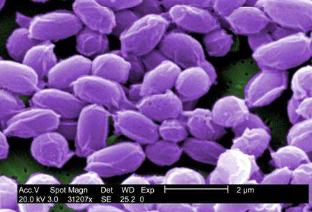
Environmental factors can drive higher incidences of anthrax cases. Temperature, precipitation, and vegetation indices are key variables that facilitate anthrax transmission and spread of the disease. Geographically, lowland areas also have higher prevalences of the disease.
For example, Dr. Blackburn and his colleagues used predictive models to quantify the theory that anthrax case rates increase during years that have wet springs followed by hot, dry summers in the region of western Texas.
Using these data would allow scientists and health care providers to predict which years would have an increased risk for anthrax cases in humans, Dr. Blackburn said, and could help hospitals and clinics effectively prepare to treat a higher influx of these cases and prevent possible outbreaks.
Although relatively large numbers of human anthrax cases persist in parts of world – particularly in Africa and central Asia – cases in the United States have been primarily relegated to livestock.
However, during the last decade, there has been a noticeable shift in cases from livestock to wildlife, particularly in western Texas and Montana, where local populations of elk, bison, and white-tailed deer have been affected. The newfound prevalence in wildlife species, along with continued presence in domestic animals such as cattle and sheep, mean that transmission to humans could become even easier.
“Human cases are usually driven by direct human interaction with mammalian hosts,” said Dr. Blackburn, citing farms and meat factories as prime examples of where the Bacillus anthracis organism would easily spread. In addition, Dr. Blackburn specifically noted a scenario in which flies can transmit the disease from sheep to humans, and have also been found to carry anthrax from carcasses to wildlife and vegetation.
From 2010 to 2012, anthrax cases in Europe, particularly Georgia and Turkey, increased, compared with numbers over a similar time frame between 2000 and 2009. While case reporting can be partly attributed to this increase, Dr. Blackburn indicated that it was most likely evidence of an associative trend between livestock and human anthrax cases.
Dr. Blackburn did not report any disclosures.
WASHINGTON– Recent isolated cases of anthrax in Minnesota and elsewhere, along with the disease’s relative ease of transmission from animals or plants to humans, should heighten U.S. physicians’ awareness of anthrax’s symptoms and treatments, warned Dr. Jason K. Blackburn.
“[Anthrax] is something that our international partners deal with on an annual basis [as] we can see the disease reemerging, or at least increasing, in annual reports on humans in a number of countries,” explained Dr. Blackburn of the University of Florida in Gainesville, at a meeting on biodefense and emerging diseases sponsored by the American Society for Microbiology. “Here in the United States, we’re seeing it shift from a livestock disease [to] a wildlife disease, where we have these populations that we can’t reach with vaccines, and where surveillance is very logistically challenging.”
Environmental factors can drive higher incidences of anthrax cases. Temperature, precipitation, and vegetation indices are key variables that facilitate anthrax transmission and spread of the disease. Geographically, lowland areas also have higher prevalences of the disease.
For example, Dr. Blackburn and his colleagues used predictive models to quantify the theory that anthrax case rates increase during years that have wet springs followed by hot, dry summers in the region of western Texas.
Using these data would allow scientists and health care providers to predict which years would have an increased risk for anthrax cases in humans, Dr. Blackburn said, and could help hospitals and clinics effectively prepare to treat a higher influx of these cases and prevent possible outbreaks.
Although relatively large numbers of human anthrax cases persist in parts of world – particularly in Africa and central Asia – cases in the United States have been primarily relegated to livestock.
However, during the last decade, there has been a noticeable shift in cases from livestock to wildlife, particularly in western Texas and Montana, where local populations of elk, bison, and white-tailed deer have been affected. The newfound prevalence in wildlife species, along with continued presence in domestic animals such as cattle and sheep, mean that transmission to humans could become even easier.
“Human cases are usually driven by direct human interaction with mammalian hosts,” said Dr. Blackburn, citing farms and meat factories as prime examples of where the Bacillus anthracis organism would easily spread. In addition, Dr. Blackburn specifically noted a scenario in which flies can transmit the disease from sheep to humans, and have also been found to carry anthrax from carcasses to wildlife and vegetation.
From 2010 to 2012, anthrax cases in Europe, particularly Georgia and Turkey, increased, compared with numbers over a similar time frame between 2000 and 2009. While case reporting can be partly attributed to this increase, Dr. Blackburn indicated that it was most likely evidence of an associative trend between livestock and human anthrax cases.
Dr. Blackburn did not report any disclosures.
AT THE ASM BIODEFENSE MEETING

Careful planning, technique, and counseling can minimize pediatric scarring
It’s a myth that children don’t scar like adults, according to Dr. Jon A. Dyer.
In fact, children often scar worse than adults, he said at the Hawaii Dermatology Seminar sponsored by Global Academy for Medical Education/Skin Disease Education Foundation.
Children have more aggressive healing and inflammatory responses, a highly elastic dermis and connective tissues, and a high activity level that increases the risk of stressing the wound, and they may pay less attention to it, he explained.
Additionally, children can be more difficult to treat for scars because they are often more fearful and anxious, they have a short attention span, and they move around – a lot, he said. This leads to greater anxiety on the part of both the dermatologist and the family, which can lead to rapidly completing a procedure, said Dr. Dyer of the University of Missouri, Columbia.
Successful treatment of a wound or lesion – that is, getting the best initial scar possible – requires good planning, proactive application of surgical principles, and careful patient and family counseling and follow-up, he said.
Among Dr. Dyer’s tips for minimizing scarring when treating lesions in children:
• Do the simplest possible procedure.
• Make the scar as small as possible, keeping in mind that pediatric skin is more elastic.
• Remember that fusiform closures in children do not always require a 3:1 ratio. Tips settle, and divots fill in in young children, he said.
• Operate before puberty when the lesion is located in a cosmetically important area.
Also, consider performing a staged excision if possible; this allows a reduction in final scar length, provides tissue expansion, and allows assessment of individual wound healing. Take as much of the lesion as possible the first time, and remember that central excision is preferable to lateral excision, Dr. Dyer said.
The ideal time to wait between stage 1 and 2 is 4-6 weeks. Longer intervals will lead to more spread and more hypertrophy, and will negate the benefits of staging, he said.
Keep in mind that purse-string sutures can be particularly effective for suturing small spaces and for closing dead spaces, he said, adding that use of a purse-string closure for a round defect can reduce the final scar length by at least 50%.
Scar prevention or minimization requires strict adherence to good surgical principles, Dr. Dyer said, explaining that attention must be paid to perfecting the excision through proper use of skin tension lines, clean wound edges, sharp corners with no or minimal boating, undermining, and removing bulk if necessary.
Track marks are more likely on nonfacial skin and can be avoided by using running subcuticular sutures; in some cases of excellent approximation with no tension, the wound surface can be secured with Dermabond, he said.
Dr. Dyer also outlined the best closure choices for specific areas. For the scalp, use running cuticular sutures; for the extremities, trunk, axilla, or groin, use running subcuticular sutures; for the face, feet, or hands, use interrupted sutures; and for a punch biopsy, consider not using any closure, he advised.
If cuticular stitches are used, minimize scarring by pulling them within 5 days on the face, and within 7-10 days on the body. If dehiscence is a concern, pull the cuticulars and follow with either Dermabond or SteriStrips, he said, but strongly consider using running subcuticular sutures.
Once a wound is closed, secure it with Dermabond or with Steri-Strips and Mastisol and provide protective dressing.
“The bulkier and more colorful the dressing, the happier and more compliant the child,” he said.
Minimize postoperative wound tension in high-risk scars or sites and restrict movement and trauma for as long as possible; 6 weeks is ideal, he added, noting that movement restriction is critical for ensuring a good outcome.
To reduce the trauma of suture removal, “cut ’em long and leave ’em long,” and be sure to use proper scissors, he said.
Running subcuticular suture removal doesn’t hurt; enlist parents’ help in convincing the child of this, he suggested.
Finally, be specific when counseling patients about restrictions, discuss possible complications, provide a written summary including contact numbers, address the scar, and give the follow-up appointment time in writing, he said.
If dehiscence occurs – and it’s not uncommon in children – let it heal and restart the process, he said, noting that prevention is best and can be achieved in many cases with aggressive pre- and postop education, restriction of movement, and proper dressing.
Dr. Dyer stressed that good surgical principles for use in children can also be utilized to improve results in adult patients.
Dr. Dyer reported having no disclosures. SDEF and this new organization are owned by the same parent company.
Meticulous planning, creativity key to management of EVAR infections
CHICAGO – Successful management of infected aortic endovascular grafts requires careful operative planning and execution, meticulous postoperative care, and a fair bit of creativity.
“Each patient is different, so surgeons have to tailor the reconstructions to the individual patient and with these specific infections, have to be creative,” Dr. Thomas C. Bower, chair of vascular and endovascular surgery at Mayo Clinic, Rochester, Minn., said. “I’ve found the operations to be more challenging and more difficult than explanting portions or total graft excision when the infection has occurred in a hand-sewn graft.”
Unlike the typical bimodal distribution seen with hand-sewn graft infections, infection following endovascular repair of aortic aneurysms (EVAR) occurs from days up to 3 years after implantation. At the Mayo Clinic, a 79-year-old man presented with an infected endograft, psoas abscess, and Salmonella septicemia 4 years after EVAR.
“These infections are uncommon, but we are seeing more of them,” Dr. Bower said at a symposium on vascular surgery sponsored by Northwestern University.
Roughly two-thirds of patients will present with fever, nonspecific abdominal or back pain, malaise, weight loss or night sweats. If time permits, preoperative assessments include echocardiography for left ventricular function, arterial blood gases for pulmonary function since many patients are smokers, and renal ultrasound if creatinine is ≥ 1.5 mg/dL after rehydration. These tests are important because preoperative chronic obstructive pulmonary disease and renal dysfunction correlate with worse postoperative outcomes, he said.
Computed tomography angiography (CTA), however, stands as the single most important step of preoperative preparation, with the sine qua non of infection being air around the graft. Unlike hand-sewn grafts where infections can be localized, typically there is total graft involvement in these cases because the device is left inside the aneurysm sac. Aneurysms or pseudoaneurysms also have been seen above the infected device, including at the top end of suprarenal stents.
“This clearly has an impact on how we approach patients, but what’s become very apparent to me is that CTA often underestimates the amount of periaortic inflammation, especially at the juxta- and pararenal locations,” Dr. Bower said.
The Mayo group initially used in situ antibiotic-soaked prosthetic grafts for explanting EVAR devices, which yielded “acceptable mortality and reinfection rates, but primarily outstanding patency rates.” However, cryopreserved aortoiliac grafts have now become their first choice, Dr. Bower said. An ABO match is not imperative, preparation takes roughly 45 minutes, branch closures done in the lab are buttressed with sutures, and the graft is turned over to keep the lumbar arteries anterior, which offers an easy fix if there is bleeding, rather than having it on the posterior wall. Cryopreserved grafts, however, can dilate 40% and lengthen 10% under pressure.
“I’ve been burned more than once where the graft elongates more than I think, and I end up having to cut a small piece out to foreshorten it,” he said.
Reconstructions are tailored to patient anatomy. Surgeons should have several plans for reconstruction, including routing a graft through a remote path, remembering that CTA will underestimate the amount of periaortic inflammation. Separate bypasses of the renal or visceral arteries are performed first before the aortic clamp is applied to reduce physiologic stress. This requires knowledge of the supraceliac and pararenal aorta exposures, which really begins with the correct choice of incisions, Dr. Bower said. This is based on the aortic segment to be treated, position of the new graft, the aortic clamp site, and patient body habitus.
Most patients with EVAR infections are approached with a midline abdominal incision extended along the xiphoid process, which is the lynch pin for allowing upward and lateral retraction of the abdominal wall, he said. Choosing an incision that allows a more vertical orientation to where the new aortic anastomosis and clamp site will be, rather than operating in a keyhole, is important.
The second step is to open up the pararenal space by moving the viscera out of the way. This begins by ligating the inferior mesenteric vein and adjacent lymphatics, which allows incision of an avascular plane along the base of the left transverse colon. Retractor blades are set to allow the upward and lateral retraction of the small bowel, the left colon, and pancreas. Exposure of the suprarenal or supramesenteric aorta requires mobilization of the left renal vein after ligation and division of its branches.
“If that vein is intensely involved in inflammation, don’t ligate the branches in case you have to divide that vein at the caval confluence. Otherwise, you’ll run into some dysfunction of that left kidney,” Dr. Bower cautioned.
To have a secure place for the aortic cross clamp, the crura must be divided on either side of the diaphragm at or above the supramesenteric aorta, he added.
Key steps in total graft explantation are to drain abscesses prior to surgery to lower the bacterial burden and thus reduce the postoperative inflammatory response, bypass renal/visceral arteries first, if needed, remove the infected graft, debride the aorta to healthy tissue, place the new graft and cover it with omentum, and repair the bowel, if needed.
A piece of the proximal aortic wall should be sent to pathology to ensure the absence of bacteria or microabscesses. Organism-specific antibiotics are administered intravenously for 6-8 weeks followed by lifelong oral antibiotics, he said.
An earlier report involving 24 patients with infected aortic endografts (21 EVARs and 3 thoracic EVARs) treated at Mayo Clinic between 1997 and 2012 revealed polymicrobial infection in 11 patients, with methicillin-resistant Staphylococcus aureus being common. Potential contributors to infection were endovascular reintervention in eight, aortoenteric fistula/erosion in four, and various remote infections (J. Vasc. Surg. 2013;58:371-9).
Rifampin-soaked grafts were used in 15 patients, cryopreserved grafts in 4, femoral vein in 2, and axillofemoral grafts in 3. At a median of 14 months follow-up, patient survival, graft-related complications, and reinfection rates were 79%, 13%, and 4%, respectively, Dr. Bower said.
Dr. Bower reported having no financial disclosures.
The expert opinion from the Northwestern Vascular Symposium regarding the management of EVAR infections reminds us of the importance of appropriate patient selection, proper performance of the planned procedure, and long-term follow-up. As EVAR has become the treatment of choice for more than 80% of patients with infrarenal AAAs in the United States, the rate of patients that return with EVAR infections, although rare, is increasing and their management can be more challenging than that of a primary or aortic graft infection as suggested by Dr. Thomas C. Bower in this opinion. The planning for these cases is critical with multiple options for treatment currently available and endorsed by a variety of investigators. From an evaluation standpoint, CTA is critical for diagnosis and case planning. Air around the graft is considered the “sine qua non” of infection but if it presents in the first month after EVAR it can be due to trapped air introduced into the sac during the intervention.

|
Dr. Luis A. Sanchez |
Patients with air in the sac at the initial postprocedure evaluation should be considered for early follow-up to make sure this finding resolves. Further assessment that will change the management of the patient includes the type of EVAR device, infra- or suprarenal, since the entire removal of a suprarenal device usually requires supraceliac cross-clamping with its associated morbidity and mortality. Drainage of the infected cavity, as suggested by Dr. Bower, can help lower the bacterial burden and provide information regarding the offending organism. That information will help the vascular surgeon decide if an in-line reconstruction or an extra-anatomical one is more appropriate in the patient’s situation as more virulent organisms tend to be associated with higher reinfection and complication rates when in-line reconstructions are performed.
The different options for aortic access need to be evaluated based on the anatomy of the patient. A transabdominal approach is best for most patients as it allows access to the iliac arteries bilaterally for removal of the entire graft, debridement of the infected bed, aortic and/or visceral reconstruction, and omental coverage of the in-line graft or aortic stump if an extra-anatomical reconstruction is selected. The retroperitoneal approach should be considered for patients that will require extensive perivisceral work, as may be necessary from suprarenal or fenestrated devices, but limitations exist accessing the right iliac system and potentially intraabdominal targets for visceral or renal reconstructions.
The best configuration to reconstruct these patients remains largely undetermined based on the literature. The published experience from the Mayo Clinic (J. Vasc. Surg. 2013;58:371-9), in which some of the opinions of Dr. Bower are based, suggested excellent results in 24 patients mostly treated with rifampin-soaked in-line reconstructions with a periprocedural mortality of 4%. Cryopreserved aortic grafts “have become the conduit of choice for the group at this time,” stated Dr. Bower, to try to further decrease the reinfection rates in their patient population. There are limited data regarding the use of cryopreserved aortoiliac segments for aortic infections and less for EVAR infections. The most recent and largest series (J. Vasc. Surg. 2014;59:669-74) included 220 patients with aortic infections with a perioperative mortality of 9% and cryopreserved graft complications in another 12%-15% of patients.
In summary, aortic infections associated with EVAR are challenging problems that should be addressed in regional centers with experience. Renal and visceral reconstructions as well as supravisceral clamping are associated with significantly higher periprocedural morbidity and mortality based on the extensive experience at the Cleveland Clinic with EVAR explants (J. Vasc. Surg. 2014;59:886-93). The choice of the reconstruction and the material used should be based on the offending organism, type of EVAR device, extent of the infectious process, and the expertise of the treating physician.
Dr. Luis A. Sanchez is chief, section of vascular surgery and Gregorio A. Sicard Distinguished Professor of Surgery and Radiology, Washington University, St. Louis, and an associate medical editor for Vascular Specialist. He had no relevant disclosures.
The expert opinion from the Northwestern Vascular Symposium regarding the management of EVAR infections reminds us of the importance of appropriate patient selection, proper performance of the planned procedure, and long-term follow-up. As EVAR has become the treatment of choice for more than 80% of patients with infrarenal AAAs in the United States, the rate of patients that return with EVAR infections, although rare, is increasing and their management can be more challenging than that of a primary or aortic graft infection as suggested by Dr. Thomas C. Bower in this opinion. The planning for these cases is critical with multiple options for treatment currently available and endorsed by a variety of investigators. From an evaluation standpoint, CTA is critical for diagnosis and case planning. Air around the graft is considered the “sine qua non” of infection but if it presents in the first month after EVAR it can be due to trapped air introduced into the sac during the intervention.
|
|
Dr. Luis A. Sanchez |
Patients with air in the sac at the initial postprocedure evaluation should be considered for early follow-up to make sure this finding resolves. Further assessment that will change the management of the patient includes the type of EVAR device, infra- or suprarenal, since the entire removal of a suprarenal device usually requires supraceliac cross-clamping with its associated morbidity and mortality. Drainage of the infected cavity, as suggested by Dr. Bower, can help lower the bacterial burden and provide information regarding the offending organism. That information will help the vascular surgeon decide if an in-line reconstruction or an extra-anatomical one is more appropriate in the patient’s situation as more virulent organisms tend to be associated with higher reinfection and complication rates when in-line reconstructions are performed.
The different options for aortic access need to be evaluated based on the anatomy of the patient. A transabdominal approach is best for most patients as it allows access to the iliac arteries bilaterally for removal of the entire graft, debridement of the infected bed, aortic and/or visceral reconstruction, and omental coverage of the in-line graft or aortic stump if an extra-anatomical reconstruction is selected. The retroperitoneal approach should be considered for patients that will require extensive perivisceral work, as may be necessary from suprarenal or fenestrated devices, but limitations exist accessing the right iliac system and potentially intraabdominal targets for visceral or renal reconstructions.
The best configuration to reconstruct these patients remains largely undetermined based on the literature. The published experience from the Mayo Clinic (J. Vasc. Surg. 2013;58:371-9), in which some of the opinions of Dr. Bower are based, suggested excellent results in 24 patients mostly treated with rifampin-soaked in-line reconstructions with a periprocedural mortality of 4%. Cryopreserved aortic grafts “have become the conduit of choice for the group at this time,” stated Dr. Bower, to try to further decrease the reinfection rates in their patient population. There are limited data regarding the use of cryopreserved aortoiliac segments for aortic infections and less for EVAR infections. The most recent and largest series (J. Vasc. Surg. 2014;59:669-74) included 220 patients with aortic infections with a perioperative mortality of 9% and cryopreserved graft complications in another 12%-15% of patients.
In summary, aortic infections associated with EVAR are challenging problems that should be addressed in regional centers with experience. Renal and visceral reconstructions as well as supravisceral clamping are associated with significantly higher periprocedural morbidity and mortality based on the extensive experience at the Cleveland Clinic with EVAR explants (J. Vasc. Surg. 2014;59:886-93). The choice of the reconstruction and the material used should be based on the offending organism, type of EVAR device, extent of the infectious process, and the expertise of the treating physician.
Dr. Luis A. Sanchez is chief, section of vascular surgery and Gregorio A. Sicard Distinguished Professor of Surgery and Radiology, Washington University, St. Louis, and an associate medical editor for Vascular Specialist. He had no relevant disclosures.
CHICAGO – Successful management of infected aortic endovascular grafts requires careful operative planning and execution, meticulous postoperative care, and a fair bit of creativity.
“Each patient is different, so surgeons have to tailor the reconstructions to the individual patient and with these specific infections, have to be creative,” Dr. Thomas C. Bower, chair of vascular and endovascular surgery at Mayo Clinic, Rochester, Minn., said. “I’ve found the operations to be more challenging and more difficult than explanting portions or total graft excision when the infection has occurred in a hand-sewn graft.”
Unlike the typical bimodal distribution seen with hand-sewn graft infections, infection following endovascular repair of aortic aneurysms (EVAR) occurs from days up to 3 years after implantation. At the Mayo Clinic, a 79-year-old man presented with an infected endograft, psoas abscess, and Salmonella septicemia 4 years after EVAR.
“These infections are uncommon, but we are seeing more of them,” Dr. Bower said at a symposium on vascular surgery sponsored by Northwestern University.
Roughly two-thirds of patients will present with fever, nonspecific abdominal or back pain, malaise, weight loss or night sweats. If time permits, preoperative assessments include echocardiography for left ventricular function, arterial blood gases for pulmonary function since many patients are smokers, and renal ultrasound if creatinine is ≥ 1.5 mg/dL after rehydration. These tests are important because preoperative chronic obstructive pulmonary disease and renal dysfunction correlate with worse postoperative outcomes, he said.
Computed tomography angiography (CTA), however, stands as the single most important step of preoperative preparation, with the sine qua non of infection being air around the graft. Unlike hand-sewn grafts where infections can be localized, typically there is total graft involvement in these cases because the device is left inside the aneurysm sac. Aneurysms or pseudoaneurysms also have been seen above the infected device, including at the top end of suprarenal stents.
“This clearly has an impact on how we approach patients, but what’s become very apparent to me is that CTA often underestimates the amount of periaortic inflammation, especially at the juxta- and pararenal locations,” Dr. Bower said.
The Mayo group initially used in situ antibiotic-soaked prosthetic grafts for explanting EVAR devices, which yielded “acceptable mortality and reinfection rates, but primarily outstanding patency rates.” However, cryopreserved aortoiliac grafts have now become their first choice, Dr. Bower said. An ABO match is not imperative, preparation takes roughly 45 minutes, branch closures done in the lab are buttressed with sutures, and the graft is turned over to keep the lumbar arteries anterior, which offers an easy fix if there is bleeding, rather than having it on the posterior wall. Cryopreserved grafts, however, can dilate 40% and lengthen 10% under pressure.
“I’ve been burned more than once where the graft elongates more than I think, and I end up having to cut a small piece out to foreshorten it,” he said.
Reconstructions are tailored to patient anatomy. Surgeons should have several plans for reconstruction, including routing a graft through a remote path, remembering that CTA will underestimate the amount of periaortic inflammation. Separate bypasses of the renal or visceral arteries are performed first before the aortic clamp is applied to reduce physiologic stress. This requires knowledge of the supraceliac and pararenal aorta exposures, which really begins with the correct choice of incisions, Dr. Bower said. This is based on the aortic segment to be treated, position of the new graft, the aortic clamp site, and patient body habitus.
Most patients with EVAR infections are approached with a midline abdominal incision extended along the xiphoid process, which is the lynch pin for allowing upward and lateral retraction of the abdominal wall, he said. Choosing an incision that allows a more vertical orientation to where the new aortic anastomosis and clamp site will be, rather than operating in a keyhole, is important.
The second step is to open up the pararenal space by moving the viscera out of the way. This begins by ligating the inferior mesenteric vein and adjacent lymphatics, which allows incision of an avascular plane along the base of the left transverse colon. Retractor blades are set to allow the upward and lateral retraction of the small bowel, the left colon, and pancreas. Exposure of the suprarenal or supramesenteric aorta requires mobilization of the left renal vein after ligation and division of its branches.
“If that vein is intensely involved in inflammation, don’t ligate the branches in case you have to divide that vein at the caval confluence. Otherwise, you’ll run into some dysfunction of that left kidney,” Dr. Bower cautioned.
To have a secure place for the aortic cross clamp, the crura must be divided on either side of the diaphragm at or above the supramesenteric aorta, he added.
Key steps in total graft explantation are to drain abscesses prior to surgery to lower the bacterial burden and thus reduce the postoperative inflammatory response, bypass renal/visceral arteries first, if needed, remove the infected graft, debride the aorta to healthy tissue, place the new graft and cover it with omentum, and repair the bowel, if needed.
A piece of the proximal aortic wall should be sent to pathology to ensure the absence of bacteria or microabscesses. Organism-specific antibiotics are administered intravenously for 6-8 weeks followed by lifelong oral antibiotics, he said.
An earlier report involving 24 patients with infected aortic endografts (21 EVARs and 3 thoracic EVARs) treated at Mayo Clinic between 1997 and 2012 revealed polymicrobial infection in 11 patients, with methicillin-resistant Staphylococcus aureus being common. Potential contributors to infection were endovascular reintervention in eight, aortoenteric fistula/erosion in four, and various remote infections (J. Vasc. Surg. 2013;58:371-9).
Rifampin-soaked grafts were used in 15 patients, cryopreserved grafts in 4, femoral vein in 2, and axillofemoral grafts in 3. At a median of 14 months follow-up, patient survival, graft-related complications, and reinfection rates were 79%, 13%, and 4%, respectively, Dr. Bower said.
Dr. Bower reported having no financial disclosures.
EXPERT OPINION FROM THE NORTHWESTERN VASCULAR SYMPOSIUM

Letters to the Editor
Editors note: The following are a selection of responses from the SVS membership sent to Dr. Peter Lawrence based upon his article in a recent issue of Vascular Specialist on the topic of the abuse of peripheral artery disease stenting in Medicare patients.
Despite the unfortunate press, we as a more global medical vascular community are unable to police our own. I have been involved in two specific instances in which inappropriate and overuse of endovascular therapy has been addressed. Unfortunately, these practitioners continue to perform unindicated procedures while hospitals and state medical boards refuse to act.
What is Medicare to do when our own medical regulatory bodies fail to act on behalf of patients and the payor? The two routes of targeting practitioners through Medicare high outliers and legal recourse for poor outcome in unindicated procedures will remain until our societies (this includes SVIR and ACC) decide to collaborate and ensure appropriate practice. Simply stating that SVS has guidelines in place will not solve the problem.
Jason M. Johanning, M.D., Omaha, Neb.
My office of five vascular surgeons actually has an in-office procedure suite. We have converted about 30%-40% of our minimally invasive patient care to this setting. In review of what we have done, we have actually decreased the cost of patient care as there is no facility or hospital add-on charge. Our cost per patient is actually about one-third of what is typically charged by the hospital, and our quality based on our independent QA is the same in our office setting as it is in the hospital. These types of settings can significantly reduce health care costs if done in the proper fashion.
Dennis Fry, M.D., West Des Moines, Iowa
The comments in the article that hospitals confer a greater degree of oversight seems to come right from the AHA. The problem is not office-based procedures but the ethics of fraudulent practices, something that occurs in and among hospitals as well. Hospitals can be as much driven by case volume, even at academic centers, as are the practices of private outpatient procedures.
Paul Gagne, M.D., Darien, Conn.
I cannot help but wonder how our specialty’s lack of identity – and thus lack of appreciation of its responsibility and role in public awareness – has contributed to this scenario. Our inclusion under the umbrella of the American Heart Association, again without any designation of our separate identity, leads only to more confusion about our specialty in the eyes of the public.
The SVS must address its lack of a public identity in a more forceful manner. Unfortunately, it’s biggest hurdle in this may well be the hospital-employed vascular surgeons who cannot fight the administrators marketing theme of “Heart and Vascular,” implying to the public that we are all one, “like the cardiologists do” as many patients state. This is not to fault anyone, but it is to awaken our leadership to the need to establish a separate, independent “awareness” vehicle to better craft our identity as a separate specialty to the entire nation.
It will take time but will be a project which, when done properly, we will never regret. It calls for a board heavily weighted toward the independent vascular surgeons, who try daily, with limited resources, to accomplish this.
Carlo Dall’Olmo, M.D., Flint, Mich.
What the article misrepresents is that this happens only in outpatient labs. The same thing occurs, albeit to a lesser degree, in our hospitals. I am glad to see no vascular surgeons were named. I am also glad they are starting to shine a light on the massive ongoing fraud in EVLT and RF ablation procedures. This is particularly bad in Florida. I wonder if SVS can come up with some response to suggest ways to police this behavior. None of us want more government oversight, but it seems like something needs to be done at the state board level to better regulate these procedures.
Geoffrey L. Risley, M.D., Jacksonville, Fla.
I think most members of SVS have intimate knowledge of a handful of physicians in their communities whose practices would be considered abusive, if not overtly fraudulent. We have struggled locally with the belief that we, as ethical and well-reasoned providers, should have some obligation to report these providers to someone. However, there are no acceptable mechanisms with which to do so, and there is a sense that this would not be accepted well by our colleagues.
We also do not want to be written off as disgruntled competitors. Physicians have never done a good job of policing themselves. Maybe articles like this can be a springboard to discuss ways to reign in the outlier providers in our communities.
Steven Merrell, M.D., Murray, Utah
I agree with Dr. Lawrence 100%. We need the SVS to be a major speaker in this debate. We have to give patients the confidence that they are being cared for by physicians who are not only capable to diagnose the problem but are also able to care for it in the most appropriate fashion. We need to silence the naysayers and the media hogs by developing a method so that surgeons who care for vascular patients in an office-based vascular suite are certified by the Society in the form a Center of Excellence designation. Initial certification would be followed by ongoing proactive reviews on a serial basis. I would ask that the leaders of our society take a step toward developing the concept of this certification body as soon as possible. We need to police ourselves and this may be the way to do it.
Thank you in advance for your attention and ongoing vigilance for the vascular surgical community.
Khash Salartash, M.D., Galloway, N.J.
Gout management best practices require getting around misconceptions
MAUI, HAWAII – Urate-lowering therapy doesn’t have to be suspended while gout patients are treated for acute attacks, according to rheumatologist Orrin Troum of Santa Monica, Calif.
In fact, there are arguments against it; stopping allopurinol or other urate-lowering therapies (ULTs) during an attack doesn’t seem to help, and there’s a chance that patients might have another attack when it’s reintroduced. Still, the practice persists despite evidence and recommendations to the contrary, Dr. Troum said at the 2015 Rheumatology Winter Clinical Symposium.
Gout is a curable or at least eminently manageable condition, but it remains a tricky problem to treat. Part of that is because referring physicians might be using an out-of-date playbook before sending patients for a rheumatology consult; another issue is that optimal care requires follow-up visits, which might not always be possible.
Gout management guidelines from the European League Against Rheumatism (Ann. Rheum. Dis. 2006;65:1312-24) and the American College of Rheumatology (Arthritis Care Res. 2012;64:1431-46) now largely concur on how best to handle the condition, which might help bring uniformity to gout management if their message filters through to other branches of medicine, said rheumatologist and copresenter Martin Bergman of the department of rheumatology at Drexel University in Philadelphia.

Another persistent misconception is that ULT can’t be started during an acute attack. “There are some very good studies showing” that it can so long as antiflare drugs are on board and patients follow up to check for hypersensitivity reactions and other potential ULT problems, said Dr. Bergman, also chief of rheumatology at Taylor Hospital in Ridley Park, Pa.
Starting low and going slow, a key concept with ULT, hasn’t fully taken hold outside the rheumatology community, either. With allopurinol, that means starting at 100 mg/day – and 50 mg/day in those with chronic kidney disease – then titrating up slowly over several follow-up visits to an effective serum uric acid (SUA)–lowering dose. The idea is to lower serum uric acid slowly, to avoid precipitating an acute attack.
Even so, patients are still sometimes started on 300 mg/day, and although more than half will need more than 300 mg/day to reach SUA targets, that dose is still sometimes considered to be the maximum allowable.
Overall, the consensus on both sides of the Atlantic is that gout patients need to have serum urate levels below 6 mg/dL, and below 5 mg/dL if they have tophi.
“So the next question is, ‘How low do you go?’ ” It’s recently been found that “lifelong maintenance on very low levels of uric acid might actually increase the risk of neurodegenerative diseases, such as Parkinson’s, multiple sclerosis, and dementia.” Uric acid is a strong antioxidant that, perhaps, has protective effects in the central nervous system, Dr. Troum said.
It might be best to go below 5 mg/dL in severe gout for only 3-5 years, then loosen the target to 5-6 mg/dL (Nat. Rev. Rheumatol. 2014;10:271-83), he added.
Among other recent developments, it’s now known that psoriasis and psoriatic arthritis substantially increase the risk of gout (Ann. Rheum. Dis. 2014 March 20 [doi:10.1136/annrheumdis-2014-205212]), so it’s important to check for gout crystals when aspirating inflamed joints in those conditions. It remains unclear, however, if psoriasis or gout should take precedence when crystals are found, Dr. Bergman said.
Also, it makes sense to screen patients for their HLA-B genotype. Carriers of the variant allele HLA-B*5801 are at high risk for severe cutaneous adverse reactions with allopurinol, so another ULT is probably a better option. The variant is most common in individuals of Korean, Han Chinese, or Thai descent (Clin. Pharmacol. Ther. 2013;93:153-8).
Dr. Troum is an adviser, consultant, speaker, or grant recipient for several companies, including AbbVie, Amgen, Bristol-Myers Squibb, Centocor, Novartis, Pfizer, and Roche. He holds shares in Theralogix. Dr. Bergman is an adviser, speaker, or consultant for several companies, as well, including AbbVie, Celgene, Amgen, and Roche. He holds shares in Bristol-Myers Squibb, Pfizer, and Johnson & Johnson.
MAUI, HAWAII – Urate-lowering therapy doesn’t have to be suspended while gout patients are treated for acute attacks, according to rheumatologist Orrin Troum of Santa Monica, Calif.
In fact, there are arguments against it; stopping allopurinol or other urate-lowering therapies (ULTs) during an attack doesn’t seem to help, and there’s a chance that patients might have another attack when it’s reintroduced. Still, the practice persists despite evidence and recommendations to the contrary, Dr. Troum said at the 2015 Rheumatology Winter Clinical Symposium.
Gout is a curable or at least eminently manageable condition, but it remains a tricky problem to treat. Part of that is because referring physicians might be using an out-of-date playbook before sending patients for a rheumatology consult; another issue is that optimal care requires follow-up visits, which might not always be possible.
Gout management guidelines from the European League Against Rheumatism (Ann. Rheum. Dis. 2006;65:1312-24) and the American College of Rheumatology (Arthritis Care Res. 2012;64:1431-46) now largely concur on how best to handle the condition, which might help bring uniformity to gout management if their message filters through to other branches of medicine, said rheumatologist and copresenter Martin Bergman of the department of rheumatology at Drexel University in Philadelphia.
Another persistent misconception is that ULT can’t be started during an acute attack. “There are some very good studies showing” that it can so long as antiflare drugs are on board and patients follow up to check for hypersensitivity reactions and other potential ULT problems, said Dr. Bergman, also chief of rheumatology at Taylor Hospital in Ridley Park, Pa.
Starting low and going slow, a key concept with ULT, hasn’t fully taken hold outside the rheumatology community, either. With allopurinol, that means starting at 100 mg/day – and 50 mg/day in those with chronic kidney disease – then titrating up slowly over several follow-up visits to an effective serum uric acid (SUA)–lowering dose. The idea is to lower serum uric acid slowly, to avoid precipitating an acute attack.
Even so, patients are still sometimes started on 300 mg/day, and although more than half will need more than 300 mg/day to reach SUA targets, that dose is still sometimes considered to be the maximum allowable.
Overall, the consensus on both sides of the Atlantic is that gout patients need to have serum urate levels below 6 mg/dL, and below 5 mg/dL if they have tophi.
“So the next question is, ‘How low do you go?’ ” It’s recently been found that “lifelong maintenance on very low levels of uric acid might actually increase the risk of neurodegenerative diseases, such as Parkinson’s, multiple sclerosis, and dementia.” Uric acid is a strong antioxidant that, perhaps, has protective effects in the central nervous system, Dr. Troum said.
It might be best to go below 5 mg/dL in severe gout for only 3-5 years, then loosen the target to 5-6 mg/dL (Nat. Rev. Rheumatol. 2014;10:271-83), he added.
Among other recent developments, it’s now known that psoriasis and psoriatic arthritis substantially increase the risk of gout (Ann. Rheum. Dis. 2014 March 20 [doi:10.1136/annrheumdis-2014-205212]), so it’s important to check for gout crystals when aspirating inflamed joints in those conditions. It remains unclear, however, if psoriasis or gout should take precedence when crystals are found, Dr. Bergman said.
Also, it makes sense to screen patients for their HLA-B genotype. Carriers of the variant allele HLA-B*5801 are at high risk for severe cutaneous adverse reactions with allopurinol, so another ULT is probably a better option. The variant is most common in individuals of Korean, Han Chinese, or Thai descent (Clin. Pharmacol. Ther. 2013;93:153-8).
Dr. Troum is an adviser, consultant, speaker, or grant recipient for several companies, including AbbVie, Amgen, Bristol-Myers Squibb, Centocor, Novartis, Pfizer, and Roche. He holds shares in Theralogix. Dr. Bergman is an adviser, speaker, or consultant for several companies, as well, including AbbVie, Celgene, Amgen, and Roche. He holds shares in Bristol-Myers Squibb, Pfizer, and Johnson & Johnson.
EXPERT ANALYSIS AT RWCS 2015
Cancer care spending doesn’t correlate to lives saved

Photo by Rhoda Baer
A new analysis suggests that although US spending on cancer treatment has increased greatly in recent years, cancer mortality rates have decreased only modestly.
The study showed that care in the US often failed to prevent cancer-related deaths as well as care in Western Europe.
And when deaths were averted in the US, there was a substantial cost attached, said study author Samir Soneji, PhD, of the Norris Cotton Cancer Center in Lebanon, New Hampshire.
He and JaeWon Yang, a former undergraduate at Dartmouth College in Hanover, New Hampshire, reported these findings in Health Affairs.
The researchers compared cancer deaths and money spent on cancer care in the US and Western Europe between 1982 and 2010. They found that costs were higher in the US than in Europe for all cancers analyzed.
And compared to Western Europe, the US had 64,560 excess leukemia deaths; 164,429 excess non-Hodgkin lymphoma (NHL) deaths; 1,119,599 excess lung cancer deaths; and 39,144 excess melanoma deaths.
On the other hand, the US averted 4859 Hodgkin lymphoma deaths; 66,797 breast cancer deaths; 4354 cervical/uterine cancer deaths; 264,632 colorectal cancer deaths; 59,882 prostate cancer deaths; 621,820 stomach cancer deaths; 3372 testicular cancer deaths; and 18,320 thyroid cancer deaths.
“The greatest number of deaths averted occurred in cancers for which decreasing mortality rates were more likely to be the result of successful prevention and screening rather than advancements in treatment,” Dr Soneji noted.
He and Yang also found that the ratio of incremental cost to quality-adjusted life years (QALYs) saved in the US was $156,045 for Hodgkin lymphoma; $402,369 for breast cancer; $110,009 for colorectal cancer; $1,978,542 for prostate cancer; $4635 for stomach cancer; $222,839 for testicular cancer; and $139,681 for thyroid cancer.
But the US lost QALYs despite additional spending for leukemia, NHL, and a few other cancers. The incremental cost divided by QALYs saved was -$30,790 for leukemia; -$41,362 for NHL; -$855,019 for cervical/uterine cancer; -$18,815 for lung cancer, and -$136,592 for melanoma.
Dr Soneji described these results as, “substantially contrary to previous findings, especially for breast and prostate cancer, despite using the same data” as a previous study published in Health Affairs.
Non-replicability is a serious problem throughout academia, Dr Soneji noted. So to promote open discussion, he makes his data and procedures available to all scholars on an open-access repository called Dataverse. ![]()
Drug shows promise for treating VOD after HSCT
![]()
Photo by Chad McNeeley
SAN DIEGO—Results of 3 studies appear to support the use of defibrotide in patients who develop hepatic veno-occlusive disease (VOD) after hematopoietic stem cell transplant (HSCT).
Defibrotide is the sodium salt of a complex mixture of single-stranded oligodeoxyribonucleotides derived from porcine mucosal DNA. It has antithrombotic, anti-inflammatory, and anti-ischemic properties.
The drug is already approved in Europe to treat VOD and is under investigation for this indication in the US.
Researchers presented data from 3 studies showing promising results with defibrotide at the 2015 BMT Tandem Meetings. The studies were sponsored by Jazz Pharmaceuticals, the company developing defibrotide.
T-IND study
Paul G. Richardson, MD, of the Dana-Farber Cancer Institute in Boston, presented data from an ongoing treatment investigational new drug (T-IND) study (abstract 111*).
The study included 641 patients with VOD who received at least one dose of defibrotide—279 who had severe VOD with multi-organ failure (MOF), 247 who had VOD without MOF, and 526 who had undergone HSCT.
Twenty-one percent of patients had at least 1 treatment-related adverse event (AE). Common treatment-related AEs included hypotension (13%), respiratory failure (8%), diarrhea (8%), pyrexia (7%), pulmonary hemorrhage (7%), renal failure (7%), vomiting (6%), gastrointestinal hemorrhage (6%), hypoxia (6%), epistaxis (5%), and nausea (5%).
At day 100 in post-HSCT patients, the overall survival rate was 52%. The rate was 45% among patients with MOF, and 60% among patients without MOF.
Dr Richardson concluded that defibrotide was generally well-tolerated, and the survival results were favorable. He noted that the higher survival rate in patients without MOF indicates a need to study the impact of treatment earlier in the course of VOD.
NNT study
Dr Richardson also presented data from a number needed to treat (NNT) analysis from a historically controlled, phase 3 trial in patients undergoing HSCT (abstract 112).
For this study, researchers used data from the phase 3 trial to evaluate the NNT with defibrotide to achieve 1 complete response (CR) or to prevent 1 death at day 100 after HSCT in patients with severe VOD (n=102) compared with untreated, historical controls (n=32).
At 100 days, 23.5% of patients in the defibrotide cohort had a CR, compared to 9.4% of historical controls (P=0.013). Survival at 100 days was 38.2% in the defibrotide cohort and 25% among historical controls (P=0.034).
Based on these data, the NNT to achieve 1 CR with defibrotide by day 100 was 7, and the NNT to prevent 1 death at day 100 was 8.
Dr Richardson concluded that defibrotide demonstrated improved CR and survival in patients with severe VOD. And the NNTs with defibrotide are comparable to or lower than those with other therapeutic medical interventions in critical care.
Compassionate use program
Selim Corbacioglu, MD, of the University of Regensburg in Germany, presented final results of an international compassionate use program for defibrotide (abstract 109).
The program included 710 patients who received at least 1 documented dose of defibrotide. In all, 628 patients had undergone HSCT, 429 had severe VOD, and 292 had MOF.
Twenty-eight percent of patients discontinued treatment with defibrotide. Fifty-three percent of patients had AEs, 18% of which were possibly treatment-related. These were primarily gastrointestinal hemorrhage (2%), hemorrhage (1%), and pulmonary hemorrhage (1%).
At day 100 (post-HSCT, chemotherapy, or radiation), the survival rate was 54%. Survival varied according to defibrotide dose. It was 43% for the 10 mg/kg/day group, 58% for the 25 mg/kg/day group, 54% for the 40 mg/kg/day group, 61% for the 60/80 mg/kg/day group, and 51% for patients whose dose was unknown.
Dr Corbacioglu noted that the side-effect profile and the survival rates in this program were consistent with those observed in prior defibrotide studies. And these data support 25 mg/kg/day as the optimal dose of the drug. ![]()
*Information in the abstract differs from that presented at the meeting.