User login
Zika virus challenges ob.gyn. practice
Viral illnesses in pregnancy are not unheard of. When a patient presents with symptoms, we often think of an influenza type of infection that will be cleared within a short period of time and with few negative consequences for the developing fetus. Other infections that can occur include TORCH – Toxoplasmosis, Other (syphilis, varicella-zoster, parvovirus B19), Rubella, Cytomegalovirus (CMV), and Herpes – infections, but these are also relatively common.
Rarely do we in the United States consider a gravida’s vulnerability to tropical infectious diseases such as dengue, chikungunya, and now Zika virus. With the popularity and ease of international travel, and the potential for women’s exposure to more exotic diseases, the practice of ob.gyn. must undergo a significant transition in perspective. It is vital for us to understand these illnesses because of their potency and reported injury to both the mother and baby, for several reasons.

First, there is the public health concern. As of June 16, 2016, the Pan American Health Organization of the World Health Organization, reported 39 countries and territories in the Americas with confirmed cases of Zika virus, with 21 of those countries having confirmed cases in pregnant women.
As of June 9, 2016, the Centers for Disease Control and Prevention reported that 234 pregnant women in the United States have laboratory evidence of possible Zika infection, along with 189 pregnant women living in U.S. territories. Since the current outbreak, which began in July 2015 in Brazil, seven countries – accounting for more than 1,600 cases – have reported babies with congenital syndrome associated with Zika virus, the majority of which have been in Brazil. With the Summer Olympics in Rio starting in August 2016, the potential spread of Zika virus is dizzying.
Second, there is the counseling and management concern. Without a treatment or vaccine available, ob.gyns. must stay current on the latest research and findings to inform their patients of the risks associated with travel to an area with confirmed, or areas at risk for developing, Zika virus transmission.
Third, there is a diagnostic concern. Women who have visited areas with Zika virus, or who have had intimate contact with someone who has traveled to these areas, must be diagnosed and then counseled immediately.
We have devoted this Master Class to a discussion of Zika virus and the work being conducted in the United States to understand this disease. We have invited Dr. Yoel Sadovsky, an expert on placental development and trophoblast function, and his colleague, Carolyn Coyne, Ph.D., a leading researcher on host-virus interactions, to address this important topic.
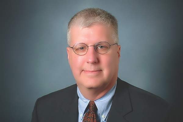
Dr. Reece, who specializes in maternal-fetal medicine, is vice president for medical affairs at the University of Maryland, Baltimore, as well as the John Z. and Akiko K. Bowers Distinguished Professor and dean of the school of medicine. Dr. Reece said he had no relevant financial disclosures. He is the medical editor of this column. Contact him at [email protected].
Viral illnesses in pregnancy are not unheard of. When a patient presents with symptoms, we often think of an influenza type of infection that will be cleared within a short period of time and with few negative consequences for the developing fetus. Other infections that can occur include TORCH – Toxoplasmosis, Other (syphilis, varicella-zoster, parvovirus B19), Rubella, Cytomegalovirus (CMV), and Herpes – infections, but these are also relatively common.
Rarely do we in the United States consider a gravida’s vulnerability to tropical infectious diseases such as dengue, chikungunya, and now Zika virus. With the popularity and ease of international travel, and the potential for women’s exposure to more exotic diseases, the practice of ob.gyn. must undergo a significant transition in perspective. It is vital for us to understand these illnesses because of their potency and reported injury to both the mother and baby, for several reasons.
First, there is the public health concern. As of June 16, 2016, the Pan American Health Organization of the World Health Organization, reported 39 countries and territories in the Americas with confirmed cases of Zika virus, with 21 of those countries having confirmed cases in pregnant women.
As of June 9, 2016, the Centers for Disease Control and Prevention reported that 234 pregnant women in the United States have laboratory evidence of possible Zika infection, along with 189 pregnant women living in U.S. territories. Since the current outbreak, which began in July 2015 in Brazil, seven countries – accounting for more than 1,600 cases – have reported babies with congenital syndrome associated with Zika virus, the majority of which have been in Brazil. With the Summer Olympics in Rio starting in August 2016, the potential spread of Zika virus is dizzying.
Second, there is the counseling and management concern. Without a treatment or vaccine available, ob.gyns. must stay current on the latest research and findings to inform their patients of the risks associated with travel to an area with confirmed, or areas at risk for developing, Zika virus transmission.
Third, there is a diagnostic concern. Women who have visited areas with Zika virus, or who have had intimate contact with someone who has traveled to these areas, must be diagnosed and then counseled immediately.
We have devoted this Master Class to a discussion of Zika virus and the work being conducted in the United States to understand this disease. We have invited Dr. Yoel Sadovsky, an expert on placental development and trophoblast function, and his colleague, Carolyn Coyne, Ph.D., a leading researcher on host-virus interactions, to address this important topic.
Dr. Reece, who specializes in maternal-fetal medicine, is vice president for medical affairs at the University of Maryland, Baltimore, as well as the John Z. and Akiko K. Bowers Distinguished Professor and dean of the school of medicine. Dr. Reece said he had no relevant financial disclosures. He is the medical editor of this column. Contact him at [email protected].

Surgical workforce shortages in rural areas
This month I write about one of the College’s current advocacy efforts directed at ensuring an adequate surgical workforce in underserved and rural areas. Evidence indicates a current and growing shortage of surgeons available to serve the needs of populations in certain parts of the country. A shortage of general surgeons is a clear component to the crisis in health care workforce. Accordingly, the American College of Surgeons (ACS) is urging policy makers to recognize that only surgeons are uniquely qualified to provide certain necessary, lifesaving procedures, which other health professionals are neither trained nor competent to provide.
To determine where these areas of shortage are located and where access to surgical care is thus potentially a challenge, the ACS is strongly supporting the efforts of Representatives Larry Bucshon, MD, FACS (R-Ind.) and Ami Bera, MD (D-Calif.) who recently introduced H.R. 4959, the Ensuring Access to General Surgery Act of 2016. This legislation serves to direct the Secretary of the Department of Health and Human Services (HHS) to conduct a study on the designation of surgical Health Professional Shortage Areas (HPSA).
A variety of federal programs use the HPSA designation to improve access to health care by focusing aid and assistance on specific geographic areas and populations with the greatest unmet needs. The division of HHS known as the Health Resources and Services Administration (HRSA) has developed criteria used to determine whether certain geographic areas, population groups, or facilities may be designated as a HPSA. HPSA designation may be applied to urban or rural geographic areas, specific population groups, medical provider groups, or other public health care facilities. Currently, HRSA limits HPSA designations to shortages in primary care services, dental services, or mental health services.
HRSA has never designated an entity as a HPSA purely based upon a shortage of surgical services. In light of the available evidence relative to the shortage of surgical providers in certain parts of the country, ACS believes that research is necessary to determine exactly what constitutes a surgical shortage area, e.g., establish definitional criteria, with subsequent application of those criteria to determine where areas so defined are located. Such would provide HRSA with a valuable tool to utilize in efforts directed at increasing patient access to surgical care. Ultimately, offering incentives to surgeons to locate or remain in HPSA communities could become critical in guaranteeing all Medicare beneficiaries, regardless of geographic location, have access to quality surgical care. Determining what constitutes a surgical shortage area will serve to help HRSA to appropriately focus its resources.
Accordingly, we need your help and urge you to take action today.
Using the information below, please call your representatives today and urge them to join their colleagues and cosponsor H.R. 4959, the Ensuring Access to General Surgery Act of 2016.
Instructions
Call toll-free: 1-877-996-4464
You will be connected to your representative‘s office. Once you are connected, provide your name and indicate that you are a constituent. You should also be prepared to provide additional contact information for follow-up purposes.
Next, we suggest you use the following message:
• As a surgeon and as your constituent, I urge you to join your colleagues and cosponsor H.R. 4959, the Ensuring Access to General Surgery Act of 2016, which would direct the Secretary of Department of Health and Human Services (HHS) to conduct a study to designate General Surgery Health Professional Shortage Areas (HPSA).
• The division of HHS known as the Health Resources and Services Administration (HRSA) has developed designation criteria in order to determine whether certain geographic areas, population groups, or facilities may be designated as a HPSA.
• HRSA has never designated an entity as a HPSA purely based upon a shortage of surgical services.
• In light of evidence relative to a shortage of surgeons, ACS believes that research is necessary to determine exactly what constitutes a surgical shortage area and subsequently where these areas exist.
Alternatively, for those who were seeking a topic on which to initiate a personal in-district meeting with representatives and their staff as was discussed in last month’s edition of this column, H.R. 4959 presents a prime subject for such in order to have a focused meeting with a specific ask on a “white hat” issue that will surely resonate with members of Congress. Currently, in-district work periods are scheduled for the last week of June, the last two weeks of July, and the entire month of August.
As always, those with questions or concerns, or those who need assistance in setting up an in-district meeting may contact staff of the Division of Advocacy and Health Policy by phone at 202-337-2701 or via e-mail at [email protected].
Thank you for taking the time to engage and take action on this critical issue.
Please encourage your colleagues to do likewise.
Until next month ...
Dr. Patrick V. Bailey is an ACS Fellow, a pediatric surgeon, and Medical Director, Advocacy, for the Division of Advocacy and Health Policy, in the ACS offices in Washington, DC.

Down Under
If you want to know how big the world really is, I suggest you take a trip from McPherson, Kansas, to Sydney, Australia, in one day. You won’t be able to do it, by the way. The construct of days prohibits you from doing this from East to West. Your vessel will pass the International Date Line and you will lose the day (sort of the opposite of seizing the day). Don’t worry. You’ll get it back on the return trip. In this way, the universe seems to enjoy a certain symmetry. But even by first class in a “Sky Couch,” your body will understand how far 8,666 miles is. Trust me: The world is a big place.
There is something unsettling about stepping out of a metal tube that was going Mach 0.7 for 13 hours into a world with “mates” and where the bathtub water drains out the “wrong” way. It’s a little like a “Twilight Zone” episode in which the guest star notes everything in this world is familiar except just different enough to make all the difference.
I entered this zone because I have the great good fortune to know John Kyngdon, MD, FRACS. Dr. Kyngdon was the convener for this year’s Rural Surgery Section of the Royal Australasian College of Surgeons, aka, RACS. I was delighted to attend the 2016 RACS annual meeting, which had the theme of technology and communication.
We Americans can be pretty smug when it comes to our health care system, our training, and our outcomes. Traveling to the other side of the world and spending time with surgeons working in Australia and New Zealand can take the smug right off one’s face. Australia is a land of immense distances and minuscule population for such a large land mass. The challenge of providing care across this gigantic continent, the center of which contains an immense desert filled with some of this most deadly insects, snakes and other creatures on the planet, is epic for sure. Yet, where an American baby boomer like me might decry the hopelessness of such a task, the Australians smile and carry on. These people just don’t understand that their task is nigh on impossible, so they succeed to a large degree against the odds.
RACS, of course, does not just include Australia and New Zealand but the South Pacific and Southeast Asia as well. It was formed in part from the efforts of Dr. Will Mayo, who supported the effort of an ACS-like organization for this part of the world. RACS members seem to have a special affection for Americans, consequently, and one feels entirely at home with them. While ACS has many more members, the quality of the presentations given at the RACS annual meeting is certainly on par with much of what one would see in October at the ACS Clinical Congress. American surgeons commonly attend, and I was delighted to see ACS Vice President Ron Maier, MD, there, as well as Gary Timmerman, MD, of South Dakota and Nathaniel Soper, MD, of Northwestern.
The striking point for me is the commonality we surgeons share worldwide. Whether trained under a UK, Australian, or American-type system, the problems we face are similar. For RACS members, the challenge of managing the EHR is about the same, and as would be expected, interoperability is a huge problem for them! Because of the distances involved in Australia, they are much more involved in telemedicine than are US surgeons, but they are just beginning to deal with privacy issues that come with the technology. They are haunted by quality metrics just as we are. Malpractice is quite different from the US in that, at least in New Zealand, surgeons are not sued for compensation, but they can lose their professional credentials over a bad outcome attributed to them. Burnout among surgeons is a problem Down Under, just as it is here. Governmental intrusions and misadventures, ditto.

I had the opportunity to observe teaching of anatomy at a medical school and learned about dissecting electronically as well as in the flesh. One of the keynote speakers at the RACS meeting was an Australian dotcom entrepreneur. From him and his cohorts on the panel I learned that, in the very near future, over 90% of health care data will likely be gathered not in medical offices but from patient-worn devices. I saw apps based on patient-generated data claiming over 97% accuracy.
Of course, I got to spend a few days touring. Who wouldn’t? I got to see animals such as kangaroos that had been just pictures to me before. By the way, have you ever noticed those sharp claws on the “cuddly” koala? Eventually, I had to return and endure the jet lag that is always worse going West to East. Naturally, my first night on call kept me up most of 30 hours. Jet lag and call lag have the same effect. You just want to get some sleep but don’t know how.
RACS and ACS have been closely aligned for decades. I cannot think of a better mind-expanding view of the surgical world than to join them at one of their meetings. Like so many surgeons, I’ve always thought I just couldn’t take so much vacation at once. Nonsense. You can’t afford not to do so. And there’s nothing like patting the head of a kangaroo to help cure burnout.
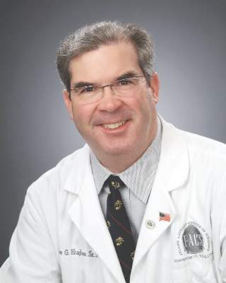
Dr. Hughes is an ACS Fellow with the department of general surgery, McPherson Hospital, McPherson, Kan., and is the Editor in Chief of ACS Communities. He is also Associate Editor for ACS Surgery News.
If you want to know how big the world really is, I suggest you take a trip from McPherson, Kansas, to Sydney, Australia, in one day. You won’t be able to do it, by the way. The construct of days prohibits you from doing this from East to West. Your vessel will pass the International Date Line and you will lose the day (sort of the opposite of seizing the day). Don’t worry. You’ll get it back on the return trip. In this way, the universe seems to enjoy a certain symmetry. But even by first class in a “Sky Couch,” your body will understand how far 8,666 miles is. Trust me: The world is a big place.
There is something unsettling about stepping out of a metal tube that was going Mach 0.7 for 13 hours into a world with “mates” and where the bathtub water drains out the “wrong” way. It’s a little like a “Twilight Zone” episode in which the guest star notes everything in this world is familiar except just different enough to make all the difference.
I entered this zone because I have the great good fortune to know John Kyngdon, MD, FRACS. Dr. Kyngdon was the convener for this year’s Rural Surgery Section of the Royal Australasian College of Surgeons, aka, RACS. I was delighted to attend the 2016 RACS annual meeting, which had the theme of technology and communication.
We Americans can be pretty smug when it comes to our health care system, our training, and our outcomes. Traveling to the other side of the world and spending time with surgeons working in Australia and New Zealand can take the smug right off one’s face. Australia is a land of immense distances and minuscule population for such a large land mass. The challenge of providing care across this gigantic continent, the center of which contains an immense desert filled with some of this most deadly insects, snakes and other creatures on the planet, is epic for sure. Yet, where an American baby boomer like me might decry the hopelessness of such a task, the Australians smile and carry on. These people just don’t understand that their task is nigh on impossible, so they succeed to a large degree against the odds.
RACS, of course, does not just include Australia and New Zealand but the South Pacific and Southeast Asia as well. It was formed in part from the efforts of Dr. Will Mayo, who supported the effort of an ACS-like organization for this part of the world. RACS members seem to have a special affection for Americans, consequently, and one feels entirely at home with them. While ACS has many more members, the quality of the presentations given at the RACS annual meeting is certainly on par with much of what one would see in October at the ACS Clinical Congress. American surgeons commonly attend, and I was delighted to see ACS Vice President Ron Maier, MD, there, as well as Gary Timmerman, MD, of South Dakota and Nathaniel Soper, MD, of Northwestern.
The striking point for me is the commonality we surgeons share worldwide. Whether trained under a UK, Australian, or American-type system, the problems we face are similar. For RACS members, the challenge of managing the EHR is about the same, and as would be expected, interoperability is a huge problem for them! Because of the distances involved in Australia, they are much more involved in telemedicine than are US surgeons, but they are just beginning to deal with privacy issues that come with the technology. They are haunted by quality metrics just as we are. Malpractice is quite different from the US in that, at least in New Zealand, surgeons are not sued for compensation, but they can lose their professional credentials over a bad outcome attributed to them. Burnout among surgeons is a problem Down Under, just as it is here. Governmental intrusions and misadventures, ditto.
I had the opportunity to observe teaching of anatomy at a medical school and learned about dissecting electronically as well as in the flesh. One of the keynote speakers at the RACS meeting was an Australian dotcom entrepreneur. From him and his cohorts on the panel I learned that, in the very near future, over 90% of health care data will likely be gathered not in medical offices but from patient-worn devices. I saw apps based on patient-generated data claiming over 97% accuracy.
Of course, I got to spend a few days touring. Who wouldn’t? I got to see animals such as kangaroos that had been just pictures to me before. By the way, have you ever noticed those sharp claws on the “cuddly” koala? Eventually, I had to return and endure the jet lag that is always worse going West to East. Naturally, my first night on call kept me up most of 30 hours. Jet lag and call lag have the same effect. You just want to get some sleep but don’t know how.
RACS and ACS have been closely aligned for decades. I cannot think of a better mind-expanding view of the surgical world than to join them at one of their meetings. Like so many surgeons, I’ve always thought I just couldn’t take so much vacation at once. Nonsense. You can’t afford not to do so. And there’s nothing like patting the head of a kangaroo to help cure burnout.
Dr. Hughes is an ACS Fellow with the department of general surgery, McPherson Hospital, McPherson, Kan., and is the Editor in Chief of ACS Communities. He is also Associate Editor for ACS Surgery News.

Setting up your own RSS feed
Last month, I discussed RSS news feeds as a useful tool for keeping abreast of frequently updated information, such as blog entries, news headlines, audio, and video, without having to visit a multitude of different Web pages each day.
This month, I’ll explain how to set up your own feed, which is useful if you want to increase the readership on your website, or publicize a podcast, or keep your patients abreast of your practice’s latest treatments and procedures. It will also alert you immediately if your name pops up in news or gossip sites.

There are several options, depending on your budget, and how involved you personally want to be in the process: Many Web hosting services will automatically create and update your feed for a monthly fee; so if you already have a professionally hosted website, check to see if your host offers that service. If not, Web services such as Feedity and Rapidfeeds allow you to manage multiple feeds, with automatic updates, so that you will not need to manually update your feed each time you update your website content. Feedity’s software can even generate an RSS file without your having to input each item. Other popular hosting options include Web Hosting Hub, Arvixe, and MyHosting, among many others. (As always, I have no financial interest in any service I mention here.)
Another option, used by many organizations that publish their own articles and news stories, is a content management system (CMS), an application designed to organize, store, and publish content, including tools for adding RSS feeds. Examples include Drupal and Plone – both free, open-source programs.
Alternatively, you can download a stand-alone RSS creation program, then create and update your feed manually. Again, there are many options to choose from. One popular example is RSS Builder, a free, open source RSS creation program that allows you to create RSS files, upload them to your website, and automatically manage them to some extent. Disadvantages of free systems include advertisements (sometimes removable for a monthly fee), scarce or nonexistent technical support, and in many cases, no option to create more than one feed. You may also have to manually add new headlines, links, and descriptive text yourself. Your “free” feed can become quite expensive if you or staffers are forced to spend an inordinate amount of time maintaining it. Paid programs such as FeedForAll allow easier creation and maintenance, and less time commitment.
Once you have chosen your service, create your first feed. The process will differ from program to program, but the general idea is the same for almost all of them. All feeds will need some basic data: A title (which should be the same as your website or podcast); the URL for your website, to help viewers link back to your home page; and a description – a sentence or two describing the general content on the feed.
Once you’ve entered this information, you can start populating the feed with content. Enter in the title of each article, blog post, podcast episode, etc., the URL that links directly to that content, and the publishing date. Each entry should have its own short but sweet description; this is what your readers will see before they choose to click your entry in their RSS readers. You can add author information and further comments if needed. Add a new entry for each piece of content that you want to broadcast.
Most RSS apps suggest that you assign each item in your feed a global unique identifier (GUID), which RSS readers use to determine if an item has been changed or updated. Each feed item should have its own GUID, particularly if multiple items are located at the same URL.
Once you’re done entering in all of your content to your feed, you need to export it to an XML file, which will allow visitors to subscribe to your feed. Then upload the XML file to your website, place it on your homepage, and click the Publish Feed button.
Once your feed is live, consider submitting it to some of the many RSS feed directories (also called aggregate sites) that collect articles from similar interests and can significantly increase your viewership. Search for medically oriented directories, and others that match the interests that your feed addresses, and submit each directory’s URL to your feed’s XML file.
Dr. Eastern practices dermatology and dermatologic surgery in Belleville, N.J. He is the author of numerous articles and textbook chapters, and is a long-time monthly columnist for Dermatology News. Write to him at [email protected].
Last month, I discussed RSS news feeds as a useful tool for keeping abreast of frequently updated information, such as blog entries, news headlines, audio, and video, without having to visit a multitude of different Web pages each day.
This month, I’ll explain how to set up your own feed, which is useful if you want to increase the readership on your website, or publicize a podcast, or keep your patients abreast of your practice’s latest treatments and procedures. It will also alert you immediately if your name pops up in news or gossip sites.
There are several options, depending on your budget, and how involved you personally want to be in the process: Many Web hosting services will automatically create and update your feed for a monthly fee; so if you already have a professionally hosted website, check to see if your host offers that service. If not, Web services such as Feedity and Rapidfeeds allow you to manage multiple feeds, with automatic updates, so that you will not need to manually update your feed each time you update your website content. Feedity’s software can even generate an RSS file without your having to input each item. Other popular hosting options include Web Hosting Hub, Arvixe, and MyHosting, among many others. (As always, I have no financial interest in any service I mention here.)
Another option, used by many organizations that publish their own articles and news stories, is a content management system (CMS), an application designed to organize, store, and publish content, including tools for adding RSS feeds. Examples include Drupal and Plone – both free, open-source programs.
Alternatively, you can download a stand-alone RSS creation program, then create and update your feed manually. Again, there are many options to choose from. One popular example is RSS Builder, a free, open source RSS creation program that allows you to create RSS files, upload them to your website, and automatically manage them to some extent. Disadvantages of free systems include advertisements (sometimes removable for a monthly fee), scarce or nonexistent technical support, and in many cases, no option to create more than one feed. You may also have to manually add new headlines, links, and descriptive text yourself. Your “free” feed can become quite expensive if you or staffers are forced to spend an inordinate amount of time maintaining it. Paid programs such as FeedForAll allow easier creation and maintenance, and less time commitment.
Once you have chosen your service, create your first feed. The process will differ from program to program, but the general idea is the same for almost all of them. All feeds will need some basic data: A title (which should be the same as your website or podcast); the URL for your website, to help viewers link back to your home page; and a description – a sentence or two describing the general content on the feed.
Once you’ve entered this information, you can start populating the feed with content. Enter in the title of each article, blog post, podcast episode, etc., the URL that links directly to that content, and the publishing date. Each entry should have its own short but sweet description; this is what your readers will see before they choose to click your entry in their RSS readers. You can add author information and further comments if needed. Add a new entry for each piece of content that you want to broadcast.
Most RSS apps suggest that you assign each item in your feed a global unique identifier (GUID), which RSS readers use to determine if an item has been changed or updated. Each feed item should have its own GUID, particularly if multiple items are located at the same URL.
Once you’re done entering in all of your content to your feed, you need to export it to an XML file, which will allow visitors to subscribe to your feed. Then upload the XML file to your website, place it on your homepage, and click the Publish Feed button.
Once your feed is live, consider submitting it to some of the many RSS feed directories (also called aggregate sites) that collect articles from similar interests and can significantly increase your viewership. Search for medically oriented directories, and others that match the interests that your feed addresses, and submit each directory’s URL to your feed’s XML file.
Dr. Eastern practices dermatology and dermatologic surgery in Belleville, N.J. He is the author of numerous articles and textbook chapters, and is a long-time monthly columnist for Dermatology News. Write to him at [email protected].
Billing for family meetings
Family meetings are never easy.
They’re difficult, and often held to discuss the case of a demented patient. In these situations, the family doesn’t want the patient to hear their concerns or is afraid they’ll be embarrassed or angry. Sometimes getting the patient to the appointment is simply too difficult.
Of course, most discussions of this type can be done by phone ... in theory. In practice, it doesn’t work that way.

It’s the subject matter that makes the impersonal nature of the phone difficult. Families have hard questions and want real answers at these times. A face-to-face meeting, with the human interaction, is often the best way to get things across clearly and still with compassion. It avoids the problem of the phone being passed around, having to repeat yourself to each person, and wondering who just asked what. Very few families, in my experience, want to do this on the phone.
These meetings are never quick, either. Depending on family and circumstances, they can take 30-60 minutes. Getting an insurance company to pay for that time is near impossible. Most plans only want to pay for visits where the patient is actually present, when in these cases the family is trying to avoid that. While there is a Medicare payment code for “advance care planning,” it doesn’t cover treatment discussions or other neurological issues they may bring up, and many patients are on non-Medicare plans.
I bill people for these times and have found that most families are willing to pay. I’m not fond of doing so, and certainly not trying to get rich off of them. But it’s still time that I’m in my office and have to pay my rent, staff, and utilities.
Part of this job – a big part – is helping patients and their loved ones understand and deal with difficult situations. Realistically, this is the best way to do it. Families understand that as well as I do.
Why won’t insurance companies cover them? I suppose their excuse is that they cover the patient, not the questions or emotional needs of their caregivers. Of course, those things are as important to the care of the patient as any treatment, but the bean counters don’t want to pay for them.
That is unfortunate, because someone has to. Good medical care depends on good communication with all involved.
Dr. Block has a solo neurology practice in Scottsdale, Ariz.
Family meetings are never easy.
They’re difficult, and often held to discuss the case of a demented patient. In these situations, the family doesn’t want the patient to hear their concerns or is afraid they’ll be embarrassed or angry. Sometimes getting the patient to the appointment is simply too difficult.
Of course, most discussions of this type can be done by phone ... in theory. In practice, it doesn’t work that way.
It’s the subject matter that makes the impersonal nature of the phone difficult. Families have hard questions and want real answers at these times. A face-to-face meeting, with the human interaction, is often the best way to get things across clearly and still with compassion. It avoids the problem of the phone being passed around, having to repeat yourself to each person, and wondering who just asked what. Very few families, in my experience, want to do this on the phone.
These meetings are never quick, either. Depending on family and circumstances, they can take 30-60 minutes. Getting an insurance company to pay for that time is near impossible. Most plans only want to pay for visits where the patient is actually present, when in these cases the family is trying to avoid that. While there is a Medicare payment code for “advance care planning,” it doesn’t cover treatment discussions or other neurological issues they may bring up, and many patients are on non-Medicare plans.
I bill people for these times and have found that most families are willing to pay. I’m not fond of doing so, and certainly not trying to get rich off of them. But it’s still time that I’m in my office and have to pay my rent, staff, and utilities.
Part of this job – a big part – is helping patients and their loved ones understand and deal with difficult situations. Realistically, this is the best way to do it. Families understand that as well as I do.
Why won’t insurance companies cover them? I suppose their excuse is that they cover the patient, not the questions or emotional needs of their caregivers. Of course, those things are as important to the care of the patient as any treatment, but the bean counters don’t want to pay for them.
That is unfortunate, because someone has to. Good medical care depends on good communication with all involved.
Dr. Block has a solo neurology practice in Scottsdale, Ariz.
Aesthetic Dermatology: Synthetic second skin
Hot off the presses, a new so-called “second skin” is being redeveloped and rebranded for use in both cosmetic and medical dermatology. But what is this substance, and will it hold up to all the claims the manufacturer and research team suggest?
Recently described in Nature Materials, the liquid polymer developed by chemical engineers at MIT is a synthetic, adherent silicone-based film that lies perfectly invisibly on the skin – providing a pulling or temporary tightening of the skin. The product was initially marketed by the company Living Proof as “Neotensil” [an acronym for (Neo) new, (T) transforming, (E) elastic, (N) non-invasive, (S) supportive, (I) invisible, and immediate (L) layer solution]. When applied to the area under the eye, the product creates a so-called “Spanx” effect or tightening of periorbital skin.

The material – called XPL – is delivered in a two-step sequential process. First, a polysiloxane cream is applied to the skin, followed by a platinum catalyst that induces the polymer to harden and tighten the skin underneath. The product uses patented Strateris technology, which is described as creating invisible “shapewear” for the eye; a film that tightens, smooths, and lifts the appearance of skin for up to 24 hours. It was briefly on the market in 2014-2015, then taken off the market to be redeveloped.
Does it work? Yes. Although it takes about an hour to take effect, the clinical results are jaw dropping. However, it also has its drawbacks. The polymer – which hardens within 2 minutes – must be applied to clean skin, with no creams or makeup whatsoever. And makeup cannot be applied over the treated area either, as the area looks irregular and uneven with makeup. This is a very difficult obstacle to overcome for many female patients.
Additionally, to take off the product, the polymer must be dissolved with a special chemical remover that is packaged with the product. Without this key component, it is very difficult and very irritating to remove. Although none of the patients I have used this product on have developed allergic reactions, any synthetic polymer, particularly one with adherent properties, has the potential to be an irritant and/or an allergen. Long-term clinical trials are needed to both validate its efficacy and side-effect profile.

The potential for clinical uses is vast. The product has been shown to provide a synthetic skin barrier that minimizes transepidermal water loss, improving skin hydration. Its uses in burns, atopic dermatitis, bullous disease, and psoriasis could help those with altered skin-barrier function. The researchers are also hoping to use this product for targeted drug delivery and for UV protection.
After a decade of research, the MIT team has developed a skinlike material that is invisible and mimics both the mechanical and elastic properties of the skin. Future clinical studies are essential to evaluating its broad applicability in both dermatology and general medicine.
1. Nature Materials 2016. doi:10.1038/nmat4635.
Dr. Wesley and Dr. Talakoub are co-contributors to this column. Dr. Talakoub is in private practice in McLean, Va. Dr. Wesley practices dermatology in Beverly Hills, Calif. This month’s column is by Dr. Talakoub. Dr. Talakoub has no disclosures related to the product. Write to them at [email protected].
Hot off the presses, a new so-called “second skin” is being redeveloped and rebranded for use in both cosmetic and medical dermatology. But what is this substance, and will it hold up to all the claims the manufacturer and research team suggest?
Recently described in Nature Materials, the liquid polymer developed by chemical engineers at MIT is a synthetic, adherent silicone-based film that lies perfectly invisibly on the skin – providing a pulling or temporary tightening of the skin. The product was initially marketed by the company Living Proof as “Neotensil” [an acronym for (Neo) new, (T) transforming, (E) elastic, (N) non-invasive, (S) supportive, (I) invisible, and immediate (L) layer solution]. When applied to the area under the eye, the product creates a so-called “Spanx” effect or tightening of periorbital skin.
The material – called XPL – is delivered in a two-step sequential process. First, a polysiloxane cream is applied to the skin, followed by a platinum catalyst that induces the polymer to harden and tighten the skin underneath. The product uses patented Strateris technology, which is described as creating invisible “shapewear” for the eye; a film that tightens, smooths, and lifts the appearance of skin for up to 24 hours. It was briefly on the market in 2014-2015, then taken off the market to be redeveloped.
Does it work? Yes. Although it takes about an hour to take effect, the clinical results are jaw dropping. However, it also has its drawbacks. The polymer – which hardens within 2 minutes – must be applied to clean skin, with no creams or makeup whatsoever. And makeup cannot be applied over the treated area either, as the area looks irregular and uneven with makeup. This is a very difficult obstacle to overcome for many female patients.
Additionally, to take off the product, the polymer must be dissolved with a special chemical remover that is packaged with the product. Without this key component, it is very difficult and very irritating to remove. Although none of the patients I have used this product on have developed allergic reactions, any synthetic polymer, particularly one with adherent properties, has the potential to be an irritant and/or an allergen. Long-term clinical trials are needed to both validate its efficacy and side-effect profile.
The potential for clinical uses is vast. The product has been shown to provide a synthetic skin barrier that minimizes transepidermal water loss, improving skin hydration. Its uses in burns, atopic dermatitis, bullous disease, and psoriasis could help those with altered skin-barrier function. The researchers are also hoping to use this product for targeted drug delivery and for UV protection.
After a decade of research, the MIT team has developed a skinlike material that is invisible and mimics both the mechanical and elastic properties of the skin. Future clinical studies are essential to evaluating its broad applicability in both dermatology and general medicine.
1. Nature Materials 2016. doi:10.1038/nmat4635.
Dr. Wesley and Dr. Talakoub are co-contributors to this column. Dr. Talakoub is in private practice in McLean, Va. Dr. Wesley practices dermatology in Beverly Hills, Calif. This month’s column is by Dr. Talakoub. Dr. Talakoub has no disclosures related to the product. Write to them at [email protected].

Driverless health care
Health care is changing. For some, this is cause for celebration: A safer, convenient, more efficient system is upon us. For others, the end is nigh: impossibly demanding patients, crushing bureaucracy, and a once sacred relationship desecrated by invasive technology.
The ascent of digital health technologies is an important driver of health care change, yet its impact is still indeterminate. Any technology that empowers patients as well as physicians will improve patients’ health outcomes. Or so it might seem. The truth is more nuanced: Some services will improve outcomes, others won’t. Fitting Fitbits into our current system is like inserting the wrong key into a lock: It might go in, but it doesn’t open anything. Fortunately, some keys are opening doors to better care – doors that that we’ve never entered – but finding the right ones is laborious. Dr. Joe Kvedar is here to help.

As a physician leader who straddles the gap between physician-centered and consumer-centered health care, Dr. Kvedar has spent his career leveraging technology to improve care delivery for both. In his new book, “The Internet of Healthy Things,” he shares what he has learned. He uses numerous examples from his experience as a physician and pioneer in digital health care with Partners HealthCare, Boston, delving deeply into the business of health care and the behavioral habits of patients.
As he notes, Partners was “prescient” in the health care landscape, introducing video conferencing in the 1990s, second opinions on the Internet in 2001, and texting as a tool for health messaging in 2008. He asks now: “What are the connected health devices and applications that our clinicians will be using in 5-10 years?” Then he uses his acumen and research to answer his own question. The ensuing chapters are more prescriptive than predictive, however. None of us knows where health care will be in 10 years, but we should think about where it ought to be.
Whether you chart on an Apple Watch or on paper, this discussion is important to you. Physicians are key players in determining where and how medicine is practiced, and we need to understand relevant risks and benefits to make the right decisions.
Confusing the matter is that desired outcomes are not absolute but relative. It depends on the frame of reference. Patients measure outcomes with service, payers with cost, and we physicians with quality. Which measurement is correct? How can we know if a remote monitoring device is worthwhile if we can’t agree on what it delivers? Is it simply sending home “the sicker even quicker?” Does a Big Pharma beyond-the-pill app really only increase consumption of the costliest medications or create more affordable alternatives?
Technologies that increase access to services such as live chat, messaging, and monitoring may be preferred by patients, but physicians see them as piling on to backbreaking loads. More artificial intelligence is needed to enable these services without requiring physician work. We need driverless health care.
Keeping patients involved has a whole other set of requirements. The tools must be easy, the information personal, the data actionable, and its use Candy-Crush-Saga addictive. This is no small feat, but there is hope.
Partner’s Text 2 Move program, which Dr. Kvedar describes as the “gold standard of what learning about your consumer means,” showed that highly personalized, targeted text messages could have a significant impact on patients’ behavior and health. It is just this type of technology that many are relentlessly pursuing to deliver care more effectively.
Dr. Kvedar devotes significant attention to the patient/consumer experience in a thoughtful, complex manner. Rather than elevate or denigrate the rise of the engaged patient, he examines this phenomenon through several lenses, addressing equally the concerns of practicing physicians and health care entrepreneurs. Nearly 20 years ago, Regina Herzlinger of Harvard Business School, Boston, predicted the rise of health care consumerism. Retail clinics, direct-to-consumer services, and the “Yelpification” of health care are signals that we’ve arrived.
It’s tough to make predictions, especially about the future, Yogi Berra warned us. The book’s mention of Theranos, the failing pinprick blood lab company founded by celebrity Stanford student Elizabeth Holmes, is an example of how risky it is to place bets on where we are headed and how quickly we will get there. DIY at-home labs are further off than they appeared, and that is the hazard of any such books: it is difficult to see more than what’s just in front of us.
Ultimately, Dr. Kvedar’s message is as realistic as it is optimistic: “The same information that could help drive healthcare costs down can be used to create highly individualized programs that will help people stay healthier and happier.” But we should resist the urge to ask “Are we there yet?” No. We’ve a ways to go.
Dr. Benabio is a partner physician in the department of dermatology of the Southern California Permanente Group in San Diego and a volunteer clinical assistant professor at the University of California, San Diego. Dr. Benabio is @Dermdoc on Twitter. Write to him at [email protected].
Health care is changing. For some, this is cause for celebration: A safer, convenient, more efficient system is upon us. For others, the end is nigh: impossibly demanding patients, crushing bureaucracy, and a once sacred relationship desecrated by invasive technology.
The ascent of digital health technologies is an important driver of health care change, yet its impact is still indeterminate. Any technology that empowers patients as well as physicians will improve patients’ health outcomes. Or so it might seem. The truth is more nuanced: Some services will improve outcomes, others won’t. Fitting Fitbits into our current system is like inserting the wrong key into a lock: It might go in, but it doesn’t open anything. Fortunately, some keys are opening doors to better care – doors that that we’ve never entered – but finding the right ones is laborious. Dr. Joe Kvedar is here to help.
As a physician leader who straddles the gap between physician-centered and consumer-centered health care, Dr. Kvedar has spent his career leveraging technology to improve care delivery for both. In his new book, “The Internet of Healthy Things,” he shares what he has learned. He uses numerous examples from his experience as a physician and pioneer in digital health care with Partners HealthCare, Boston, delving deeply into the business of health care and the behavioral habits of patients.
As he notes, Partners was “prescient” in the health care landscape, introducing video conferencing in the 1990s, second opinions on the Internet in 2001, and texting as a tool for health messaging in 2008. He asks now: “What are the connected health devices and applications that our clinicians will be using in 5-10 years?” Then he uses his acumen and research to answer his own question. The ensuing chapters are more prescriptive than predictive, however. None of us knows where health care will be in 10 years, but we should think about where it ought to be.
Whether you chart on an Apple Watch or on paper, this discussion is important to you. Physicians are key players in determining where and how medicine is practiced, and we need to understand relevant risks and benefits to make the right decisions.
Confusing the matter is that desired outcomes are not absolute but relative. It depends on the frame of reference. Patients measure outcomes with service, payers with cost, and we physicians with quality. Which measurement is correct? How can we know if a remote monitoring device is worthwhile if we can’t agree on what it delivers? Is it simply sending home “the sicker even quicker?” Does a Big Pharma beyond-the-pill app really only increase consumption of the costliest medications or create more affordable alternatives?
Technologies that increase access to services such as live chat, messaging, and monitoring may be preferred by patients, but physicians see them as piling on to backbreaking loads. More artificial intelligence is needed to enable these services without requiring physician work. We need driverless health care.
Keeping patients involved has a whole other set of requirements. The tools must be easy, the information personal, the data actionable, and its use Candy-Crush-Saga addictive. This is no small feat, but there is hope.
Partner’s Text 2 Move program, which Dr. Kvedar describes as the “gold standard of what learning about your consumer means,” showed that highly personalized, targeted text messages could have a significant impact on patients’ behavior and health. It is just this type of technology that many are relentlessly pursuing to deliver care more effectively.
Dr. Kvedar devotes significant attention to the patient/consumer experience in a thoughtful, complex manner. Rather than elevate or denigrate the rise of the engaged patient, he examines this phenomenon through several lenses, addressing equally the concerns of practicing physicians and health care entrepreneurs. Nearly 20 years ago, Regina Herzlinger of Harvard Business School, Boston, predicted the rise of health care consumerism. Retail clinics, direct-to-consumer services, and the “Yelpification” of health care are signals that we’ve arrived.
It’s tough to make predictions, especially about the future, Yogi Berra warned us. The book’s mention of Theranos, the failing pinprick blood lab company founded by celebrity Stanford student Elizabeth Holmes, is an example of how risky it is to place bets on where we are headed and how quickly we will get there. DIY at-home labs are further off than they appeared, and that is the hazard of any such books: it is difficult to see more than what’s just in front of us.
Ultimately, Dr. Kvedar’s message is as realistic as it is optimistic: “The same information that could help drive healthcare costs down can be used to create highly individualized programs that will help people stay healthier and happier.” But we should resist the urge to ask “Are we there yet?” No. We’ve a ways to go.
Dr. Benabio is a partner physician in the department of dermatology of the Southern California Permanente Group in San Diego and a volunteer clinical assistant professor at the University of California, San Diego. Dr. Benabio is @Dermdoc on Twitter. Write to him at [email protected].

Discrimination, Dignity, and Duty
This spring, Tennessee became the first state in the union to pass legislation that gives a mental health care professional the right to refuse to see a patient based on “sincerely held principles.” HB 1840 reads:
No counselor or therapist providing counseling or therapy services shall be required to counsel or serve a client as to goals, outcomes, or behaviors that conflict with a sincerely held principles of the counselor or therapist; provided, that the counselor or therapist coordinates a referral of the client to another counselor or therapist who will provide the counseling or therapy.1
Although Tennessee was the first, many other states have enacted similar laws over the past year, which critics believe represent discrimination against lesbian, gay, bisexual, and transgender (LGBT) individuals.2
In Tennessee, a late amendment substituted “sincerely held principles” for “sincerely held religious beliefs.” The change from religious beliefs to principles may seem to attenuate the most discriminatory aspects of the law, while actually expanding both its application and acceptance. Although most civil rights advocates believe the law was designed to target LGBT individuals, “sincerely held principles” could potentially affect veterans and active-duty service members (ADSMs) with any socially stigmatized condition or circumstance from domestic violence to addiction.
No doubt, we will see the law’s constitutionality argued in court. We do know that the Supremacy Clause (Article VI, Clause 2) of the U.S. Constitution states that the federal constitution and laws take precedence over state laws and constitutions.3 Not being a lawyer, I cannot opine on how this particular piece of legislation will interact with federal law. But as a physician and an ethicist, I can say that it challenges the foundational commitment of all health care practitioners to place the good of the patient above all other considerations.
In an interview shortly before signing the law, Tennessee governor Bill Haslam discussed his decision-making process. It is “all about values,” he said, “therapists cannot and should not be expected to leave those values out of their work.”4 The governor’s statements are based on a fundamental misunderstanding of the ethical obligations and values intrinsic to the health care practitioner-patient relationship.
As citizens and human beings practitioners are entitled to hold any beliefs, preferences, and principles. But it is the respect for patient’s beliefs, preferences, and values over and above those of the practitioner that make health care practice a profession. Professional relationships in health care are not like those in commerce or industry, entertainment, or advertising; these relationships are of a special fiduciary nature that mandates a duty to care that is expressed in both ancient and modern ethical codes.
The American Counseling Association specifically mentioned its ethics code in the organization’s response to the Tennessee legislation. “HB 1840 is an unprecedented attack on the American Counseling Association’s Code of Ethics, something to which nearly 60,000 counselors abide.”5 In addition, more than half a million counselors and thousands of social workers, physicians, nurses, and pharmacists also abide by codes of ethics, professional principles, and organizational policies contained in provisions explicitly prohibiting the clinician from discriminating against any patient. The American Medical Association, for example, states:
A physician may decline to undertake the care of a patient whose medical condition is not within the physician’s current competence. However, physicians who offer their services to the public may not decline to accept patients because of race, color, religion, national origin, sexual orientation, gender identity or any other basis that would constitute invidious discrimination.6
Governor Haslam reportedly signed the law once it addressed 2 of his concerns.7 The first was that the bill must require that practitioners who object to treating LGBT patients or any other patient on principled grounds refer them to another counselor. Undoubtedly, this requirement is of little comfort for those denied mental health care that live in small communities. Many of these communities already face shortages of mental health professionals and higher levels of prejudice and ostracism, which the law will only amplify.
The second provision insisted on by the governor required a counselor to continue treating a patient if the patient is a danger to himself or herself or others. This provision hardly seems exculpatory when we consider that LGBT military and veteran populations already have higher risks of suicide and struggle to access mental health care, problems the DoD and VA are addressing.8 A referral cannot psychologically mitigate or ethically defend the devastating impact of having a therapist refuse help to an already wounded and isolated patient and may well trigger a suicide attempt.
The VA has made an impressive commitment to end health care disparities for veteran LGBT patients, but much less is known about the health care quality gaps for ADSMs. In both cases, legislation like that enacted in Tennessee only places additional obstacles on that steep climb to health care equity.
Not even the most impassioned advocate of social justice would likely say that a counselor or a nurse is not entitled to have personal beliefs and values. Codes of ethics, hospital policies, and state and federal laws often contain “conscience clauses” allowing persons to decline to participate in procedures that violate their religious and moral beliefs—we will explore this further in a later editorial. But a procedure, for example, prescribing a medication or doing a surgery is different from refusing to serve an entire group of persons on the basis of a characteristic that is neither chosen nor changeable.
A conscience clause summoned to defend or disguise clearly discriminatory actions is invalidated and self-contradictory because it violates the most essential ethical principle, that all human beings are worthy of respect and dignity. Conscientious objection on the grounds of personal religion or morality in health care ends where discrimination against a class of persons denying clinically indicated treatment begins.9
There are multiple clinically concerning implications of this legislation for federal practice. We are only too aware that our organizations historically have failed to safeguard the rights and dignity of ADSMs or veterans who belong to many types of minority groups. But recently we have made progress in addressing these health care disparities especially for the VA LGBT community.10 Legislation like that passed in Tennessee and proposed in other states threatens to undermine these gains for those who served honorably and those who still put their life on the line to defend “liberty and justice for all.”
Even if we continue to uphold high moral and legal principles in regards to the patients we treat in our institutions, millions of ADSM and veterans receive their care in the community, especially with the advent of the Choice Act. Our duty to care must begin within our federal auspices to ensure that all those we treat receive health care with dignity, but it must extend outside the walls of our institutions to protect those who have been or are now in uniform against discrimination in health care.
1. An Act to amend Tennessee Code , Title 4; Title 49 and Title 63, relative to conscientious objections to the provision of counseling and therapy. Tennessee Annotated Code. HB 1840 109th Leg (Tenn 2016).
2. Dobuzinskis A. Tennessee law to allow counselors to deny service based on beliefs. Reuters website. http://www.reuters.com/article/us-tennessee-counseling-law-idUSKCN0XO2VH. Published April 28, 2016. Accessed May 9, 2016.
3. Alexander MC. A Short and Happy Guide to Constitutional Law. St. Paul, MN: West Academic; 2013.
4. Inskeep S. For Tennessee governor weighing religious objection bill, it’s all about values. National Public Radio website. http://wnpr.org/post/tenn-gov-weighing-therapist-religious-objection-bill-its-all-about-values#stream/0. Published April 21, 2016. Accessed May 9, 2016.
5. American Counseling Association. Tennessee advances bill that tells counselors to discriminate. American Counseling Association website. https://www.counseling.org/news/updates/2016/03/24/tennessee-advances-bill-that-tells-counselors-to-discriminate. Published March 24, 2016. Accessed May 8, 2016.
6. American Medical Association. E-9.12 patient-physician relationship: respect for law and human rights. American Medical Association website. http://www.ama-assn.org/ama/pub/about-ama/our-people/member-groups-sections/glbt-advisory-committee/ama-policy-regarding-sexual-orientation.page? Adopted November 2007. Accessed May 9, 2016.
7. Wagner L. Tennessee enacts law letting therapists refuse patients on religious grounds. National Public Radio website. http://wnpr.org/post/tennessee-enacts-law-letting-therapists-refuse-patients-religious-grounds#stream/0. Published April 27, 2016. Accessed May 9, 2016.
8. Matarazzo BB, Barnes SM, Pease JL, et al. Suicide risk among lesbian, gay, bisexual, and transgender military personnel and veterans: what does the literature tell us? Suicide Life Threat Behav. 2014;44(2):200-217.
9. Dickens BM. Legal protection and limits of conscientious objection: when conscientious objection is unethical. Med Law. 2009;28(2):337-347.
10. Sharpe VA, Uchendu US. Ensuring appropriate care for LGBT veterans in the Veterans Health Administration. Hastings Cent Rep. 2014;44(suppl 4):S53-S55.
This spring, Tennessee became the first state in the union to pass legislation that gives a mental health care professional the right to refuse to see a patient based on “sincerely held principles.” HB 1840 reads:
No counselor or therapist providing counseling or therapy services shall be required to counsel or serve a client as to goals, outcomes, or behaviors that conflict with a sincerely held principles of the counselor or therapist; provided, that the counselor or therapist coordinates a referral of the client to another counselor or therapist who will provide the counseling or therapy.1
Although Tennessee was the first, many other states have enacted similar laws over the past year, which critics believe represent discrimination against lesbian, gay, bisexual, and transgender (LGBT) individuals.2
In Tennessee, a late amendment substituted “sincerely held principles” for “sincerely held religious beliefs.” The change from religious beliefs to principles may seem to attenuate the most discriminatory aspects of the law, while actually expanding both its application and acceptance. Although most civil rights advocates believe the law was designed to target LGBT individuals, “sincerely held principles” could potentially affect veterans and active-duty service members (ADSMs) with any socially stigmatized condition or circumstance from domestic violence to addiction.
No doubt, we will see the law’s constitutionality argued in court. We do know that the Supremacy Clause (Article VI, Clause 2) of the U.S. Constitution states that the federal constitution and laws take precedence over state laws and constitutions.3 Not being a lawyer, I cannot opine on how this particular piece of legislation will interact with federal law. But as a physician and an ethicist, I can say that it challenges the foundational commitment of all health care practitioners to place the good of the patient above all other considerations.
In an interview shortly before signing the law, Tennessee governor Bill Haslam discussed his decision-making process. It is “all about values,” he said, “therapists cannot and should not be expected to leave those values out of their work.”4 The governor’s statements are based on a fundamental misunderstanding of the ethical obligations and values intrinsic to the health care practitioner-patient relationship.
As citizens and human beings practitioners are entitled to hold any beliefs, preferences, and principles. But it is the respect for patient’s beliefs, preferences, and values over and above those of the practitioner that make health care practice a profession. Professional relationships in health care are not like those in commerce or industry, entertainment, or advertising; these relationships are of a special fiduciary nature that mandates a duty to care that is expressed in both ancient and modern ethical codes.
The American Counseling Association specifically mentioned its ethics code in the organization’s response to the Tennessee legislation. “HB 1840 is an unprecedented attack on the American Counseling Association’s Code of Ethics, something to which nearly 60,000 counselors abide.”5 In addition, more than half a million counselors and thousands of social workers, physicians, nurses, and pharmacists also abide by codes of ethics, professional principles, and organizational policies contained in provisions explicitly prohibiting the clinician from discriminating against any patient. The American Medical Association, for example, states:
A physician may decline to undertake the care of a patient whose medical condition is not within the physician’s current competence. However, physicians who offer their services to the public may not decline to accept patients because of race, color, religion, national origin, sexual orientation, gender identity or any other basis that would constitute invidious discrimination.6
Governor Haslam reportedly signed the law once it addressed 2 of his concerns.7 The first was that the bill must require that practitioners who object to treating LGBT patients or any other patient on principled grounds refer them to another counselor. Undoubtedly, this requirement is of little comfort for those denied mental health care that live in small communities. Many of these communities already face shortages of mental health professionals and higher levels of prejudice and ostracism, which the law will only amplify.
The second provision insisted on by the governor required a counselor to continue treating a patient if the patient is a danger to himself or herself or others. This provision hardly seems exculpatory when we consider that LGBT military and veteran populations already have higher risks of suicide and struggle to access mental health care, problems the DoD and VA are addressing.8 A referral cannot psychologically mitigate or ethically defend the devastating impact of having a therapist refuse help to an already wounded and isolated patient and may well trigger a suicide attempt.
The VA has made an impressive commitment to end health care disparities for veteran LGBT patients, but much less is known about the health care quality gaps for ADSMs. In both cases, legislation like that enacted in Tennessee only places additional obstacles on that steep climb to health care equity.
Not even the most impassioned advocate of social justice would likely say that a counselor or a nurse is not entitled to have personal beliefs and values. Codes of ethics, hospital policies, and state and federal laws often contain “conscience clauses” allowing persons to decline to participate in procedures that violate their religious and moral beliefs—we will explore this further in a later editorial. But a procedure, for example, prescribing a medication or doing a surgery is different from refusing to serve an entire group of persons on the basis of a characteristic that is neither chosen nor changeable.
A conscience clause summoned to defend or disguise clearly discriminatory actions is invalidated and self-contradictory because it violates the most essential ethical principle, that all human beings are worthy of respect and dignity. Conscientious objection on the grounds of personal religion or morality in health care ends where discrimination against a class of persons denying clinically indicated treatment begins.9
There are multiple clinically concerning implications of this legislation for federal practice. We are only too aware that our organizations historically have failed to safeguard the rights and dignity of ADSMs or veterans who belong to many types of minority groups. But recently we have made progress in addressing these health care disparities especially for the VA LGBT community.10 Legislation like that passed in Tennessee and proposed in other states threatens to undermine these gains for those who served honorably and those who still put their life on the line to defend “liberty and justice for all.”
Even if we continue to uphold high moral and legal principles in regards to the patients we treat in our institutions, millions of ADSM and veterans receive their care in the community, especially with the advent of the Choice Act. Our duty to care must begin within our federal auspices to ensure that all those we treat receive health care with dignity, but it must extend outside the walls of our institutions to protect those who have been or are now in uniform against discrimination in health care.
1. An Act to amend Tennessee Code , Title 4; Title 49 and Title 63, relative to conscientious objections to the provision of counseling and therapy. Tennessee Annotated Code. HB 1840 109th Leg (Tenn 2016).
2. Dobuzinskis A. Tennessee law to allow counselors to deny service based on beliefs. Reuters website. http://www.reuters.com/article/us-tennessee-counseling-law-idUSKCN0XO2VH. Published April 28, 2016. Accessed May 9, 2016.
3. Alexander MC. A Short and Happy Guide to Constitutional Law. St. Paul, MN: West Academic; 2013.
4. Inskeep S. For Tennessee governor weighing religious objection bill, it’s all about values. National Public Radio website. http://wnpr.org/post/tenn-gov-weighing-therapist-religious-objection-bill-its-all-about-values#stream/0. Published April 21, 2016. Accessed May 9, 2016.
5. American Counseling Association. Tennessee advances bill that tells counselors to discriminate. American Counseling Association website. https://www.counseling.org/news/updates/2016/03/24/tennessee-advances-bill-that-tells-counselors-to-discriminate. Published March 24, 2016. Accessed May 8, 2016.
6. American Medical Association. E-9.12 patient-physician relationship: respect for law and human rights. American Medical Association website. http://www.ama-assn.org/ama/pub/about-ama/our-people/member-groups-sections/glbt-advisory-committee/ama-policy-regarding-sexual-orientation.page? Adopted November 2007. Accessed May 9, 2016.
7. Wagner L. Tennessee enacts law letting therapists refuse patients on religious grounds. National Public Radio website. http://wnpr.org/post/tennessee-enacts-law-letting-therapists-refuse-patients-religious-grounds#stream/0. Published April 27, 2016. Accessed May 9, 2016.
8. Matarazzo BB, Barnes SM, Pease JL, et al. Suicide risk among lesbian, gay, bisexual, and transgender military personnel and veterans: what does the literature tell us? Suicide Life Threat Behav. 2014;44(2):200-217.
9. Dickens BM. Legal protection and limits of conscientious objection: when conscientious objection is unethical. Med Law. 2009;28(2):337-347.
10. Sharpe VA, Uchendu US. Ensuring appropriate care for LGBT veterans in the Veterans Health Administration. Hastings Cent Rep. 2014;44(suppl 4):S53-S55.

A review of 2015 drug approvals: Safety in pregnancy and lactation
The Food and Drug Administration approved 45 new drugs in 2015. Currently, lesinurad (Zurampic) to treat high blood uric levels associated with gout, is not yet available. Another agent, aripiprazole (Aristada) for the treatment of schizophrenia, was initially approved in 2004 and is included in “Drugs in Pregnancy and Lactation,” 10th ed. (Philadelphia: Walters Kluwer: 2014, pp. 83-5).
There are three new multidrug combinations. Ceftazidime-avibactam (Avycaz) is indicated for complicated intra-abdominal and urinary tract infections, including pyelonephritis, when there are limited or no alternative treatment options. Lumacaftor-ivacaftor (Orkambi) is used to treat cystic fibrosis. There are no human pregnancy data for these two combination products but, based on animal data, they can be classified as low risk (no developmental toxicity in two or more animal species). There also is a four-drug combination that is a complete regimen for the treatment of HIV-1 infections: elvitegravir, cobicistat, emtricitabine, and tenofovir alafenamide (Genvoya). There is one reference citing the use of this combination drug in human pregnancy. It resulted in a healthy full-term infant. Because of its indication, this drug product should not be withheld because of pregnancy.

Asfotase (Strensiq) is a tissue nonspecific alkaline phosphatase. It is indicated for the treatment of perinatal/infantile and juvenile-onset hypophosphatasia. The animal data suggest low risk but it is doubtful that this drug will be used in pregnancy. In studies conducted by the manufacturer, the oldest patient age was 12.6 years.
Dinutuximab (Unituxin) is indicated for the treatment of neuroblastoma, an extracranial solid cancer that occurs in children. There are no studies in pregnant animals or humans.
The remaining 38 agents can be classified into the following categories: anticoagulant (2), antidiarrheal (1), antidiabetic (1), antidote (3), antiemetic (1), antifungal (1), antilipemic (2), antineoplastic (13), antipsychotic (2), antiviral (1), bile acid (2), cardiovascular (2), female sexual dysfunction (1), immunomodulator (1), lysosomal acid lipase deficiency (1), parathyroid hormone (1), pyrimidine analog (1), and respiratory (2).
Only two (cholic acid and ivabradine) of these 38 drugs have any human pregnancy data. Thus, the embryo-fetal risk has to be estimated using only animal data. However, an analysis of 1,154 drugs found that animal data raised the possibility of human developmental toxicity in 311 drugs, which was eventually confirmed in 75 drugs (Am J Obstet Gynecol. 2015 Dec;213[6]:810-5). Nevertheless, in some cases the maternal benefit will outweigh the risk based on animal data.
Anticoagulants
The two anticoagulants are cangrelor (Kengreal), indicated as an adjunct to percutaneous coronary intervention to reduce the risk of myocardial infarction, and edoxaban (Savaysa), to reduce the risk of stroke and embolism in patients with atrial fibrillation and for the treatment of deep vein thrombosis and pulmonary embolism. Although the animal data for cangrelor suggest moderate risk (developmental toxicity in one animal species), the potential benefit to the mother outweighs the risk. The animal data for edoxaban suggest low risk.
Antidiarrheal
Eluxadoline (Viberzi) is indicated for the treatment of irritable bowel syndrome with diarrhea. The animal data suggest low risk.
Antidiabetic
The animal data also suggest low risk for insulin degludec (Tresiba), a long-acting human insulin analog.
Antidote
Sugammadex (Bridion), an antidote, is indicated for the reversal of neuromuscular blockade induced by rocuronium and vecuronium in adults undergoing surgery. The animal data suggest risk (developmental toxicity in two or more animal species). Another antidote, idarucizumab (Praxbind) is a humanized monoclonal antibody fragment. It is indicated to reverse the anticoagulant effects of dabigatran. Animal reproduction studies have not been conducted. If indicated, the drug should not be withheld because of pregnancy. The third antidote is patiromer (Veltassa). It is a potassium binder used for the treatment of hyperkalemia. It should not be used for life-threatening hyperkalemia because of its delayed onset of action. It is not absorbed systemically. Because it can bind other drugs in the gut, other oral medications should be given at least 6 hours before or after patiromer.
Antiemetic

The antiemetic drug rolapitant (Varubi) is used, in combination with other antiemetics, for patients receiving antineoplastics. Animal data suggest moderate risk.
Antifungal
The triazole antifungal, isavuconazonium (Cresemba) is used for the treatment of invasive aspergillosis or invasive mucormycosis. Low doses of the drug caused perinatal death and skeletal defects in rats, similar to other azole antifungal agents.
Antilipemic agents
There are two antilipemic agents; alirocumab (Praluent) and evolocumab (Repatha). Alirocumab, a monoclonal antibody, is combined with statin therapy. Statins are contraindicated in pregnancy because interruption of cholesterol-lowering therapy during pregnancy should have no effect on the long-term treatment of hyperlipidemia and there is potential for embryo-fetal risk. Thus, alirocumab is also contraindicated during pregnancy. The same reasoning applies to evolocumab, an immunoglobulin, that is also given with statins.
Antineoplastic
There are 13 new antineoplastic agents that are often combined with other agents. They all can be classified as contraindicated in pregnancy, but the maternal benefit has to be weighed against the potential for embryo-fetal harm. Moreover, they are usually indicated when older drugs and treatments have not been effective. If the drug must be used in pregnancy, avoiding the first trimester should be a priority.
Sonidegib (Odomzo) is used for the treatment of basal cell carcinoma. The new agent for breast cancer is palbociclib (Ibrance). It is combined with letrozole, an antineoplastic that has caused spontaneous abortions and birth defects. A combination drug indicated for metastatic colorectal cancer is trifluridine/tipiracil (Lonsurf).
There are three new drugs that are indicated for non–small-cell lung cancer: alectinib (Alecensa), necitumumab (Portrazza), and osimertinib (Tagrisso). Alectinib and osimertinib are kinase inhibitors, whereas necitumumab is a epidermal growth factor receptor antagonist. The latter agent is a first-line drug that is combined with gemcitabine and cisplatin, both of which can cause birth defects.
The drug used for metastatic liposarcoma or leiomyosarcoma is trabectedin (Yondelis), an alkylating drug. There are no animal data, but the drug’s mechanism of action suggests that it can cause fetal harm. Cobimetinib (Cotellic), a kinase inhibitor, is indicated for the treatment of metastatic melanoma in combination with vemurafenib, another kinase inhibitor. The animal data and mechanism of action suggest that the drug could cause fetal harm.
There are four new agents for the treatment of multiple myeloma: panobinostat (Farydak), daratumumab (Darzalex), elotuzumab (Empliciti), and ixazomib (Ninlaro). Panobinostat, a histone deacetylase inhibitor, is combined with bortezomib (a proteasome inhibitor) and dexamethasone. The animal data suggest risk. The manufacturer recommends effective contraception while taking the drug and for at least 1 month after the last dose. Daratumumab is a monoclonal antibody. There are no animal data, but based on its mechanism of action, the drug may cause fetal myeloid or lymphoid-cell depletion and decreased bone density. The immunostimulatory antibody elotuzumab is given in combination with lenalidomide, an analog of thalidomide, and dexamethasone. There are no pregnancy data in animals or humans with lenalidomide, but this agent is contraindicated in pregnancy because of its relationship to thalidomide. Ixazomib, a proteasome inhibitor, is also given in combination with lenalidomide and dexamethasone, so the concerns are the same.
A new drug for thyroid cancer is lenvatinib (Lenvima), a tyrosine kinase inhibitor. The animal data suggest risk.
Antipsychotics
The two atypical antipsychotics are brexpiprazole (Rexulti) and cariprazine (Vraylar). Both drugs are included in National Pregnancy Registry for Atypical Antipsychotics. Information concerning the registry can be obtained by calling 1-866-961-2388 or at http://womensmentalhealth.org/clinical-and-research-programs/pregnancyregistry/.
Brexpiprazole is indicated for use as an adjunctive therapy to antidepressants for the treatment of major depressive disorder and for the treatment of schizophrenia. The animal data suggest low risk. Cariprazine is indicated for the treatment of schizophrenia and for the treatment of manic or mixed episodes associated with bipolar I disorder. The animal data suggest moderate risk.
Antiviral
Daclatasvir (Daklinza) is an antiviral agent indicated for use with sofosbuvir, with or without ribavirin, for the treatment of chronic hepatitis C virus genotype 1 or 3 infection. The animal data suggest low risk but, if ribavirin is used, the combination is contraindicated.
Bile acid
There are two new bile acid products. Cholic acid (Cholbam), is indicated for the treatment of bile acid synthesis disorders due to single enzyme defects and for peroxisomal disorders, including Zellweger spectrum disorders. Although studies in pregnant women or animals have not been conducted, the manufacturer is aware of some case reports that resulted in healthy infants.
Deoxycholic acid (Kybella) is a cytolytic agent that is indicated for improvement in the appearance of moderate to severe convexity or fullness associated with submental fat in adults. The drug caused no fetal harm in rats but did in rabbits, which may have been due to maternal toxicity.
Cardiovascular agents
There are two new cardiovascular agents. Ivabradine (Corlanor) is a hyperpolarization-activated cyclic nucleotide-gated channel blocker that is indicated to reduce the risk of hospitalization for worsening heart failure. The animal data suggest risk. A 2001 case report described the use of the drug in a pregnant woman. She suffered a cardiopulmonary arrest in her 10th week of pregnancy and was successfully treated with several drugs, including ivabradine. Ivabradine was stopped 1 month later. She gave birth at term to a normal healthy male infant.
Entresto is a combination of sacubitril, a neprilysin inhibitor, and valsartan, an angiotensin II receptor blocker. The combination is indicated to reduce the risk of cardiovascular death and hospitalization for heart failure in patients with chronic heart failure and reduced ejection fraction. There is ample evidence that exposure to valsartan in the second half of pregnancy can cause severe fetal/neonatal toxicity including death.
Sexual dysfunction
Flibanserin (Addyi) is indicated for the treatment of premenopausal women with acquired, generalized hypoactive sexual desire disorder. The animal data suggest moderate risk. The simultaneous use of alcohol increases the risk of severe hypotension and syncope. The drug is available only through a restricted program called the Addyi REMS Program.
Immunomodulator
The new immunomodulator is secukinumab (Cosentyx). It is a human interleukin-17A antagonist indicated for the treatment of moderate to severe plaque psoriasis in adult patients who are candidates for systemic therapy of phototherapy, adults with psoriatic arthritis, and adults with active ankylosing spondylitis. The animal data suggest low risk.
Lysosomal acid lipase deficiency treatment
Sebelipase (Kanuma) is a lysosomal cholesteryl ester and triacylglycerol-specific enzyme that is indicated for the treatment of lysosomal acid lipase deficiency. The animal data suggest low risk.
Parathyroid hormone
Parathyroid hormone (Natpara) is used as an adjunct to calcium and vitamin D to control hypocalcemia in patients with hypoparathryoidism. The animal data in one species suggest moderate risk. Moreover, because of the risk of osteosarcoma, the drug should be used only in patients who cannot be well controlled on calcium and active forms of vitamin D alone.
Pyrimidine analog
Uridine (Xuriden) is a pyrimidine analog for uridine replacement. It is indicated for the treatment of hereditary orotic aciduria. The animal data suggest low risk.
Respiratory agents
The two respiratory agents are mepolizumab (Nucala) and selexipag (Uptravi). Mepolizumab is an interleukin-5 monoclonal antibody. It is indicated for the add-on maintenance treatment of patients with severe asthma aged 12 years and older and with an eosinophilic phenotype. The animal data suggest low risk. Selexipag is a prostacyclin receptor agonist that is used for the treatment of pulmonary arterial hypertension to delay disease progression and reduce the risk of hospitalization. The animal data suggest low risk.
Lactation
Use of the above drugs during human lactation has not been reported. It is likely that many of these agents will be excreted into breast milk. If a new agent is being taken by a woman who is or will be breastfeeding, the drug’s product information should be checked for the most common adverse reactions noted in nonpregnant patients and the nursing infant should be monitored for these effects.
Mr. Briggs is clinical professor of pharmacy at the University of California, San Francisco, and adjunct professor of pharmacy at the University of Southern California, Los Angeles, and Washington State University, Spokane. He is coauthor of “Drugs in Pregnancy and Lactation,” and coeditor of “Diseases, Complications, and Drug Therapy in Obstetrics.” He has no relevant financial disclosures.
The Food and Drug Administration approved 45 new drugs in 2015. Currently, lesinurad (Zurampic) to treat high blood uric levels associated with gout, is not yet available. Another agent, aripiprazole (Aristada) for the treatment of schizophrenia, was initially approved in 2004 and is included in “Drugs in Pregnancy and Lactation,” 10th ed. (Philadelphia: Walters Kluwer: 2014, pp. 83-5).
There are three new multidrug combinations. Ceftazidime-avibactam (Avycaz) is indicated for complicated intra-abdominal and urinary tract infections, including pyelonephritis, when there are limited or no alternative treatment options. Lumacaftor-ivacaftor (Orkambi) is used to treat cystic fibrosis. There are no human pregnancy data for these two combination products but, based on animal data, they can be classified as low risk (no developmental toxicity in two or more animal species). There also is a four-drug combination that is a complete regimen for the treatment of HIV-1 infections: elvitegravir, cobicistat, emtricitabine, and tenofovir alafenamide (Genvoya). There is one reference citing the use of this combination drug in human pregnancy. It resulted in a healthy full-term infant. Because of its indication, this drug product should not be withheld because of pregnancy.
Asfotase (Strensiq) is a tissue nonspecific alkaline phosphatase. It is indicated for the treatment of perinatal/infantile and juvenile-onset hypophosphatasia. The animal data suggest low risk but it is doubtful that this drug will be used in pregnancy. In studies conducted by the manufacturer, the oldest patient age was 12.6 years.
Dinutuximab (Unituxin) is indicated for the treatment of neuroblastoma, an extracranial solid cancer that occurs in children. There are no studies in pregnant animals or humans.
The remaining 38 agents can be classified into the following categories: anticoagulant (2), antidiarrheal (1), antidiabetic (1), antidote (3), antiemetic (1), antifungal (1), antilipemic (2), antineoplastic (13), antipsychotic (2), antiviral (1), bile acid (2), cardiovascular (2), female sexual dysfunction (1), immunomodulator (1), lysosomal acid lipase deficiency (1), parathyroid hormone (1), pyrimidine analog (1), and respiratory (2).
Only two (cholic acid and ivabradine) of these 38 drugs have any human pregnancy data. Thus, the embryo-fetal risk has to be estimated using only animal data. However, an analysis of 1,154 drugs found that animal data raised the possibility of human developmental toxicity in 311 drugs, which was eventually confirmed in 75 drugs (Am J Obstet Gynecol. 2015 Dec;213[6]:810-5). Nevertheless, in some cases the maternal benefit will outweigh the risk based on animal data.
Anticoagulants
The two anticoagulants are cangrelor (Kengreal), indicated as an adjunct to percutaneous coronary intervention to reduce the risk of myocardial infarction, and edoxaban (Savaysa), to reduce the risk of stroke and embolism in patients with atrial fibrillation and for the treatment of deep vein thrombosis and pulmonary embolism. Although the animal data for cangrelor suggest moderate risk (developmental toxicity in one animal species), the potential benefit to the mother outweighs the risk. The animal data for edoxaban suggest low risk.
Antidiarrheal
Eluxadoline (Viberzi) is indicated for the treatment of irritable bowel syndrome with diarrhea. The animal data suggest low risk.
Antidiabetic
The animal data also suggest low risk for insulin degludec (Tresiba), a long-acting human insulin analog.
Antidote
Sugammadex (Bridion), an antidote, is indicated for the reversal of neuromuscular blockade induced by rocuronium and vecuronium in adults undergoing surgery. The animal data suggest risk (developmental toxicity in two or more animal species). Another antidote, idarucizumab (Praxbind) is a humanized monoclonal antibody fragment. It is indicated to reverse the anticoagulant effects of dabigatran. Animal reproduction studies have not been conducted. If indicated, the drug should not be withheld because of pregnancy. The third antidote is patiromer (Veltassa). It is a potassium binder used for the treatment of hyperkalemia. It should not be used for life-threatening hyperkalemia because of its delayed onset of action. It is not absorbed systemically. Because it can bind other drugs in the gut, other oral medications should be given at least 6 hours before or after patiromer.
Antiemetic
The antiemetic drug rolapitant (Varubi) is used, in combination with other antiemetics, for patients receiving antineoplastics. Animal data suggest moderate risk.
Antifungal
The triazole antifungal, isavuconazonium (Cresemba) is used for the treatment of invasive aspergillosis or invasive mucormycosis. Low doses of the drug caused perinatal death and skeletal defects in rats, similar to other azole antifungal agents.
Antilipemic agents
There are two antilipemic agents; alirocumab (Praluent) and evolocumab (Repatha). Alirocumab, a monoclonal antibody, is combined with statin therapy. Statins are contraindicated in pregnancy because interruption of cholesterol-lowering therapy during pregnancy should have no effect on the long-term treatment of hyperlipidemia and there is potential for embryo-fetal risk. Thus, alirocumab is also contraindicated during pregnancy. The same reasoning applies to evolocumab, an immunoglobulin, that is also given with statins.
Antineoplastic
There are 13 new antineoplastic agents that are often combined with other agents. They all can be classified as contraindicated in pregnancy, but the maternal benefit has to be weighed against the potential for embryo-fetal harm. Moreover, they are usually indicated when older drugs and treatments have not been effective. If the drug must be used in pregnancy, avoiding the first trimester should be a priority.
Sonidegib (Odomzo) is used for the treatment of basal cell carcinoma. The new agent for breast cancer is palbociclib (Ibrance). It is combined with letrozole, an antineoplastic that has caused spontaneous abortions and birth defects. A combination drug indicated for metastatic colorectal cancer is trifluridine/tipiracil (Lonsurf).
There are three new drugs that are indicated for non–small-cell lung cancer: alectinib (Alecensa), necitumumab (Portrazza), and osimertinib (Tagrisso). Alectinib and osimertinib are kinase inhibitors, whereas necitumumab is a epidermal growth factor receptor antagonist. The latter agent is a first-line drug that is combined with gemcitabine and cisplatin, both of which can cause birth defects.
The drug used for metastatic liposarcoma or leiomyosarcoma is trabectedin (Yondelis), an alkylating drug. There are no animal data, but the drug’s mechanism of action suggests that it can cause fetal harm. Cobimetinib (Cotellic), a kinase inhibitor, is indicated for the treatment of metastatic melanoma in combination with vemurafenib, another kinase inhibitor. The animal data and mechanism of action suggest that the drug could cause fetal harm.
There are four new agents for the treatment of multiple myeloma: panobinostat (Farydak), daratumumab (Darzalex), elotuzumab (Empliciti), and ixazomib (Ninlaro). Panobinostat, a histone deacetylase inhibitor, is combined with bortezomib (a proteasome inhibitor) and dexamethasone. The animal data suggest risk. The manufacturer recommends effective contraception while taking the drug and for at least 1 month after the last dose. Daratumumab is a monoclonal antibody. There are no animal data, but based on its mechanism of action, the drug may cause fetal myeloid or lymphoid-cell depletion and decreased bone density. The immunostimulatory antibody elotuzumab is given in combination with lenalidomide, an analog of thalidomide, and dexamethasone. There are no pregnancy data in animals or humans with lenalidomide, but this agent is contraindicated in pregnancy because of its relationship to thalidomide. Ixazomib, a proteasome inhibitor, is also given in combination with lenalidomide and dexamethasone, so the concerns are the same.
A new drug for thyroid cancer is lenvatinib (Lenvima), a tyrosine kinase inhibitor. The animal data suggest risk.
Antipsychotics
The two atypical antipsychotics are brexpiprazole (Rexulti) and cariprazine (Vraylar). Both drugs are included in National Pregnancy Registry for Atypical Antipsychotics. Information concerning the registry can be obtained by calling 1-866-961-2388 or at http://womensmentalhealth.org/clinical-and-research-programs/pregnancyregistry/.
Brexpiprazole is indicated for use as an adjunctive therapy to antidepressants for the treatment of major depressive disorder and for the treatment of schizophrenia. The animal data suggest low risk. Cariprazine is indicated for the treatment of schizophrenia and for the treatment of manic or mixed episodes associated with bipolar I disorder. The animal data suggest moderate risk.
Antiviral
Daclatasvir (Daklinza) is an antiviral agent indicated for use with sofosbuvir, with or without ribavirin, for the treatment of chronic hepatitis C virus genotype 1 or 3 infection. The animal data suggest low risk but, if ribavirin is used, the combination is contraindicated.
Bile acid
There are two new bile acid products. Cholic acid (Cholbam), is indicated for the treatment of bile acid synthesis disorders due to single enzyme defects and for peroxisomal disorders, including Zellweger spectrum disorders. Although studies in pregnant women or animals have not been conducted, the manufacturer is aware of some case reports that resulted in healthy infants.
Deoxycholic acid (Kybella) is a cytolytic agent that is indicated for improvement in the appearance of moderate to severe convexity or fullness associated with submental fat in adults. The drug caused no fetal harm in rats but did in rabbits, which may have been due to maternal toxicity.
Cardiovascular agents
There are two new cardiovascular agents. Ivabradine (Corlanor) is a hyperpolarization-activated cyclic nucleotide-gated channel blocker that is indicated to reduce the risk of hospitalization for worsening heart failure. The animal data suggest risk. A 2001 case report described the use of the drug in a pregnant woman. She suffered a cardiopulmonary arrest in her 10th week of pregnancy and was successfully treated with several drugs, including ivabradine. Ivabradine was stopped 1 month later. She gave birth at term to a normal healthy male infant.
Entresto is a combination of sacubitril, a neprilysin inhibitor, and valsartan, an angiotensin II receptor blocker. The combination is indicated to reduce the risk of cardiovascular death and hospitalization for heart failure in patients with chronic heart failure and reduced ejection fraction. There is ample evidence that exposure to valsartan in the second half of pregnancy can cause severe fetal/neonatal toxicity including death.
Sexual dysfunction
Flibanserin (Addyi) is indicated for the treatment of premenopausal women with acquired, generalized hypoactive sexual desire disorder. The animal data suggest moderate risk. The simultaneous use of alcohol increases the risk of severe hypotension and syncope. The drug is available only through a restricted program called the Addyi REMS Program.
Immunomodulator
The new immunomodulator is secukinumab (Cosentyx). It is a human interleukin-17A antagonist indicated for the treatment of moderate to severe plaque psoriasis in adult patients who are candidates for systemic therapy of phototherapy, adults with psoriatic arthritis, and adults with active ankylosing spondylitis. The animal data suggest low risk.
Lysosomal acid lipase deficiency treatment
Sebelipase (Kanuma) is a lysosomal cholesteryl ester and triacylglycerol-specific enzyme that is indicated for the treatment of lysosomal acid lipase deficiency. The animal data suggest low risk.
Parathyroid hormone
Parathyroid hormone (Natpara) is used as an adjunct to calcium and vitamin D to control hypocalcemia in patients with hypoparathryoidism. The animal data in one species suggest moderate risk. Moreover, because of the risk of osteosarcoma, the drug should be used only in patients who cannot be well controlled on calcium and active forms of vitamin D alone.
Pyrimidine analog
Uridine (Xuriden) is a pyrimidine analog for uridine replacement. It is indicated for the treatment of hereditary orotic aciduria. The animal data suggest low risk.
Respiratory agents
The two respiratory agents are mepolizumab (Nucala) and selexipag (Uptravi). Mepolizumab is an interleukin-5 monoclonal antibody. It is indicated for the add-on maintenance treatment of patients with severe asthma aged 12 years and older and with an eosinophilic phenotype. The animal data suggest low risk. Selexipag is a prostacyclin receptor agonist that is used for the treatment of pulmonary arterial hypertension to delay disease progression and reduce the risk of hospitalization. The animal data suggest low risk.
Lactation
Use of the above drugs during human lactation has not been reported. It is likely that many of these agents will be excreted into breast milk. If a new agent is being taken by a woman who is or will be breastfeeding, the drug’s product information should be checked for the most common adverse reactions noted in nonpregnant patients and the nursing infant should be monitored for these effects.
Mr. Briggs is clinical professor of pharmacy at the University of California, San Francisco, and adjunct professor of pharmacy at the University of Southern California, Los Angeles, and Washington State University, Spokane. He is coauthor of “Drugs in Pregnancy and Lactation,” and coeditor of “Diseases, Complications, and Drug Therapy in Obstetrics.” He has no relevant financial disclosures.
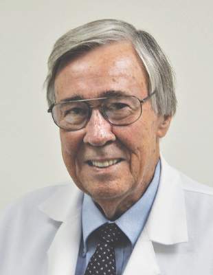
Reactions to reactions
When patients say they had “a reaction” to some treatment, they often don’t mean what we would call allergy or even intolerance. This doesn’t make their reluctance – or refusal – to consider what we suggest any less insistent.
I’ve lost track, for insistence, of the number of patients who called 2 days after I prescribed clindamycin lotion to complain that it “dried me out completely.”

When a customer in a cosmetic store or pharmacy returns a skin care cream because of that kind of “reaction,” the store takes it back, no questions asked. This is not just good business practice; in that context, both the customer and the salesperson use the word “reaction” the way laymen do, not the way doctors do.
If you doubt a reaction is real and say so, however gently, the response you may get is, “That may be true, Doctor, but my body is sensitive. I get strange reactions.”
Like most everyone else in practice, I use negotiating tactics, such as: “Stop the cream for 3 days, then try it again, but just in one spot in front of your left ear.”
Some patients agree to try this. Mostly, nothing happens, they resume the medication, and life goes on. For fearful people, we have to use something else. Sometimes patients and I run through a succession of options, each of which produces an inscrutable “reaction.”
As the years pass, I’ve gotten better at gauging the range of true reactions to medications. At the same time, the list of perplexing things patients complain about gets longer all the time. Lately, there’s been a run of them:
• The woman who used low-strength tretinoin cream all of twice and stopped 2 weeks ago who insisted that “my face is still all dried out.” I assured her that 2 days of tretinoin cannot damage the skin forever, but she remains unconvinced.
• The man who says that putting tretinoin on his face “made me dry out all over.”
• The mother who said her daughter stopped my steroid cream because “every time we used it, her tongue broke out in blisters.”
• The middle-aged woman who claimed, “I can’t take minocycline because it changed the color of my teeth and my eyes.”
• The young woman who puts clindamycin pads only on her right cheek, where she still has pimples, but not on the left, “because it makes the pimples go away on the cheek that has them, but on the other side it makes pimples come out.”
When I hear complaints like these, I just sigh and make a lateral move.
But sometimes I run out of lateral moves. The other month a woman told me that doxycycline made her “tired.” We tried minocycline, but that made her “irritable.” “My body is prone to strange reactions that no one else gets,” she explained. Then she asked for a third option.
“I honestly don’t know what to suggest,” I apologized. “Because your body reacts in ways that other people don’t, I can’t predict what some other treatment will do to you, even if it hasn’t done it to anyone else.”
She asked for her records, to get another opinion. I happily complied.
Speaking of weird “reactions,” there are of course the ones listed on e-scribing programs. Here are a couple:
• Hydrocortisone valerate has been associated with WARTS. (Capitalized)
• Hydrocortisone valerate has been associated with tinea cruris.
Make any sense to you? Me neither.
I end with a call I got the other day from a pharmacist. This concerned a patient for whom I had prescribed oral doxycycline and topical tretinoin. The message was, “Please call regarding drug interaction.”
When I got through, I politely asked the nature of this interaction. The pharmacist said she didn’t know exactly, but would look into it and fax me the details.
Half an hour later I got a sheet explaining the there is an increased risk of pseudotumor cerebri when patients take both oral tretinoin and oral tetracyclines.
That is correct: Not isotretinoin. Oral tretinoin. Used any oral tretinoin lately?
At any rate, the helpful pharmacist wrote at the bottom, “This interaction is flagged by several insurers for topical tretinoin too.”
Thank heavens for computer-generated warnings. Whatever would we do without them?
Besides, it’s nice to know that even after all these years, I can still react myself, with surprise.
Dr. Rockoff practices dermatology in Brookline, Mass. He is on the clinical faculty at Tufts University School of Medicine, Boston, and has taught senior medical students and other trainees for 30 years. Dr. Rockoff has contributed to the Under My Skin column in Dermatology News since January 2002. Write to him at [email protected].
When patients say they had “a reaction” to some treatment, they often don’t mean what we would call allergy or even intolerance. This doesn’t make their reluctance – or refusal – to consider what we suggest any less insistent.
I’ve lost track, for insistence, of the number of patients who called 2 days after I prescribed clindamycin lotion to complain that it “dried me out completely.”
When a customer in a cosmetic store or pharmacy returns a skin care cream because of that kind of “reaction,” the store takes it back, no questions asked. This is not just good business practice; in that context, both the customer and the salesperson use the word “reaction” the way laymen do, not the way doctors do.
If you doubt a reaction is real and say so, however gently, the response you may get is, “That may be true, Doctor, but my body is sensitive. I get strange reactions.”
Like most everyone else in practice, I use negotiating tactics, such as: “Stop the cream for 3 days, then try it again, but just in one spot in front of your left ear.”
Some patients agree to try this. Mostly, nothing happens, they resume the medication, and life goes on. For fearful people, we have to use something else. Sometimes patients and I run through a succession of options, each of which produces an inscrutable “reaction.”
As the years pass, I’ve gotten better at gauging the range of true reactions to medications. At the same time, the list of perplexing things patients complain about gets longer all the time. Lately, there’s been a run of them:
• The woman who used low-strength tretinoin cream all of twice and stopped 2 weeks ago who insisted that “my face is still all dried out.” I assured her that 2 days of tretinoin cannot damage the skin forever, but she remains unconvinced.
• The man who says that putting tretinoin on his face “made me dry out all over.”
• The mother who said her daughter stopped my steroid cream because “every time we used it, her tongue broke out in blisters.”
• The middle-aged woman who claimed, “I can’t take minocycline because it changed the color of my teeth and my eyes.”
• The young woman who puts clindamycin pads only on her right cheek, where she still has pimples, but not on the left, “because it makes the pimples go away on the cheek that has them, but on the other side it makes pimples come out.”
When I hear complaints like these, I just sigh and make a lateral move.
But sometimes I run out of lateral moves. The other month a woman told me that doxycycline made her “tired.” We tried minocycline, but that made her “irritable.” “My body is prone to strange reactions that no one else gets,” she explained. Then she asked for a third option.
“I honestly don’t know what to suggest,” I apologized. “Because your body reacts in ways that other people don’t, I can’t predict what some other treatment will do to you, even if it hasn’t done it to anyone else.”
She asked for her records, to get another opinion. I happily complied.
Speaking of weird “reactions,” there are of course the ones listed on e-scribing programs. Here are a couple:
• Hydrocortisone valerate has been associated with WARTS. (Capitalized)
• Hydrocortisone valerate has been associated with tinea cruris.
Make any sense to you? Me neither.
I end with a call I got the other day from a pharmacist. This concerned a patient for whom I had prescribed oral doxycycline and topical tretinoin. The message was, “Please call regarding drug interaction.”
When I got through, I politely asked the nature of this interaction. The pharmacist said she didn’t know exactly, but would look into it and fax me the details.
Half an hour later I got a sheet explaining the there is an increased risk of pseudotumor cerebri when patients take both oral tretinoin and oral tetracyclines.
That is correct: Not isotretinoin. Oral tretinoin. Used any oral tretinoin lately?
At any rate, the helpful pharmacist wrote at the bottom, “This interaction is flagged by several insurers for topical tretinoin too.”
Thank heavens for computer-generated warnings. Whatever would we do without them?
Besides, it’s nice to know that even after all these years, I can still react myself, with surprise.
Dr. Rockoff practices dermatology in Brookline, Mass. He is on the clinical faculty at Tufts University School of Medicine, Boston, and has taught senior medical students and other trainees for 30 years. Dr. Rockoff has contributed to the Under My Skin column in Dermatology News since January 2002. Write to him at [email protected].
