User login
Measuring Cognition in Migraine, One Patient at a Time
SAN DIEGO —
In fact, these effects may appear in the prodromal phase and carry through the headache and into the post-headache period, according to Richard Lipton, MD, who spoke about cognition and migraine at the annual meeting of the American Headache Society.

He pointed out existing evidence that migraine patients have cognitive impairment relative to the general population even during the interictal period. Such studies suggest that migraine, especially with aura, could be a risk factor for later dementia.
One important limitation of studies that compare people with migraines with controls is that a range of factors could explain an association between lower cognitive function and migraines, including socioeconomic factors, education, severe headaches requiring specialty care, and comorbidities, among others. Acute and preventative treatments could also affect cognition.
However, longitudinal studies of cognitive function in individual patients have been sparse. Questions remain, like whether cognitive performance differs between the headache period and the interictal period, as well as similar questions about the premonitory and post-drome phases. “And then there’s a long-term question: Do people with migraine show more interictal or ictal decline in cognitive performance relative to migraine-free controls?” said Dr. Lipton, professor of neurology at Albert Einstein College of Medicine in the Bronx, New York.
He showed evidence from a retrospective study by Lundbeck conducted at four sites that asked patients with chronic migraine about bothersome symptoms both before and after treatment. More than three-fourths (77.7%) rated “difficulty concentrating or thinking clearly” as a bothersome symptom.
Following treatment, 5.0% said their cognitive issues had completely improved, 32.0% that they were “very much” improved, 26.0% moderately improved, 23.0% slightly improved, and 14.0% not at all improved.
“I am not saying this is a rigorous study, but I am saying that it illustrates two points that are important for us today: One is that brain fog is very common in a subspecialty care sample of headache patients like the ones many of us treat, and it also suggests that there’s hope that treatment can improve cognitive impairment as migraine gets better,” said Dr. Lipton.
Cognition has received less attention than other migraine symptoms, and treatment can be a two-edged sword: “There’s some evidence that some treatments can reduce cognitive impairment, and obvious evidence that some treatments, topiramate and tricyclics, can induce cognitive impairment,” said Dr. Lipton.
Studies that compare cognition within the same patient at different time periods can get around some of the limitations of comparisons between populations, but face their own challenges. “Single shot” cognitive measures may not be reliably repeatable and differences seen on “good” versus “bad” days or proximity to recent headaches.
The solution, Dr. Lipton believes, is intensive repeated measures that avoid the practice effect, in which a participant improves at a test due to repetition.
He summarized a study that was presented later in the day at a poster session, which used smartphones or other devices to test 19 participants five times per day, over 5 days, in natural environments. Devices gathered both subjective and objective assessments of cognition, along with information on mood, stress, and status and fluctuations in pain, and have the potential to go further by measuring things like physical exertion, heart rate, pollution levels, and other variables.
“It clearly improves the reliability and the validity of cognitive assessment and makes it possible to link cognition to the stage of the headache cycle,” said Dr. Lipton.
The researchers found worse cognitive performance during the headache phase as compared with the interictal phase. “Objective cognitive performance measurably declines during the headache phase, and the next step is to fully control for acute medications that people may take during the headache phase,” said Dr. Lipton.
He expressed hope that improved measurements can improve outcomes, if it’s possible to identify therapies that don’t impact cognition. “We think it’s very likely that certain classes of acute and preventive medications may not cause cognitive impairment, and there is a strong hope that they may actually reduce the cognitive burden of disease and potentially even reduce cognitive decline. Those are areas that I’m very excited to explore in the future,” said Dr. Lipton.
The results emphasize the need to treat patients early, according to Nada Hindiyeh, MD, who attended the session and was asked for comment. “Generally, patients are going to come to you with episodic migraines. When migraines start to increase in frequency and severity, that means all of these other symptoms are going to come along with it and be increased in frequency and severity, so it’s important to recognize this early so you can get patients on the right treatments and preventives to really prevent these episodes from happening and prevent that cognitive decline,” said Dr. Hindiyeh, director of headache neurology at Metrodora Institute, West Valley City, Utah.
Dr. Lipton has financial relationships with Aeon, AbbVie/Allergan, Amgen, Biohaven, Dr. Reddy’s Laboratories, electroCore, Eli Lilly, GlaxoSmithKline, Lundbeck, Merck, Novartis, Pfizer, Teva, Axon, CoolTech, and Manistee. Dr. Hindiyeh has no relevant financial disclosures.
SAN DIEGO —
In fact, these effects may appear in the prodromal phase and carry through the headache and into the post-headache period, according to Richard Lipton, MD, who spoke about cognition and migraine at the annual meeting of the American Headache Society.
He pointed out existing evidence that migraine patients have cognitive impairment relative to the general population even during the interictal period. Such studies suggest that migraine, especially with aura, could be a risk factor for later dementia.
One important limitation of studies that compare people with migraines with controls is that a range of factors could explain an association between lower cognitive function and migraines, including socioeconomic factors, education, severe headaches requiring specialty care, and comorbidities, among others. Acute and preventative treatments could also affect cognition.
However, longitudinal studies of cognitive function in individual patients have been sparse. Questions remain, like whether cognitive performance differs between the headache period and the interictal period, as well as similar questions about the premonitory and post-drome phases. “And then there’s a long-term question: Do people with migraine show more interictal or ictal decline in cognitive performance relative to migraine-free controls?” said Dr. Lipton, professor of neurology at Albert Einstein College of Medicine in the Bronx, New York.
He showed evidence from a retrospective study by Lundbeck conducted at four sites that asked patients with chronic migraine about bothersome symptoms both before and after treatment. More than three-fourths (77.7%) rated “difficulty concentrating or thinking clearly” as a bothersome symptom.
Following treatment, 5.0% said their cognitive issues had completely improved, 32.0% that they were “very much” improved, 26.0% moderately improved, 23.0% slightly improved, and 14.0% not at all improved.
“I am not saying this is a rigorous study, but I am saying that it illustrates two points that are important for us today: One is that brain fog is very common in a subspecialty care sample of headache patients like the ones many of us treat, and it also suggests that there’s hope that treatment can improve cognitive impairment as migraine gets better,” said Dr. Lipton.
Cognition has received less attention than other migraine symptoms, and treatment can be a two-edged sword: “There’s some evidence that some treatments can reduce cognitive impairment, and obvious evidence that some treatments, topiramate and tricyclics, can induce cognitive impairment,” said Dr. Lipton.
Studies that compare cognition within the same patient at different time periods can get around some of the limitations of comparisons between populations, but face their own challenges. “Single shot” cognitive measures may not be reliably repeatable and differences seen on “good” versus “bad” days or proximity to recent headaches.
The solution, Dr. Lipton believes, is intensive repeated measures that avoid the practice effect, in which a participant improves at a test due to repetition.
He summarized a study that was presented later in the day at a poster session, which used smartphones or other devices to test 19 participants five times per day, over 5 days, in natural environments. Devices gathered both subjective and objective assessments of cognition, along with information on mood, stress, and status and fluctuations in pain, and have the potential to go further by measuring things like physical exertion, heart rate, pollution levels, and other variables.
“It clearly improves the reliability and the validity of cognitive assessment and makes it possible to link cognition to the stage of the headache cycle,” said Dr. Lipton.
The researchers found worse cognitive performance during the headache phase as compared with the interictal phase. “Objective cognitive performance measurably declines during the headache phase, and the next step is to fully control for acute medications that people may take during the headache phase,” said Dr. Lipton.
He expressed hope that improved measurements can improve outcomes, if it’s possible to identify therapies that don’t impact cognition. “We think it’s very likely that certain classes of acute and preventive medications may not cause cognitive impairment, and there is a strong hope that they may actually reduce the cognitive burden of disease and potentially even reduce cognitive decline. Those are areas that I’m very excited to explore in the future,” said Dr. Lipton.
The results emphasize the need to treat patients early, according to Nada Hindiyeh, MD, who attended the session and was asked for comment. “Generally, patients are going to come to you with episodic migraines. When migraines start to increase in frequency and severity, that means all of these other symptoms are going to come along with it and be increased in frequency and severity, so it’s important to recognize this early so you can get patients on the right treatments and preventives to really prevent these episodes from happening and prevent that cognitive decline,” said Dr. Hindiyeh, director of headache neurology at Metrodora Institute, West Valley City, Utah.
Dr. Lipton has financial relationships with Aeon, AbbVie/Allergan, Amgen, Biohaven, Dr. Reddy’s Laboratories, electroCore, Eli Lilly, GlaxoSmithKline, Lundbeck, Merck, Novartis, Pfizer, Teva, Axon, CoolTech, and Manistee. Dr. Hindiyeh has no relevant financial disclosures.
FROM AHS 2024
Emergency Department Visits for Suicide Attempts Rise Across the United States
TOPLINE:
Emergency department (ED) visits in the United States for suicide attempts and intentional self-harm show an increasing trend from 2011 to 2020, with visits being most common among adolescents and the largest increase in visits being seen in adults aged 65 years or older.
METHODOLOGY:
- This study used data from the National Hospital Ambulatory Medical Care Survey, an annual nationwide cross-sectional survey, to track trends in ED visits for suicide attempts and intentional self-harm in the United States from 2011 to 2020.
- Researchers identified visits for suicide attempts and intentional self-harm, along with diagnoses of any co-occurring mental health conditions, using discharge diagnosis codes or reason-for-visit codes.
- The focus was to identify the percentages of ED visits for suicide attempts and intentional self-harm, with analyses done per 100,000 persons and for changes possibly linked to the COVID-19 pandemic in 2019-2020.
TAKEAWAY:
- The number of ED visits owing to suicide attempts and intentional self-harm increased from 1.43 million in 2011-2012 to 5.37 million in 2019-2020 (average annual percent change, 19.5%; 95% confidence interval, 16.9-22.2).
- The rate of ED visits for suicide attempts and intentional self-harm was higher among adolescents and young adults, particularly women, and lower among children.
- Despite a surge in ED visits for self-harm, less than 16% included a mental health evaluation, with visits among patients with mood disorders decreasing by 5.5% annually and those among patients with drug-related disorders increasing by 6.8% annually.
- In 2019-2020, those aged 15-20 years had the highest rate of ED visits (1552 visits per 100,000 persons), with a significant increase seen across all age groups; the largest increase was among those aged 65 years or older.
IN PRACTICE:
“Given that suicide attempts are the single greatest risk factor for suicide, evidence-based management of individuals presenting to emergency departments with suicide attempts and intentional self-harm is a critical component of comprehensive suicide prevention strategies,” the authors wrote.
SOURCE:
The investigation, led by Tanner J. Bommersbach, MD, MPH, Department of Psychiatry and Psychology, Mayo Clinic, Rochester, Minnesota, was published online in The American Journal of Psychiatry.
LIMITATIONS:
Visits for suicide attempts and intentional self-harm were identified based on discharge diagnostic and reason-for-visit codes, which may have led to an underestimation of visits for suicide attempts. ED visits for suicidal vs nonsuicidal self-injury could not be distinguished due to reliance on discharge diagnostic codes. Visits for suicidal ideation, which was not the focus of the study, may have been miscoded as suicide attempts and intentional self-harm.
DISCLOSURES:
No funding source was reported for the study. Some authors received funding grants from various institutions, and one author disclosed receiving honoraria for service as a review committee member and serving as a stakeholder/consultant and as an advisory committee member for some institutes and agencies.
A version of this article appeared on Medscape.com.
Continuous Glucose Monitors Should Not Be Normalized
Should we now recommend continuous glucose monitoring to all our patients, even those without diabetes? Most of us would instinctively say “no” to this question, but we are seeing opinions from doctors recommending it, and in recent years, scientific literature has focused on the subject.
Today, anyone can get an arm patch that continuously measures interstitial glucose, which is closely related to blood sugar. The information can be read on a dedicated reader or on a mobile phone by scanning the patch or, with some models, without even doing anything.
There is a consensus for prescribing continuous glucose monitoring for patients with type 1 or type 2 diabetes who are treated with at least three insulin injections. Not only is the use of continuous glucose monitoring much more comfortable than self-monitoring with finger sticks, but continuous monitoring also helps reduce glycosylated hemoglobin while decreasing the risk for hypoglycemia. Recently, another indication has begun to be reimbursed in France: Type 2 diabetes under mono-insulin injection when the diabetes is not well controlled.
But alongside these situations, there are two questions that are worth considering.
Untreated Type 2 Diabetes
First, is continuous glucose monitoring desirable for all patients with diabetes, even those not treated with insulin and even when blood sugar levels are well managed? Intuitively, one might think that it can’t hurt and that continuous monitoring of blood sugar can only improve things. We have some evidence supporting this idea, but the level of proof is quite weak. It is not clear that continuous monitoring can improve patients’ awareness of the impact of dietary choices or physical activity on blood sugar. Obviously, one can imagine that continuously monitoring glucose will encourage a shift toward more beneficial behaviors. But honestly, today, we do not have proof that wearing a continuous glucose monitor can improve behaviors in patients with type 2 diabetes who are treated with noninsulin antidiabetic medications.
Furthermore, a significant study has shown that while the effectiveness is more evident in patients treated with insulin, strong evidence suggests that continuous glucose monitoring could also reduce glycosylated hemoglobin in patients with type 2 diabetes who are not treated with insulin. A close examination of the results suggests that the benefits generally are less than those observed in insulin-treated patients with diabetes.
When we look at the scientific literature, two factors seem particularly important to consider if choosing to prescribe a continuous glucose monitoring sensor. The first is the method used, because the results can vary depending on the method. It appears that only self-monitoring that allows the patient to follow glucose in real time is effective, unlike blind monitoring that allows only a retrospective analysis of blood sugar levels. In the latter case, the patient wears the sensor, and after a week, 10 days, or 15 days, the results are analyzed, possibly with a health care provider. It seems that this is not very effective in improving glycosylated hemoglobin and dietary and physical activity behavior.
The second essential factor to consider is the need for an education program for the use of these sensors to be helpful. If sensors are used but nothing else is done, it does not seem logical. Seeing blood sugar levels without being able to understand them and act accordingly seems of little use. Scientific literature seems to confirm this idea.
Patients Without Diabetes
Now there is another question. We have discussed patients with type 2 diabetes without insulin. It’s trendy to talk about the potential benefits of continuous glucose monitors in patients without diabetes. The idea is emerging that these monitors could be used to refine the diagnosis of diabetes or to better predict the onset of diabetes in the subsequent years.
Others claim that continuous glucose monitors are an effective way to induce a change in dietary and physical activity behaviors in patients with prediabetes. One can, for example, tell a patient, “You are at risk of developing diabetes, so by monitoring your glucose, you will change your behavior.” Honestly, the scientific data we have today do not support these ideas, and I sincerely believe that it is not advisable today to recommend, as some would like, the mass use of monitors, whether in patients with overweight or obesity, or in patients with prediabetes. This goes for suggestions for using the monitor for 7-10 days per year, in the form of a session to try to reduce the risk for diabetes by motivating patients to change their behavior. We have no evidence at all that this can work. And in my opinion, with this kind of discourse, we ultimately risk, as usual, encouraging patients who are already “fans” of self-checks and self-monitoring to get health data, even if they do not know how to interpret it. Maybe even the doctor they ask for interpretation will not be trained to interpret the results of these monitors.
Spreading the idea that monitors are useful for preventing diabetes has a side effect: It hinders progress on the essential issue. Today, one of the problems in diabetes and prediabetes is that screening is not done often enough, and 20% of patients with diabetes are still unaware of their diagnosis. The management of early diabetes or prediabetes, in my opinion, is not optimal in routine care today. So, I think that adding the idea that using monitors could be beneficial dilutes the main information.
Having said that, I sometimes offer continuous glucose monitoring to some of my patients on a case-by-case basis. I believe that with proper support and an educational program, it can be beneficial for certain patients.
In Practice
In summary, I am totally opposed to the normalization of the use of monitors. I think it is our role as health care professionals to warn the public that even if it is accessible — anyone can buy a reader, a sensor — it is not necessarily beneficial, and it may even distract us from what is essential. But as a specialist, I think that using a monitor within a genuine care plan seems reasonable. Ultimately, it’s just personalized medicine.
Dr. Hansel is an endocrinologist-diabetologist and nutritionist, Department of Diabetology-Endocrinology-Nutrition, Hôpital Bichat, and a university lecturer and hospital practitioner, Université Paris-Diderot, France. He discloses ties with Iriade, Sanofi-Aventis, and Amgen.
This story was translated from the Medscape French edition using several editorial tools, including AI, as part of the process. Human editors reviewed this content before publication. A version of this article first appeared on Medscape.com.
GLP-1s Reduced Secondary Stroke Risk in Patients With Diabetes, Obesity
, according to authors of a recent meta-analysis. With benefits across administration routes, dosing regimens, type 2 diabetes status, and total and nonfatal strokes, the findings could improve GLP-1 RA implementation by stroke specialists in patients with stroke history and concurrent type 2 diabetes or obesity, authors said. The study was published online in the International Journal of Stoke.
Extending Longevity
Agents including GLP-1 RAs that have been found to reduce cardiovascular events among patients with type 2 diabetes and patients who are overweight or obese also reduce risk of recurrent stroke among patients with a history of stroke who are overweight, obese, or have metabolic disease, said American Heart Association (AHA) Chief Clinical Science Officer Mitchell S. V. Elkind, MD, who was not involved with the study but was asked to comment.
“Stroke is a leading cause of mortality and the leading cause of serious long-term disability,” he added, “so medications that help to reduce that risk can play an important role in improving overall health and well-being and hopefully reducing premature mortality.”
Investigators Anastasia Adamou, MD, an internal medicine resident at AHEPA University Hospital in Thessaloniki, Greece, and colleagues searched MEDLINE and Scopus for cardiovascular outcome trials involving adults randomly assigned to GLP-1 RAs or placebo through November 2023, ultimately analyzing 11 randomized controlled trials (RCTs).
Among 60,380 participants in the nine studies that assessed total strokes, 2.5% of the GLP-1 RA group experienced strokes during follow-up, versus 3% in the placebo group (relative risk [RR] 0.85, 95% confidence interval [CI] 0.77-0.93). Regarding secondary outcomes, the GLP-1 RA group showed a significantly lower rate of nonfatal strokes versus patients on placebo (RR 0.87, 95% CI 0.79-0.95). Conversely, investigators observed no significant risk difference among the groups regarding fatal strokes, probably due to the low rate of events — 0.3% and 0.4% for treated and untreated patients, respectively.
Subgroup analyses revealed no interaction between dosing frequency and total, nonfatal, or fatal strokes. The investigators observed no difference in nonfatal strokes among participants by type 2 diabetes status and medication administration route (oral versus subcutaneous).
“The oral administration route could provide the advantage of lower local ecchymoses and allergic reactions due to subcutaneous infusions,” Dr. Adamou said in an interview. But because oral administration demands daily intake, she added, treatment adherence might be affected. “For this reason, our team performed another subgroup analysis to compare the once-a-day to the once-a-month administration. No interaction effect was again presented between the two subgroups. This outcome allows for personalization of the administration method for each patient.”

Addressing Underutilization
Despite more than 2 decades of widespread use and well-established effects on body weight, HbA1c, and cardiovascular risk, GLP-1 RAs remain underutilized, authors wrote. This is especially true in primary care, noted one study published in Clinical Diabetes.
“GLP-1 RAs have been used for many years to treat diabetic patients,” said Dr. Adamou. But because their impact on cardiovascular health regardless of diabetic status is only recently known, she said, physicians are exercising caution when prescribing this medication to patients without diabetes. “This is why more studies need to be available, especially RCTs.”
Most neurologists traditionally have left management of type 2 diabetes and other metabolic disorders to primary care doctors, said Dr. Elkind. “However, these medications are increasingly important to vascular risk reduction and should be considered part of the stroke specialist’s armamentarium.”
Vascular neurologists can play an important role in managing metabolic disease and obesity by recommending GLP-1 RAs for patients with a history of stroke, or by initiating these medications themselves, Dr. Elkind said. “These drugs are likely to become an important part of stroke patients’ medication regimens, along with antithrombotic agents, blood pressure control, and statins. Neurologists are well-positioned to educate other physicians about the important connections among brain, heart, and metabolic health.”
To that end, he said, the AHA will update guidelines for both primary and secondary stroke prevention as warranted by evidence supporting GLP-1 RAs and other medications that could impact stroke risk in type 2 diabetes and related metabolic disorders. However, no guidelines concerning use of GLP-1 RAs for secondary stroke prevention in obesity exist. Here, said Dr. Elkind, the AHA will continue building on its innovative Cardiovascular-Kidney Metabolic Health program, which includes clinical suggestions and may include more formal clinical practice guidelines as the evidence evolves.
Among the main drivers of the initiative, he said, is the recognition that cardiovascular disease — including stroke — is the major cause of death and morbidity among patients with obesity, type 2 diabetes, and metabolic disorders. “Stroke should be considered an important part of overall cardiovascular risk, and the findings that these drugs can help to reduce the risk of stroke specifically is an important additional reason for their use.”
Dr. Elkind and Dr. Adamou reported no conflicting interests. The authors received no financial support for the study.
, according to authors of a recent meta-analysis. With benefits across administration routes, dosing regimens, type 2 diabetes status, and total and nonfatal strokes, the findings could improve GLP-1 RA implementation by stroke specialists in patients with stroke history and concurrent type 2 diabetes or obesity, authors said. The study was published online in the International Journal of Stoke.
Extending Longevity
Agents including GLP-1 RAs that have been found to reduce cardiovascular events among patients with type 2 diabetes and patients who are overweight or obese also reduce risk of recurrent stroke among patients with a history of stroke who are overweight, obese, or have metabolic disease, said American Heart Association (AHA) Chief Clinical Science Officer Mitchell S. V. Elkind, MD, who was not involved with the study but was asked to comment.
“Stroke is a leading cause of mortality and the leading cause of serious long-term disability,” he added, “so medications that help to reduce that risk can play an important role in improving overall health and well-being and hopefully reducing premature mortality.”
Investigators Anastasia Adamou, MD, an internal medicine resident at AHEPA University Hospital in Thessaloniki, Greece, and colleagues searched MEDLINE and Scopus for cardiovascular outcome trials involving adults randomly assigned to GLP-1 RAs or placebo through November 2023, ultimately analyzing 11 randomized controlled trials (RCTs).
Among 60,380 participants in the nine studies that assessed total strokes, 2.5% of the GLP-1 RA group experienced strokes during follow-up, versus 3% in the placebo group (relative risk [RR] 0.85, 95% confidence interval [CI] 0.77-0.93). Regarding secondary outcomes, the GLP-1 RA group showed a significantly lower rate of nonfatal strokes versus patients on placebo (RR 0.87, 95% CI 0.79-0.95). Conversely, investigators observed no significant risk difference among the groups regarding fatal strokes, probably due to the low rate of events — 0.3% and 0.4% for treated and untreated patients, respectively.
Subgroup analyses revealed no interaction between dosing frequency and total, nonfatal, or fatal strokes. The investigators observed no difference in nonfatal strokes among participants by type 2 diabetes status and medication administration route (oral versus subcutaneous).
“The oral administration route could provide the advantage of lower local ecchymoses and allergic reactions due to subcutaneous infusions,” Dr. Adamou said in an interview. But because oral administration demands daily intake, she added, treatment adherence might be affected. “For this reason, our team performed another subgroup analysis to compare the once-a-day to the once-a-month administration. No interaction effect was again presented between the two subgroups. This outcome allows for personalization of the administration method for each patient.”
Addressing Underutilization
Despite more than 2 decades of widespread use and well-established effects on body weight, HbA1c, and cardiovascular risk, GLP-1 RAs remain underutilized, authors wrote. This is especially true in primary care, noted one study published in Clinical Diabetes.
“GLP-1 RAs have been used for many years to treat diabetic patients,” said Dr. Adamou. But because their impact on cardiovascular health regardless of diabetic status is only recently known, she said, physicians are exercising caution when prescribing this medication to patients without diabetes. “This is why more studies need to be available, especially RCTs.”
Most neurologists traditionally have left management of type 2 diabetes and other metabolic disorders to primary care doctors, said Dr. Elkind. “However, these medications are increasingly important to vascular risk reduction and should be considered part of the stroke specialist’s armamentarium.”
Vascular neurologists can play an important role in managing metabolic disease and obesity by recommending GLP-1 RAs for patients with a history of stroke, or by initiating these medications themselves, Dr. Elkind said. “These drugs are likely to become an important part of stroke patients’ medication regimens, along with antithrombotic agents, blood pressure control, and statins. Neurologists are well-positioned to educate other physicians about the important connections among brain, heart, and metabolic health.”
To that end, he said, the AHA will update guidelines for both primary and secondary stroke prevention as warranted by evidence supporting GLP-1 RAs and other medications that could impact stroke risk in type 2 diabetes and related metabolic disorders. However, no guidelines concerning use of GLP-1 RAs for secondary stroke prevention in obesity exist. Here, said Dr. Elkind, the AHA will continue building on its innovative Cardiovascular-Kidney Metabolic Health program, which includes clinical suggestions and may include more formal clinical practice guidelines as the evidence evolves.
Among the main drivers of the initiative, he said, is the recognition that cardiovascular disease — including stroke — is the major cause of death and morbidity among patients with obesity, type 2 diabetes, and metabolic disorders. “Stroke should be considered an important part of overall cardiovascular risk, and the findings that these drugs can help to reduce the risk of stroke specifically is an important additional reason for their use.”
Dr. Elkind and Dr. Adamou reported no conflicting interests. The authors received no financial support for the study.
FROM THE INTERNATIONAL JOURNAL OF STROKE
DEA Training Mandate: 8 Hours of My Life I’d Like Back
It’s time to renew two of my three narcotic prescribing licenses. For the first time in my career, I’ve waffled on whether the financial outlay to the US Drug Enforcement Agency (DEA) is worth it.
At $888 each, I’ve considered letting two licenses lapse because I only work part-time in Montana. But several friends advised me to keep a “spare” in case I transfer to a new location.
I thought about just paying the fees until I could do a little more research, but there is no mechanism for a refund unless I die within the first year of the 3-year cycle, provide incorrect credit card digits, or accidentally duplicate payments.
The renewal fee is just part of the issue.
Mandatory 8-Hour Training
I also received an alert about the requirement for more “narcotics prescribing education” thanks to the Medication Access and Training Expansion Act (MATE).
The requirement seems counterintuitive because opioid prescribing has decreased for the 10th consecutive year, according to the AMA Overdose Epidemic Report. The continuing rise in overdose deaths is largely due to illegitimate manufacturing of synthetic opioids.
I’ve written zero outpatient narcotics prescriptions in the past 6 years, and I’ve written very few in my 33 years of practice. My use is limited to intravenous morphine for flash pulmonary edema or refractory angina, but unless you graduated from a training program within 5 years of the June 2023 mandate or are boarded in addiction medicine, there is no way to escape the 8-hour education requirement.
The problem is that these courses are never just 8 hours in duration. After signing up for one such CME course that cost $150, I was still dying of boredom and at risk for DVT 4 days later. That’s how long it took to sit through.
Instead of the 30 seconds it should have taken to review the simple instructions to deliver Narcan, there were scores of screens followed by juvenile quizlets and cartoons. All but about 2 hours out of the 4 days is now relegated to that category of “hours of my life that I can never get back.” Additionally, none of that mandatory “education” will change my prescribing habits one whit.
And beware the penalty. 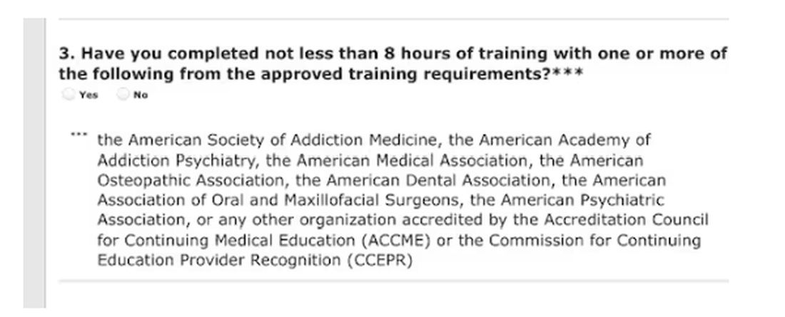
Of course, I would always be truthful when asked to check the box on the DEA renewal application attesting to my having completed the required education. On the outside chance that you plan to check the yes box without completing the relevant courses, those found guilty of such false claims could be fined up to $250,000 and subject to “not more than four years in prison,” or both. Yikes! 
Larry Houck, a former DEA investigator, explained that “[t]here are lot of people who are coming up for renewal and log on but still don’t know this is a requirement.” Neither ignorance nor complacency is an acceptable defense.
Changes Needed
The only good thing that came of those 4 long days of opioid education was a motivation to drive change in our current licensing and educational experience. Why not use this opportunity to reform the DEA-physician/prescriber relationship?
The educational requirements should be curtailed for those of us who do not provide outpatient narcotic prescriptions even if we use inpatient opioids. Meds with low abuse potential should be rescheduled to minimize who gets caught in the broad net of the education requirement.
We should reduce overregulation of the legitimate prescribers by lowering, instead of increasing, licensing fees. We should change to a single license number that covers every state. In this digital age, there is no legitimate excuse to prevent this from happening.
After all, the settlements from opioid manufacturers and distributors will in time total $50 billion. It seems that at least some of the responsibilities of the DEA could shift to states, cities, and towns.
My friend Siamak Karimian, MD, who provides locum services in multiple states, pays for seven active DEA licenses every 3 years. He pointed out the hypocrisy in the current regulatory system: “It’s funny that you can have only one DEA or state license and work for the government in all other states or territories with no limits, including the VA, Indian healthcare systems, or prison systems.”
All other prescribers require a separate DEA number for every state. Ultimately, you’d think tracking prescriptions for a single DEA number should be far simpler than tracking someone with seven.
Competent physicians not guilty of criminal overprescribing seem to be the last to be considered in nearly every healthcare endeavor these days. It would be refreshing if they would reduce our fees and prevent this waste of our time.
And while we are at it, perhaps a more fitting punishment is due for Richard Sackler and all the Purdue Pharma–affiliated family members. The Sacklers will pay out $6 billion in exchange for immunity against civil litigation. That doesn’t seem like much when they are worth $11 billion.
Perhaps they should be made to take an 8-hour course on opioid prescribing, annually and in perpetuity. Let’s see them complete a few quizlets and sit through screens of instruction on how to administer Naloxone. Of course, that would be a mild punishment for those who manufactured a drug that killed hundreds of thousands. But it would be a start.
Dr. Walton-Shirley, a clinical cardiologist in Nashville, Tennessee, has disclosed no relevant financial relationships.
A version of this article appeared on Medscape.com.
It’s time to renew two of my three narcotic prescribing licenses. For the first time in my career, I’ve waffled on whether the financial outlay to the US Drug Enforcement Agency (DEA) is worth it.
At $888 each, I’ve considered letting two licenses lapse because I only work part-time in Montana. But several friends advised me to keep a “spare” in case I transfer to a new location.
I thought about just paying the fees until I could do a little more research, but there is no mechanism for a refund unless I die within the first year of the 3-year cycle, provide incorrect credit card digits, or accidentally duplicate payments.
The renewal fee is just part of the issue.
Mandatory 8-Hour Training
I also received an alert about the requirement for more “narcotics prescribing education” thanks to the Medication Access and Training Expansion Act (MATE).
The requirement seems counterintuitive because opioid prescribing has decreased for the 10th consecutive year, according to the AMA Overdose Epidemic Report. The continuing rise in overdose deaths is largely due to illegitimate manufacturing of synthetic opioids.
I’ve written zero outpatient narcotics prescriptions in the past 6 years, and I’ve written very few in my 33 years of practice. My use is limited to intravenous morphine for flash pulmonary edema or refractory angina, but unless you graduated from a training program within 5 years of the June 2023 mandate or are boarded in addiction medicine, there is no way to escape the 8-hour education requirement.
The problem is that these courses are never just 8 hours in duration. After signing up for one such CME course that cost $150, I was still dying of boredom and at risk for DVT 4 days later. That’s how long it took to sit through.
Instead of the 30 seconds it should have taken to review the simple instructions to deliver Narcan, there were scores of screens followed by juvenile quizlets and cartoons. All but about 2 hours out of the 4 days is now relegated to that category of “hours of my life that I can never get back.” Additionally, none of that mandatory “education” will change my prescribing habits one whit.
And beware the penalty.
Of course, I would always be truthful when asked to check the box on the DEA renewal application attesting to my having completed the required education. On the outside chance that you plan to check the yes box without completing the relevant courses, those found guilty of such false claims could be fined up to $250,000 and subject to “not more than four years in prison,” or both. Yikes!
Larry Houck, a former DEA investigator, explained that “[t]here are lot of people who are coming up for renewal and log on but still don’t know this is a requirement.” Neither ignorance nor complacency is an acceptable defense.
Changes Needed
The only good thing that came of those 4 long days of opioid education was a motivation to drive change in our current licensing and educational experience. Why not use this opportunity to reform the DEA-physician/prescriber relationship?
The educational requirements should be curtailed for those of us who do not provide outpatient narcotic prescriptions even if we use inpatient opioids. Meds with low abuse potential should be rescheduled to minimize who gets caught in the broad net of the education requirement.
We should reduce overregulation of the legitimate prescribers by lowering, instead of increasing, licensing fees. We should change to a single license number that covers every state. In this digital age, there is no legitimate excuse to prevent this from happening.
After all, the settlements from opioid manufacturers and distributors will in time total $50 billion. It seems that at least some of the responsibilities of the DEA could shift to states, cities, and towns.
My friend Siamak Karimian, MD, who provides locum services in multiple states, pays for seven active DEA licenses every 3 years. He pointed out the hypocrisy in the current regulatory system: “It’s funny that you can have only one DEA or state license and work for the government in all other states or territories with no limits, including the VA, Indian healthcare systems, or prison systems.”
All other prescribers require a separate DEA number for every state. Ultimately, you’d think tracking prescriptions for a single DEA number should be far simpler than tracking someone with seven.
Competent physicians not guilty of criminal overprescribing seem to be the last to be considered in nearly every healthcare endeavor these days. It would be refreshing if they would reduce our fees and prevent this waste of our time.
And while we are at it, perhaps a more fitting punishment is due for Richard Sackler and all the Purdue Pharma–affiliated family members. The Sacklers will pay out $6 billion in exchange for immunity against civil litigation. That doesn’t seem like much when they are worth $11 billion.
Perhaps they should be made to take an 8-hour course on opioid prescribing, annually and in perpetuity. Let’s see them complete a few quizlets and sit through screens of instruction on how to administer Naloxone. Of course, that would be a mild punishment for those who manufactured a drug that killed hundreds of thousands. But it would be a start.
Dr. Walton-Shirley, a clinical cardiologist in Nashville, Tennessee, has disclosed no relevant financial relationships.
A version of this article appeared on Medscape.com.
Solving Restless Legs: Largest Genetic Study to Date May Help
For decades, scientists have been trying to unravel the mysteries of restless legs syndrome (RLS), a poorly understood and underdiagnosed neurological disorder causing itching, crawling, and aching sensations in the limbs that can only be relieved with movement.
A sweeping new genetic study, coauthored by an international team of 70 — including the world’s leading RLS experts — marks a significant advance in that pursuit. Published in Nature Genetics, it is the largest genetic study of the disease to date.
“It’s a huge step forward for patients as well as the scientific community,” said lead author Juliane Winkelmann, MD, a neurologist and geneticist with the Technical University of Munich, Munich, Germany, who’s been studying and treating patients with RLS for 30 years. “We believe it will allow us to better predict the likelihood of developing RLS and investigate new ways to prevent and modify it.”
The common condition, affecting about 1 in 10 adults, was first described centuries ago — by English physician Thomas Willis in the late 1600s. And while we know a lot more about it today — it’s familial in about half of all patients and has been linked to iron deficiency, among other conditions — its exact cause remains unknown.
With preferred drugs long prescribed to quell symptoms shown in recent years to actually worsen the disorder over time, doctors and patients are hungry for alternatives to treat or prevent the sleep-sabotaging condition.
“The main treatments that everybody continues to use are actually making people worse,” said Andrew Berkowski, MD, a Michigan-based neurologist and RLS specialist not involved in the study. These drugs — dopamine agonists such as levodopa and pramipexole — can also potentially cause drug dependence, Dr. Berkowski said.
How This Could Lead to New Treatments
In the new study, the group analyzed three genome-wide association studies, collectively including genetic information from 116,647 patients with RLS and more than 1.5 million people without it.
They identified 161 gene regions believed to contribute to RLS, about a dozen of which are already targets for existing drugs for other conditions. Previously, scientists knew of only 22 associated genes.
“It’s useful in that it identifies new genes we haven’t looked at yet and reinforces the science behind some of the older genes,” said Dr. Berkowski. “It’s given us some ideas for different things we should look into more closely.”
Among the top candidates are genes that influence glutamate — a key chemical messenger that helps move signals between nerve cells in the brain.
Several anticonvulsant and antiseizure drugs, including perampanel, lamotrigine, and gabapentin, target glutamate receptors. And at least one small study has shown perampanel prescribed off-label can improve RLS symptoms.
“Compared to starting at the beginning and developing an entirely new chemical entity, we could run clinical trials using these alternatives in RLS patients,” said the study’s first author, Steven Bell, PhD, an epidemiologist with the University of Cambridge, Cambridge, England.
The study also confirmed the MIES1 gene, which is related to dopamine expression and iron homeostasis, as a key genetic contributor to RLS risk. Low levels of iron in the blood have long been thought to trigger RLS.
The Role of Gene-Environment Interactions
Through additional data analysis, the team confirmed that many of the genes associated with RLS play a role in development of the central nervous system.
“This strongly supports the hypothesis that restless legs syndrome is a neurodevelopmental disorder that develops during the embryo stage but doesn’t clinically manifest until later in life,” said Dr. Winkelmann.
About half of people with RLS report some family history of it.
But not all with a genetic predisposition will develop symptoms.
For instance, the study found that while the same gene regions seem to be associated with risk in both men and women, in practice, RLS is twice as common among women. This suggests that something about women’s lives — menstruation, childbirth, metabolism — may switch a preexisting risk into a reality.
“We know that genetic factors play an important role in making people susceptible to the disease,” said Dr. Winkelmann, “but in the end, it is the interaction between genetic and environmental factors that may lead to its manifestation.”
The study also found associations between RLS and depression and suggests that RLS may increase the risk for type 2 diabetes.
Improving RLS Care
A potentially useful tool coming out of the study was a “polygenic risk score,” which the researchers developed based on the genes identified. When they tested how accurately the score could predict whether someone would develop RLS within the next 5 years, the model got it right about 90% of the time.
Dr. Winkelmann imagines a day when someone could use such a polygenic risk score to flag the high risk for RLS early enough to take action to try to prevent it. More research is necessary to determine precisely what that action would be.
As for treatments, Dr. Berkowski thinks it’s unlikely that doctors will suddenly begin using existing, glutamate-targeting drugs off-label to treat RLS, as many are prohibitively expensive and wouldn’t be covered by insurance. But he’s optimistic that the study can spawn new research that could ultimately help fill the treatment gap.
Shalini Paruthi, MD, an adjunct professor at Saint Louis University, St. Louis, Missouri, and chair of the Restless Legs Syndrome Foundation’s board of directors, sees another benefit.
“The associations found in this study between RLS and other medical disorders may help patients and their physicians take RLS more seriously,” Dr. Paruthi said, “as treating RLS can lead to multiple other downstream improvements in their health.”
A version of this article appeared on Medscape.com.
Yoga May Augment Medical Therapy in Heart Failure
LISBON, PORTUGAL — The addition of a yearlong customized yoga therapy intervention to guideline-directed medical therapy (GDMT) appears to significantly improve heart failure measures associated with long-term prognosis, findings from an Indian study suggested.
The research, presented at the Heart Failure Association of the European Society of Cardiology (HFA-ESC) 2024 congress, involved 105 patients assigned to yoga plus GDMT or GDMT alone and demonstrated that there was a large shift in the New York Heart Association (NYHA) functional class from baseline to the 52-week follow-up.
“Yoga therapy has a beneficial impact on heart failure patients on optimal medical management,” said study presenter Ajit Singh, MD, Department of Medicine, Kasturba Medical College, Manipal, Karnataka, India, and the study “demonstrated an overall improvement in left ventricle dimensions and function.”
However, because patients were followed every day and almost a quarter had dropped out by 6 months, the study was “a challenge,” he noted. Nevertheless, the addition of yoga to GDMT could be a “game changer if we try for longer duration.”
For yoga therapy to be considered in clinical practice, a randomized study is required, said session cochair Dana Dawson, MD, PhD, professor of cardiovascular medicine and lead of the Cardiology and Cardiovascular Research Unit, University of Aberdeen, Scotland.
Patients in the current analysis, however, were not randomly allocated to treatment group, which resulted in baseline discrepancies that made the groups “incomparable,” Dr. Dawson explained.
Still, the study showed that yoga is feasible in this patient group and that, even just comparing baseline and follow-up outcomes in the yoga group, there were some significant results.
“It is effective in implementing a change,” she said, “and whether that change is clinically effective needs to be tested in a clinic in a randomized study.”
Why Yoga May Be Particularly Effective
Yoga may be different from other exercise and lifestyle interventions because it is “also about meditation and meeting with your own self,” which corresponds to a form of cognitive behavioral therapy, albeit “conducted in singular manner,” she added.
“It’s not going to be everyone’s cup of tea, and not everyone is going to be inclined to do it,” but it could be suitable in countries where yoga is more commonly practiced as a behavioral, as opposed to lifestyle, intervention, said Dr. Dawson.
Heart failure is a “complex chronic disease” that is a “prime cause of concern for healthcare sectors worldwide,” not least in India, where there is a “very high prevalence” of the disease, Dr. Singh noted.
Evidence from the literature indicates that yoga and other lifestyle modifications can improve the quality of life of patients with heart failure, alongside measures such as left ventricular ejection fraction (LVEF) and NYHA functional class, he said. However, the researchers did not find any study that looked at yoga therapy as an adjunct to standard-of-care treatment.
How Yoga Was Applied
They recruited patients aged 30-70 years with persistent heart failure symptoms, an LVEF of < 45%, and NYHA class III or lower heart failure. All participants had undergone a cardiac procedure 6-12 months previously, and all were receiving optimal GDMT.
Patients were assigned in a nonrandomized fashion to GDMT with or without a customized yoga program. Eight forms of pranayama breath work, meditation, and relaxation techniques were taught to patients in the yoga group by experienced hospital faculty.
They were supervised for 1 week and then advised to continue self-administered yoga at home once a week for 45 minutes. After each home session, an instructor followed up with each study participant to monitor progress.
All participants were assessed with echocardiography and other measures, including physical activities, to determine NYHA functional status at baseline, 6 months, and 1 year.
Of the 110 patients recruited, 25 had dropped out by 6 months. Of the remaining 85 patients included in the analysis, 40 were assigned to the yoga group. The average age was 49 years, and 70 (82%) of the participants were men. The lack of women in the study is a “major drawback,” Dr. Singh noted.
Women did not want to participate, he explained, “because they were afraid to get the follow-up,” saying, “We will not be able to follow this yoga therapy for 1 year.”
After 52 weeks, patients in the yoga group had significantly greater reductions from baseline in systolic and diastolic blood pressure, heart rate, and body mass index than those in the GDMT-alone group (P < .05 for all).
Patients in the yoga group also experienced significantly greater improvements in ejection fraction, increasing from an average of 41.5% to 44.4% over the course of the study. In contrast, ejection fraction decreased from 42.3% to 41.6% in the GDMT-alone group (P < .05).
Crucially, there was a marked improvement in the NYHA class in the yoga group.
With yoga, the proportion of patients with class I heart failure increased from 12% to 47% over the 52 weeks of the study, whereas the proportion with class II heart failure decreased from 57% to 30%, and the proportion with class III heart failure decreased from 30% to 12% (P < .001). In both the yoga and GDMT-alone groups, the proportion of patients with class IV disease increased from 0% to about 10%.
No funding was declared. No relevant financial relationships were declared.
A version of this article appeared on Medscape.com.
Beta Thalassemia: Pricey Gene Therapy Hits The Mark
With luck, maybe Ms. Ahmed’s son could follow in his aunt’s footsteps and get a stem cell transplant from a compatible family donor. But while little Yusuf Saeed has a twin sister of his own, she wasn’t a match. Without another treatment option, he’d face the prospect of a lifetime not only cut short but burdened by multiple monthly transfusions and severe limitations.
Then came glimpses of hope. One of Yusuf’s physicians at Cohen Children’s Medical Center in Long Island, New York, told Yusuf’s mother about a new kind of gene therapy on the horizon. But it took time to get FDA approval. Yusuf grew older, heading toward his teenage years, when regular transfusions would be a huge burden. “He’s turning 5 and 6, and there’s nothing,” Ms. Ahmed recalled, and the family worried.
Finally, the FDA approved the one-time treatment — betibeglogene autotemcel (beti-cel, Zynteglo) in 2022. By January 2024, the hospital was ready to treat Yusuf. At age 8, he became the first patient in the state of New York to undergo gene therapy for beta thalassemia.
A medical team infused Yusuf with his own stem cells, which had been genetically engineered to boost production of hemoglobin and prevent thalassemia’s devastating effects.
There are caveats about the treatment. It’s an extraordinarily expensive therapy that can be performed at only a few institutions. And it’s so brand new that caveats may not even have appeared yet. Yet, for kids like Yusuf, the gene therapy could transform a life.
“We feel like a weight has been lifted,” Ms. Ahmed said in an interview. “It’s something we’ve been waiting for.”
Anemia Becomes a Lifetime Threat
Among all genetic diseases, thalassemia stands alone. It’s the most common condition caused by a single gene, according to Hanny Al-Samkari, MD, a hematologist/clinical investigator at Massachusetts General Hospital and associate professor of medicine at Harvard Medical School, in Boston, Massachusetts.
Millions of people have the thalassemia trait, especially in southern Europe, the Middle East, southeast Asia, and Africa, Dr. Al-Samkari said. (Yusuf’s parents are from Pakistan.)
The trait, which appears to provide protection against malaria, may cause mild anemia in some cases but is otherwise harmless. However, a child born to parents with the same kind of trait has a high risk of developing alpha thalassemia or beta thalassemia. Like his aunt, Yusuf developed beta thalassemia, which is generally more severe. Yusuf’s bleeding disorder requires him to be transfusion-dependent.
In these patients, the disease disrupts the production of red blood cells in the bone marrow, Dr. Al-Samkari said. Hemoglobin levels can fall to 7 or 8 g/dL, compared with the normal levels of 12-16 g/dL in adults. “They’re chronically anemic, and that low hemoglobin that leads to things you associate with anemia: fatigue, reduced exercise tolerance, mind fog, challenges with work or school, and hypersomnolence.”
In addition, the bones become thinner and more brittle, he said, leading to fractures.
Transfusions are one treatment option, but they’re needed for a lifetime and cause their own problems, such as iron overload. Care of thalassemia patients “becomes quite complex and quite challenging for both families and medical institutions,” Alexis A. Thompson MD, MPH, chief of hematology at Children’s Hospital of Philadelphia, Pennsylvania, said in an interview.
Yusara Ahmed remembers her sister’s endless visits to the hospital after she was diagnosed at age 4. “We were all very traumatized by the hospital environment,” she said. But good news came in 2008, a few years later, when her sister was able to get a stem cell transplant from their brother.
But while stem cell transplants can be curative, most children don’t have a relative who can be a suitable match as a donor, Dr. Thompson said. Now, gene therapy offers another option, by turning a patient into his or her own matched donor.
Stem Cells Out, Stem Cells In
Last year, Yusuf went to Cohen Children’s Medical Center to donate stem cells, which were sent to a laboratory where they were genetically engineered to add copies of the beta-globin gene. Then, in January 2024, the modified stem cells were infused back into Yusuf after he underwent chemotherapy to make room for them in his bone marrow.
In April, a bald-headed Yusuf played with toy dinosaurs while his mother and clinicians met the media at a hospital press conference about his so-far-successful treatment. Early reports about the efficacy of the treatment suggest it may be the proverbial “game changer” for many of the estimated 100,000-plus people in the world who are diagnosed with transfusion-dependent beta thalassemia each year.
Over a median follow-up of 29.5 months, 20 of 22 patients treated with beti-cel no longer needed transfusions, according to a 2022 open-label phase 3 study published in the New England Journal of Medicine. Only one adverse event — thrombocytopenia in one patient — was considered both serious and related to the treatment, the industry-funded trial reported.
Costly Treatment Seems to Be Cost-Effective
As of 2022, gene therapy for transfusion-dependent beta thalassemia was listed as $2.8 million per treatment making it the most expensive single-treatment therapy ever approved in the United States. The price is “extraordinary,” said Dr. Thompson. “For some families, it gives them pause when they first hear about it.”
The hospital makes the case to insurers that covering the treatment is cost-effective in the long run, considering the high cost of traditional treatment, she said. “We’ve been very successful in getting coverage.”
In addition, the independent Institute for Clinical and Economic Review reported in 2022 that the treatment will be cost-effective at the “anticipated price of $2.1 million with an 80% payback option for patients who do not achieve and maintain transfusion independence over a 5-year period.”
Moving Forward, Clinicians Want to Reduce Complications
What’s next for transfusion-dependent beta thalassemia treatment? Earlier this year, the FDA approved a second gene therapy treatment called exagamglogene autotemcel (exa-cel, Casgevy). “We’re just beginning to evaluate individuals for the product, and we intend to make it available for families as well,” Dr. Thompson said.
In the bigger picture, she said gene therapy still has room for improvement. The need for chemotherapy is one target. According to her, it causes most of the complications related to gene therapy.
“Chemotherapy is a part of all gene therapies today because one has to make space in the bone marrow in order to have modified stem cells to come back to settle in and grow,” she said.
One strategy is to reduce the number of stem cells that are required for the therapy to work. “That would essentially eliminate the need for chemotherapy,” she said. “We’re not there yet.”
Another goal is to reduce the small risk of complications from gene therapy itself, she said. “Overall, though, this doesn’t detract us at all from being very excited about how well children are doing with the current approach. We’re very enthusiastic and very confident in recommending it to families.”
Back on Long Island, a Sense of Relief
Several months after his treatment, Yusuf is doing well. His hemoglobin levels are increasing, and his bone marrow has grown back, his mother said. He’s being home-schooled for the time being because he still faces a risk of infection. (Ms. Ahmed, a stay-at-home mom, has worked a teacher and mosque volunteer. Her husband runs a consumer electronics business.)
As Yusuf gets better, his parents hope they’ll soon be able to take a long trip back home to Pakistan to see relatives. They’ll be able to share their son with family along with something else: a sense of relief.
Dr. Al-Samkari discloses consulting for Agios. Dr. Thompson discloses research for Beam, Bluebird Bio, Editas, Novartis, and Novo Nordisk and consulting for Beam, Bluebird Bio, Editas, Roche, and Vertex.
What’s in a Name: Defining Difficult-to-Treat axSpA and PsA
Despite an expanding arsenal of disease-modifying antirheumatic drugs (DMARDs), many patients with axial spondyloarthritis (axSpA) and psoriatic arthritis (PsA) still struggle to reach remission even after trying multiple advanced treatments.
Now, international groups of experts are working to better define these “difficult-to-treat” patients to both inform care and improve selection of participants for future clinical trials.
“The idea is rather simple, and the need is relatively ubiquitous,” Denis Poddubnyy, MD, of the Charité – Universitätsmedizin Berlin and the German Rheumatism Research Center Berlin, both in Berlin, Germany, said in an interview. He is the co-primary investigator for the ongoing Assessment of SpondyloArthritis International Society (ASAS) project to develop a consensus definition of difficult-to-treat axSpA.
According to ASAS, only 40%-50% of patients with axSpA achieve a 40% improvement in ASAS response criteria (ASAS40), and few (10%-20%) achieve remission in the first 4-6 months of treatment.

“If you look into current clinical guidelines, you will see that there is no clear guidance,” on how to manage these patients, Dr. Poddubnyy continued. “In other similar recommendations for the treatment of axSpA, the only point which is clearly made with regards to nonresponders to effective anti-inflammatory treatment is to ‘check the diagnosis.’”
Multiple Reasons for Nonresponse
“While the term difficult-to-treat can refer to refractory disease, that is not the only reason why a patient might not be responding to medication. In fact, it’s likely that truly biologically refractory disease makes up only a fraction of cases that respond inadequately to treatment,” said Shikha Singla, MD, who directs the psoriatic arthritis program at the Medical College of Wisconsin in Milwaukee. She is also involved with the Group for Research and Assessment of Psoriasis and Psoriatic Arthritis (GRAPPA) initiative to define Difficult-to-Treat and Complex-to-Manage PsA.

“Apart from the persistent articular and periarticular inflammation, there could be multiple noninflammatory factors that may be contributing to this treatment-resistant disease, including comorbid conditions such as obesity, cardiovascular disease, fibromyalgia, and even social factors such as limited access to medications,” she told this news organization. “Given these complexities, it is a matter of supreme importance to recognize and carefully delineate the elements that contribute to treatment refractory disease: Is it truly the inflammation, or are there noninflammatory components that are causing the treatment failure, or a combination of the two?”
Other contributing factors could be depression, hypersensitization, and comorbidities that prevent certain treatment approaches, added Fabian Proft, MD, also of Charité – Universitätsmedizin Berlin. Dr. Proft discussed these difficult-to-treat definition efforts at the recent Spondyloarthritis Research and Treatment Network (SPARTAN) annual meeting held in Cleveland. Patients also might not be taking their medication regularly and may be seeking alternative medicine approaches, he said.

“There is a quite clear consensus within the community” that differentiation between these two groups is needed, Dr. Proft said.
The Definitions
Terminology for these two groups can vary by professional society. The European Alliance of Associations for Rheumatology (EULAR) published a definition for “difficult-to-treat” rheumatoid arthritis (RA) that includes cases with “both inflammatory activity and/or noninflammatory complaints.”
The definition includes three criteria:
1) Treatment according to EULAR recommendation and failure of at least two biologic DMARDs (bDMARDs) or targeted synthetic DMARDs (tsDMARDs) (with different mechanisms of action) after failing conventional synthetic DMARD therapy (unless contraindicated)
2) Signs suggestive of active/progressive disease, including at least one of the following:
- Moderate disease activity (according to validated composite measures including joint counts)
- Signs (including acute phase reactants and imaging) and/or symptoms suggestive of active disease, whether joint-related or other
- Inability to taper glucocorticoid treatment
- Rapid radiographic progression (with or without signs of active disease)
- RA symptoms that are causing a reduction in quality of life
3) Symptom/sign management perceived as problematic by the rheumatologist or the patient
All three criteria must be met.
Both GRAPPA and ASAS plan to use the term “difficult-to-treat” or “treatment refractory” to describe true biologically refractory inflammatory disease and are categorizing the larger, heterogeneous group of nonresponders as “difficult-to-manage” (ASAS) or “complex-to-manage” (GRAPPA).
According to Dr. Poddubnyy, the agreed ASAS definition of difficult-to-manage has several similarities with EULAR’s RA definition, including three pillars:
- Treatment according to existing recommendations and failure of at least two different bDMARDs or tsDMARDs with different mechanisms
- Having signs and symptoms of disease (measured by high disease activity by certain disease activity indexes, persistently elevated C-reactive protein, inflammation on MRI, or rapid radiographic spinal progression)
- Symptoms/signs of disease that are considered problematic by the provider or patient
The definition was approved in January, and the manuscript is in the works, Dr. Poddubnyy said.
The GRAPPA project on PsA is still in its early stages, which so far has included a comprehensive literature review as well as a survey of GRAPPA members across 47 countries. The group is generally in agreement that two separate definitions for nonresponse to treatment are necessary, and that the “difficult-to-treat” definition — which identifies true refractory disease — should include objective signs of inflammation, Dr. Singla said.
Looking Forward
The next step of the ASAS project is to “define the pathway” from difficult-to-manage axSpA to treatment refractory disease, Dr. Poddubnyy said.
“What should be ruled out in order to exclude so-called noninflammatory causes of pain?” he continued. “It will require some Delphi exercises and [a] consensus approach.”
Proft anticipates that this treatment refractory definition in both axSpA and PsA will be most useful in research, rather than clinical practice.
“It is really important to have unified definition criteria to shape as homogeneous a cohort as possible,” he said, for future clinical trials in this population.
On the other hand, the complex/difficult-to-manage definition may be more useful for clinical practice, Dr. Proft thought.
“If you see a patient not responding to treatment, the easiest thing you can do would be to change treatment,” like swapping one biologic for another, Dr. Poddubnyy added, “but this would not be the right approach in every patient.” One goal of these initiatives is to give guidance on “what things should be looked after or excluded before you conclude this is biological [nonresponse],” he said.
Dr. Singla consults for AbbVie, Janssen, and UCB and received research funding from Eli Lilly. Dr. Poddubnyy disclosed serving as a speaker, consultant, and/or research grant recipient for multiple companies including AbbVie, Lilly, Merck Sharp and Dohme, Novartis, Pfizer, GlaxoSmithKline, Novartis, and UCB. Dr. Proft reported receiving research grants, consultant fees, or support for attending meetings and/or travel from Amgen, AbbVie, Bristol Myers Squibb, Celgene, Eli Lilly, Janssen, Merck Sharp and Dohme, Novartis, Pfizer, Roche, UCB, Medscape Medical News, Galapagos, and Hexal. Dr. Proft also participants on a data safety monitoring board or advisory board for AbbVie, Celgene, Janssen, Novartis, and UCB.
A version of this article appeared on Medscape.com.
Despite an expanding arsenal of disease-modifying antirheumatic drugs (DMARDs), many patients with axial spondyloarthritis (axSpA) and psoriatic arthritis (PsA) still struggle to reach remission even after trying multiple advanced treatments.
Now, international groups of experts are working to better define these “difficult-to-treat” patients to both inform care and improve selection of participants for future clinical trials.
“The idea is rather simple, and the need is relatively ubiquitous,” Denis Poddubnyy, MD, of the Charité – Universitätsmedizin Berlin and the German Rheumatism Research Center Berlin, both in Berlin, Germany, said in an interview. He is the co-primary investigator for the ongoing Assessment of SpondyloArthritis International Society (ASAS) project to develop a consensus definition of difficult-to-treat axSpA.
According to ASAS, only 40%-50% of patients with axSpA achieve a 40% improvement in ASAS response criteria (ASAS40), and few (10%-20%) achieve remission in the first 4-6 months of treatment.
“If you look into current clinical guidelines, you will see that there is no clear guidance,” on how to manage these patients, Dr. Poddubnyy continued. “In other similar recommendations for the treatment of axSpA, the only point which is clearly made with regards to nonresponders to effective anti-inflammatory treatment is to ‘check the diagnosis.’”
Multiple Reasons for Nonresponse
“While the term difficult-to-treat can refer to refractory disease, that is not the only reason why a patient might not be responding to medication. In fact, it’s likely that truly biologically refractory disease makes up only a fraction of cases that respond inadequately to treatment,” said Shikha Singla, MD, who directs the psoriatic arthritis program at the Medical College of Wisconsin in Milwaukee. She is also involved with the Group for Research and Assessment of Psoriasis and Psoriatic Arthritis (GRAPPA) initiative to define Difficult-to-Treat and Complex-to-Manage PsA.
“Apart from the persistent articular and periarticular inflammation, there could be multiple noninflammatory factors that may be contributing to this treatment-resistant disease, including comorbid conditions such as obesity, cardiovascular disease, fibromyalgia, and even social factors such as limited access to medications,” she told this news organization. “Given these complexities, it is a matter of supreme importance to recognize and carefully delineate the elements that contribute to treatment refractory disease: Is it truly the inflammation, or are there noninflammatory components that are causing the treatment failure, or a combination of the two?”
Other contributing factors could be depression, hypersensitization, and comorbidities that prevent certain treatment approaches, added Fabian Proft, MD, also of Charité – Universitätsmedizin Berlin. Dr. Proft discussed these difficult-to-treat definition efforts at the recent Spondyloarthritis Research and Treatment Network (SPARTAN) annual meeting held in Cleveland. Patients also might not be taking their medication regularly and may be seeking alternative medicine approaches, he said.
“There is a quite clear consensus within the community” that differentiation between these two groups is needed, Dr. Proft said.
The Definitions
Terminology for these two groups can vary by professional society. The European Alliance of Associations for Rheumatology (EULAR) published a definition for “difficult-to-treat” rheumatoid arthritis (RA) that includes cases with “both inflammatory activity and/or noninflammatory complaints.”
The definition includes three criteria:
1) Treatment according to EULAR recommendation and failure of at least two biologic DMARDs (bDMARDs) or targeted synthetic DMARDs (tsDMARDs) (with different mechanisms of action) after failing conventional synthetic DMARD therapy (unless contraindicated)
2) Signs suggestive of active/progressive disease, including at least one of the following:
- Moderate disease activity (according to validated composite measures including joint counts)
- Signs (including acute phase reactants and imaging) and/or symptoms suggestive of active disease, whether joint-related or other
- Inability to taper glucocorticoid treatment
- Rapid radiographic progression (with or without signs of active disease)
- RA symptoms that are causing a reduction in quality of life
3) Symptom/sign management perceived as problematic by the rheumatologist or the patient
All three criteria must be met.
Both GRAPPA and ASAS plan to use the term “difficult-to-treat” or “treatment refractory” to describe true biologically refractory inflammatory disease and are categorizing the larger, heterogeneous group of nonresponders as “difficult-to-manage” (ASAS) or “complex-to-manage” (GRAPPA).
According to Dr. Poddubnyy, the agreed ASAS definition of difficult-to-manage has several similarities with EULAR’s RA definition, including three pillars:
- Treatment according to existing recommendations and failure of at least two different bDMARDs or tsDMARDs with different mechanisms
- Having signs and symptoms of disease (measured by high disease activity by certain disease activity indexes, persistently elevated C-reactive protein, inflammation on MRI, or rapid radiographic spinal progression)
- Symptoms/signs of disease that are considered problematic by the provider or patient
The definition was approved in January, and the manuscript is in the works, Dr. Poddubnyy said.
The GRAPPA project on PsA is still in its early stages, which so far has included a comprehensive literature review as well as a survey of GRAPPA members across 47 countries. The group is generally in agreement that two separate definitions for nonresponse to treatment are necessary, and that the “difficult-to-treat” definition — which identifies true refractory disease — should include objective signs of inflammation, Dr. Singla said.
Looking Forward
The next step of the ASAS project is to “define the pathway” from difficult-to-manage axSpA to treatment refractory disease, Dr. Poddubnyy said.
“What should be ruled out in order to exclude so-called noninflammatory causes of pain?” he continued. “It will require some Delphi exercises and [a] consensus approach.”
Proft anticipates that this treatment refractory definition in both axSpA and PsA will be most useful in research, rather than clinical practice.
“It is really important to have unified definition criteria to shape as homogeneous a cohort as possible,” he said, for future clinical trials in this population.
On the other hand, the complex/difficult-to-manage definition may be more useful for clinical practice, Dr. Proft thought.
“If you see a patient not responding to treatment, the easiest thing you can do would be to change treatment,” like swapping one biologic for another, Dr. Poddubnyy added, “but this would not be the right approach in every patient.” One goal of these initiatives is to give guidance on “what things should be looked after or excluded before you conclude this is biological [nonresponse],” he said.
Dr. Singla consults for AbbVie, Janssen, and UCB and received research funding from Eli Lilly. Dr. Poddubnyy disclosed serving as a speaker, consultant, and/or research grant recipient for multiple companies including AbbVie, Lilly, Merck Sharp and Dohme, Novartis, Pfizer, GlaxoSmithKline, Novartis, and UCB. Dr. Proft reported receiving research grants, consultant fees, or support for attending meetings and/or travel from Amgen, AbbVie, Bristol Myers Squibb, Celgene, Eli Lilly, Janssen, Merck Sharp and Dohme, Novartis, Pfizer, Roche, UCB, Medscape Medical News, Galapagos, and Hexal. Dr. Proft also participants on a data safety monitoring board or advisory board for AbbVie, Celgene, Janssen, Novartis, and UCB.
A version of this article appeared on Medscape.com.
FROM SPARTAN 2024
Autoantibodies Nonspecific to Systemic Sclerosis May Play Role in ILD Prediction
VIENNA — Anti-Ro/SSA antibodies may help predict which patients with systemic sclerosis (SSc) are at a greater risk for interstitial lung disease (ILD) and may serve as a biomarker to guide screening, according to an analysis of data from a large European cohort.
The researchers were led by Blaž Burja, MD, PhD, a physician-scientist at the Center of Experimental Rheumatology, University Hospital Zürich, Switzerland, who reported that anti-Ro/SSA antibodies are a risk factor for ILD, with an odds ratio of 1.24, in patients with SSc.
At the annual European Congress of Rheumatology, he presented the findings of the study that aimed to find out if SSc-nonspecific antibodies might help better risk-stratify patients with SSc, focusing on lung involvement. “Among them, anti-Ro/SSA antibodies have been shown to be associated with interstitial lung disease in different connective tissue diseases,” Dr. Burja pointed out.
“A total of 15% of all patients in the SSc cohort presented with anti-Ro/SSA antibodies, and this subgroup presented with distinct clinical features: Importantly, higher prevalence of ILD and lower DLCO% [diffusing capacity of the lungs for carbon monoxide] in patients with established ILD,” reported Dr. Burja. “However, these anti-Ro/SSA antibodies do not predict ILD progression, death, or overall disease progression.”
Based on the findings, Dr. Burja suggested that these antibodies be incorporated into routine clinical practice to identify patients with SSc who have a high risk for ILD. He noted that “this has specific importance in clinical settings without availability of high-resolution computed tomography (HRCT), where anti-Ro/SSA antibodies could represent an additional biomarker to guide the screening process, in particular, in patients without SSc-specific antibodies.”
Caroline Ospelt, MD, PhD, co-moderator of the session and scientific program chair of EULAR 2024, told this news organization that the study was unique in its approach to studying ILD risk by “looking outside the box, so not just at specific antibodies but whether cross-disease antibodies may have value in stratifying patients and help predict risk of lung involvement and possibly monitor these patients.”
Dr. Ospelt, professor of experimental rheumatology at University Hospital Zürich, who was not involved in the study, noted: “It might also be the case that we could adapt this concept and use these antibodies in other rheumatic diseases, too, not just systemic sclerosis, to predict lung involvement.”
Risk-Stratifying With SSc-Nonspecific Antibodies
Dr. Burja explained that despite better stratification of patients with SSc with SSc-specific antibodies, “in clinical practice, we see large heterogeneity, and individual prognosis with regards to outcomes is still unpredictable, so we wanted to know whether by using nonspecific autoantibodies we might be better able to risk-stratify these patients.”
A study population of 4421 with at least one follow-up visit, including 3060 patients with available follow-up serologic data, was drawn from the European Scleroderma Trials and Research group database (n = 22,482). Of these 3060 patients, 461 were positive for anti-Ro/SSA antibodies and 2599 were negative. The researchers analyzed the relationships between baseline characteristics and the development or progression of ILD over 2.7 years of follow-up. Incident, de novo ILD was defined based on its presence on HRCT, and progression was defined by whether the percentage of predicted forced vital capacity (FVC%) dropped ≥ 10%, FVC% dropped 5%-9% in association with a DLCO% drop ≥ 15%, or FVC% dropped > 5%. Deaths from all causes and prognostic factors for the progression of lung fibrosis during follow-up were recorded.
High Prevalence of ILD With Anti-Ro/SSA Antibodies in SSc
At baseline, patients with anti-Ro/SSA antibodies were aged 55-56 years, 84%-87% were women, and muscular involvement was present in 18% of patients positive for anti-Ro/SSA antibodies and 12.5% of those who were negative (P < .001). According to HRCT, ILD was present in 56.2% of patients positive for anti-Ro/SSA antibodies and in 47.8% of those who were negative (P = .001). FVC% was 92.5% in patients positive for anti-Ro/SSA antibodies and 95.7% in those who were negative (P = .002). DLCO% was 66.9% in patients positive for anti-Ro/SSA antibodies and 71% in those who were negative (P < .001).
“A total of 15% of all SSc patients presented as positive for anti-Ro/SSA antibodies, and these patients all presented with higher prevalence of SSA-nonspecific antibodies, too: Of note, those with anti-La/SSB and anti-U1/RNP and rheumatoid factor,” Dr. Burja reported.
In patients with anti-U1/RNP autoantibodies, 1% were positive and 4% were negative for anti-Ro/SSA antibodies; in those with anti-La/SSB autoantibodies, 17% were positive and 1% were negative for anti-Ro/SSA antibodies; and in those with rheumatoid factor, 28% were positive and 14% were negative for anti-Ro/SSA antibodies.
Dr. Burja pointed out that the average disease duration in the study cohort at baseline was 7 years, “and at this timepoint, we expect to see some common disease manifestations. Specifically, higher muscular involvement and higher ILD based on HRCT.
“We decided to focus on patients with established ILD at baseline,” said Dr. Burja. “Anti-Ro/SSA-positive patients with established ILD at baseline presented with lower DLCO values at 59% in patients positive for anti-Ro/SSA antibodies and 61% for those who were negative.”
After conducting a multivariable analysis of 14,066 healthcare visits and adjusting for known risk factors for ILD, the researchers concluded that anti-Ro/SSA antibodies are an independent risk factor for ILD, with an odds ratio of 1.24 (95% CI, 1.07-1.44; P = .006). They also determined that anti-Ro/SSA antibodies are a risk factor for lower DLCO values in patients with ILD, with a regression coefficient of −1.93.
The researchers then explored the progression of ILD and overall disease progression and survival during the follow-up period in a longitudinal analysis. “However, anti-Ro/SSA antibodies were not found to predict the progression of ILD,” reported Dr. Burja, adding that this was true regardless of the definition of ILD progression used. “Nor did anti-Ro/SSA antibodies do not predict survival or overall disease progression.”
Dr. Burja pointed out the limitations in his study, including the lack of standardized criteria for all centers to assess anti-Ro/SSA positivity; there was a lack of discrimination between anti-Ro52 and anti-Ro60 subtypes, and there were no standardized applicable criteria to study lung progression in SSc.
Dr. Burja and Dr. Ospelt had no relevant financial disclosures.
A version of this article appeared on Medscape.com.
FROM EULAR 2024