User login
Sleep EEG may predict later antidepressant response
A change in rapid eye movement sleeping pattern as measured by quantitative EEG in patients with major depressive disorder after just a single week on a first-line antidepressant predicts eventual clinical response or nonresponse to the medication weeks later, Thorsten Mikoteit, MD, reported at the virtual congress of the European College of Neuropsychopharmacology.

This finding from a small, randomized, controlled trial opens the door to a novel biomarker-based treatment strategy: namely, an immediate switch to a different antidepressant in predicted nonresponders to the first agent. The goal is to improve the final treatment response rate while collapsing the time required to get there, explained Dr. Mikoteit, a psychiatrist affiliated with the University of Basel (Switzerland).
“In real terms, it means that patients, often in the depths of despair, might not need to wait weeks to see if their therapy is working before modifying their treatment,” he observed.
There is a huge unmet need for a biomarker predictive of response to antidepressant medication in patients with major depression, the psychiatrist added. At present, the treatment response rate is unsatisfactory. Moreover, clinical improvement takes a long time to achieve, often requiring several rounds of therapeutic trials during which patients are exposed to weeks of unpleasant side effects of drugs that are ultimately switched out for lack of efficacy or poor tolerance.
The quantitative EEG biomarker under investigation is prefrontal theta cordance (PTC) during REM sleep. It is computed from the absolute and relative theta power in tonic REM sleep. PTC has been shown to correlate with frontocingulate brain activity and cerebral blood perfusion. In an earlier pilot study, Dr. Mikoteit and coinvestigators demonstrated in 33 patients who were experiencing a depressive episode that an increase in PTC after their first week on an antidepressant was associated a significantly increased treatment response rate at the end of the fourth week on the drug, while nonresponders failed to show such increase (J Psychiatr Res. 2017 Sep;92:64-73).
At ECNP 2020, Dr. Mikoteit presented preliminary results from an ongoing randomized, controlled trial including 37 patients hospitalized for major depressive disorder. All underwent baseline evaluation using the Hamilton Depression Rating Scale (HAMD) and were placed on the first-line antidepressant of their psychiatrist’s choice. After 1 week of therapy, participants underwent polysomnography with PTC measurement during tonic REM sleep.
Twenty-two patients were randomized to the intervention arm, in which investigators informed treating psychiatrists of the PTC results. The clinicians were instructed to change to another antidepressant if the biomarker predicted nonresponse or stay the course if the PTC results were favorable. Polysomnography was repeated 1 week later in the intervention arm, and the second-line antidepressant was either continued or switched out depending on the PTC findings. In the control arm, psychiatrists weren’t informed of the PTC results and patients continued on their initial antidepressant. The intervention and control groups were comparable in terms of age, sex, and severity of depression, with an average baseline HAMD score of 22.
A treatment response was defined as at least a 50% reduction in HAMD score from baseline to week 5. About 86% of patients who switched antidepressants based upon their 1-week quantitative EEG findings were categorized as treatment responders at week 5, compared with 20% of controls.
The overall 5-week response rate in the intervention group was 73%, compared with 60% in the control arm. This favorable trend didn’t achieve statistical significance, presumably because of the study’s sample size; however, the study is continuing to enroll participants in order to achieve a definitive result.
Dr. Mikoteit noted that the cost and inconvenience of spending a night in a sleep laboratory would be worthwhile if it resulted in the ability to give effective treatment much sooner. This would be particularly advantageous in patients at increased risk for suicide.
he said.
Study could have “enormous implications”
Of note, in the landmark National Institute of Mental Health–sponsored Sequenced Treatment Alternatives to Relieve Depression (STAR*D) study, slightly less than half of patients with major depressive disorder achieved a treatment response to their first-line antidepressant, and it took an average of 6 weeks of therapy to do. About one in four nonresponders who chose to switch to a different antidepressant got better.

“The STAR*D trial is still the gold standard for understanding antidepressant response, and so being able to see if an antidepressant works within 1 week would be a real breakthrough,” Catherine Harmer, DPhil, said in an interview.
“Most of the time, patients need to wait for around 4 weeks before they can tell if they are responding to a particular antidepressant or not. This is a hugely disabling and lengthy process, and often a different treatment then needs to be started,” added Dr. Harmer, professor of cognitive neuroscience and director of the Psychopharmacology and Emotional Research Lab at the University of Oxford (England).
“If the study results presented by Dr. Mikoteit are replicated in a larger blinded study, then it would have enormous implications for the future treatment of individuals with depression,” according to Dr. Harmer, who was not involved in the study and has no conflicts of interest related to it.
Dr. Mikoteit reported having no financial conflicts regarding the study, funded by the Psychiatric University Hospital of Basel.
SOURCE: Mikoteit T et al. ECNP 2020, Abstract P.733.
A change in rapid eye movement sleeping pattern as measured by quantitative EEG in patients with major depressive disorder after just a single week on a first-line antidepressant predicts eventual clinical response or nonresponse to the medication weeks later, Thorsten Mikoteit, MD, reported at the virtual congress of the European College of Neuropsychopharmacology.
This finding from a small, randomized, controlled trial opens the door to a novel biomarker-based treatment strategy: namely, an immediate switch to a different antidepressant in predicted nonresponders to the first agent. The goal is to improve the final treatment response rate while collapsing the time required to get there, explained Dr. Mikoteit, a psychiatrist affiliated with the University of Basel (Switzerland).
“In real terms, it means that patients, often in the depths of despair, might not need to wait weeks to see if their therapy is working before modifying their treatment,” he observed.
There is a huge unmet need for a biomarker predictive of response to antidepressant medication in patients with major depression, the psychiatrist added. At present, the treatment response rate is unsatisfactory. Moreover, clinical improvement takes a long time to achieve, often requiring several rounds of therapeutic trials during which patients are exposed to weeks of unpleasant side effects of drugs that are ultimately switched out for lack of efficacy or poor tolerance.
The quantitative EEG biomarker under investigation is prefrontal theta cordance (PTC) during REM sleep. It is computed from the absolute and relative theta power in tonic REM sleep. PTC has been shown to correlate with frontocingulate brain activity and cerebral blood perfusion. In an earlier pilot study, Dr. Mikoteit and coinvestigators demonstrated in 33 patients who were experiencing a depressive episode that an increase in PTC after their first week on an antidepressant was associated a significantly increased treatment response rate at the end of the fourth week on the drug, while nonresponders failed to show such increase (J Psychiatr Res. 2017 Sep;92:64-73).
At ECNP 2020, Dr. Mikoteit presented preliminary results from an ongoing randomized, controlled trial including 37 patients hospitalized for major depressive disorder. All underwent baseline evaluation using the Hamilton Depression Rating Scale (HAMD) and were placed on the first-line antidepressant of their psychiatrist’s choice. After 1 week of therapy, participants underwent polysomnography with PTC measurement during tonic REM sleep.
Twenty-two patients were randomized to the intervention arm, in which investigators informed treating psychiatrists of the PTC results. The clinicians were instructed to change to another antidepressant if the biomarker predicted nonresponse or stay the course if the PTC results were favorable. Polysomnography was repeated 1 week later in the intervention arm, and the second-line antidepressant was either continued or switched out depending on the PTC findings. In the control arm, psychiatrists weren’t informed of the PTC results and patients continued on their initial antidepressant. The intervention and control groups were comparable in terms of age, sex, and severity of depression, with an average baseline HAMD score of 22.
A treatment response was defined as at least a 50% reduction in HAMD score from baseline to week 5. About 86% of patients who switched antidepressants based upon their 1-week quantitative EEG findings were categorized as treatment responders at week 5, compared with 20% of controls.
The overall 5-week response rate in the intervention group was 73%, compared with 60% in the control arm. This favorable trend didn’t achieve statistical significance, presumably because of the study’s sample size; however, the study is continuing to enroll participants in order to achieve a definitive result.
Dr. Mikoteit noted that the cost and inconvenience of spending a night in a sleep laboratory would be worthwhile if it resulted in the ability to give effective treatment much sooner. This would be particularly advantageous in patients at increased risk for suicide.
he said.
Study could have “enormous implications”
Of note, in the landmark National Institute of Mental Health–sponsored Sequenced Treatment Alternatives to Relieve Depression (STAR*D) study, slightly less than half of patients with major depressive disorder achieved a treatment response to their first-line antidepressant, and it took an average of 6 weeks of therapy to do. About one in four nonresponders who chose to switch to a different antidepressant got better.
“The STAR*D trial is still the gold standard for understanding antidepressant response, and so being able to see if an antidepressant works within 1 week would be a real breakthrough,” Catherine Harmer, DPhil, said in an interview.
“Most of the time, patients need to wait for around 4 weeks before they can tell if they are responding to a particular antidepressant or not. This is a hugely disabling and lengthy process, and often a different treatment then needs to be started,” added Dr. Harmer, professor of cognitive neuroscience and director of the Psychopharmacology and Emotional Research Lab at the University of Oxford (England).
“If the study results presented by Dr. Mikoteit are replicated in a larger blinded study, then it would have enormous implications for the future treatment of individuals with depression,” according to Dr. Harmer, who was not involved in the study and has no conflicts of interest related to it.
Dr. Mikoteit reported having no financial conflicts regarding the study, funded by the Psychiatric University Hospital of Basel.
SOURCE: Mikoteit T et al. ECNP 2020, Abstract P.733.
FROM ECNP 2020
High plasma GFAP level predicts disability accumulation in secondary progressive MS
according to investigators. The biomarker appears to have a stronger correlation with disability in people with nonactive disease, compared with those with active disease. These data were presented at the Joint European Committee for Treatment and Research in Multiple Sclerosis–Americas Committee for Treatment and Research in Multiple Sclerosis (ECTRIMS–ACTRIMS) 2020, this year known as MSVirtual2020.

Astroglial injury and activation is one of the characteristic features of progressive MS. Following such injury, GFAP is released into the cerebrospinal fluid and blood.
“It may be that GFAP plays an especially important role in patients without focal inflammatory activity and is more associated with insidious progression,” said Jens Kuhle, MD, PhD, head of the MS center at University Hospital Basel (Switzerland). “This [finding] needs to be investigated further within the same cohort, but also [within] additional well-characterized other cohorts.”
Dr. Kuhle and colleagues examined GFAP as a prognostic biomarker of disability worsening by analyzing data for patients with active or nonactive secondary progressive MS who participated in the phase 3 EXPAND study, which compared siponimod with placebo. In this post hoc analysis, the investigators quantified baseline GFAP in plasma samples using single-molecule array technology. They categorized GFAP as high or low according to the gender-stratified 80th percentile.
Dr. Kuhle’s group assessed the effect of GFAP on time to an Expanded Disability Status Scale score of 7 (i.e., restriction to wheelchair) using a Cox regression model adjusted for age, gender, disease duration, treatment, relapses in the 24 months prior to study start, and baseline EDSS. In addition, they performed subgroup analyses in patients with active secondary progressive MS and those with nonactive secondary progressive MS. They defined active disease as having relapses at 24 or fewer months before study entry or gadolinium-enhancing T1 lesions at baseline. Participants without these characteristics were classified as having nonactive disease. The investigators also stratified the results by gender.
Correlation was strongest in nonactive disease
The current analysis included samples for 1,405 of the 1,651 patients who had been randomly assigned to treatment in the EXPAND study. The median GFAP level was 119.6 pg/mL among men and 141.4 pg/mL among women.
The risk of reaching an EDSS score of 7 was higher in patients with a high baseline GFAP level. Of 281 (12.1%) participants with a high baseline GFAP level, 34 reached this endpoint, compared with 54 of 1,117 (4.8%) participants with a low baseline GFAP level. For patients with a high GFAP level at baseline, the hazard ratio of this outcome was 1.96.
Subgroup analyses indicated that the increased risk of reaching an EDSS score of 7 was seen mainly in women. Of 169 women (13.6%) with high baseline GFAP level, 23 reached this endpoint, compared with 34 of 673 women (5.1%) without a high baseline GFAP level (HR, 2.22). Among men, the difference was not significant. Of 112 men (9.8%) with a high baseline GFAP level, 11 reached an EDSS score of 7, compared with 20 of 444 men (4.5%) without a high baseline GFAP level (HR, 1.45). The reason for this sex difference is unknown, said Dr. Kuhle. “A next important step is to ensure this [finding] is not influenced by other hidden factors.”
Dr. Kuhle and colleagues also found that the increase in risk of reaching an EDSS score of 7 was mainly observed in patients with nonactive secondary progressive MS. Among 133 such patients with a high baseline GFAP level, 14 (10.5%) reached this endpoint, compared with 22 of 570 patients (3.9%) without a high baseline GFAP level (HR, 3.40). The difference among patients with active secondary progressive MS was not significant (20 of 144 patients [13.9%] with high baseline GFAP level, compared with 30 of 521 patients [5.8%] without a high baseline GFAP level; HR, 1.58). Dr. Kuhle and colleagues found similar trends in the associations between baseline GFAP levels and time to 6-month confirmed disability progression, but these trends were less pronounced.
“The measurement of plasma or blood neurofilament light chain [NfL] is certainly closer to a potential clinical application than [the measurement of] GFAP,” Dr. Kuhle admitted. “However, highly sensitive platforms open the field to the fascinating possibility of finding meaningful biomarkers in the blood compartment in MS.” This development should be developed further. It is necessary to validate the significance of GFAP measures in individual patients and describe them with greater precision before they can be applied clinically. It also is necessary to create normative data and explore for the impact of other variables like age and comorbidities, he added.
“We are currently analyzing the EXPAND data further to see which characteristics at baseline and at end of study are driving plasma GFAP concentrations,” said Dr. Kuhle. “We also need to investigate whether progression events are captured accurately by GFAP in plasma. It will also be important to combine the GFAP data with NfL measures that are already available in this cohort.”
Study addresses a clinical need
“There is great need for a reliable, easy-to-measure, and relevant fluid biomarker for use in MS,” said Robert J. Fox, MD, staff neurologist at the Cleveland Clinic’s Mellen Center for MS. Neurofilaments have been a leading candidate among biomarkers, but researchers are exploring other candidates as well. An advantage of the present study is that Dr. Kuhle and colleagues examined a large number of patients with secondary progressive MS who underwent highly structured follow-up over several years, Dr. Fox said.
“What is most interesting is that the predictive capacity was greater in nonrelapsing secondary progressive MS, and so may have advantages over neurofilament in this group of patients,” he added. “Currently, GFAP is a research test and isn’t available for clinical practice.”
Researchers should investigate other ways in which GFAP is related to future disease activity (e.g., in the form of relapses or new MRI lesions) as well as to other measures of disability progression besides restriction to a wheelchair, said Dr. Fox. “Future research needs to examine whether this biomarker is helpful at the individual patient level. Can it guide a patient’s clinician toward treatment recommendations?”
This study was funded by Novartis. Neither Dr. Kuhle nor Dr. Fox had no relevant disclosures to report.
This article was updated 9/14/2020.
according to investigators. The biomarker appears to have a stronger correlation with disability in people with nonactive disease, compared with those with active disease. These data were presented at the Joint European Committee for Treatment and Research in Multiple Sclerosis–Americas Committee for Treatment and Research in Multiple Sclerosis (ECTRIMS–ACTRIMS) 2020, this year known as MSVirtual2020.
Astroglial injury and activation is one of the characteristic features of progressive MS. Following such injury, GFAP is released into the cerebrospinal fluid and blood.
“It may be that GFAP plays an especially important role in patients without focal inflammatory activity and is more associated with insidious progression,” said Jens Kuhle, MD, PhD, head of the MS center at University Hospital Basel (Switzerland). “This [finding] needs to be investigated further within the same cohort, but also [within] additional well-characterized other cohorts.”
Dr. Kuhle and colleagues examined GFAP as a prognostic biomarker of disability worsening by analyzing data for patients with active or nonactive secondary progressive MS who participated in the phase 3 EXPAND study, which compared siponimod with placebo. In this post hoc analysis, the investigators quantified baseline GFAP in plasma samples using single-molecule array technology. They categorized GFAP as high or low according to the gender-stratified 80th percentile.
Dr. Kuhle’s group assessed the effect of GFAP on time to an Expanded Disability Status Scale score of 7 (i.e., restriction to wheelchair) using a Cox regression model adjusted for age, gender, disease duration, treatment, relapses in the 24 months prior to study start, and baseline EDSS. In addition, they performed subgroup analyses in patients with active secondary progressive MS and those with nonactive secondary progressive MS. They defined active disease as having relapses at 24 or fewer months before study entry or gadolinium-enhancing T1 lesions at baseline. Participants without these characteristics were classified as having nonactive disease. The investigators also stratified the results by gender.
Correlation was strongest in nonactive disease
The current analysis included samples for 1,405 of the 1,651 patients who had been randomly assigned to treatment in the EXPAND study. The median GFAP level was 119.6 pg/mL among men and 141.4 pg/mL among women.
The risk of reaching an EDSS score of 7 was higher in patients with a high baseline GFAP level. Of 281 (12.1%) participants with a high baseline GFAP level, 34 reached this endpoint, compared with 54 of 1,117 (4.8%) participants with a low baseline GFAP level. For patients with a high GFAP level at baseline, the hazard ratio of this outcome was 1.96.
Subgroup analyses indicated that the increased risk of reaching an EDSS score of 7 was seen mainly in women. Of 169 women (13.6%) with high baseline GFAP level, 23 reached this endpoint, compared with 34 of 673 women (5.1%) without a high baseline GFAP level (HR, 2.22). Among men, the difference was not significant. Of 112 men (9.8%) with a high baseline GFAP level, 11 reached an EDSS score of 7, compared with 20 of 444 men (4.5%) without a high baseline GFAP level (HR, 1.45). The reason for this sex difference is unknown, said Dr. Kuhle. “A next important step is to ensure this [finding] is not influenced by other hidden factors.”
Dr. Kuhle and colleagues also found that the increase in risk of reaching an EDSS score of 7 was mainly observed in patients with nonactive secondary progressive MS. Among 133 such patients with a high baseline GFAP level, 14 (10.5%) reached this endpoint, compared with 22 of 570 patients (3.9%) without a high baseline GFAP level (HR, 3.40). The difference among patients with active secondary progressive MS was not significant (20 of 144 patients [13.9%] with high baseline GFAP level, compared with 30 of 521 patients [5.8%] without a high baseline GFAP level; HR, 1.58). Dr. Kuhle and colleagues found similar trends in the associations between baseline GFAP levels and time to 6-month confirmed disability progression, but these trends were less pronounced.
“The measurement of plasma or blood neurofilament light chain [NfL] is certainly closer to a potential clinical application than [the measurement of] GFAP,” Dr. Kuhle admitted. “However, highly sensitive platforms open the field to the fascinating possibility of finding meaningful biomarkers in the blood compartment in MS.” This development should be developed further. It is necessary to validate the significance of GFAP measures in individual patients and describe them with greater precision before they can be applied clinically. It also is necessary to create normative data and explore for the impact of other variables like age and comorbidities, he added.
“We are currently analyzing the EXPAND data further to see which characteristics at baseline and at end of study are driving plasma GFAP concentrations,” said Dr. Kuhle. “We also need to investigate whether progression events are captured accurately by GFAP in plasma. It will also be important to combine the GFAP data with NfL measures that are already available in this cohort.”
Study addresses a clinical need
“There is great need for a reliable, easy-to-measure, and relevant fluid biomarker for use in MS,” said Robert J. Fox, MD, staff neurologist at the Cleveland Clinic’s Mellen Center for MS. Neurofilaments have been a leading candidate among biomarkers, but researchers are exploring other candidates as well. An advantage of the present study is that Dr. Kuhle and colleagues examined a large number of patients with secondary progressive MS who underwent highly structured follow-up over several years, Dr. Fox said.
“What is most interesting is that the predictive capacity was greater in nonrelapsing secondary progressive MS, and so may have advantages over neurofilament in this group of patients,” he added. “Currently, GFAP is a research test and isn’t available for clinical practice.”
Researchers should investigate other ways in which GFAP is related to future disease activity (e.g., in the form of relapses or new MRI lesions) as well as to other measures of disability progression besides restriction to a wheelchair, said Dr. Fox. “Future research needs to examine whether this biomarker is helpful at the individual patient level. Can it guide a patient’s clinician toward treatment recommendations?”
This study was funded by Novartis. Neither Dr. Kuhle nor Dr. Fox had no relevant disclosures to report.
This article was updated 9/14/2020.
FROM MSVIRTUAL2020
ISCHEMIA trial revisited: Some MAY benefit from invasive strategy
The landmark ISCHEMIA trial rattled the cardiology world with its message that clinical outcomes weren’t significantly better with a routine initial invasive strategy than with medical therapy alone in patients with stable coronary artery disease and moderate or severe myocardial ischemia on noninvasive testing. But

ISCHEMIA participants in the sweet spot for an initial invasive strategy were the ones with a baseline history of mild to moderate heart failure symptoms and a left ventricular ejection fraction (LVEF) of 35%-45%, Renato Lopes, MD, PhD, reported at the virtual annual congress of the European Society of Cardiology.
“An invasive approach may be beneficial in this subgroup of high-risk patients with moderate to severe ischemia and a history of heart failure and left ventricular dysfunction,” declared Dr. Lopes, professor of medicine at Duke University, Durham, N.C.
He was quick to add, however, that this finding from ISCHEMIA should be considered hypothesis generating in light of the small sample size in the subgroup analysis.
The ISCHEMIA trial randomized 5,179 patients with stable CAD and at least moderate myocardial ischemia on noninvasive testing to a routine invasive or conservative management strategy. At 4 years of follow-up there was no significant between-group difference in cardiovascular outcomes (N Engl J Med. 2020 Apr 9;382[15]:1395-407).
Patients with a baseline LVEF below 35% weren’t eligible for enrollment in ISCHEMIA. That’s because the prior Surgical Treatment for Ischemic Heart Failure Extension Study (STICHES) showed patients with ischemic cardiomyopathy and an LVEF below 35% had a significantly lower cardiovascular death rate with surgical revascularization plus medical therapy than optimal medical therapy alone at 10 years of follow-up (N Engl J Med. 2016 Apr 21;374[16]:1511-20).
But what about the impact of immediate revascularization as compared with medical management alone in patients with milder impairment of LVEF in the 35%-45% range and/or a history of symptomatic heart failure? Theoretically, the improved blood flow to ischemic myocardium obtained via revascularization in such patients might activate hibernating myocardium and reduce ventricular dysfunction, thereby reducing the risk of cardiovascular events. The ISCHEMIA trial provided a unique opportunity to prospectively examine this question in 398 affected study participants, Dr. Lopes explained.
This 398-patient subgroup with a baseline history of heart failure and/or left ventricular dysfunction (HF/LVD) was at higher risk than patients without those features. Indeed, the primary outcome in ISCHEMIA – a composite of cardiovascular death, nonfatal MI, or hospitalization for unstable angina, heart failure, or resuscitated cardiac arrest – occurred in 22.7% of the HF/LVD group at 4 years of follow-up, compared with 13.8% of the much larger group without HF/LVD. The HF/LVD group also had significantly higher rates of the secondary composite endpoints of cardiovascular death or MI (19.7% vs. 12.3%) and all-cause mortality or heart failure (15% vs. 6.9%).
The provocative central finding in this new subanalysis was that patients in the HF/LVD subgroup fared significantly better in terms of cardiovascular events if randomized to the initial invasive approach. Indeed, their 4-year rate of the primary outcome was 17.2%, compared with 29.3% with an initial conservative approach. The various secondary outcomes followed suit. In contrast, the primary outcome occurred in 13% of patients without HF/LVD who were randomized to the invasive strategy, not significantly different from the 14.6% rate with conservative management.
Drilling deeper into the data, Dr. Lopes and coinvestigators found that the enhanced event-free survival benefit of an initial invasive strategy was restricted to the 28 patients having both a baseline history of symptomatic heart failure and an LVEF of 35%-45%. There was no significant difference in outcomes with an invasive versus conservative strategy in the 177 patients with a history of heart failure whose LVEF was greater than 50% – that is, patients with heart failure with preserved ejection fraction – nor in the 193 participants with an LVEF of 35%-45% but no history of symptomatic heart failure.
In an interview, Mark H. Drazner, MD, commented, “this is an interesting hypothesis, for sure, that warrants further study to confirm whether it’s valid. And if it is valid, there could be real implications. If this is true, I think there could be a decent number of patients out there that this would have implications for.
“The ISCHEMIA trial was a heroic effort. While there are certainly logistical hurdles involved in anybody doing an ISCHEMIA 2 trial based on this small subgroup analysis, other people could start looking at retrospective datasets and see if they can confirm these findings to build momentum to study this further,” said Dr. Drazner, professor of medicine and chief of clinical cardiology at the University of Texas Southwestern Medical Center, Dallas, as well as an associate editor at Circulation.
Dr. Lopes reported receiving research grants from Amgen, Bristol-Myers Squibb, GlaxoSmithKline, Medtronic, Pfizer, and Sanofi-Aventis as well as serving as a consultant to a handful of pharmaceutical companies, none relevant to his presentation.
Simultaneous with his presentation at ESC Congress 2020, Dr. Lopes’ study was published online in Circulation.
SOURCE: Lopes R et al. Circulation. 2020 Aug 29. doi: 10.1161/CIRCULATIONAHA.120.050304.
The landmark ISCHEMIA trial rattled the cardiology world with its message that clinical outcomes weren’t significantly better with a routine initial invasive strategy than with medical therapy alone in patients with stable coronary artery disease and moderate or severe myocardial ischemia on noninvasive testing. But
ISCHEMIA participants in the sweet spot for an initial invasive strategy were the ones with a baseline history of mild to moderate heart failure symptoms and a left ventricular ejection fraction (LVEF) of 35%-45%, Renato Lopes, MD, PhD, reported at the virtual annual congress of the European Society of Cardiology.
“An invasive approach may be beneficial in this subgroup of high-risk patients with moderate to severe ischemia and a history of heart failure and left ventricular dysfunction,” declared Dr. Lopes, professor of medicine at Duke University, Durham, N.C.
He was quick to add, however, that this finding from ISCHEMIA should be considered hypothesis generating in light of the small sample size in the subgroup analysis.
The ISCHEMIA trial randomized 5,179 patients with stable CAD and at least moderate myocardial ischemia on noninvasive testing to a routine invasive or conservative management strategy. At 4 years of follow-up there was no significant between-group difference in cardiovascular outcomes (N Engl J Med. 2020 Apr 9;382[15]:1395-407).
Patients with a baseline LVEF below 35% weren’t eligible for enrollment in ISCHEMIA. That’s because the prior Surgical Treatment for Ischemic Heart Failure Extension Study (STICHES) showed patients with ischemic cardiomyopathy and an LVEF below 35% had a significantly lower cardiovascular death rate with surgical revascularization plus medical therapy than optimal medical therapy alone at 10 years of follow-up (N Engl J Med. 2016 Apr 21;374[16]:1511-20).
But what about the impact of immediate revascularization as compared with medical management alone in patients with milder impairment of LVEF in the 35%-45% range and/or a history of symptomatic heart failure? Theoretically, the improved blood flow to ischemic myocardium obtained via revascularization in such patients might activate hibernating myocardium and reduce ventricular dysfunction, thereby reducing the risk of cardiovascular events. The ISCHEMIA trial provided a unique opportunity to prospectively examine this question in 398 affected study participants, Dr. Lopes explained.
This 398-patient subgroup with a baseline history of heart failure and/or left ventricular dysfunction (HF/LVD) was at higher risk than patients without those features. Indeed, the primary outcome in ISCHEMIA – a composite of cardiovascular death, nonfatal MI, or hospitalization for unstable angina, heart failure, or resuscitated cardiac arrest – occurred in 22.7% of the HF/LVD group at 4 years of follow-up, compared with 13.8% of the much larger group without HF/LVD. The HF/LVD group also had significantly higher rates of the secondary composite endpoints of cardiovascular death or MI (19.7% vs. 12.3%) and all-cause mortality or heart failure (15% vs. 6.9%).
The provocative central finding in this new subanalysis was that patients in the HF/LVD subgroup fared significantly better in terms of cardiovascular events if randomized to the initial invasive approach. Indeed, their 4-year rate of the primary outcome was 17.2%, compared with 29.3% with an initial conservative approach. The various secondary outcomes followed suit. In contrast, the primary outcome occurred in 13% of patients without HF/LVD who were randomized to the invasive strategy, not significantly different from the 14.6% rate with conservative management.
Drilling deeper into the data, Dr. Lopes and coinvestigators found that the enhanced event-free survival benefit of an initial invasive strategy was restricted to the 28 patients having both a baseline history of symptomatic heart failure and an LVEF of 35%-45%. There was no significant difference in outcomes with an invasive versus conservative strategy in the 177 patients with a history of heart failure whose LVEF was greater than 50% – that is, patients with heart failure with preserved ejection fraction – nor in the 193 participants with an LVEF of 35%-45% but no history of symptomatic heart failure.
In an interview, Mark H. Drazner, MD, commented, “this is an interesting hypothesis, for sure, that warrants further study to confirm whether it’s valid. And if it is valid, there could be real implications. If this is true, I think there could be a decent number of patients out there that this would have implications for.
“The ISCHEMIA trial was a heroic effort. While there are certainly logistical hurdles involved in anybody doing an ISCHEMIA 2 trial based on this small subgroup analysis, other people could start looking at retrospective datasets and see if they can confirm these findings to build momentum to study this further,” said Dr. Drazner, professor of medicine and chief of clinical cardiology at the University of Texas Southwestern Medical Center, Dallas, as well as an associate editor at Circulation.
Dr. Lopes reported receiving research grants from Amgen, Bristol-Myers Squibb, GlaxoSmithKline, Medtronic, Pfizer, and Sanofi-Aventis as well as serving as a consultant to a handful of pharmaceutical companies, none relevant to his presentation.
Simultaneous with his presentation at ESC Congress 2020, Dr. Lopes’ study was published online in Circulation.
SOURCE: Lopes R et al. Circulation. 2020 Aug 29. doi: 10.1161/CIRCULATIONAHA.120.050304.
FROM ESC CONGRESS 2020
No one-size-fits-all approach to tissue-tightening devices
.

“There are many devices on the market, but their efficacy is not consistent,” Catherine M. DiGiorgio, MS, MD, said during the virtual annual Masters of Aesthetics Symposium. “The key to maximizing patient satisfaction is patient selection and setting realistic expectations.”
She avoids recommending the use of tissue-tightening devices for patients who require surgical correction and for those who find the idea of minimal improvement unacceptable. “These are not the treatments for them,” she said. “I also find that when a patient uses her fingers to pull her face back and says, ‘I want to look like this,’ this is not the right patient for these devices. They can get a good amount of improvement, but efficacy is not consistent.”
Still, patients favor noninvasive or minimally invasive procedures for skin tightening now more than ever before. “They are not willing to undergo surgical treatments, and they want something with low downtime,” she said.
Dr. DiGiorgio, who practices at the Boston Center for Facial Rejuvenation, began a review of tissue-tightening devices on the market by discussing the role of ablative fractional lasers such as the carbon dioxide 10,600-nm laser and the Erbium:YAG 2,940-nm laser, which carry risks and downtime. “I don’t view these lasers as a tissue-tightening devices, but they are included because they can provide a little bit of tightening,” she said.
The ideal candidate is someone with skin type I-II and mild skin laxity. “These lasers are really good at improving rhytides,” she noted. “The patient needs to be able to tolerate the discomfort and manage the healing process. Sometimes you can get blepharoplastylike results with some patients. This can be combined with vascular lasers and pigment-targeting lasers to improve the overall texture and tone of the skin. Many combine this with a face-lift or a blepharoplasty. You should wait at least 6-8 weeks after a face-lift before performing this procedure. Some plastic surgeons do combine this with blepharoplasty in the same visit.”
A less invasive option for skin tightening is the delivery of radiofrequency energy, which disrupts hydrogen bonds of the collagen triple helix. This occurs in temperatures greater than 60° C and results in collagen contraction and tightening and neocollagenesis. There are several devices available including transcutaneous monopolar radiofrequency (Thermage, TempSure), subsurface thermistor–controlled monopolar radiofrequency (ThermiTight), and fractional microneedling radiofrequency (Profound RF, Genius RF, Vivace, and Secret RF). The transcutaneous monopolar radiofrequency device delivers energy uniformly via a treatment tip that has contact cooling and coupling fluid. Collagen is denatured at 65° C and fibroblasts are stimulated to form new collagen. The healing process provides additional tightening.
“These treatments are noninvasive; there’s no downtime, and there’s mild discomfort,” Dr. DiGiorgio commented. “Treatments can be done around the eyes, on the face and body. When treating around the eyes with these devices you want to use a corneal plastic eye shield. Contraindications include having a pacemaker, defibrillator, or other electronic implantable device.”
In her opinion, the ideal patient for this device has mild skin laxity or is younger and seeking to maintain a youthful appearance. “It’s great for mild upper eyelid laxity and for temporary improvement of cellulite appearance,” she said. “The patient should not require surgical intervention and the patient should also agree to undergo multiple treatment sessions. Just one treatment session is not going to cut it.”
Another device in this class of technology is subsurface thermistor–controlled monopolar radiofrequency, “which is basically a probe that’s inserted into the skin, most commonly in the submental area,” Dr. DiGiorgio said. An external infrared camera monitors the epidermal temperature, which should not exceed 45°C. This results in a controlled deep dermal and subdermal delivery of thermal energy. “It requires light tumescent anesthesia, and it can be combined with liposuction,” she said. “Common side effects include erythema, edema, and bruising, and sometimes contour irregularities or nodules.” In her opinion, the ideal candidate for this device is someone with mild to moderate skin laxity who does not require surgical correction. “You can combine this with liposuction, but you can achieve good results without it,” she said.
The next device in this class of technology that Dr. DiGiorgio discussed is fractional microneedling radiofrequency. Of several such devices on the market, some have adjustable depths up to 4 mm while others have fixed depths. The energy is adjustable, and the tips can be insulated or noninsulated. “Insulated tips make it safer to perform in darker skin types because the proximal portion of the needle is insulated and the epidermis is spared from damage,” she explained. “Some devices are a bit more painful than others. It does require topical anesthesia; some require local injection anesthesia. Patients have erythema for about 24 hours, and treatments are recommended monthly.” In her opinion, the ideal candidate for this device is someone with mild to moderate skin laxity who does not require surgical intervention but who seeks to maintain a youthful appearance. “Patients should understand that multiple treatments will be required to achieve optimal results,” she said. “I find that there is less improvement in older patients. This can be combined with thread lifts, vascular lasers, pigment-targeting lasers, and CO2 lasers.”
The next device for skin tightening that she discussed is microfocused ultrasound (Ultherapy), which delivers millisecond domain pulses at three different depths that are determined by the transducer that you use. It can go as deep as 4.5 mm. “Each pulse delivers a focal zone of coagulation to achieve tissue contraction,” Dr. DiGiorgio said. “There’s an ultrasound-imaging device attached to it to ensure proper skin contact and the delivery of energy at an appropriate depth. Patients can have a little bit of pain and erythema and edema, sometime bruising. Usually there is not much downtime with these treatments.”
A newcomer in this class of technology is SoftWave, an intense ultrasound beam array (IUB), which delivers energy precisely to the middermis at a depth of 1.5 mm. “With each pulse, the hand piece has seven transducers that deliver energy in 3-dimensional cylindrical thermal zones,” Dr. DiGiorgio said. “You get greater than 25% tissue coverage in one treatment, and there is no injury to the epidermis or deeper structures. It has unique vectors that are along the lines of facial wrinkles, so you get tightening along those lines.”
The procedure takes about 30 minutes, there is no downtime, and it causes no pain, she said. Pretreatment, patients receive topical anesthesia. “This device has active skin cooling and has an ultrasound gel,” she added. “It does not have an imaging platform like the microfocused ultrasound does, because the depth is fixed. You get significant wrinkle reduction and decrease in submental fullness with improvement in jawline definition, eyebrow position, fine lines, and texture.” In her opinion, the ideal candidate for this device is a patient in the mid-40s to early 50s with mild to moderate elastosis, fullness, texture irregularities, laxity, rhytids, elastosis, and photoaging.
She reported having no financial disclosures.
.
“There are many devices on the market, but their efficacy is not consistent,” Catherine M. DiGiorgio, MS, MD, said during the virtual annual Masters of Aesthetics Symposium. “The key to maximizing patient satisfaction is patient selection and setting realistic expectations.”
She avoids recommending the use of tissue-tightening devices for patients who require surgical correction and for those who find the idea of minimal improvement unacceptable. “These are not the treatments for them,” she said. “I also find that when a patient uses her fingers to pull her face back and says, ‘I want to look like this,’ this is not the right patient for these devices. They can get a good amount of improvement, but efficacy is not consistent.”
Still, patients favor noninvasive or minimally invasive procedures for skin tightening now more than ever before. “They are not willing to undergo surgical treatments, and they want something with low downtime,” she said.
Dr. DiGiorgio, who practices at the Boston Center for Facial Rejuvenation, began a review of tissue-tightening devices on the market by discussing the role of ablative fractional lasers such as the carbon dioxide 10,600-nm laser and the Erbium:YAG 2,940-nm laser, which carry risks and downtime. “I don’t view these lasers as a tissue-tightening devices, but they are included because they can provide a little bit of tightening,” she said.
The ideal candidate is someone with skin type I-II and mild skin laxity. “These lasers are really good at improving rhytides,” she noted. “The patient needs to be able to tolerate the discomfort and manage the healing process. Sometimes you can get blepharoplastylike results with some patients. This can be combined with vascular lasers and pigment-targeting lasers to improve the overall texture and tone of the skin. Many combine this with a face-lift or a blepharoplasty. You should wait at least 6-8 weeks after a face-lift before performing this procedure. Some plastic surgeons do combine this with blepharoplasty in the same visit.”
A less invasive option for skin tightening is the delivery of radiofrequency energy, which disrupts hydrogen bonds of the collagen triple helix. This occurs in temperatures greater than 60° C and results in collagen contraction and tightening and neocollagenesis. There are several devices available including transcutaneous monopolar radiofrequency (Thermage, TempSure), subsurface thermistor–controlled monopolar radiofrequency (ThermiTight), and fractional microneedling radiofrequency (Profound RF, Genius RF, Vivace, and Secret RF). The transcutaneous monopolar radiofrequency device delivers energy uniformly via a treatment tip that has contact cooling and coupling fluid. Collagen is denatured at 65° C and fibroblasts are stimulated to form new collagen. The healing process provides additional tightening.
“These treatments are noninvasive; there’s no downtime, and there’s mild discomfort,” Dr. DiGiorgio commented. “Treatments can be done around the eyes, on the face and body. When treating around the eyes with these devices you want to use a corneal plastic eye shield. Contraindications include having a pacemaker, defibrillator, or other electronic implantable device.”
In her opinion, the ideal patient for this device has mild skin laxity or is younger and seeking to maintain a youthful appearance. “It’s great for mild upper eyelid laxity and for temporary improvement of cellulite appearance,” she said. “The patient should not require surgical intervention and the patient should also agree to undergo multiple treatment sessions. Just one treatment session is not going to cut it.”
Another device in this class of technology is subsurface thermistor–controlled monopolar radiofrequency, “which is basically a probe that’s inserted into the skin, most commonly in the submental area,” Dr. DiGiorgio said. An external infrared camera monitors the epidermal temperature, which should not exceed 45°C. This results in a controlled deep dermal and subdermal delivery of thermal energy. “It requires light tumescent anesthesia, and it can be combined with liposuction,” she said. “Common side effects include erythema, edema, and bruising, and sometimes contour irregularities or nodules.” In her opinion, the ideal candidate for this device is someone with mild to moderate skin laxity who does not require surgical correction. “You can combine this with liposuction, but you can achieve good results without it,” she said.
The next device in this class of technology that Dr. DiGiorgio discussed is fractional microneedling radiofrequency. Of several such devices on the market, some have adjustable depths up to 4 mm while others have fixed depths. The energy is adjustable, and the tips can be insulated or noninsulated. “Insulated tips make it safer to perform in darker skin types because the proximal portion of the needle is insulated and the epidermis is spared from damage,” she explained. “Some devices are a bit more painful than others. It does require topical anesthesia; some require local injection anesthesia. Patients have erythema for about 24 hours, and treatments are recommended monthly.” In her opinion, the ideal candidate for this device is someone with mild to moderate skin laxity who does not require surgical intervention but who seeks to maintain a youthful appearance. “Patients should understand that multiple treatments will be required to achieve optimal results,” she said. “I find that there is less improvement in older patients. This can be combined with thread lifts, vascular lasers, pigment-targeting lasers, and CO2 lasers.”
The next device for skin tightening that she discussed is microfocused ultrasound (Ultherapy), which delivers millisecond domain pulses at three different depths that are determined by the transducer that you use. It can go as deep as 4.5 mm. “Each pulse delivers a focal zone of coagulation to achieve tissue contraction,” Dr. DiGiorgio said. “There’s an ultrasound-imaging device attached to it to ensure proper skin contact and the delivery of energy at an appropriate depth. Patients can have a little bit of pain and erythema and edema, sometime bruising. Usually there is not much downtime with these treatments.”
A newcomer in this class of technology is SoftWave, an intense ultrasound beam array (IUB), which delivers energy precisely to the middermis at a depth of 1.5 mm. “With each pulse, the hand piece has seven transducers that deliver energy in 3-dimensional cylindrical thermal zones,” Dr. DiGiorgio said. “You get greater than 25% tissue coverage in one treatment, and there is no injury to the epidermis or deeper structures. It has unique vectors that are along the lines of facial wrinkles, so you get tightening along those lines.”
The procedure takes about 30 minutes, there is no downtime, and it causes no pain, she said. Pretreatment, patients receive topical anesthesia. “This device has active skin cooling and has an ultrasound gel,” she added. “It does not have an imaging platform like the microfocused ultrasound does, because the depth is fixed. You get significant wrinkle reduction and decrease in submental fullness with improvement in jawline definition, eyebrow position, fine lines, and texture.” In her opinion, the ideal candidate for this device is a patient in the mid-40s to early 50s with mild to moderate elastosis, fullness, texture irregularities, laxity, rhytids, elastosis, and photoaging.
She reported having no financial disclosures.
REPORTING FROM MOA 2020
Innovator banks on ‘truly smart’ robotic lasers in dermatology

Dr. Anderson, director of the Wellman Center for Photomedicine at Massachusetts General Hospital, Boston, conceived and developed many of the nonscarring laser treatments now widely used in dermatology. These include selective photothermolysis for birthmarks, microvascular and pigmented lesions, and tattoo and permanent hair removal. He also contributed to laser lithotripsy, laser angioplasty, photodynamic therapy, and optical diagnostics. The highest-resolution imaging device approved for human use, an infrared confocal microscope, came from his laboratory. Dr. Anderson has also contributed to basic knowledge of human photobiology, drug photosensitization mechanisms, tissue optics and laser-tissue interactions. In this Q&A with Doug Brunk, he reflects on his achievements and on the future of lasers in dermatology.
In published interviews you have described yourself as more of a problem solver than an inventor. How did your upbringing foster your affinity for problem solving?
I grew up in Central Illinois during the 1950s and early 1960s, an area known for corn, soybeans, and hogs. At an early age I learned to be interested in other things because it’s possible to die of boredom there. By the time I was 12 years old, I was an amateur radio operator, and I was building rockets to see how high they would go.
Problem solving comes naturally to me. I enjoy very much finding a problem that is worth solving, which means getting passionate about it and brainstorming. Half the time you don’t come up with a potential route to solve the problem. I attended the Massachusetts Institute of Technology at the age of 17, which was a real eye-opener. I had never been east of the Wabash River prior to that. I studied physics for a while, then decided to flip into biology. That combination has served me well. My special sauce is to have some intuitive and academic rigorous feeling for physical processes. But we physicians have a front row seat to nature’s human drama. There is no lack of problems to solve. I can sit around and obsess about things theoretically, but at the end of the day I want to work on things that ultimately benefit people.
What inspired you most early in your career as a physician scientist?
After I made a commitment to medicine, Dr. John A. Parrish, and Dr. Thomas B. Fitzpatrick were key mentors to me. I was 30 years old when I started medical school, but they took me under their wing even before that. I took a part-time, temporary job with them, which turned into a permanent job. That turned into a love for the work they did. Instead of going into a graduate program in a laboratory and studying bacteria and genetics, the whole idea of working with people and on people was awesome. Dr. Parrish really mentored me. I won a lifetime achievement award from the American Academy of Dermatology a few years ago. I found myself on stage and it rolled out of my mouth that John Parrish believed in me before I believed in myself. It’s really true. He somehow recognized that I had some talents. I was very young and a combination of naive and humble, I guess.
What was the initial genesis for your idea of selective photothermolysis?
I was interested in going to medical school and working with Dr. Fitzpatrick and Dr. Parrish on things related to light. They were mostly interested in PUVA and UVB; it was the heyday of modern phototherapy. I attended a lecture at the Beth Israel Hospital in Boston given by a plastic surgeon, Dr. Joel Mark Noe. He was talking about using lasers to treat port-wine stains in children. The gist of the talk was that argon lasers were being used, and that the results were sometimes decent, but not great. Often children would have burn scars after the treatment. Dr. Noe was talking about how you had to choose the color of the wavelength of the laser to be absorbed by hemoglobin, but he wasn’t talking about what happens to the heat once it’s created. My background in physics led me to recognize that he wasn’t capturing the full picture. Selective disruption of a target in the skin by light is half of the story. The confinement of heat in the target is the other half of the story. Literally on a bus on the way home from that lecture to my apartment in Cambridge, I hatched the idea for selective photothermolysis and wrote down some equations. I also wrote down the ideal wavelength region, how much energy was needed, and what the pulse duration would have to be like to damage target vessels that small. I showed John Parrish what I had written. He took me seriously and said, “Let’s see if we can find a light source that can accomplish this.” We traveled around the country looking at various lasers, but we wound up building the first pulsed dye laser for treating port-wine stains. To me, the surprise was that we didn’t kill the skin. If you treat an area of skin with a laser and hurt all the blood vessels, you think, “Wait a minute. Are we going to kill the skin because it has no blood supply?” The questions of the day were so basic, and we just got lucky. It took 6-8 years before we ramped up the clinical studies showing efficacy and safety of this technology.
I presume that you experimented on your own skin while developing some of the nonscarring laser treatments now widely used in dermatology. What “war story” stands out to you most from that part of your work?
I’m right handed, so I’d grab a laser with my right hand and treat my left arm, so that arm sports a bit of history. In 1994, while working with Dr. Melanie Grossman on the development of laser hair removal, I used a ruby laser to self-treat a patch of hair on my left arm. I still have the world’s oldest laser-induced bald spot on that arm. It’s been 26 years now. I still look at it and count the hairs, because one of the big questions is, is laser hair removal permanent? In all these years I have grown two hairs.
What technology that you conceived of or developed has most surprised you, in term of its ultimate clinical impact?
I would say confocal microscopy. In the mid-1990s I worked with a physicist named Robert H. Webb, who invented an imaging system for the retina. We got together, noodled about it, and decided we would modify his ophthalmoscope system to see if we could get images from inside the skin. It worked pretty well. It was truly surprising from many points of view. First, it wasn’t clear at all that we’d get any images this way. Now, reflectance confocal microscopy is a standard tool in both clinical and research dermatology. But there were odd discoveries early on. For example, the darker your skin, the brighter it appeared in the microscope. You might think that melanin absorbs light and that you would get poor images in dark skin. It was the exact opposite; melanin acts as a natural contrast agent.
We worked with a small company to make the first confocal microscope. Initially, it had no clinical applications but what was fascinating to me was the incredible value of being able to see inside human skin harmlessly, and just see what’s going on. It became a potent research tool, and recently CPT codes were established for its use in evaluating skin cancer margins. I wouldn’t be surprised if 30 years from now, taking a skin biopsy is seemingly barbaric. A forerunner of all these new imaging tools for the skin was the confocal microscope developed in my lab in 1994.
During a 2011 TED talk, you said that nevus of Ota is your favorite thing to treat, because the outcome is usually perfect skin. Are there other technologies or devices you played a role in developing that make you proud at this stage in your career?
The reason I love treating nevus of Ota is that you have a lifelong facial disfigurement, and the only treatment for it is a laser we came up with, and it always works. How perfect could it be? The flip side of the same coin is, there are lesions of the skin that just don’t respond. One of the things we don’t know enough about is the connection between the biologic aspects of repair of various lesions and the treatments that we come up with. The most recent example of selective photothermolysis is a new laser we’re building right now for acne that is based on sebaceous gland injury. You’ll see this coming out in the next year or two. My heart goes out to people with nodular cystic acne. For young men it’s highly associated with suicide. So, I’m excited about optimizing and learning what happens when we target sebaceous glands.
One of the other big stories in laser dermatology is the fractional laser. I developed this with Dr. Dieter Manstein when he was a postdoc in my lab. One of the most pleasing things from this technology is how well you can rehabilitate scars, particularly burn scars in children. Over the last few years, I have trained plastic surgeons at the Shriners Hospital for Children in Boston on how to use fractional lasers to improve the lives of these kids. Another technology I developed with Dr. Manstein is cryolipolysis, which is removing fat from the body by cooling it. There are no lasers involved with this technology. I like to say that I’ve spent most of my career studying light and heat, and now we’ve come up with something that’s cold in the dark. We are now working on derivatives of cryolipolysis, to determine if what we’ve learned about targeting fat that might be applicable elsewhere.
Who inspires you most in your work today?
In addition to Dr. John Parrish and Dr. Thomas Fitzpatrick, the late Dr. Albert M. Kligman also influenced me. He never accepted dogma and he loved to ask questions, like, “What if?” as opposed to just accumulating a fund of knowledge. Understanding things is not just based on how much you know. It’s based on critical thinking and the ability to question. I also admire Albert Einstein, his ability to sit down with nothing more than pencil and paper and change our view of the universe. I love physics because it’s the science of everything. I also love poetry. My favorite poet is Stanley Kunitz. He had amazing insight and was named United States Poet Laureate in 1974 and in 2000. I have plenty of antiheroes as well, mostly politicians.
I understand that you play the banjo. How long have you been playing, and what do you enjoy about it?
You cannot sit down and play the banjo and have your mind on much else. It’s a wonderful moving meditation. Before my medical career, I was a schoolteacher in Vermont. There was a guy on the staff there who played banjo. He came from a small town in Georgia. I just picked it up and started plunking. It’s a happy instrument. It’s awfully hard to make the banjo sound melancholy.
What novel use of lasers and light in dermatology are you most excited about in the next 5 years?
The marriage of therapeutic devices with diagnostic and imaging devices has not happened yet. They are not even in the honeymoon moment. But I think that having truly smart robotic systems in our hands for treating patients will become a reality. These days, dermatologists have to buy a certain type of laser to treat a certain type of lesion. For example, the Q-switched alexandrite laser you buy for treating Nevus of Ota won’t do anything for a port-wine stain; it’s the wrong pulse duration. This means that clinicians who practice a lot of laser dermatology end up with a dozen lasers in their practice. In the future, I think it will be possible to have a software laser, so when you want to acquire another target, you load an App as opposed to buying a new laser. This means that you would have software programmable targeting, and you would not have the requirement of having selective absorption. So, I’m excited by the idea of guided fractional lasers. None of them exist now. We have to start from scratch.
Visionary reflects on the importance of teamwork in advancing technology
When John A. Parrish, MD, worked with R. Rox Anderson, MD, and a team of clinicians and scientists in the early 1980s to develop the first pulsed dye laser for dermatologic use, it became clear that the Food and Drug Administration required convincing that their prototype would be safe.
“Laser medicine was new, and lasers had some specific frightening risks like blindness and bleeding from laser suturing,” recalled Dr. Parrish, founder of the Wellman Center for Photomedicine at Massachusetts General Hospital, Boston. “The main issue was eye risk. Because the operator and the patient were at risk for eye injury, the FDA was reluctant to press on with laser treatments of skin.”
To make the FDA more comfortable with their efforts, Dr. Parrish and his colleagues drew from the published work of ophthalmologists, who were ahead of dermatologists in the clinical use of lasers. “A lot of the animal experiments and the human understanding of laser-tissue interactions came from ophthalmologists,” he said. “We worked with a fellow named David H. Sliney, PhD. He was very interested in laser safety of the eye, so we worked closely with him to measure the boundary conditions that could be used without injuring the eye.”
To Dr. Parrish, forging that partnership illustrated a key principle in developing novel diagnostics and therapeutics that use lasers and light: You need a multidisciplinary team. “You need a pathologist, clinicians, physicists, technologists, and engineers, because all of the barriers to figure out how to deliver a new treatment safely often don’t rest in one person’s mind, so early on we had to be very collaborative and find experts who would help us solve problems,” he said. “That’s how the Wellman Labs got started. All of the new treatments were explored by multidisciplinary teams so that we didn’t have to hope that the expertise to get past all the barriers was in one person’s mind. That was often not the case.”
Dr. Parrish credits his mentor, the late Thomas B. Fitzpatrick, MD, PhD, who in 1975 devised the Fitzpatrick scale of skin phototypes, with inspiring his career path. Dr. Fitzpatrick, who is widely considered the father of modern academic dermatology, was professor and chief of dermatology at Harvard Medical School when Dr. Parrish began his dermatology training there. “He was a great clinician who loved patient care and he was a very curious investigator,” said Dr. Parrish, who cofounded the Consortia for Improving Medicine with Innovation and Technology (CIMIT). “He not only trained me, but I became his collaborator during my early faculty time. What I learned most from him was the joy of work, curiosity, and serious commitment to patient care. It was almost contagious.”

Of all the devices he’s played a role in developing in the past 50 years, Dr. Parrish said that he remains most surprised by the impact of pulsed lasers in dermatology. “It took us a while to understand the capabilities of pulsed lasers in that they could confine injury to small spots and treat multiple areas at once,” he said. “A lot of that did not come because we were so wise to think about that, but we did a lot of work in the early days with a free-electron laser, a pulsed laser which had a tunable wavelength and a tunable pulse duration. That gave us the capability of looking at very specific injuries and the host responses that heal without scarring.”
Dr. Parrish’s interest in dermatology was piqued in 1968, when he was assigned to Oak Knoll Naval Hospital in Oakland, Calif., after a year of serving in the U.S. Marine Corps as a battlefield doctor in Vietnam. (He wrote about his wartime experience in two books, most recently “Autopsy of War: A Personal History” [New York: Thomas Dunne Books, 2012].) Prior to serving in Vietnam he had completed early internal medicine training, but once at Oak Knoll he discovered that he had a propensity for diagnosing and treating disorders of the skin. “When I came back to resume my residency, I asked if I could train in dermatology,” he said. “It was by happenstance. I felt like I could understand skin disease and that I could make a difference. In internal medicine you often change blood pressure medicines around. I felt like I was a better diagnostician than in internal medicine and that I could most often make a difference. I liked seeing all ages of patients, and most of them got better, so it was more fun.”
When John A. Parrish, MD, worked with R. Rox Anderson, MD, and a team of clinicians and scientists in the early 1980s to develop the first pulsed dye laser for dermatologic use, it became clear that the Food and Drug Administration required convincing that their prototype would be safe.
“Laser medicine was new, and lasers had some specific frightening risks like blindness and bleeding from laser suturing,” recalled Dr. Parrish, founder of the Wellman Center for Photomedicine at Massachusetts General Hospital, Boston. “The main issue was eye risk. Because the operator and the patient were at risk for eye injury, the FDA was reluctant to press on with laser treatments of skin.”
To make the FDA more comfortable with their efforts, Dr. Parrish and his colleagues drew from the published work of ophthalmologists, who were ahead of dermatologists in the clinical use of lasers. “A lot of the animal experiments and the human understanding of laser-tissue interactions came from ophthalmologists,” he said. “We worked with a fellow named David H. Sliney, PhD. He was very interested in laser safety of the eye, so we worked closely with him to measure the boundary conditions that could be used without injuring the eye.”
To Dr. Parrish, forging that partnership illustrated a key principle in developing novel diagnostics and therapeutics that use lasers and light: You need a multidisciplinary team. “You need a pathologist, clinicians, physicists, technologists, and engineers, because all of the barriers to figure out how to deliver a new treatment safely often don’t rest in one person’s mind, so early on we had to be very collaborative and find experts who would help us solve problems,” he said. “That’s how the Wellman Labs got started. All of the new treatments were explored by multidisciplinary teams so that we didn’t have to hope that the expertise to get past all the barriers was in one person’s mind. That was often not the case.”
Dr. Parrish credits his mentor, the late Thomas B. Fitzpatrick, MD, PhD, who in 1975 devised the Fitzpatrick scale of skin phototypes, with inspiring his career path. Dr. Fitzpatrick, who is widely considered the father of modern academic dermatology, was professor and chief of dermatology at Harvard Medical School when Dr. Parrish began his dermatology training there. “He was a great clinician who loved patient care and he was a very curious investigator,” said Dr. Parrish, who cofounded the Consortia for Improving Medicine with Innovation and Technology (CIMIT). “He not only trained me, but I became his collaborator during my early faculty time. What I learned most from him was the joy of work, curiosity, and serious commitment to patient care. It was almost contagious.”
Of all the devices he’s played a role in developing in the past 50 years, Dr. Parrish said that he remains most surprised by the impact of pulsed lasers in dermatology. “It took us a while to understand the capabilities of pulsed lasers in that they could confine injury to small spots and treat multiple areas at once,” he said. “A lot of that did not come because we were so wise to think about that, but we did a lot of work in the early days with a free-electron laser, a pulsed laser which had a tunable wavelength and a tunable pulse duration. That gave us the capability of looking at very specific injuries and the host responses that heal without scarring.”
Dr. Parrish’s interest in dermatology was piqued in 1968, when he was assigned to Oak Knoll Naval Hospital in Oakland, Calif., after a year of serving in the U.S. Marine Corps as a battlefield doctor in Vietnam. (He wrote about his wartime experience in two books, most recently “Autopsy of War: A Personal History” [New York: Thomas Dunne Books, 2012].) Prior to serving in Vietnam he had completed early internal medicine training, but once at Oak Knoll he discovered that he had a propensity for diagnosing and treating disorders of the skin. “When I came back to resume my residency, I asked if I could train in dermatology,” he said. “It was by happenstance. I felt like I could understand skin disease and that I could make a difference. In internal medicine you often change blood pressure medicines around. I felt like I was a better diagnostician than in internal medicine and that I could most often make a difference. I liked seeing all ages of patients, and most of them got better, so it was more fun.”
Experts reflect on the past 50 years of lasers in dermatology
During her dermatology residency at Yale University in the late 1980s, Tina S. Alster, MD, met a 44-year-old woman who changed the trajectory of her professional career.

During her clinic visit, the woman explained that she always wore heavy facial makeup to hide her port-wine stain birthmark – a vascular malformation that she kept secret from her husband and teenage son. “She was very good about covering it,” recalled Dr. Alster, who is the founding director of the Washington Institute of Dermatologic Laser Surgery and clinical professor of dermatology at Georgetown University, Washington. “She removed a small amount of makeup for me so I could take a look at it. I had just finished reading an article about using a laser for birthmarks; it had just been published. I told her, ‘There’s something new; we don’t have it at Yale, but I read about treatment that could hone in on birthmarks.’ I promised her I would find out more details.”
A few days later, Dr. Alster pored through stacks of medical journals at Yale’s library and relocated the article she’d seen by first author Oon Tian Tan, MD, PhD, of the department of dermatology at Boston University Medical Center. It described use of the flashpump-pulsed tunable dye laser to treat port-wine stains in 35 children (N Engl J Med. 1989;320:416-21). After giving the article a more thorough read, Dr. Alster became so intrigued by the technology it described that she moved to Boston the following year for a dermatology fellowship with Dr. Tan and joined the ranks of early clinicians who used lasers for treating port-wine stains and other dermatologic conditions.
“That was at a time when there were only a handful of pulsed dye lasers in the world, and the first time I used it was when I went to Boston,” she said. “It was life-changing. You think, ‘Isn’t this great for children with port-wine stains.’ Your heart breaks for them, but I also felt compassion for adults who had suffered a lifetime of stares, including the woman who propelled me to look into this. She ended up coming to Boston during my fellowship and had her birthmark removed, so I changed her life, but she changed mine as well.”
The real credit for that series of events, Dr. Alster continued, belongs to John A. Parrish, MD, and R. Rox Anderson, MD, who in 1983 published the concept of selective photothermolysis, a seminal work that shifted the paradigm for how lasers and other light sources are designed for skin diseases and conditions (Science. 1983 Apr 29;220(4596):524-7). The first pulsed dye laser was built on this concept, an approach that minimized or eliminated the unwanted tissue damage and significant scarring that impeded therapeutic use of laser energy for port-wine stains and other lesions prior to that time. “Lasers that were built subsequent to that seminal paper focused our attention on building lasers that were specific for treatment of certain skin conditions,” Dr. Alster said. “Selective photothermolysis catapulted not only our understanding of how lasers interact with the skin, but allowed us to identify things in the skin that we could potentially target with this new laser technology, and to build laser systems that were specific to those purposes.”
In the late 1970s, Dr. Parrish, who played a key role in making psoralens plus ultraviolet A safe and effective for patients with severe psoriasis, turned his attention to studying lasers in his lab at Harvard Medical School. He hired R. Rox Anderson, a recent graduate of the Massachusetts Institute of Technology, as a technician. “Rox then got interested in medicine and went to medical school at Harvard, got interested in dermatology, and then worked in my lab a little bit more,” said Dr. Parrish, who founded the Wellman Center for Photomedicine at Massachusetts General Hospital, Boston.
“Rox was interested in port-wine stains because of his rotation through pediatrics and was theorizing about how lasers could improve port-wine stains and hemangiomas. I think he first thought of that through the physics of what would be needed. He was thinking, ‘What are these hemangiomas under the microscope? What does the target look like, and what do you need to do to promote healing without scarring? You would have to be able to heat for this duration and this time and at this wavelength.’ He matched the physics of lasers with the pathophysiology of port-wine stains, and together we figured out how to deliver the right energy at the right wavelength at the right time. In fact, at the time, there was no ideal laser. We had to convince a laser manufacturer to build a tunable dye laser, which is what we ended up using around the specifications that we wanted for this treatment.”
Prior to the theory of selective photothermolysis, lasers were a blunt instrument. “They would target the skin but you wouldn’t just selectively target something; you’d get a result you didn’t want,” said Mathew M. Avram, MD, JD, director of laser, cosmetics, and dermatologic surgery at Massachusetts General Hospital.

Once pulsed dye lasers that incorporated principles of selective photothermolysis hit the marketplace, clinicians could treat and improve port-wine stains without scarring the skin. They could even improve scarring from port-wine stains that had been previously treated with the argon laser, the subject of early published work by Dr. Alster (Lasers Surg Med. 1993;13[3]:368-73). “When we treated port-wine stains with the pulsed dye laser on top of the argon laser scars, we observed that the scars were looking better,” Dr. Alster said. “From that observation, we were able to demonstrate improvement of a wide range of scars: traumatic and burn scars, surgical scars, acne scars, and scars caused by other lasers. But it all started with the pulsed dye laser for treating port-wine stains that had scars in them.”
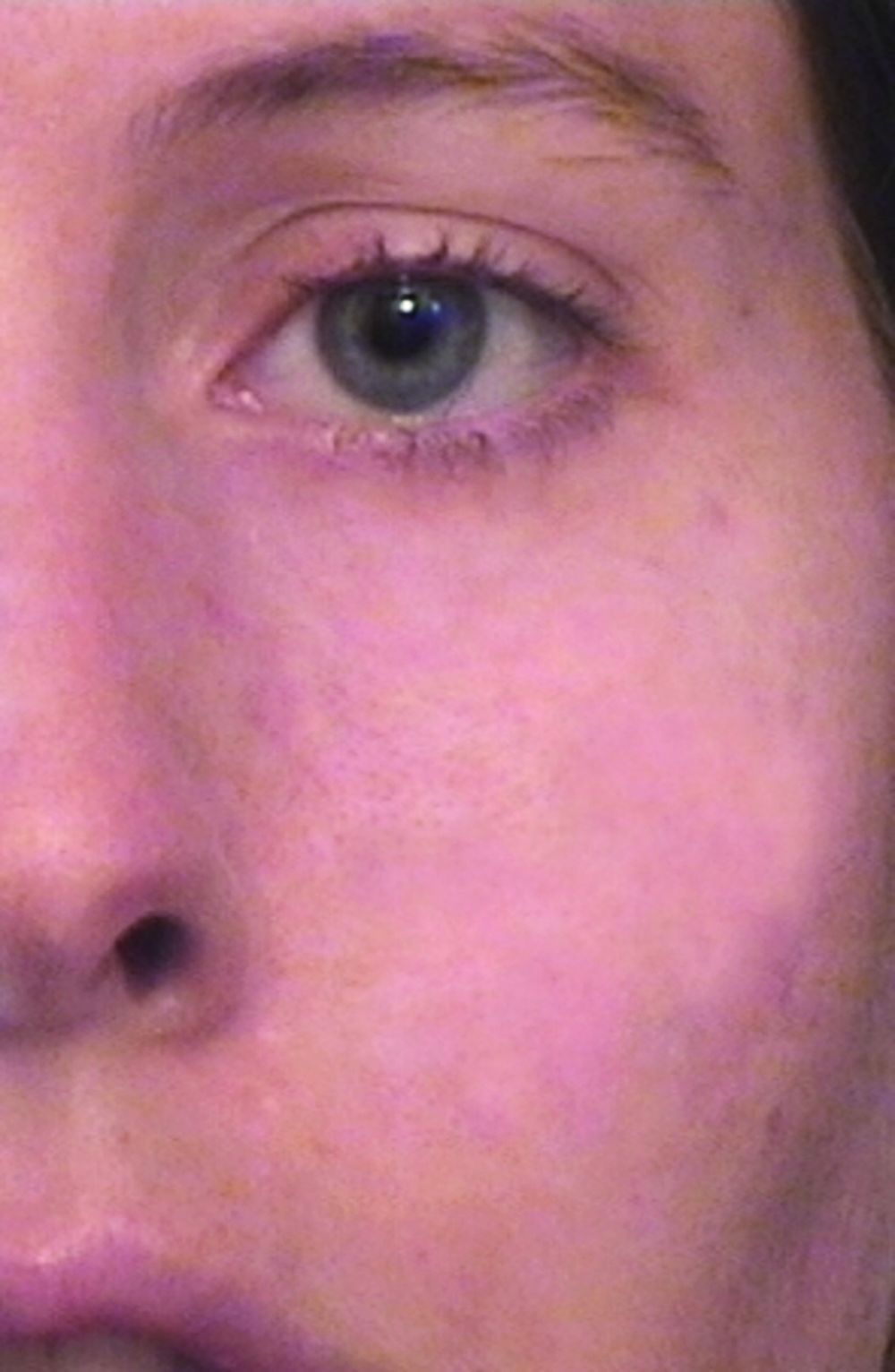
, which enabled the user to deliver even shorter pulse widths in the nanosecond domain. “That changed tattoo treatment,” said Dr. Avram, who is also a past president of the American Society for Laser Medicine and Surgery. “Prior to that, for tattoos and brown spots you would use ablative lasers like CO2 or dermabrasion. They would cause scarring and not really get rid of the tattoo ink or the brown spots. With the Q-switched nanosecond lasers and the picosecond lasers, which came about 15 years later, you had the ability to remove spots with a week of down time, and [they worked] for things like Nevus of Ota, where someone has a disfiguring blue-brown discoloration of their cheek. There’s no surgical treatment for that whatsoever. It’s not like you can take it out.”
Another key advancement was the introduction of “scanning” technology in the early 1990s for CO2 and erbium YAG lasers, which enabled precise computerized control of laser beams. Dr. Avram characterized the CO2 laser as “the gold standard for facial rejuvenation, for sun-damaged skin. The downside of CO2 lasers is that they really need to be in skilled hands. There can be serious side effects such as scarring if it’s not done appropriately or there is not appropriate follow-up. The CO2 lasers have been used in fractional modes for scars. I think it’s the best treatment for scars.”
Dr. Anderson and Melanie Grossman, MD, who practices in New York City, developed the ruby laser for hair removal in the 1990s, and today that procedure ranks as the most common laser treatment in medicine, according to Dr. Avram. He described it as “safe and effective in skilled hands,” requiring about six treatments. Indications are for hypertrichosis, hirsutism (sometimes in the setting of polycystic ovary syndrome), pseudofolliculitis barbae, pilonidal cysts, and gender reassignment surgery.
Another game-changing technology developed by Dr. Anderson came in the early 2000s with the Food and Drug Administration clearance of the Fraxel laser, which is based on the concept of fractional photothermolysis. With this technology, “instead of treating skin to a certain depth, you treat a fraction of it, anywhere from 5% to 40% of the skin,” Dr. Avram explained. “You go in deeper, but you leave surrounding viable tissue that is not affected by the laser. That serves as viable tissue to promote healing. The laser goes in deeper but it’s fractional, so there are skip zones in between the lasers that are going into the skin. You can do this with the CO2 and erbium YAG lasers.” Since hitting the marketplace, the FDA has cleared the use of Fraxel for a number of indications, from periorbital wrinkles and acne scars to surgical scars and melasma.
Dr. Parrish predicted that the next frontier for the advancement of lasers in dermatology will involve the treatment of photodamaged skin. “I’m not sure which technology is going to win,” he said.
Dr. Avram anticipates that dermatologic lasers of the future are going to be more effective, safer, and result in less downtime for patients. “I think we are going to be able to treat skin of color more safely and more effectively, and I think we’re going to become much more successful,” he said. “At some point, the standard of care of treatment for skin cancer will involve lasers and light sources. With all the advances that have happened in the last 50 years, sometimes you wonder, are we at a time to pause, or is most of the story behind us? I think that the advances in innovation that are occurring are going to accelerate greatly as we pass the 50th anniversary. In due credit, laser therapy has completely revolutionized the field of dermatology and has completely revolutionized the way we practice medicine. That will only accelerate in the future.”
Dr. Alster emphasized a “safety first” approach to her hopes for the future. “My wish is that we educate people to know that, while lasers have become ubiquitous and we’ve made them safe, they’re still only safe in the right hands,” she said. “There’s not a day that goes by when I don’t have somebody referred to me who’s been mishandled. There’s no reason for that. With proper training, the risk of bad side effects or complications is markedly reduced.”
During her dermatology residency at Yale University in the late 1980s, Tina S. Alster, MD, met a 44-year-old woman who changed the trajectory of her professional career.
During her clinic visit, the woman explained that she always wore heavy facial makeup to hide her port-wine stain birthmark – a vascular malformation that she kept secret from her husband and teenage son. “She was very good about covering it,” recalled Dr. Alster, who is the founding director of the Washington Institute of Dermatologic Laser Surgery and clinical professor of dermatology at Georgetown University, Washington. “She removed a small amount of makeup for me so I could take a look at it. I had just finished reading an article about using a laser for birthmarks; it had just been published. I told her, ‘There’s something new; we don’t have it at Yale, but I read about treatment that could hone in on birthmarks.’ I promised her I would find out more details.”
A few days later, Dr. Alster pored through stacks of medical journals at Yale’s library and relocated the article she’d seen by first author Oon Tian Tan, MD, PhD, of the department of dermatology at Boston University Medical Center. It described use of the flashpump-pulsed tunable dye laser to treat port-wine stains in 35 children (N Engl J Med. 1989;320:416-21). After giving the article a more thorough read, Dr. Alster became so intrigued by the technology it described that she moved to Boston the following year for a dermatology fellowship with Dr. Tan and joined the ranks of early clinicians who used lasers for treating port-wine stains and other dermatologic conditions.
“That was at a time when there were only a handful of pulsed dye lasers in the world, and the first time I used it was when I went to Boston,” she said. “It was life-changing. You think, ‘Isn’t this great for children with port-wine stains.’ Your heart breaks for them, but I also felt compassion for adults who had suffered a lifetime of stares, including the woman who propelled me to look into this. She ended up coming to Boston during my fellowship and had her birthmark removed, so I changed her life, but she changed mine as well.”
The real credit for that series of events, Dr. Alster continued, belongs to John A. Parrish, MD, and R. Rox Anderson, MD, who in 1983 published the concept of selective photothermolysis, a seminal work that shifted the paradigm for how lasers and other light sources are designed for skin diseases and conditions (Science. 1983 Apr 29;220(4596):524-7). The first pulsed dye laser was built on this concept, an approach that minimized or eliminated the unwanted tissue damage and significant scarring that impeded therapeutic use of laser energy for port-wine stains and other lesions prior to that time. “Lasers that were built subsequent to that seminal paper focused our attention on building lasers that were specific for treatment of certain skin conditions,” Dr. Alster said. “Selective photothermolysis catapulted not only our understanding of how lasers interact with the skin, but allowed us to identify things in the skin that we could potentially target with this new laser technology, and to build laser systems that were specific to those purposes.”
In the late 1970s, Dr. Parrish, who played a key role in making psoralens plus ultraviolet A safe and effective for patients with severe psoriasis, turned his attention to studying lasers in his lab at Harvard Medical School. He hired R. Rox Anderson, a recent graduate of the Massachusetts Institute of Technology, as a technician. “Rox then got interested in medicine and went to medical school at Harvard, got interested in dermatology, and then worked in my lab a little bit more,” said Dr. Parrish, who founded the Wellman Center for Photomedicine at Massachusetts General Hospital, Boston.
“Rox was interested in port-wine stains because of his rotation through pediatrics and was theorizing about how lasers could improve port-wine stains and hemangiomas. I think he first thought of that through the physics of what would be needed. He was thinking, ‘What are these hemangiomas under the microscope? What does the target look like, and what do you need to do to promote healing without scarring? You would have to be able to heat for this duration and this time and at this wavelength.’ He matched the physics of lasers with the pathophysiology of port-wine stains, and together we figured out how to deliver the right energy at the right wavelength at the right time. In fact, at the time, there was no ideal laser. We had to convince a laser manufacturer to build a tunable dye laser, which is what we ended up using around the specifications that we wanted for this treatment.”
Prior to the theory of selective photothermolysis, lasers were a blunt instrument. “They would target the skin but you wouldn’t just selectively target something; you’d get a result you didn’t want,” said Mathew M. Avram, MD, JD, director of laser, cosmetics, and dermatologic surgery at Massachusetts General Hospital.
Once pulsed dye lasers that incorporated principles of selective photothermolysis hit the marketplace, clinicians could treat and improve port-wine stains without scarring the skin. They could even improve scarring from port-wine stains that had been previously treated with the argon laser, the subject of early published work by Dr. Alster (Lasers Surg Med. 1993;13[3]:368-73). “When we treated port-wine stains with the pulsed dye laser on top of the argon laser scars, we observed that the scars were looking better,” Dr. Alster said. “From that observation, we were able to demonstrate improvement of a wide range of scars: traumatic and burn scars, surgical scars, acne scars, and scars caused by other lasers. But it all started with the pulsed dye laser for treating port-wine stains that had scars in them.”
, which enabled the user to deliver even shorter pulse widths in the nanosecond domain. “That changed tattoo treatment,” said Dr. Avram, who is also a past president of the American Society for Laser Medicine and Surgery. “Prior to that, for tattoos and brown spots you would use ablative lasers like CO2 or dermabrasion. They would cause scarring and not really get rid of the tattoo ink or the brown spots. With the Q-switched nanosecond lasers and the picosecond lasers, which came about 15 years later, you had the ability to remove spots with a week of down time, and [they worked] for things like Nevus of Ota, where someone has a disfiguring blue-brown discoloration of their cheek. There’s no surgical treatment for that whatsoever. It’s not like you can take it out.”
Another key advancement was the introduction of “scanning” technology in the early 1990s for CO2 and erbium YAG lasers, which enabled precise computerized control of laser beams. Dr. Avram characterized the CO2 laser as “the gold standard for facial rejuvenation, for sun-damaged skin. The downside of CO2 lasers is that they really need to be in skilled hands. There can be serious side effects such as scarring if it’s not done appropriately or there is not appropriate follow-up. The CO2 lasers have been used in fractional modes for scars. I think it’s the best treatment for scars.”
Dr. Anderson and Melanie Grossman, MD, who practices in New York City, developed the ruby laser for hair removal in the 1990s, and today that procedure ranks as the most common laser treatment in medicine, according to Dr. Avram. He described it as “safe and effective in skilled hands,” requiring about six treatments. Indications are for hypertrichosis, hirsutism (sometimes in the setting of polycystic ovary syndrome), pseudofolliculitis barbae, pilonidal cysts, and gender reassignment surgery.
Another game-changing technology developed by Dr. Anderson came in the early 2000s with the Food and Drug Administration clearance of the Fraxel laser, which is based on the concept of fractional photothermolysis. With this technology, “instead of treating skin to a certain depth, you treat a fraction of it, anywhere from 5% to 40% of the skin,” Dr. Avram explained. “You go in deeper, but you leave surrounding viable tissue that is not affected by the laser. That serves as viable tissue to promote healing. The laser goes in deeper but it’s fractional, so there are skip zones in between the lasers that are going into the skin. You can do this with the CO2 and erbium YAG lasers.” Since hitting the marketplace, the FDA has cleared the use of Fraxel for a number of indications, from periorbital wrinkles and acne scars to surgical scars and melasma.
Dr. Parrish predicted that the next frontier for the advancement of lasers in dermatology will involve the treatment of photodamaged skin. “I’m not sure which technology is going to win,” he said.
Dr. Avram anticipates that dermatologic lasers of the future are going to be more effective, safer, and result in less downtime for patients. “I think we are going to be able to treat skin of color more safely and more effectively, and I think we’re going to become much more successful,” he said. “At some point, the standard of care of treatment for skin cancer will involve lasers and light sources. With all the advances that have happened in the last 50 years, sometimes you wonder, are we at a time to pause, or is most of the story behind us? I think that the advances in innovation that are occurring are going to accelerate greatly as we pass the 50th anniversary. In due credit, laser therapy has completely revolutionized the field of dermatology and has completely revolutionized the way we practice medicine. That will only accelerate in the future.”
Dr. Alster emphasized a “safety first” approach to her hopes for the future. “My wish is that we educate people to know that, while lasers have become ubiquitous and we’ve made them safe, they’re still only safe in the right hands,” she said. “There’s not a day that goes by when I don’t have somebody referred to me who’s been mishandled. There’s no reason for that. With proper training, the risk of bad side effects or complications is markedly reduced.”
App for MS aims to capture elusive signals of progression

At the Joint European Committee for Treatment and Research in Multiple Sclerosis–Americas Committee for Treatment and Research in Multiple Sclerosis (ECTRIMS–ACTRIMS) 2020, this year known as MSVirtual2020, researchers at the University Hospital and University of Basel in Switzerland, presented data on their dreaMS app. The investigators are validating the app in a nonblinded cohort of 30 people with MS in the early to middle stages of progression and 30 controls without MS.
The application comprises a series of active tests measuring movement, fine motor skills, cognition, and vision, as well as questionnaires to assess quality of life, walking ability, and fatigue in people with Expanded Disability Status Scale (EDSS) scores of 6.5 or lower. A wrist device, used concurrently with the app, passively monitors subjects’ step count, heart rate, and different measures of activity.
If validated, such smartphone-based “digital biomarkers” will provide clinicians and investigators with a steadier flow of information for assessing MS disease progression and informing clinical decision-making. In June, Ludwig Kappos, MD, the app study’s senior researcher, co-authored an analysis of randomized trial data that argued for discarding the standard categories of relapsing and progressive MS in favor of seeing the disease as a continuum, in which progression can and does occur in the absence of relapses.
The digital biomarker work builds on that more unified view of the disease, Dr. Kappos said in an interview.
Outside of disease exacerbations or relapses, “progression can be very difficult to capture, especially in the first stage of the disease because of compensation in the central nervous system,” he said. “Our ability to see these very slight changes during a neurological examination is limited even if we do it very thoroughly. But by having these more frequent assessments we may be able to.”
Smartphone-gleaned biomarkers may have implications for prognosis and for choice of therapy, Dr. Kappos added. “We expect that these digital biomarkers will be even more sensitive and to be able to recognize before severe deficits are evident who is a candidate for a more intensive treatment and who is not.”
At the MSVirtual2020 congress, Dr. Kappos’s colleagues at the university Johannes Lorscheider, MD, and Yvonne Naegelin, MD, presented their feasibility and acceptance study currently underway in 60 volunteers. One of the concerns the investigators have had was whether engaged users would remain with the app. “We have designed the tests as little challenges to help keep people interested—we want to make these tests as appealing as possible,” Dr. Kappos said.
In this study, the reliability of each test is determined by intra-class correlation and median coefficient of variation. Preliminary reliability testing with healthy controls showed intra-class correlation coefficients of greater than 60% for the digital biomarkers and greater than 80% for at least one in every domain.
Once the best tests are selected and the app is fine-tuned, the group intends to embark on larger studies of the digital biomarkers. The next, planned for 2021, will recruit approximately 400 patients from the Swiss MS cohort, whose 1,000-some MS participants are followed with standardized examination and imaging protocols across healthcare centers.
“This is a very well characterized group of patients who are followed continuously with state-of-the-art neurological examinations, high-end MRI, and blood biomarkers,” Dr. Kappos said. “We want to see if we can add value by using digital biomarkers.”
The dreaMS app project is an independent investigator-initiated venture in cooperation with a technological partner. The study was supported by the Swiss Innovation Agency. The University Hospital Basel has received research funding for clinical trials from a number of pharmaceutical manufacturers.
SOURCE: Lorscheider J, et al. MSVirtual2020. Abstract P0069.
FROM MSVirtual2020
Satralizumab reduces risk of severe NMOSD relapse
(NMOSD), according to investigators. The drug also was associated with a lower likelihood of using acute relapse therapy.
These results were presented at the Joint European Committee for Treatment and Research in Multiple Sclerosis–Americas Committee for Treatment and Research in Multiple Sclerosis (ECTRIMS–ACTRIMS) 2020, this year known as MSVirtual2020.

NMOSD is characterized by acute relapses that are unpredictable and lead to the accumulation of disability. “Patients with NMOSD often recover poorly from relapses, therefore, the primary goal for disease management is to reduce attack frequency,” said Ingo Kleiter, MD, medical director of Marianne-Strauß-Klinik in Berg, Germany. “In the two phase 3 trials SAkuraSky and SAkuraStar, the IL-6 receptor inhibitor satralizumab was found to significantly reduce the risk of relapses versus placebo.” Satralizumab is a humanized, monoclonal, recycling antibody that targets the interleukin-6 receptor.
Dr. Kleiter and colleagues examined pooled data from the two phase 3 trials of satralizumab to determine the treatment’s effect on relapse severity in patients with NMOSD. Participants in those trials received placebo or 120 mg of satralizumab at weeks 0, 2, 4, and every 4 weeks thereafter.
For their research, the investigators analyzed data from the pooled intention-to-treat population in the double-blind periods of both studies. To evaluate the severity of protocol-defined relapses, they compared patients’ Expanded Disability Status Scale (EDSS) scores at the time of relapse with their scores before the relapse (i.e., their scores at the last scheduled study visit). Using the visual Functional Systems Score (FSS), Dr. Kleiter and colleagues performed a similar analysis on optic neuritis relapses. They categorized a protocol-defined relapse as severe if it entailed a change of two or more points on the EDSS or visual FSS. The investigators conducted Kaplan-Meier analyses to evaluate the time to first severe protocol-defined relapse. They also compared the number of patients receiving acute therapy for any relapse between treatment groups.
Safety profile confirmed
Dr. Kleiter and colleagues included 178 patients in their analyses. A total of 27 of 104 patients (26%) who received satralizumab had a protocol-defined relapse, compared with 34 of 74 patients (46%) who received placebo. The number and proportion of severe protocol-defined relapses were lower in the satralizumab group (5 of 27 events [19%]), compared with the placebo group (12 of 34 events [35%]). In addition, the number and proportion of severe protocol-defined optic neuritis relapses were lower in patients receiving satralizumab (2 of 8 events [25%]), compared with those receiving placebo (5 of 13 events [39%]). Compared with placebo, satralizumab was associated with a 79% reduction in the risk of severe protocol-defined relapse (hazard ratio, 0.21).
A lower proportion of patients receiving satralizumab was prescribed acute relapse therapy (38%), compared with patients receiving placebo (58%). The odds ratio of receiving a prescription of acute relapse therapy was 0.46 among patients receiving satralizumab.
The activity of IL-6 may cause neurologic damage in patients with NMOSD through astrocytic damage, disruption of the blood–brain barrier, and T cell polarization. “It is proposed that through inhibiting IL-6 across these multiple mechanisms, satralizumab reduces the risk and severity of NMOSD attacks,” Dr. Kleiter said.
To date, the rates of infection and serious infection for patients treated with satralizumab in the combined double-blind and open-label extension periods have been consistent with those for patients treated with satralizumab in the double-blind portion. These rates have not increased over time. Satralizumab is administered as a subcutaneous injection every 4 weeks, and treatment can be self-administered at the discretion of the managing physician. “These data provide reassurance to physicians about the overall profile of satralizumab, with respect to efficacy and safety in the longer term,” said Dr. Kleiter.
Does satralizumab differ from other new agents?
The main strength of the study is that sufficient numbers of relapses were available for analysis in the active and control groups, said Achim Berthele, MD, associate professor of neurology at the Technical University of Munich. This allowed the researchers to examine whether satralizumab led to a better outcome after each relapse, which it did. “A weakness is how the severity of relapses was quantified,” said Dr. Berthele. “The EDSS as a measure is not linear, and its functional systems are not clinically equivalent. However, the whole NMOSD community is struggling with this problem.”
The study’s implications for neurologists’ clinical practice are unclear, however. “Although the results presented are encouraging, the data are still too small to say with certainty that satralizumab does indeed improve the outcome of relapses,” said Dr. Berthele. “It is also an open question whether satralizumab differs in this respect from the other new immunotherapeutic agents.”
Investigators must collect further data on the outcome of relapses that occur during treatment with modern immunomodulatory therapy, Dr. Berthele added. Future research could examine whether the new anti-inflammatory immunotherapeutic agents also are suitable drugs for relapse therapy. Another salient question is whether clinical vigilance or relapse therapy in NMOSD has improved in general. “This is what Kleiter and colleagues show as well: The number of severe relapses under placebo was much lower than expected,” said Dr. Berthele.
Chugai/Roche funded the study. Dr. Kleiter has received compensation for consulting, speaking, or serving on advisory boards for Alexion, Biogen, Celgene, Merck, and Roche. Dr. Berthele was not involved in any of the satralizumab trials, but is an investigator and coauthor of the PREVENT trial of eculizumab.
SOURCE: Kleiter I, et al. MSVirtual2020. Abstract FC01.03.
(NMOSD), according to investigators. The drug also was associated with a lower likelihood of using acute relapse therapy.
These results were presented at the Joint European Committee for Treatment and Research in Multiple Sclerosis–Americas Committee for Treatment and Research in Multiple Sclerosis (ECTRIMS–ACTRIMS) 2020, this year known as MSVirtual2020.
NMOSD is characterized by acute relapses that are unpredictable and lead to the accumulation of disability. “Patients with NMOSD often recover poorly from relapses, therefore, the primary goal for disease management is to reduce attack frequency,” said Ingo Kleiter, MD, medical director of Marianne-Strauß-Klinik in Berg, Germany. “In the two phase 3 trials SAkuraSky and SAkuraStar, the IL-6 receptor inhibitor satralizumab was found to significantly reduce the risk of relapses versus placebo.” Satralizumab is a humanized, monoclonal, recycling antibody that targets the interleukin-6 receptor.
Dr. Kleiter and colleagues examined pooled data from the two phase 3 trials of satralizumab to determine the treatment’s effect on relapse severity in patients with NMOSD. Participants in those trials received placebo or 120 mg of satralizumab at weeks 0, 2, 4, and every 4 weeks thereafter.
For their research, the investigators analyzed data from the pooled intention-to-treat population in the double-blind periods of both studies. To evaluate the severity of protocol-defined relapses, they compared patients’ Expanded Disability Status Scale (EDSS) scores at the time of relapse with their scores before the relapse (i.e., their scores at the last scheduled study visit). Using the visual Functional Systems Score (FSS), Dr. Kleiter and colleagues performed a similar analysis on optic neuritis relapses. They categorized a protocol-defined relapse as severe if it entailed a change of two or more points on the EDSS or visual FSS. The investigators conducted Kaplan-Meier analyses to evaluate the time to first severe protocol-defined relapse. They also compared the number of patients receiving acute therapy for any relapse between treatment groups.
Safety profile confirmed
Dr. Kleiter and colleagues included 178 patients in their analyses. A total of 27 of 104 patients (26%) who received satralizumab had a protocol-defined relapse, compared with 34 of 74 patients (46%) who received placebo. The number and proportion of severe protocol-defined relapses were lower in the satralizumab group (5 of 27 events [19%]), compared with the placebo group (12 of 34 events [35%]). In addition, the number and proportion of severe protocol-defined optic neuritis relapses were lower in patients receiving satralizumab (2 of 8 events [25%]), compared with those receiving placebo (5 of 13 events [39%]). Compared with placebo, satralizumab was associated with a 79% reduction in the risk of severe protocol-defined relapse (hazard ratio, 0.21).
A lower proportion of patients receiving satralizumab was prescribed acute relapse therapy (38%), compared with patients receiving placebo (58%). The odds ratio of receiving a prescription of acute relapse therapy was 0.46 among patients receiving satralizumab.
The activity of IL-6 may cause neurologic damage in patients with NMOSD through astrocytic damage, disruption of the blood–brain barrier, and T cell polarization. “It is proposed that through inhibiting IL-6 across these multiple mechanisms, satralizumab reduces the risk and severity of NMOSD attacks,” Dr. Kleiter said.
To date, the rates of infection and serious infection for patients treated with satralizumab in the combined double-blind and open-label extension periods have been consistent with those for patients treated with satralizumab in the double-blind portion. These rates have not increased over time. Satralizumab is administered as a subcutaneous injection every 4 weeks, and treatment can be self-administered at the discretion of the managing physician. “These data provide reassurance to physicians about the overall profile of satralizumab, with respect to efficacy and safety in the longer term,” said Dr. Kleiter.
Does satralizumab differ from other new agents?
The main strength of the study is that sufficient numbers of relapses were available for analysis in the active and control groups, said Achim Berthele, MD, associate professor of neurology at the Technical University of Munich. This allowed the researchers to examine whether satralizumab led to a better outcome after each relapse, which it did. “A weakness is how the severity of relapses was quantified,” said Dr. Berthele. “The EDSS as a measure is not linear, and its functional systems are not clinically equivalent. However, the whole NMOSD community is struggling with this problem.”
The study’s implications for neurologists’ clinical practice are unclear, however. “Although the results presented are encouraging, the data are still too small to say with certainty that satralizumab does indeed improve the outcome of relapses,” said Dr. Berthele. “It is also an open question whether satralizumab differs in this respect from the other new immunotherapeutic agents.”
Investigators must collect further data on the outcome of relapses that occur during treatment with modern immunomodulatory therapy, Dr. Berthele added. Future research could examine whether the new anti-inflammatory immunotherapeutic agents also are suitable drugs for relapse therapy. Another salient question is whether clinical vigilance or relapse therapy in NMOSD has improved in general. “This is what Kleiter and colleagues show as well: The number of severe relapses under placebo was much lower than expected,” said Dr. Berthele.
Chugai/Roche funded the study. Dr. Kleiter has received compensation for consulting, speaking, or serving on advisory boards for Alexion, Biogen, Celgene, Merck, and Roche. Dr. Berthele was not involved in any of the satralizumab trials, but is an investigator and coauthor of the PREVENT trial of eculizumab.
SOURCE: Kleiter I, et al. MSVirtual2020. Abstract FC01.03.
FROM MSVirtual2020
Cardiovascular risk factors linked to brain atrophy in MS
The presence of cardiovascular risk factors in patients with multiple sclerosis (MS) is associated with a greater degree of brain atrophy even in young patients who are unlikely to have small vessel disease, a new study has shown.
The results were presented by Raffaello Bonacchi, MD, Vita-Salute San Raffaele University, Milan, Italy, at at the Joint European Committee for Treatment and Research in Multiple Sclerosis–Americas Committee for Treatment and Research in Multiple Sclerosis (ECTRIMS–ACTRIMS) 2020, this year known as MSVirtual2020. .
“Our results suggest that even low levels of exposure to cardiovascular risk factors are important in MS and might affect brain atrophy—and therefore long-term disability—even in young patients,” Dr. Bonacchi said.
“It is not only smoking,” he added. “Other cardiovascular risk factors also appear to be implicated. We found a synergistic effect of the different risk factors.”
These are only preliminary data and need to be confirmed in other studies,” he said, “but it does suggest that MS neurologists need to pay attention to comprehensive care—not just MS disease activity.
“They also need to be discussing lifestyle with their patients, evaluating their cardiovascular risk factors, and giving advice on stopping smoking, lowering blood pressure, cholesterol, etc.”
Brain changes
Dr. Bonacchi explained that previous studies have suggested a relationship between cardiovascular risk factors and changes on magnetic resonance imaging (MRI) and clinical outcomes in patients with MS that may be mediated by small vessel disease and/or inflammation.
“Small vessel disease is widespread in the population over 50 years of age, but in this study we wanted to look at the impact of cardiovascular risk factors in younger patients with MS who are not likely to have much small vessel disease to try and see whether there is still a relationship with brain atrophy or white/gray matter lesions,” he said.
Previous studies have not set an age limit for examining this relationship and they have also assessed the presence versus absence of cardiovascular risk factors, without attempting to grade the strength of exposure, he noted.
For the current study, the researchers examined several cardiovascular risk factors and in addition to just being present or absent. They also graded each risk factor as being stringent or not depending on a certain threshold.
For example, smoking was defined as a threshold of 5 pack-years (smoking 5 cigarettes a day for 20 years or 20 cigarettes a day for 5 years). And the more stringent definition was 10 pack-years.
For hypertension, the stringent definition was consistently high blood pressure levels and use of antihypertensive medication, with similar definitions used for cholesterol and diabetes.
This was a cross-sectional observational study in 124 patients with MS and 95 healthy controls. The researchers examined MRI scans and neurological exams and investigated whether the amount of cardiovascular risk factors a patient was exposed to was associated with degree of brain atrophy and white matter/gray matter volume. Results were adjusted for age, sex, disease duration, phenotype (relapsing-remitting versus progressive MS) and treatment.
Results showed no significant difference if patients were exposed to at least one classical risk factor versus no risk factors. But if a patient had at least two classical risk factors, significant differences were found in gray matter, white matter, and total brain volume.
Patients with MS and no risk factors had a mean brain volume of 1524 mL versus 1481 mL in those with at least two risk factors, a difference that was significant (P = 0.003). Mean gray matter volume was 856 mL in MS patients without cardiovascular risk factors and 836 mL in those with at least two risk factors (P = 0.01) Mean white matter volume was 668 mL in MS patients without cardiovascular risk factors and 845 mL in those with at least two risk factors (P = 0.03).
“This is one of the first studies to have graded degrees of risk factors and we found one stringent risk factor was associated with the same effects on brain atrophy as two less stringent risk factors,” Dr. Bonacchi reported.
Healthy controls showed no differences in any of the brain volume outcomes in those with or without cardiovascular risk factors.
“As our population was under aged 50 years, who are unlikely to have much small vessel disease, our results suggest that the influence of cardiovascular risk factors on brain atrophy in MS is not just mediated through small vessel disease and is probably also mediated by increased inflammation,” Dr. Bonacchi suggested.
Impact of CV risk factors
Commenting on the study, Dalia Rotstein, MD, assistant professor, department of neurology, University of Toronto, Ontario, Canada, session cochair, said: “This is an interesting study that captures the impact of cardiovascular risk factors on various measures of brain atrophy in MS.”
The cohort was quite young, under age 50, and the effect on brain atrophy was increased with more severe cardiovascular risk factors, she noted.
“The investigators compared these effects to a population of healthy controls and did not observe as substantial an effect in controls. However, they were likely underpowered for the analysis in the healthy controls because of a relatively small number of subjects with cardiovascular risk factors in this group,” Dr. Rotstein noted.
“More research is needed to determine whether the observed relationship is unique to MS and whether treating cardiovascular risk factors may help protect against neurodegeneration in MS,” she added.
Dr. Bonacchi has reported no relevant financial relationships. Dr. Rotstein has reported acting as a consultant for Roche, Alexion, Novartis, EMD Serono, and Sanofi Aventis.
SOURCE: Bonacchi R. et al. MSVirtual2020. Session PS04.05.
This article originally appeared on Medscape.com .
FROM MSVirtual2020