User login
Post-parathyroidectomy follow-up may need to be open-ended
BALTIMORE – Patients who have had parathyroidectomy for primary hyperparathyroidism can have disease recurrence 10 years or longer after surgery, raising the possibility that postop follow-up should never end, according to a study presented at the annual meeting of the American Association of Endocrine Surgeons.
Dr. Irene Lou of the University of Wisconsin–Madison reported on results of a retrospective study of 196 patients who had a presumably “curative” parathyroidectomy at the institution between November 2000 and June 2005. The mean age of the study population was 61 years.

“The long-term recurrences of primary hyperparathyroidism after curative parathyroidectomy is likely higher than previously reported, with over a third of recurrences occurring 10 years after their operation,” Dr. Lou said.
The study also identified independent predictors of recurrence, among them younger age, a drop in intraoperative parathyroid hormone less than 70%, and double adenoma, Dr. Lou said. All patients after parathyroidectomy should have at minimum an annual serum calcium test, especially younger patients with longer life expectancies, she said. This recommendation, however, may be altered for older patients or those with additional comorbidities.
The study defined recurrence as serum calcium of 10.2 mg/dL or greater 6 months or longer after the initial operation. The overall 10-year recurrence rate was 14.8% and the median time to recurrence was 6.3 years. “We found that 41.4% of patients who recurred did so by 5 years and 65.5% by 10 years,” Dr. Lou said.
The University of Wisconsin and University of Alabama at Birmingham investigators undertook the study because the recent data on recurrence was limited, with the longest study topping out at 7 years, Dr. Lou said. “We previously looked at this problem in other perspectives and we found that a lot of curves separated at around 8 years,” she said.
With regard to the type of operation the patients had, whether unilateral minimally invasive parathyroidectomy or bilateral open surgery, the study found no significant differences in recurrence rates, Dr. Lou said. “This is an excellent study,” Dr. Samuel K. Snyder of Temple, Tex., said during the discussion. “You’re telling us we need to follow patients much longer than perhaps we did previously, but we all see patients who have normal calcium and still have a residual elevated parathyroid hormone level.” He asked if the study considered parathyroid hormone levels at 6 months or more after surgery or vitamin D levels, but Dr. Lou said this information was not available, therefore could not be evaluated.
Dr. Lou and her coauthors had no financial relationships to disclose.
BALTIMORE – Patients who have had parathyroidectomy for primary hyperparathyroidism can have disease recurrence 10 years or longer after surgery, raising the possibility that postop follow-up should never end, according to a study presented at the annual meeting of the American Association of Endocrine Surgeons.
Dr. Irene Lou of the University of Wisconsin–Madison reported on results of a retrospective study of 196 patients who had a presumably “curative” parathyroidectomy at the institution between November 2000 and June 2005. The mean age of the study population was 61 years.
“The long-term recurrences of primary hyperparathyroidism after curative parathyroidectomy is likely higher than previously reported, with over a third of recurrences occurring 10 years after their operation,” Dr. Lou said.
The study also identified independent predictors of recurrence, among them younger age, a drop in intraoperative parathyroid hormone less than 70%, and double adenoma, Dr. Lou said. All patients after parathyroidectomy should have at minimum an annual serum calcium test, especially younger patients with longer life expectancies, she said. This recommendation, however, may be altered for older patients or those with additional comorbidities.
The study defined recurrence as serum calcium of 10.2 mg/dL or greater 6 months or longer after the initial operation. The overall 10-year recurrence rate was 14.8% and the median time to recurrence was 6.3 years. “We found that 41.4% of patients who recurred did so by 5 years and 65.5% by 10 years,” Dr. Lou said.
The University of Wisconsin and University of Alabama at Birmingham investigators undertook the study because the recent data on recurrence was limited, with the longest study topping out at 7 years, Dr. Lou said. “We previously looked at this problem in other perspectives and we found that a lot of curves separated at around 8 years,” she said.
With regard to the type of operation the patients had, whether unilateral minimally invasive parathyroidectomy or bilateral open surgery, the study found no significant differences in recurrence rates, Dr. Lou said. “This is an excellent study,” Dr. Samuel K. Snyder of Temple, Tex., said during the discussion. “You’re telling us we need to follow patients much longer than perhaps we did previously, but we all see patients who have normal calcium and still have a residual elevated parathyroid hormone level.” He asked if the study considered parathyroid hormone levels at 6 months or more after surgery or vitamin D levels, but Dr. Lou said this information was not available, therefore could not be evaluated.
Dr. Lou and her coauthors had no financial relationships to disclose.
FROM AAES 2016
Key clinical point: Long-term recurrence rates for hyperparathyroidism (HPT) after “curative” parathyroidectomy are likely higher than previously reported.
Major finding: Approximately one-third of patients were found to have recurrences 10 or more years after the initial operation.
Data source: Single-institution cohort of 196 patients who had initial parathyroidectomy for HPT between November 2000 and June 2005.
Disclosures: Dr. Lou and her study coauthors reported having no financial disclosures.

Reintubation avoided by majority of patients on noninvasive ventilation therapy, high-flow oxygen
Extubated patients who either received noninvasive ventilation (NIV) therapy or high-flow nasal cannula oxygen had a lower risk of reintubation, compared with extubated patients who received some form of standard oxygen therapy, according to the results of two multicenter, randomized clinical trials published online in JAMA.
Participants in one of the studies, which included abdominal surgery patients diagnosed with respiratory failure within 7 days following surgery, either received NIV or standard oxygen therapy for 30 days or until ICU discharge, whichever came first. While NIV has been effectively used to treat nonsurgical patients with acute exacerbations of chronic obstructive pulmonary disease and cardiogenic pulmonary edema, there is no evidence to support the use of NIV in surgical patients with hypoxemic acute respiratory failure after abdominal surgery, according to Dr. Samir Jaber of the Saint Eloi University Hospital and Montpellier School of Medicine, both in Montpellier, France, and his colleagues (JAMA. 2016 Apr 5;315[13]:1345-53).
The second study included adult patients who had received mechanical ventilation for more than 12 hours and who met criteria for being considered at low risk for reintubation. Patients were administered either high-flow oxygen therapy through nasal cannula immediately after extubation or continuous conventional oxygen therapy through nasal cannula or nonrebreather facemask; the patients were observed for 72 hours. High-flow therapy has been shown to improve oxygenation and survival in clinical studies of critically ill patients in the acute phase of respiratory failure. “[A study by S.M. Maggiore and his colleagues (Am J Respir Crit Care Med. 2014;190(3):282-8)] suggested that high-flow therapy after planned extubation decreased the reintubation rate in a general population of critical patients, but the benefits might be mainly attributable to improvements in high-risk patients,” said Dr. Gonzalo Hernandez, of the Hospital Virgen de la Salud, Toledo, Spain, and his colleagues (JAMA. 2016 Apr 5;315[13]:1354-61).
In the first study, 148 patients received NIV and 145 patients received standard oxygen therapy only. NIV was administered through a facemask connected to an ICU- or a NIV-dedicated ventilator, using either a heated humidifier or heat and moisture exchanger to warm and humidify inspired gases. Patients were encouraged to use NIV for 6 hours during the first 24 hours of the study and received standard oxygen therapy at a rate of up to 15 L/minute to maintain an arterial oxygen saturation estimate (SpO2) of at least 94% in between NIV sessions. NIV was started at an inspiratory positive airway pressure of 5 cm H2O, increasing to a maximum inspiratory pressure of 15 cm H2O, aiming to achieve an expiratory tidal volume between 6 and 8 mL/kg of predicted body weight and a respiratory rate lower than 25/min. The patients in this study’s control group only received the standard oxygen therapy.
In the other study, 263 patients received conventional therapy, with the oxygen flow having been adjusted to maintain an arterial oxygen saturation estimate of greater than 92%. This study’s other 264 patients received high-flow oxygen therapy, with the flow having been initially set at 10 L/min and titrated upward in 5-L/min steps until patients experienced discomfort. The high-flow therapy was stopped after 24 hours and was followed by conventional oxygen therapy, when needed.
The primary outcome measure in the study involving NIV was cause for reintubation within 7 days of randomization.
Secondary outcome measures included gas exchange, healthcare-associated infection rate within 30 days, number of ventilator-free days between days 1 and 30, antibiotic use duration, ICU and in-hospital length of stay, and 30- and 90-day mortality.
Reintubation occurred in 49 patients in the NIV group and 66 patients in the standard oxygen therapy group, a significant difference (P = .03). Among the reintubated patients, those who had received NIV spent less time under invasive mechanical ventilation as did the patients given standard oxygen therapy. The interquartile ranges of days of invasive mechanical ventilation were 0-3 for patients in the NIV group and 0-5 for patients in the standard oxygen therapy group (P = .05). At 30 days, NIV was associated with significantly more ventilator-free days than standard oxygen therapy (25.4 vs. 23.2; P = .04). At 90 days, 22 patients in the NIV group and 31 patients in the standard oxygen therapy group had died (P = .15).
“Recent high-impact trials have demonstrated the benefits in nonsurgical hypoxemic respiratory failure or equivalence of high-flow nasal cannula compared with NIV in patients after cardiothoracic surgery with moderate to severe hypoxemia. Future studies comparing use of high-flow oxygen cannula vs standard oxygen therapy and NIV for patients after abdominal surgery as preventive (prophylactic) or curative applications are needed,” according to Dr. Jaber and his colleagues.
The primary outcome measure for the study of patients receiving high-flow oxygen therapy was reintubation within 72 hours after extubation; this occurred in fewer patients in the high-flow oxygen group than in the conventional therapy group (13 or 4.9% vs. 32 or 12.2%.) This statistically significant difference was mainly attributable to a lower incidence of respiratory-related reintubation in the high-flow group, compared with the conventional therapy group (1.5% vs. 8.7%), said Dr. Hernandez and his colleagues.
Secondary outcome measures included postextubation respiratory failure, respiratory infection, sepsis, multiorgan failure, ICU and hospital length of stay and mortality, time to reintubation, and adverse effects. Postintubation respiratory failure was less common in the high-flow therapy group than in the conventional therapy group (22 patients or 8.3% vs. 38 or 14.4%). Differences between the two groups in other secondary outcomes were not statistically significant.
“The main finding of this study was that high-flow oxygen significantly reduced the reintubation rate in critically ill patients at low risk for extubation failure ... High-flow therapy improves oxygenation, and the lower rate of reintubation secondary to hypoxia in the high-flow group corroborates this finding. High-flow oxygen also seems to reduce other causes of respiratory failure such as increased work of breathing and respiratory muscle fatigue, which are frequently associated with reintubation secondary to hypoxia. Another way in which high-flow therapy improves extubation outcome is by conditioning the inspired gas,” said Dr. Hernandez and his colleagues.
No adverse events were reported in either study.
Dr. Hernandez and his colleagues reported no conflicts of interest. Dr. Jaber and his colleagues disclosed no potential conflicts of interest with their study’s sponsors, Montpellier (France) University Hospital and the APARD Foundation.
Dr. Eric Gartman, FCCP, comments: These two studies augment a growing body of literature supporting the use of adjunctive therapies immediately following extubation to prevent reintubation for respiratory failure.
It has been known for several years that the use of noninvasive ventilation (NIV) immediately after extubation in COPD patients prevents reintubation rates, and these new data demonstrate efficacy in an expanded population. Further, the use of high-flow humidified oxygen therapy in acute respiratory failure has been shown to prevent progression to initial intubation, and now these data expand potential use to prevent reintubation, as well.
While not studied, if high-flow oxygen therapy is found to be equivalent to NIV to prevent reintubation (similar to the previously-published prevention of intubation studies), that would be clinically important since there is a significant difference in tolerance to these two therapies. Across these trials, the very important point to remember is that these therapies were found to be effective if put on directly after extubation, and one cannot wait to apply them at the point where the patient shows signs of respiratory decline.
Extubated patients who either received noninvasive ventilation (NIV) therapy or high-flow nasal cannula oxygen had a lower risk of reintubation, compared with extubated patients who received some form of standard oxygen therapy, according to the results of two multicenter, randomized clinical trials published online in JAMA.
Participants in one of the studies, which included abdominal surgery patients diagnosed with respiratory failure within 7 days following surgery, either received NIV or standard oxygen therapy for 30 days or until ICU discharge, whichever came first. While NIV has been effectively used to treat nonsurgical patients with acute exacerbations of chronic obstructive pulmonary disease and cardiogenic pulmonary edema, there is no evidence to support the use of NIV in surgical patients with hypoxemic acute respiratory failure after abdominal surgery, according to Dr. Samir Jaber of the Saint Eloi University Hospital and Montpellier School of Medicine, both in Montpellier, France, and his colleagues (JAMA. 2016 Apr 5;315[13]:1345-53).
The second study included adult patients who had received mechanical ventilation for more than 12 hours and who met criteria for being considered at low risk for reintubation. Patients were administered either high-flow oxygen therapy through nasal cannula immediately after extubation or continuous conventional oxygen therapy through nasal cannula or nonrebreather facemask; the patients were observed for 72 hours. High-flow therapy has been shown to improve oxygenation and survival in clinical studies of critically ill patients in the acute phase of respiratory failure. “[A study by S.M. Maggiore and his colleagues (Am J Respir Crit Care Med. 2014;190(3):282-8)] suggested that high-flow therapy after planned extubation decreased the reintubation rate in a general population of critical patients, but the benefits might be mainly attributable to improvements in high-risk patients,” said Dr. Gonzalo Hernandez, of the Hospital Virgen de la Salud, Toledo, Spain, and his colleagues (JAMA. 2016 Apr 5;315[13]:1354-61).
In the first study, 148 patients received NIV and 145 patients received standard oxygen therapy only. NIV was administered through a facemask connected to an ICU- or a NIV-dedicated ventilator, using either a heated humidifier or heat and moisture exchanger to warm and humidify inspired gases. Patients were encouraged to use NIV for 6 hours during the first 24 hours of the study and received standard oxygen therapy at a rate of up to 15 L/minute to maintain an arterial oxygen saturation estimate (SpO2) of at least 94% in between NIV sessions. NIV was started at an inspiratory positive airway pressure of 5 cm H2O, increasing to a maximum inspiratory pressure of 15 cm H2O, aiming to achieve an expiratory tidal volume between 6 and 8 mL/kg of predicted body weight and a respiratory rate lower than 25/min. The patients in this study’s control group only received the standard oxygen therapy.
In the other study, 263 patients received conventional therapy, with the oxygen flow having been adjusted to maintain an arterial oxygen saturation estimate of greater than 92%. This study’s other 264 patients received high-flow oxygen therapy, with the flow having been initially set at 10 L/min and titrated upward in 5-L/min steps until patients experienced discomfort. The high-flow therapy was stopped after 24 hours and was followed by conventional oxygen therapy, when needed.
The primary outcome measure in the study involving NIV was cause for reintubation within 7 days of randomization.
Secondary outcome measures included gas exchange, healthcare-associated infection rate within 30 days, number of ventilator-free days between days 1 and 30, antibiotic use duration, ICU and in-hospital length of stay, and 30- and 90-day mortality.
Reintubation occurred in 49 patients in the NIV group and 66 patients in the standard oxygen therapy group, a significant difference (P = .03). Among the reintubated patients, those who had received NIV spent less time under invasive mechanical ventilation as did the patients given standard oxygen therapy. The interquartile ranges of days of invasive mechanical ventilation were 0-3 for patients in the NIV group and 0-5 for patients in the standard oxygen therapy group (P = .05). At 30 days, NIV was associated with significantly more ventilator-free days than standard oxygen therapy (25.4 vs. 23.2; P = .04). At 90 days, 22 patients in the NIV group and 31 patients in the standard oxygen therapy group had died (P = .15).
“Recent high-impact trials have demonstrated the benefits in nonsurgical hypoxemic respiratory failure or equivalence of high-flow nasal cannula compared with NIV in patients after cardiothoracic surgery with moderate to severe hypoxemia. Future studies comparing use of high-flow oxygen cannula vs standard oxygen therapy and NIV for patients after abdominal surgery as preventive (prophylactic) or curative applications are needed,” according to Dr. Jaber and his colleagues.
The primary outcome measure for the study of patients receiving high-flow oxygen therapy was reintubation within 72 hours after extubation; this occurred in fewer patients in the high-flow oxygen group than in the conventional therapy group (13 or 4.9% vs. 32 or 12.2%.) This statistically significant difference was mainly attributable to a lower incidence of respiratory-related reintubation in the high-flow group, compared with the conventional therapy group (1.5% vs. 8.7%), said Dr. Hernandez and his colleagues.
Secondary outcome measures included postextubation respiratory failure, respiratory infection, sepsis, multiorgan failure, ICU and hospital length of stay and mortality, time to reintubation, and adverse effects. Postintubation respiratory failure was less common in the high-flow therapy group than in the conventional therapy group (22 patients or 8.3% vs. 38 or 14.4%). Differences between the two groups in other secondary outcomes were not statistically significant.
“The main finding of this study was that high-flow oxygen significantly reduced the reintubation rate in critically ill patients at low risk for extubation failure ... High-flow therapy improves oxygenation, and the lower rate of reintubation secondary to hypoxia in the high-flow group corroborates this finding. High-flow oxygen also seems to reduce other causes of respiratory failure such as increased work of breathing and respiratory muscle fatigue, which are frequently associated with reintubation secondary to hypoxia. Another way in which high-flow therapy improves extubation outcome is by conditioning the inspired gas,” said Dr. Hernandez and his colleagues.
No adverse events were reported in either study.
Dr. Hernandez and his colleagues reported no conflicts of interest. Dr. Jaber and his colleagues disclosed no potential conflicts of interest with their study’s sponsors, Montpellier (France) University Hospital and the APARD Foundation.
FROM JAMA
Key clinical point: Extubated patients who either received noninvasive ventilation (NIV) therapy or high-flow nasal cannula oxygen reduced their risk of reintubation, compared with patients who received some form of standard oxygen therapy.
Major finding: In one study, significantly fewer of the patients who received NIV needed to be reintubated than the patients who received standard oxygen therapy.
Data source: Two multicenter, randomized clinical trials published online in JAMA.
Disclosures: Dr. Hernandez and his colleagues reported no conflicts of interest. Dr. Jaber and his colleagues disclosed no potential conflicts of interest with Montpellier (France) University Hospital and the APARD Foundation, who funded their study.
Cochrane Review nixes specific allergen immunotherapy for atopic dermatitis
LOS ANGELES – A new Cochrane systematic review and meta-analysis has concluded there is no consistent evidence that specific allergen immunotherapy is beneficial in patients with atopic dermatitis, Dr. Herman H. Tam reported at the annual meeting of the American Academy of Allergy, Asthma, and Immunology.
“We know that for specific allergen immunotherapy, there have been really good results in allergic rhinitis and venom allergy. For atopic dermatitis, however, dating back almost 40 years, we found there have only been 12 randomized trials using standardized allergen extracts. The quality of evidence was low, and the study findings have been inconsistent,” Dr. Tam, first author of the Cochrane review, said in an interview.

The dozen trials included a total of 733 children and adults in nine countries. Because of insufficient follow-up in most of the studies, coupled with the use of a variety of endpoints, the analysis concluded that “specific allergen immunotherapy cannot be recommended for atopic eczema at present” (Cochrane Database Syst Rev. 2016 Feb 12;2:CD008774).
“We found no consistent evidence that specific allergen immunotherapy provides a treatment benefit for people with allergic eczema, compared with placebo or no treatment. The message of this review is that we need more large randomized trials with better controls, modern high-quality allergen extracts that have proven themselves in other allergic diseases, and patient-centered outcome measures,” according to Dr. Tam, who participated in the Cochrane review while at Imperial College London and is now a pediatric resident at the University of Manitoba, Winnipeg.
He reported having no relevant financial conflicts.
LOS ANGELES – A new Cochrane systematic review and meta-analysis has concluded there is no consistent evidence that specific allergen immunotherapy is beneficial in patients with atopic dermatitis, Dr. Herman H. Tam reported at the annual meeting of the American Academy of Allergy, Asthma, and Immunology.
“We know that for specific allergen immunotherapy, there have been really good results in allergic rhinitis and venom allergy. For atopic dermatitis, however, dating back almost 40 years, we found there have only been 12 randomized trials using standardized allergen extracts. The quality of evidence was low, and the study findings have been inconsistent,” Dr. Tam, first author of the Cochrane review, said in an interview.
The dozen trials included a total of 733 children and adults in nine countries. Because of insufficient follow-up in most of the studies, coupled with the use of a variety of endpoints, the analysis concluded that “specific allergen immunotherapy cannot be recommended for atopic eczema at present” (Cochrane Database Syst Rev. 2016 Feb 12;2:CD008774).
“We found no consistent evidence that specific allergen immunotherapy provides a treatment benefit for people with allergic eczema, compared with placebo or no treatment. The message of this review is that we need more large randomized trials with better controls, modern high-quality allergen extracts that have proven themselves in other allergic diseases, and patient-centered outcome measures,” according to Dr. Tam, who participated in the Cochrane review while at Imperial College London and is now a pediatric resident at the University of Manitoba, Winnipeg.
He reported having no relevant financial conflicts.
AT 2016 AAAAI ANNUAL MEETING
Key clinical point: The available evidence doesn’t support using specific allergen immunotherapy for atopic dermatitis.
Major finding: A new Cochrane systematic review and meta-analysis has found “no consistent evidence” that specific allergen immunotherapy is more effective than placebo in treating atopic dermatitis.
Data source: This was a Cochrane systematic review and meta-analysis of 12 randomized controlled trials of specific allergen immunotherapy in 733 pediatric and adult atopic dermatitis patients.
Disclosures: The presenter reported having no relevant financial conflicts of interest.
Negative sestamibi scan for primary hyperparathyroidism can mean no referral or surgery
BALTIMORE – In the treatment of primary hyperparathyroidism, clinical guidelines recommend using sestamibi scan for localizing adenoma, but increasingly endocrinologists are using sestamibi results to determine whether or not to refer a patient for parathyroidectomy surgery, while surgeons are using the scans as a factor in deciding whether to perform the operation.
That was the conclusion of a paper Dr. Susana Wu presented at the American Association of Endocrine Surgeons annual meeting. Dr. Wu reported on behalf of her colleagues at Kaiser Permanente Los Angeles Medical Center and at Scripps Clinic in San Diego.

“This study suggests that negative sestamibi scan (SS) results influence management of patients with primary hyperparathyroidism,” Dr. Wu said. “Endocrinologists were less likely to refer to surgeons and surgeons were less likely to offer parathyroidectomy to a patient with a negative sestamibi scan.”
The study involved a retrospective chart review of all 539 patients with primary hyperparathyroidism in the Kaiser Permanente Southern California database from December 2011 to December 2013, 452 of whom were seen by 63 endocrinologists at 14 centers. Among these patients, 260 had SS – 120 negative and 140 positive. The study identified statistically significant variations in how both endocrinologists and surgeons managed patients depending on SS results. The researchers used Kaiser Permanente’s electronic referral system to track referrals.
“The most significant negative predictor for endocrinologists referring to surgeons was a negative sestamibi scan, with an odds ratio of 0.36,” Dr. Wu said.
Endocrinologists referred 86% of patients with positive SS to surgeons, but only 68% of those with negative SS. Surgeons exhibited a similar practice pattern. “Surgeons were less likely to recommend parathyroidectomy for patients with a negative sestamibi scan, with an odds ratio of 0.20,” Dr. Wu said. Surgeons operated on 87% of patients with a negative SS scan but 96% with a positive SS.
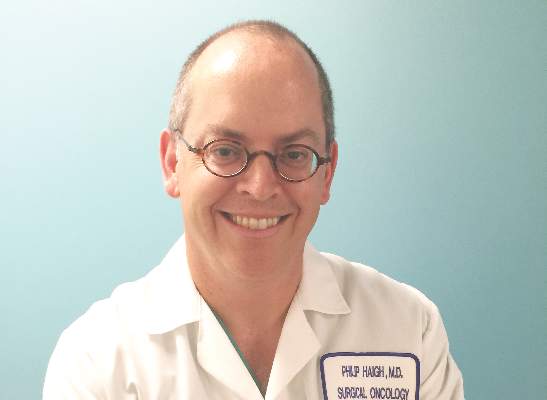
In an interview, study coauthor Dr. Philip Haigh explained that parathyroidectomy when the SS is negative is a more difficult operation for the surgeon, and that might make some physicians hesitate before going forward with surgery. “It has been previously shown by other studies that it is a more difficult operation when the sestamibi scan is negative because you have to look at four glands instead of removing just one, but if the surgeon is experienced, it should achieve a high success rate,” Dr. Haigh said. He said that parathyroidectomy in sestamibi-negative hyperparathyroidism had a cure rate as high as 98% in the study presented.
He offered two thoughts on how clinicians should use the study results. “To the endocrinologist, if you’re going to order a sestamibi scan, don’t change your referral practice depending on the result,” Dr. Haigh said. “To the surgeon, if you’re not comfortable operating on a patient with a negative sestamibi scan, then find someone who is.”
The study had a few limitations, Dr. Wu said. Along with its retrospective nature, the study also did not account for potential disparity in radiological vs. surgeon interpretation of the scans.
During the discussion, Dr. Samuel Snyder, of Baylor Scott & White Health, Temple, Tex., said he concurred with the results Dr. Wu reported. “It really worries me about what is happening to patients who have negative scans,” he said. “What I’ve seen in patients referred for surgery is a lot of variation in how the sestamibi scan is done.” He asked if the study accounted for the different types of sestamibi scans and how they were performed, but Dr. Wu said it did not.
Dr. Christopher McHenry of MetroHealth Medical Center, Cleveland, also concurred. “I think this is a phenomenon that occurs more often than we think or we’re aware of,” he said. “I continue to be amazed with how clinicians equate a negative sestamibi scan with not having primary hyperparathyroidism. I think it needs to reemphasized that the sestamibi scan is not diagnostic; it’s for localization.”
He asked Dr. Wu, “How do we change behavior to deal with this problem?”
Dr. Wu said her institution is developing a safety-net program that would aim to increase the identification and chart coding of patients with primary hyperparathyroidism, automate essential labs to be ordered in patients with high calcium, and automate referral to endocrinologists. The study and its findings will be disseminated to endocrinologists in the region.
The study authors had no disclosures.
BALTIMORE – In the treatment of primary hyperparathyroidism, clinical guidelines recommend using sestamibi scan for localizing adenoma, but increasingly endocrinologists are using sestamibi results to determine whether or not to refer a patient for parathyroidectomy surgery, while surgeons are using the scans as a factor in deciding whether to perform the operation.
That was the conclusion of a paper Dr. Susana Wu presented at the American Association of Endocrine Surgeons annual meeting. Dr. Wu reported on behalf of her colleagues at Kaiser Permanente Los Angeles Medical Center and at Scripps Clinic in San Diego.
“This study suggests that negative sestamibi scan (SS) results influence management of patients with primary hyperparathyroidism,” Dr. Wu said. “Endocrinologists were less likely to refer to surgeons and surgeons were less likely to offer parathyroidectomy to a patient with a negative sestamibi scan.”
The study involved a retrospective chart review of all 539 patients with primary hyperparathyroidism in the Kaiser Permanente Southern California database from December 2011 to December 2013, 452 of whom were seen by 63 endocrinologists at 14 centers. Among these patients, 260 had SS – 120 negative and 140 positive. The study identified statistically significant variations in how both endocrinologists and surgeons managed patients depending on SS results. The researchers used Kaiser Permanente’s electronic referral system to track referrals.
“The most significant negative predictor for endocrinologists referring to surgeons was a negative sestamibi scan, with an odds ratio of 0.36,” Dr. Wu said.
Endocrinologists referred 86% of patients with positive SS to surgeons, but only 68% of those with negative SS. Surgeons exhibited a similar practice pattern. “Surgeons were less likely to recommend parathyroidectomy for patients with a negative sestamibi scan, with an odds ratio of 0.20,” Dr. Wu said. Surgeons operated on 87% of patients with a negative SS scan but 96% with a positive SS.
In an interview, study coauthor Dr. Philip Haigh explained that parathyroidectomy when the SS is negative is a more difficult operation for the surgeon, and that might make some physicians hesitate before going forward with surgery. “It has been previously shown by other studies that it is a more difficult operation when the sestamibi scan is negative because you have to look at four glands instead of removing just one, but if the surgeon is experienced, it should achieve a high success rate,” Dr. Haigh said. He said that parathyroidectomy in sestamibi-negative hyperparathyroidism had a cure rate as high as 98% in the study presented.
He offered two thoughts on how clinicians should use the study results. “To the endocrinologist, if you’re going to order a sestamibi scan, don’t change your referral practice depending on the result,” Dr. Haigh said. “To the surgeon, if you’re not comfortable operating on a patient with a negative sestamibi scan, then find someone who is.”
The study had a few limitations, Dr. Wu said. Along with its retrospective nature, the study also did not account for potential disparity in radiological vs. surgeon interpretation of the scans.
During the discussion, Dr. Samuel Snyder, of Baylor Scott & White Health, Temple, Tex., said he concurred with the results Dr. Wu reported. “It really worries me about what is happening to patients who have negative scans,” he said. “What I’ve seen in patients referred for surgery is a lot of variation in how the sestamibi scan is done.” He asked if the study accounted for the different types of sestamibi scans and how they were performed, but Dr. Wu said it did not.
Dr. Christopher McHenry of MetroHealth Medical Center, Cleveland, also concurred. “I think this is a phenomenon that occurs more often than we think or we’re aware of,” he said. “I continue to be amazed with how clinicians equate a negative sestamibi scan with not having primary hyperparathyroidism. I think it needs to reemphasized that the sestamibi scan is not diagnostic; it’s for localization.”
He asked Dr. Wu, “How do we change behavior to deal with this problem?”
Dr. Wu said her institution is developing a safety-net program that would aim to increase the identification and chart coding of patients with primary hyperparathyroidism, automate essential labs to be ordered in patients with high calcium, and automate referral to endocrinologists. The study and its findings will be disseminated to endocrinologists in the region.
The study authors had no disclosures.
AT AAES 2016
Key clinical point: Endocrinologists and surgeons are less likely to order surgery when patients with primary hyperparathyroidism have negative sestamibi scan (SS) results.
Major finding: Endocrinologists referred 86% of patients with positive SS to surgeons, but only 68% of those with negative SS.
Data source: A retrospective chart review of all 539 patients with primary hyperparathyroidism in the Kaiser Permanente Southern California database over a 2-year period.
Disclosures: Dr. Wu and her coauthors reported having no financial disclosures.

Tool Offers Hand Hygiene Help
The healthcare industry is not yet at zero when it comes to healthcare-associated infections—and that’s a problem. Hand hygiene compliance remains a major cause.

The Joint Commission addresses that problem with the Hand Hygiene Targeted Solutions Tool (TST), an online application that guides the user through collecting and analyzing data, with suggested solutions based on the root causes revealed. “It’s based on robust process improvement, what we refer to as RPI, that brings in Lean, Six Sigma, and change management,” says Erin DuPree, MD, chief medical officer and vice president, The Joint Commission Center for Transforming Healthcare.
The tool was tested in a pilot program summarized in an article in the January 2016 issue of The Joint Commission Journal on Quality and Safety, “Hand Hygiene Tool Linked to Decrease in Health Care-Associated Infections at Memorial Hermann Health System,” by M. Michael Shabot, MD, of Memorial Hermann Health System, Mark R. Chassin, MD, MPP, MPH, of The Joint Commission, and their co-authors. In more than 31,600 observations, the organization’s average hand hygiene compliance improved from 58.1% to 95.6%. Rates of central line–associated bloodstream infections and ventilator-associated pneumonia in adult ICUs decreased by 49% and 45%, respectively.
Dr. DuPree encourages hospitalists to champion hand hygiene at their own organizations. “The more physicians lead and participate, the higher performing the organization is.”
The healthcare industry is not yet at zero when it comes to healthcare-associated infections—and that’s a problem. Hand hygiene compliance remains a major cause.
The Joint Commission addresses that problem with the Hand Hygiene Targeted Solutions Tool (TST), an online application that guides the user through collecting and analyzing data, with suggested solutions based on the root causes revealed. “It’s based on robust process improvement, what we refer to as RPI, that brings in Lean, Six Sigma, and change management,” says Erin DuPree, MD, chief medical officer and vice president, The Joint Commission Center for Transforming Healthcare.
The tool was tested in a pilot program summarized in an article in the January 2016 issue of The Joint Commission Journal on Quality and Safety, “Hand Hygiene Tool Linked to Decrease in Health Care-Associated Infections at Memorial Hermann Health System,” by M. Michael Shabot, MD, of Memorial Hermann Health System, Mark R. Chassin, MD, MPP, MPH, of The Joint Commission, and their co-authors. In more than 31,600 observations, the organization’s average hand hygiene compliance improved from 58.1% to 95.6%. Rates of central line–associated bloodstream infections and ventilator-associated pneumonia in adult ICUs decreased by 49% and 45%, respectively.
Dr. DuPree encourages hospitalists to champion hand hygiene at their own organizations. “The more physicians lead and participate, the higher performing the organization is.”
When Introducing Innovations, Context Matters

“We are missing an important opportunity to learn from what is going on in health systems every day that could tell us how to make healthcare better and cheaper,” says Dr. Fisher, lead author of a January 2016 JAMA “Viewpoint” called “Implementation Science: A Potential Catalyst for Delivery System Reform.” “That’s the argument for the the field of implementation science.”
Implementation science studies ways to promote the integration of research findings and evidence into the healthcare system. Dr. Fisher says that integration is influenced by multiple factors: the characteristic of the innovation itself, the organizational setting, and the policy or community environment within which that organization is working. Context matters.
“We tend to think about adopting innovations like a new blood pressure medication or a new device,” Dr. Fisher says. “Those decisions rest almost entirely on the shoulders of physicians, so adoption requires thinking about the attributes of biomedical innovations and how physicians think.”
With care delivery innovations—for example, how to provide optimal care for people with heart failure across home, hospital, and nursing home—those are often developed with clinical input but by people who are fundamentally managers.
“It’s a more complex set of actors,” he says, “so you have to think about those decision makers if you’re going to get the best evidence-based practice into their setting.”
A third category of innovation focuses on individual behavior change, where the decision makers are the clinician and the patient and family. “You’ve got to persuade the patient the innovation is worth doing, so different factors may influence the successful adoptions of those interventions,” Dr. Fisher says.
Reference
1. Fisher ES, Shortell SM, Savitz LA. Implementation science: a potential catalyst for delivery system reform. JAMA. 2016;315(4):339-340. doi:10.1001/jama.2015.17949.

HHS provides funding for trial of Zika test

Photo by Juan D. Alfonso
The US Department of Health and Human Services (HHS) is providing financial support for a clinical trial of the cobas® Zika test, which is designed to screen blood donations for Zika virus.
The US Food and Drug Administration (FDA) recently authorized the use of this test, under an investigational new drug application protocol, for screening donated blood in areas with active, mosquito-borne transmission of Zika virus.
This means the cobas® Zika test can be used by US blood screening laboratories, but the laboratories will need to be enrolled in and contracted into the clinical trial as specified and agreed with the FDA’s Center for Biologics Evaluation and Research.
Now, the HHS has announced that the Biomedical Advanced Research and Development Authority (BARDA) is supporting the trial, which will evaluate the sensitivity and specificity of the test in its actual use.
The trial is necessary for Roche, the company developing the cobas® Zika test, to apply for FDA approval for commercial marketing.
“BARDA staff has worked closely with our partners at FDA and the Office of the Assistant Secretary of Health to ensure the continuity and safety of the US blood supply,” said Richard Hatchett, BARDA’s acting director.
“Today’s award to Roche is an important step towards securing the safety of the blood supply in Puerto Rico and in the rest of the United States.”
Under the 1-year, $354,500 contract, Roche will study blood samples to confirm whether the test accurately and reliably detects and identifies Zika virus, even when present in very low concentrations in donor blood. ![]()
Antithrombotics appear safe in BCVI with concomitant injuries
SAN ANTONIO – Don’t withhold antiplatelet or heparin therapy in patients with blunt cerebrovascular injury, even if they have concomitant traumatic brain or solid organ injuries, advised researchers from the University of Tennessee Health Science Center, Memphis.
With close monitoring, “initiation of early antithrombotic therapy for patients with BCVI [blunt cerebrovascular injury] and concomitant TBI [traumatic brain injury] or SOI [solid organ injury] does not increase the risk of worsening TBI or SOI above baseline.” It is safe, effective, and “should not be withheld,” the researchers concluded after a review of 119 BCVI patients with concomitant injuries.

Seventy four (62%) had TBIs, 26 (22%) had SOIs, and 19 (16%) had both. At some institutions, antithrombotic therapy – the mainstay for BCVI to prevent secondary ischemic stroke – would have been delayed or withheld for fear of triggering hemorrhagic complications.
But at the Health Science Center in Memphis, “we have an extremely cooperative group of neurosurgeons who take BCVI as seriously as we do, and actually allow us, more often than not, to start antithrombotic therapy pretty much immediately after the injury is identified,” investigator and surgery resident Dr. Charles Shahan said at the annual scientific assembly of the Eastern Association for the Surgery of Trauma.
As a result, 85 patients (71%) received heparin infusions with goal-activated partial thromboplastin times of 45-60 seconds, and the rest antiplatelet therapy, typically 81-mg aspirin. The center generally uses heparin for TBI patients because of its short half-life, and aspirin for others.
Antithrombosed BCVI patients did as well as did historical controls. TBIs deteriorated – meaning worsening on clinical or CT exam, or delayed operative intervention – in 7%, vs. 10% of TBI patients without BCVI (P = .34). Three percent of SOI patients had delayed laparotomies vs. 5% of SOI patients without BCVI (P = .54). None of the BCVI patients stopped antithrombotics because of complications.
The results held regardless of the type of TBI, SOI, or antithrombotic used.
Overall, 11 patients (9%) had BCVI-related strokes. Without antithrombotic therapy, stroke rates in BCVI can approach 40%.
“Our extremely early use of antithrombotic therapy does not appear to increase our rate of worsening of our hemorrhagic injures and also gets our stroke rate within acceptable limits,” Dr. Shahan said.
The mean age in the study was 38, and just over half the subjects were men.
Dr. Shahan had no disclosures
SAN ANTONIO – Don’t withhold antiplatelet or heparin therapy in patients with blunt cerebrovascular injury, even if they have concomitant traumatic brain or solid organ injuries, advised researchers from the University of Tennessee Health Science Center, Memphis.
With close monitoring, “initiation of early antithrombotic therapy for patients with BCVI [blunt cerebrovascular injury] and concomitant TBI [traumatic brain injury] or SOI [solid organ injury] does not increase the risk of worsening TBI or SOI above baseline.” It is safe, effective, and “should not be withheld,” the researchers concluded after a review of 119 BCVI patients with concomitant injuries.
Seventy four (62%) had TBIs, 26 (22%) had SOIs, and 19 (16%) had both. At some institutions, antithrombotic therapy – the mainstay for BCVI to prevent secondary ischemic stroke – would have been delayed or withheld for fear of triggering hemorrhagic complications.
But at the Health Science Center in Memphis, “we have an extremely cooperative group of neurosurgeons who take BCVI as seriously as we do, and actually allow us, more often than not, to start antithrombotic therapy pretty much immediately after the injury is identified,” investigator and surgery resident Dr. Charles Shahan said at the annual scientific assembly of the Eastern Association for the Surgery of Trauma.
As a result, 85 patients (71%) received heparin infusions with goal-activated partial thromboplastin times of 45-60 seconds, and the rest antiplatelet therapy, typically 81-mg aspirin. The center generally uses heparin for TBI patients because of its short half-life, and aspirin for others.
Antithrombosed BCVI patients did as well as did historical controls. TBIs deteriorated – meaning worsening on clinical or CT exam, or delayed operative intervention – in 7%, vs. 10% of TBI patients without BCVI (P = .34). Three percent of SOI patients had delayed laparotomies vs. 5% of SOI patients without BCVI (P = .54). None of the BCVI patients stopped antithrombotics because of complications.
The results held regardless of the type of TBI, SOI, or antithrombotic used.
Overall, 11 patients (9%) had BCVI-related strokes. Without antithrombotic therapy, stroke rates in BCVI can approach 40%.
“Our extremely early use of antithrombotic therapy does not appear to increase our rate of worsening of our hemorrhagic injures and also gets our stroke rate within acceptable limits,” Dr. Shahan said.
The mean age in the study was 38, and just over half the subjects were men.
Dr. Shahan had no disclosures
AT THE EAST SCIENTIFIC ASSEMBLY
Key clinical point: Antithrombotics for BCVI do not make concomitant brain and solid organ injuries worse.
Major finding: TBIs deteriorated in 7% of BCVI patients on heparin infusion, versus 10% of TBI patients without BCVI (P = .34).
Data source: Review of 119 BCVI patients.
Disclosures: The lead investigator had no disclosures.
Save the Date for ACS Clinical Congress 2016, October 16−20
Save the date for the American College of Surgeons Clinical Congress 2016, October 16−20 in Washington, DC, at the Walter E. Washington Convention Center. The Marriot Marquis Washington, DC, located next to the convention center, will serve as the headquarters hotel.
The theme of this year’s meeting, Challenges for the Second Century, recognizes the College’s second 100 years of ensuring quality surgical patient care. Clinical Congress 2016 will present hundreds of educational sessions, including Panel Sessions, Postgraduate Didactic and Skills-Oriented Courses, Meet-the-Expert Luncheons, Names Lectures, Scientific Paper Sessions, and Poster Presentations. View an ACS press release at https://www.facs.org/media/press-releases/2016/clincon0316#sthash.SnHNsOJB.dpuf for more information on Clinical Congress 2016.
ART cycles, live births up in 2014
More than 65,000 babies were born following about 190,000 cycles of IVF and other assisted reproductive technologies in 2014, according to data from the Society for Assisted Reproductive Technology.
The report – which includes data from 375 member clinics of the Society for Assisted Reproductive Technology (SART) – shows an increase both in assisted reproductive technology (ART) procedures performed and in the number of subsequent live births, compared with 2013.

Another trend in the 2014 data is a decline in the number of multiple births, due largely to increased use of single-embryo transfer.
Singleton births reached 78% from the 2014 cycles, up from 75.5% the previous year. Single-embryo transfer cycles accounted for 27.2% of all cycles in 2014, compared with 20.6% in 2013.
“The latest data reflect the success of our efforts to improve ART practice from ovarian stimulation through transfer,” Dr. Bradley Van Voorhis, SART president, said in a statement. “We are proud of the progress we have made improving live birth rates – the true measure of a cycle’s success – and in improving maternal and child health outcomes for our patients through lower multiple birth rates.”
Read the national summary report for SART clinics here.
On Twitter @maryellenny
More than 65,000 babies were born following about 190,000 cycles of IVF and other assisted reproductive technologies in 2014, according to data from the Society for Assisted Reproductive Technology.
The report – which includes data from 375 member clinics of the Society for Assisted Reproductive Technology (SART) – shows an increase both in assisted reproductive technology (ART) procedures performed and in the number of subsequent live births, compared with 2013.
Another trend in the 2014 data is a decline in the number of multiple births, due largely to increased use of single-embryo transfer.
Singleton births reached 78% from the 2014 cycles, up from 75.5% the previous year. Single-embryo transfer cycles accounted for 27.2% of all cycles in 2014, compared with 20.6% in 2013.
“The latest data reflect the success of our efforts to improve ART practice from ovarian stimulation through transfer,” Dr. Bradley Van Voorhis, SART president, said in a statement. “We are proud of the progress we have made improving live birth rates – the true measure of a cycle’s success – and in improving maternal and child health outcomes for our patients through lower multiple birth rates.”
Read the national summary report for SART clinics here.
On Twitter @maryellenny
