User login
Study finds COVID-19 boosters don’t increase miscarriage risk
COVID-19 boosters are not linked to an increased chance of miscarriage, according to a new study in JAMA Network Open.
Researchers were seeking to learn whether a booster in early pregnancy, before 20 weeks, was associated with greater likelihood of spontaneous abortion.
They examined more than 100,000 pregnancies at 6-19 weeks from eight health systems in the Vaccine Safety Datalink (VSD). They found that receiving a COVID-19 booster shot in a 28-day or 42-day exposure window did not increase the chances of miscarriage.
The VSD is a collaboration between the Centers for Disease Control and Prevention’s Immunization Safety Office and large health care systems. The “observational, case-control, surveillance study” was conducted from Nov. 1, 2021, to June 12, 2022.
“COVID infection during pregnancy increases risk of poor outcomes, yet many people who are pregnant or thinking about getting pregnant are hesitant to get a booster dose because of questions about safety,” said Elyse Kharbanda, MD, senior investigator at HealthPartners Institute and lead author of the study in a press release.
The University of Minnesota reported that “previous studies have shown COIVD-19 primary vaccination is safe in pregnancy and not associated with an increased risk for miscarriage. Several studies have also shown COVID-19 can be more severe in pregnancy and lead to worse outcomes for the mother.”
The study was funded by the CDC. Five study authors reported conflicts of interest with Pfizer, Merck, GlaxoSmithKline, AbbVie, and Sanofi Pasteur.
A version of this article first appeared on Medscape.com.
FROM JAMA NETWORK OPEN
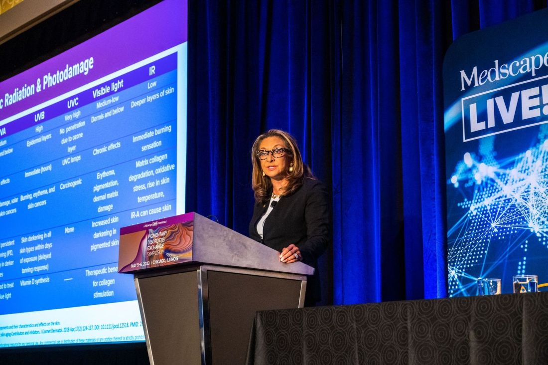
SPF is only the start when recommending sunscreens
CHICAGO – at the inaugural Pigmentary Disorders Exchange Symposium.
Among the first factors physicians should consider before recommending sunscreen are a patient’s Fitzpatrick skin type, risks for burning or tanning, underlying skin disorders, and medications the patient is taking, Dr. Taylor, professor of dermatology at the University of Pennsylvania, Philadelphia, said at the meeting, provided by MedscapeLIVE! If patients are on hypertensives, for example, medications can make them more photosensitive.
Consider skin type
Dr. Taylor said she was dismayed by the results of a recent study, which found that 43% of dermatologists who responded to a survey reported that they never, rarely, or only sometimes took a patient’s skin type into account when making sunscreen recommendations. The article is referenced in a 2022 expert panel consensus paper she coauthored on photoprotection “for skin of all color.” But she pointed out that considering skin type alone is inadequate.
Questions for patients in joint decision-making should include lifestyle and work choices such as whether they work inside or outside, and how much sun exposure they get in a typical day. Heat and humidity levels should also be considered as should a patient’s susceptibility to dyspigmentation. “That could be overall darkening of the skin, mottled hyperpigmentation, actinic dyspigmentation, and, of course, propensity for skin cancer,” she said.
Use differs by race
Dr. Taylor, who is also vice chair for diversity, equity and inclusion in the department of dermatology at the University of Pennsylvania, pointed out that sunscreen use differs considerably by race.
In study of 8,952 adults in the United States who reported that they were sun sensitive found that a subset of adults with skin of color were significantly less likely to use sunscreen when compared with non-Hispanic White adults: Non-Hispanic Black (adjusted odds ratio, 0.43); non-Hispanic Asian (aOR. 0.54); and Hispanic (aOR, 0.70) adults.
In the study, non-Hispanic Black and Hispanic adults were significantly less likely to use sunscreens with an SPF greater than 15. In addition, non-Hispanic Black, non-Hispanic Asian, and Hispanic adults were significantly more likely than non-Hispanic Whites to wear long sleeves when outside. Such differences are important to keep in mind when advising patients about sunscreens, she said.
Protection for lighter-colored skin
Dr. Taylor said that, for patients with lighter skin tones, “we really want to protect against ultraviolet B as well as ultraviolet A, particularly ultraviolet A2. Ultraviolet radiation is going to cause DNA damage.” Patients with Fitzpatrick skin types I, II, or III are most susceptible to the effects of UVB with sunburn inflammation, which will cause erythema and tanning, and immunosuppression.
“For those who are I, II, and III, we do want to recommend a broad-spectrum, photostable sunscreen with a critical wavelength of 370 nanometers, which is going to protect from both UVB and UVA2,” she said.
Sunscreen recommendations are meant to be paired with advice to avoid midday sun from 10 a.m. to 2 p.m., wearing protective clothing and accessories, and seeking shade, she noted.
Dr. Taylor said, for those patients with lighter skin who are more susceptible to photodamage and premature aging, physicians should recommend sunscreens that contain DNA repair enzymes such as photolyases and sunscreens that contain antioxidants that can prevent or reverse DNA damage. “The exogenous form of these lyases have been manufactured and added to sunscreens,” Dr. Taylor said. “They’re readily available in the United States. That is something to consider for patients with significant photodamage.”
Retinoids can also help alleviate or reverse photodamage, she added.
Protection for darker-colored skin
“Many people of color do not believe they need sunscreen,” Dr. Taylor said. But studies show that, although there may be more intrinsic protection, sunscreen is still needed.
Over 30 years ago, Halder and colleagues reported that melanin in skin of color can filter two to five times more UV radiation, and in a paper on the photoprotective role of melanin, Kaidbey and colleagues found that skin types V and VI had an intrinsic SPF of 13 when compared with those who have lighter complexions, which had an SPF of 3.
Sunburns seem to occur less frequently in people with skin of color, but that may be because erythema is less apparent in people with darker skin tones or because of differences in personal definitions of sunburn, Dr. Taylor said.
“Skin of color can and does sustain sunburns and sunscreen will help prevent that,” she said, adding that a recommendation of an SPF 30 is likely sufficient for these patients. Dr. Taylor noted that sunscreens for patients with darker skin often cost substantially more than those for lighter skin, and that should be considered in recommendations.
Tinted sunscreens
Dr. Taylor said that, while broad-spectrum photostable sunscreens protect against UVB and UVA 2, they don’t protect from visible light and UVA1. Two methods to add that protection are using inorganic tinted sunscreens that contain iron oxide or pigmentary titanium dioxide. Dr. Taylor was a coauthor of a practical guide to tinted sunscreens published in 2022.
“For iron oxide, we want a concentration of 3% or greater,” she said, adding that the percentage often is not known because if it is contained in a sunscreen, it is listed as an inactive ingredient.
Another method to address visible light and UVA1 is the use of antioxidant-containing sunscreens with vitamin E, vitamin C, or licochalcone A, Dr. Taylor said.
During the question-and-answer period following her presentation, Amit Pandya, MD, adjunct professor of dermatology at University of Texas Southwestern Medical Center, Dallas, asked why “every makeup, every sunscreen, just says iron oxide,” since it is known that visible light will cause pigmentation, especially in those with darker skin tones.
He urged pushing for a law that would require listing the percentage of iron oxide on products to assure it is sufficient, according to what the literature recommends.
Conference Chair Pearl Grimes, MD, director of the Vitiligo and Pigmentation Institute of Southern California, Los Angeles, said that she recommends tinted sunscreens almost exclusively for her patients, but those with darker skin colors struggle to match color.
Dr. Taylor referred to an analysis published in 2022 of 58 over-the counter sunscreens, which found that only 38% of tinted sunscreens was available in more than one shade, “which is a problem for many of our patients.” She said that providing samples with different hues and tactile sensations may help patients find the right product.
Dr. Taylor disclosed being on the advisory boards for AbbVie, Avita Medical, Beiersdorf, Biorez, Eli Lily, EPI Health, Evolus, Galderma, Hugel America, Johnson and Johnson, L’Oreal USA, MedScape, Pfizer, Scientis US, UCB, Vichy Laboratories. She is a consultant for Arcutis Biothermapeutics, Beiersdorf, Bristol-Myers Squibb, Cara Therapeutics, Dior, and Sanofi. She has done contracted research for Allergan Aesthetics, Concert Pharmaceuticals, Croma-Pharma, Eli Lilly, and Pfizer, and has an ownership interest in Armis Scientific, GloGetter, and Piction Health.
Medscape and this news organization are owned by the same parent company.
AT THE MEDSCAPELIVE! PIGMENTARY DISORDERS SYMPOSIUM
FDA warns people to avoid compounded semaglutide medicines
Compounded medicines are not FDA approved but are allowed to be made during an official drug shortage. Ozempic and Wegovy are currently on the FDA’s shortage list, but the federal agency warned that it has received reports of people experiencing “adverse events” after using compounded versions of the drugs. (The FDA did not provide details of those events or where the drugs involved were compounded.)
Agency officials are concerned that the compounded versions may contain ingredients that sound like the brand name drugs’ active ingredient, semaglutide, but are different because the ingredients are in salt form.
“Patients should be aware that some products sold as ‘semaglutide’ may not contain the same active ingredient as FDA-approved semaglutide products and may be the salt formulations,” the FDA warning stated. “Products containing these salts, such as semaglutide sodium and semaglutide acetate, have not been shown to be safe and effective.”
The agency said salt forms don’t meet the criteria for compounding during a shortage and sent a letter to the National Association of Boards of Pharmacy expressing “concerns with use of the salt forms in compounded products.”
Patients and health care providers should be aware that “compounded drugs are not FDA approved, and the agency does not verify the safety or effectiveness of compounded drugs,” the FDA explained in its statement.
The Alliance for Pharmacy Compounding’s board of directors said in a statement that some compounders’ arguments for the suitability of semaglutide sodium are “worthy of discussion,” but the board did not endorse those arguments.
For people who use an online pharmacy, the FDA recommends checking the FDA’s website BeSafeRx to check its credentials.
A version of this article first appeared on WebMD.com.
Sucralose damages DNA, linked to leaky gut: Study
Sucralose is sold under the brand name Splenda and is also used as an ingredient in packaged foods and beverages.
The findings were published in the Journal of Toxicology and Environmental Health, Part B. The researchers conducted a series of laboratory experiments exposing human blood cells and gut tissue to sucralose-6-acetate. The findings build on previous research that linked sucralose to gut health problems.
The researchers found that sucralose causes DNA to break apart, putting people at risk for disease. They also linked sucralose to leaky gut syndrome, which means the lining of the intestines are worn down and become permeable. Symptoms are a burning sensation, painful digestion, diarrhea, gas, and bloating.
When a substance damages DNA, it is called genotoxic. Researchers have found that eating sucralose results in the body producing a substance called sucralose-6-acetate, which the new study now shows is genotoxic. The researchers also found sucralose-6-acetate in trace amounts in off-the-shelf products that are so high, they would exceed the safety levels currently allowed in Europe.
“It’s time to revisit the safety and regulatory status of sucralose because the evidence is mounting that it carries significant risks. If nothing else, I encourage people to avoid products containing sucralose,” researcher Susan Schiffman, PhD, adjunct professor of biomedical engineering at North Carolina State University, Raleigh, said in a statement. “It’s something you should not be eating.”
The FDA says sucralose is safe, describing it as 600 times sweeter than table sugar and used in “baked goods, beverages, chewing gum, gelatins, and frozen dairy desserts.”
“To determine the safety of sucralose, the FDA reviewed more than 110 studies designed to identify possible toxic effects, including studies on the reproductive and nervous systems, carcinogenicity, and metabolism,” the agency explained on its website. “The FDA also reviewed human clinical trials to address metabolism and effects on patients with diabetes.”
The study authors reported that they had no conflicts of interest.A version of this article first appeared on WebMD.com.
FROM THE JOURNAL OF TOXICOLOGY AND ENVIRONMENTAL HEALTH, PART B
Low-dose atropine improves myopia in children
Myopia affects roughly one-third of the population worldwide - a figure that is projected to reach 50% by 2050. Low-dose atropine, which helps curb the condition, currently is available in the United States only through compounding pharmacies. The products contain preservatives – raising questions about potential toxicities to the eye – and may not be of pharmaceutical grade. In a new study published in JAMA Ophthalmology,
Methodology
- The CHAMP study was a double-masked, placebo-controlled, randomized, phase 3 trial conducted between Nov. 20, 2017, and Aug. 22, 2022, that involved children at 26 sites in North America and 5 centers in Europe.
- Children received either 0.01% or 0.02% atropine drops once per day.
- Patients were aged 3-16 years. They demonstrated a spherical equivalent refractive error (SER) of −0.50 diopter (D) to −6.00 D astigmatism no worse than −1.50 D.
- Of these patients, 573 were included in a safety analysis, and 489 were included in a modified intention-to-treat analysis.
Takeaways
- After 36 months, the 0.01% dose of atropine was associated with a significantly lower responder proportion (odds ratio, 4.54) and slower progression of SER and axial elongation.
- The effect of the 0.02% dose on responder proportion and SER progression was not statistically significant, but the treatment was associated with slower axial elongation.
- The researchers observed no serious ocular adverse events and few serious nonocular events, none of which was determined to be associated with the treatment.
In practice: According to the researchers, “from a risk/benefit perspective, the efficacy and safety observed suggests that low-dose atropine may provide a treatment option for children aged 3-17 years with myopia progression, which may lead to less frequent or delayed change in glasses, progression to less severe correction, and potentially reduce long-term sequelae, which could lead to vision loss later in life, such as myopic maculopathy.”
Study details: The CHAMP study was led by Karla Zadnik, OD, PhD, of Ohio State University, Columbus, and was funded by Vylulma.
Limitations: The researchers said the trial was potentially limited by the fact that patients switched from the study drug to confounding treatments. In addition, patients at the low and high age ranges were not well represented.
Disclosures: Dr. Zadnik received consultant fees from Vyluma during the study.
A version of this article first appeared on Medscape.com.
FROM JAMA OPHTHALMOLOGY
Potential new treatment for REM sleep behavior disorder
Dual orexin receptor antagonists (DORAs), a class of drugs approved to treat insomnia, may also be effective for rapid eye movement sleep behavior disorder (RBD), a study suggests.
About 3 million people in the United States have RBD, which is often a precursor to Parkinson’s disease. People with the disorder act out their dreams by talking, flailing their arms and legs, punching, kicking, and exhibiting other behaviors while asleep.
Researchers used an animal model for the study, which they say is the first to identify a new form of treatment for RBD.
“REM behavior disorder is difficult to treat, and the treatments are mostly limited to clonazepam and melatonin,” which may have side effects, senior investigator Andrew Varga, MD, PhD, associate professor of pulmonary, critical care, and sleep medicine at the Icahn School of Medicine at Mount Sinai, New York, told this news organization. “We’re using something completely different, which raises the possibility this might be something useful for REM behavior disorders.”
The findings, with Mount Sinai assistant professor Korey Kam, PhD, as lead author, were published online in the Journal of Neuroscience.
A new model for RBD?
RBD can signal risk for synucleinopathies, a group of neurological conditions such as Parkinson’s disease that involve the formation of clumps of alpha-synuclein protein in the brain.
Prior research on RBD was done in synucleinopathy mouse models. For this study, however, researchers used a tauopathy mouse model to investigate how the abnormal accumulation of tau protein might affect RBD.
Researchers collected data on biophysical properties when the mice were awake and in REM and non-REM sleep. They examined length of sleep, transitions from waking to sleep, and how some factors are related to age.
Nearly a third of the older animals showed behaviors similar to REM sleep behavior disorder in humans, including chewing and limb extension.
But after researchers administered a DORA medication twice during a 24-hour period, they noted that the medication not only helped the animals fall asleep faster and for longer, it also reduced levels of dream enactment that are a hallmark of RBD.
The ‘bigger highlight’
Finding RBD behaviors in a tauopathy animal model was surprising, Dr. Varga said, because RBD has been previously linked to synucleinopathies. There was no known correlation between RBD and abnormal accumulation of tau.
Another unexpected finding was the detection of RBD in some of the younger animals, who had not yet shown evidence of tau accumulation.
“It appears to be a biomarker or a signature of something that’s going on that predicts the impending tauopathy at a time where there is very little, or no, tau pathology going on in the brain,” Dr. Varga said.
If RBD is an early predictor of future tau accumulation, the model could guide future prevention and treatment. However, the more important finding is the potential new treatment for the condition.
“The bigger highlight here is less about what’s causing the RBD [than about] what you can do to make it better,” he said.
The next step in the work is to study whether the effect of DORAs on RBD seen in this tauopathy mouse model is evidenced in other animals and whether it is effective in humans with RBD, Dr. Varga said.
The study was funded by the Alzheimer’s Association and Merck Investigator Studies Program. Dr. Kam, Dr. Varga, and coauthors report no relevant financial relationships.
A version of this article first appeared on Medscape.com.
FROM THE JOURNAL OF NEUROSCIENCE
APA launches online eating disorder assessment tool
The American Psychiatric Association has released an online screening and assessment tool for eating disorders.
“People with eating disorders have a high rate of mortality and [the disorder is] growing in prevalence among young adults and adolescents,” APA CEO and medical director Saul Levin, MD, MPA, said in a news release.
“It is vital that we equip our clinicians, especially primary care clinicians, with the latest evidence from the APA to empower their decision-making and improve care for their patients,” Dr. Levin added.
The clinical decision support tool was developed by the APA’s guideline writing group in collaboration with AvoMD, a software company that translates clinical evidence into the workflow.
The tool guides clinicians through the screening, assessing, diagnosing, and treatment planning of patients with anorexia nervosa, bulimia nervosa, binge-eating disorder, and other eating disorders.
It’s available free in the electronic health record, on the APA website, and on the AvoMD mobile app.
The tool is based on the APA’s updated practice guidelines for the management of eating disorders, which were released in March.
The tool incorporates guidance on screening tools, levels of care, nutrition, exercise, psychotherapy, and more. Additionally, the tool provides a summary of responses that can be leveraged for easy documentation.
It is intended for use by pediatricians, family physicians, and other primary care and mental health clinicians, including psychiatrists and therapists.
Data published earlier this year show that more than 1 in 5 children worldwide are at risk of developing an eating disorder. Girls are significantly more likely than boys to have disordered eating.
Early diagnosis and treatment are associated with a higher rate of recovery, and extended illness is associated with potentially devastating consequences, the APA notes.
“As an internal medicine physician myself, I see a wide variety of patients and clinical issues,” Joongheum Park, MD, head of product and engineering at AvoMD, said in the release.
“Easy access to the expertise in me and my peers’ workflow is essential to ensuring high-quality care, and partnering with the leading authority on eating disorders to provide this tool will improve clinician efficiency and most importantly, patient outcomes,” Dr. Park added.
A version of this article first appeared on Medscape.com.
Lomitapide shows promise in pediatric homozygous FH
MANNHEIM, Germany – Lomitapide, which reduces lipoprotein production in the liver, could help manage pediatric homozygous familial hypercholesterolemia (HoFH), suggest results of a trial that showed large reductions in circulating lipids.
The research was presented May 23 at the 91st European Atherosclerosis Society Congress.
Lomitapide inhibits microsomal triglyceride transfer protein, which plays a key role in apolipoprotein B-containing lipoprotein assembly and secretion in the liver and intestines. Crucially, the drug acts independently of the LDL cholesterol receptor.
It was approved in December 2012 by the U.S. Food and Drug Administration for use in adults with HoFH, sold under the name Juxtapid, and by the European Medicines Agency, where the brand name is Lojuxta.
The current trial involved more than 40 children and teenagers with HoFH aged 5-17 years; they were treated with the drug for 24 weeks, resulting in reductions of low density lipoprotein cholesterol of almost 54%, with nearly 42% reaching target levels.
The drug was also associated with marked reductions in other key lipids of at least 50%. However, 67% of patients also experienced gastrointestinal adverse events, and around 25% saw their levels of liver enzymes increase.
Early diagnosis ‘imperative’
The findings show that the “early diagnosis and treatment of HoFH is imperative,” said study presenter Luis Masana, MD, PhD, director of the Vascular Medicine and Metabolism Unit at Sant Joan de Reus University Hospital, Tarragona, Spain.
“I think that, with these results, we are bringing a new hope for this group of patients,” he continued. “I also think we will increase the quality of life, not just of the patients but also all the families involved in [managing] this problem.”
Session co-chair Andreas Zirlik, MD, PhD, head of the department of cardiology and chairman of the University Heart Center Graz, LKH-University Hospital, and Medical University of Graz (Austria), was more circumspect in his appraisal of the results.
He told this news organization that it is “always very difficult to establish therapy in pediatrics,” and believes that the drug “will give us an additional option” in managing HoFH.
However, Dr. Zirlik warned that he is a “little bit concerned” about lomitapide’s adverse event profile, and “would need to see a little bit deeper into the safety data.”
Highlighting the elevations in liver enzymes of around 25%, he asked: “What does it mean?” And how will it “play out in the long run?”
Beyond lomitapide, Dr. Zirlik pointed out that there are other drugs that have shown potential in managing HoFH and could potentially be used in the pediatric population, such as angiopoietin-like 3 protein (ANGPTL3) inhibitors and small interfering RNA (siRNA) compounds that target upstream production. “So, let’s see how they pan out,” he said.
Life-limiting condition
HoFH is an “ultra-rare, life-limiting condition,” with an estimated prevalence of approximately 3 per 1 million people, and a life expectancy in untreated patients of just 18 years, Dr. Masana said during his presentation.
Case series of lomitapide use in pediatric HoFH patients have shown encouraging results that are consistent with those seen in adults, he noted, with many able to achieve their LDL cholesterol target and stop or reduce apheresis.
To investigate further, a phase 3, single arm, open-label study was conducted. Following screening, 46 children and teenagers with HoFH underwent a 6- to 12-week run-in period, during which they were put on a low-fat diet with nutritional supplements.
“As you can imagine,” Dr. Masana said, “we are reducing the capacity for fat absorption with lomitapide, so the supplements and low-fat diet are necessary.”
Of these, 43 participants then entered a 24-week treatment period in which they were started on one of three doses, before undergoing dose escalation to the maximally tolerated dose. This was followed by an 80-week open-label safety phase, in which they continued on the maximally tolerated dose, then a follow-up period.
For the current presentation, Dr. Masana focused on the efficacy phase, showing that the mean age of participants was 10.7 years and that 55.8% were female. The HoFH diagnosis was confirmed genetically in 88.4% of cases.
Results showed that lomitapide was associated with a significant reduction in LDL cholesterol levels, from 435.8 mg/dL at baseline to 176.5 mg/dL at Week 24, which corresponded to a 53.5% overall reduction (P < .0001).
This meant that 41.9% of patients achieved their EAS LDL cholesterol target of less than 135 mg/dL at some point during the 24-week treatment period.
Stratifying by age, the reduction between baseline and week 24 was 538.5 mg/dL to 207.2 mg/dL, or 56.5%, in the 20 children aged 5-10 years, and 346.5 mg/dL to 149.9 mg/L, or 50.9%, in the 23 patients aged 11-17 years.
Dr. Masana explained that the results were “a little bit better in the younger group because they were receiving less treatment at this stage of the disease” than the older group.
He showed that lomitapide was associated with significant reductions in other lipid markers, including a 53.9% reduction in non–HDL cholesterol (P < .0001), a 50.1% drop in total cholesterol (P < .0001), and a 50.2% fall in very-low-density lipoprotein cholesterol (P < .0001).
Results showed 93% of patients experienced treatment-related adverse events, with 11.6% having serious events and 4.7% having events that led to study discontinuation. There was one (2.3%) major adverse cardiac event but no deaths.
He said that, despite these figures, the adverse events were “mostly mild or moderate.”
The majority (67%) of patients nevertheless had gastrointestinal adverse events, which were, “in general, associated with a lack of adherence to the low-fat diet.”
Aspartate aminotransferase levels were elevated in 23% of patients, while 28% had elevations in alanine aminotransferase, which were described by Dr. Masana as “moderate.”
The study was sponsored by Amryt Pharma. Dr. Masana declares relationships with Amarin, Amryt, Daiichi-Sankyo, Novartis, Sanofi, Servier, Servier, and Viatrix.
A version of this article first appeared on Medscape.com.
United Healthcare ditches prior authorization in favor of new policy
Instead, the giant health insurer will adopt an “advance notification” program for nonscreening and nonemergent gastrointestinal procedures.
The company has not made any changes to their policy regarding screening colonoscopies for preventive care, and the advance notification policy does not impact screening colonoscopies.
UHC alerted physicians to changes to the program yesterday, including updated notices on UHCProvider.com with a new Frequently Asked Questions document.
The advance notification program “will not result in the denial of care for clinical reasons or for failure to notify and will help educate physicians who are not following clinical best practices. Provider groups who do not submit advance notification during this period will not be eligible for the United Healthcare Gold Card program,” a spokesperson for the company said.
The previously announced Gold Card program, which is scheduled to start in early 2024, would eliminate prior authorization requirements for providers that meet certain eligibility requirements.
The American Gastroenterological Association remains “extremely concerned” that UHC’s advance notification program is a “temporary patch” likely to have significant repercussions for patient access. The organization says the program only temporarily postpones prior authorization requirements set to impact the insurer’s 27.4 million commercial beneficiaries while increasing the administrative burden on clinicians.
The AGA called the program “nebulous” and “poorly defined.” It would ostensibly require physicians to input “copious” amounts of highly complex and granular patient data prior to performing colonoscopies and endoscopies, the AGA says.
AGA President Barbara H. Jung, MD, AGAF, said UHC’s “slap-dash approach to rolling out a policy that will ultimately control patient access to critical, often life-saving, medical procedures flies in the face of common sense and responsible medical practice.”
“It also indicates that UHC does not currently have data that show any significant overutilization of critical endoscopy and colonoscopy procedures that would ostensibly justify this program or prior authorization. UHC is not acting in good faith, and its actions will compromise patient access to potentially lifesaving procedures,” Dr. Jung added.
Recent data show 62% of high-risk patients in the United States who had polyps removed had evidence of delayed or no use of surveillance colonoscopies after 10 years.
“If other prior authorization requirements imposed on patients for specialty care are any indication, we expect to see negative patient outcomes with an enormous cost to patient well-being and physician resources,” AGA Vice President Lawrence Kim, MD, wrote in a news release.
“Given the high percentage of eventual approvals by insurers mandating prior authorization, we anticipate there will be little to no benefit from this prior authorization requirement. When utilized this way, it becomes a nonsensical and harmful policy,” Dr. Kim added.
AGA says it will continue to work closely with its members to assess the full impact of the new requirements and urges UHC to make endoscopy procedures more accessible to patients.
A recent American Medical Association survey on prior authorization found that one-third (33%) of doctors said the insurance barrier has led to a serious adverse event such as hospitalization, permanent disability, or death for a patient in their care. Nearly half (46%) of physicians reported that prior authorization has led to immediate care and/or emergency department visits.
In a 2023 survey of AGA membership, conducted before UHC announced its proposed prior authorization policy, 95% of respondents said prior authorization restrictions have impacted patient access to clinically appropriate treatments and patient clinical outcomes. And 84% said the burdens associated with prior authorization policies have increased “significantly” (60%) or “somewhat” (24%) over the last 5 years.
A version of this article first appeared on Medscape.com.
Encouraging telitacicept results reported in phase 3 for lupus, phase 2 for Sjögren’s
MILAN – Results of a phase 3 trial with the investigational drug telitacicept show that patients with systemic lupus erythematosus have a significantly greater rate of response to SLE response criteria, compared with placebo, while results from a phase 2 trial of the drug in patients with primary Sjögren’s syndrome (pSS) also show significant improvements versus placebo.
“With only a limited number of treatments available for patients with lupus, this additional option is certainly an advance and the trial shows a strong efficacy result,” said Ronald van Vollenhoven, MD, PhD, who was not an investigator for either trial but presented the results for both at the annual European Congress of Rheumatology. He is professor of clinical immunology and rheumatology at Amsterdam University Medical Center and VU University Medical Center, also in Amsterdam.
Telitacicept is a recombinant fusion protein that targets B-lymphocyte stimulator and a proliferating-inducing ligand. It is currently undergoing testing in another phase 3 trial (REMESLE-1) at sites in the United States, Europe, and Asia. The current SLE results relate to the phase 3 study conducted in China, Dr. van Vollenhoven clarified.
SLE trial
The double-blind, placebo-controlled trial included 335 patients with SLE who had an average age of 35 years, a body mass index of 22-23 kg/m2, and a mean SELENA-SLEDAI (Safety of Estrogens in Systemic Lupus Erythematosus National Assessment–Systemic Lupus Erythematosus Disease Activity Index) score of at least 11.5, indicating high disease activity. Most patients were on glucocorticoids and immunosuppressants.
Patients were randomized 1:1 to weekly subcutaneous injections of telitacicept (160 mg; n = 167) or placebo (n = 168) in combination with standard therapy for 52 weeks. The primary endpoint was the SLE Responder Index-4 (SRI4) response rate at week 52, while key secondary endpoints included SELENA-SLEDAI, physician global assessment, and levels of immunologic biomarkers including C3, C4, IgM, IgG, IgA, and CD19+ B cells. Safety was also assessed.
At week 52, Dr. van Vollenhoven reported that significantly more patients taking telitacicept achieved a SRI4 response, compared with placebo, at 67.1% versus 32.7%, respectively (P < .001). “The difference was seen at 4-8 weeks and stabilized at around 20 weeks,” he said.
Time to first SLE flare was also reduced in patients on the trial drug at a median of 198 days (95% confidence interval, 169-254 days), compared with placebo at 115 days (95% CI, 92-140 days).
“The secondary outcomes also supported efficacy in these patients,” Dr. van Vollenhoven added, noting that there was a rapid and sustained increase of C3 and C4, the latter being significantly greater than placebo, and reduction of IgM, IgG, IgA, and CD19+ B cells observed following telitacicept treatment.
A significantly higher proportion of patients in the telitacicept group showed improvement in SELENA-SLEDAI at week 52, defined as a 4-point or greater reduction, compared with placebo (70.1% vs. 40.5%).
Telitacicept did not increase the risk of infections. Treatment-emergent adverse events occurred in 84.5% with telitacicept versus 91.6% with placebo, with infections (mostly upper respiratory) seen in 65.3% and 60.1%, respectively.
Sjögren’s trial
The second trial was a phase 2, randomized, placebo-controlled, 24-week study in 42 patients with pSS. Patients (18-65 years) received telitacicept at 160 mg or 240 mg subcutaneously once a week, or placebo, for a total of 24 doses. Patients had a EULAR Sjögren’s Syndrome Disease Activity Index (ESSDAI) score of 5 points or more, and were anti-SSA antibody positive.
“Compared with placebo, telitacicept treatment resulted in significant improvement in ESSDAI and MFI-20 [20-item Multidimensional Fatigue Inventory],” Dr. van Vollenhoven reported, adding that, “there was a trend for improvement in salivary gland function and lacrimal gland function relative to placebo, as well as a favorable safety profile.”
ESSDAI change from baseline was 0.5, –3.8, and –2.3 in placebo, 160-mg, and 240-mg telitacicept doses, respectively. MFI-20 change from baseline was 7.0, –4.0, and –5.1, respectively. Dr. Van Vollenhoven said the difference between the doses was not statistically significant.
“If these results are confirmed, it could be the first time a biologic is proven efficacious in this disease,” Dr. Van Vollenhoven said in an interview. “It’s encouraging to know that a new treatment is showing promise in this phase 2 trial. A phase 3 trial is warranted.”
Studies yield promising but confusing results
In an interview, Roy Fleischmann, MD, who was not involved with either study, wondered whether the results of the SLE study could be race specific given the magnitude of response to the drug and that the trial was conducted only in China, and whether the positive results of the small Sjögren’s study will pan out in a larger trial.

“The SLE study was very interesting, but the problem is that it’s a Chinese drug in Chinese patients with Chinese doctors, so they are very dramatic results,” he said, questioning whether “these results are race specific,” and that “we will find out when they do the multinational study, but we haven’t seen this type of separation before [in response]. It’s interesting.
“The Sjögren’s was a positive study, but it was confusing because the low dose seemed to be better than the higher dose, and there were very few patients,” said Dr. Fleischmann, clinical professor of medicine at the University of Texas Southwestern Medical Center and codirector of the Metroplex Clinical Research Center, both in Dallas. The left and right eyes gave different results, which was strange, and the salivary gland test was the same [mixed results], so what can we conclude? All in all, it was a small study with a suggestion of efficacy, but we have to do the phase 3 and see what it shows.”
Both trials were sponsored by RemeGen. Dr. van Vollenhoven reported serving as a paid adviser to AbbVie, AstraZeneca, Biogen, Bristol-Myers Squibb, Galapagos, GlaxoSmithKline, Janssen, Pfizer, RemeGen, and UCB. He has received research funding from Bristol-Myers Squibb and UCB and educational support from AstraZeneca, Galapagos, Merck Sharp & Dohme, Novartis, Pfizer, Roche, Sanofi, and UCB. Dr. Fleischmann said he had has no relevant financial relationships.
MILAN – Results of a phase 3 trial with the investigational drug telitacicept show that patients with systemic lupus erythematosus have a significantly greater rate of response to SLE response criteria, compared with placebo, while results from a phase 2 trial of the drug in patients with primary Sjögren’s syndrome (pSS) also show significant improvements versus placebo.
“With only a limited number of treatments available for patients with lupus, this additional option is certainly an advance and the trial shows a strong efficacy result,” said Ronald van Vollenhoven, MD, PhD, who was not an investigator for either trial but presented the results for both at the annual European Congress of Rheumatology. He is professor of clinical immunology and rheumatology at Amsterdam University Medical Center and VU University Medical Center, also in Amsterdam.
Telitacicept is a recombinant fusion protein that targets B-lymphocyte stimulator and a proliferating-inducing ligand. It is currently undergoing testing in another phase 3 trial (REMESLE-1) at sites in the United States, Europe, and Asia. The current SLE results relate to the phase 3 study conducted in China, Dr. van Vollenhoven clarified.
SLE trial
The double-blind, placebo-controlled trial included 335 patients with SLE who had an average age of 35 years, a body mass index of 22-23 kg/m2, and a mean SELENA-SLEDAI (Safety of Estrogens in Systemic Lupus Erythematosus National Assessment–Systemic Lupus Erythematosus Disease Activity Index) score of at least 11.5, indicating high disease activity. Most patients were on glucocorticoids and immunosuppressants.
Patients were randomized 1:1 to weekly subcutaneous injections of telitacicept (160 mg; n = 167) or placebo (n = 168) in combination with standard therapy for 52 weeks. The primary endpoint was the SLE Responder Index-4 (SRI4) response rate at week 52, while key secondary endpoints included SELENA-SLEDAI, physician global assessment, and levels of immunologic biomarkers including C3, C4, IgM, IgG, IgA, and CD19+ B cells. Safety was also assessed.
At week 52, Dr. van Vollenhoven reported that significantly more patients taking telitacicept achieved a SRI4 response, compared with placebo, at 67.1% versus 32.7%, respectively (P < .001). “The difference was seen at 4-8 weeks and stabilized at around 20 weeks,” he said.
Time to first SLE flare was also reduced in patients on the trial drug at a median of 198 days (95% confidence interval, 169-254 days), compared with placebo at 115 days (95% CI, 92-140 days).
“The secondary outcomes also supported efficacy in these patients,” Dr. van Vollenhoven added, noting that there was a rapid and sustained increase of C3 and C4, the latter being significantly greater than placebo, and reduction of IgM, IgG, IgA, and CD19+ B cells observed following telitacicept treatment.
A significantly higher proportion of patients in the telitacicept group showed improvement in SELENA-SLEDAI at week 52, defined as a 4-point or greater reduction, compared with placebo (70.1% vs. 40.5%).
Telitacicept did not increase the risk of infections. Treatment-emergent adverse events occurred in 84.5% with telitacicept versus 91.6% with placebo, with infections (mostly upper respiratory) seen in 65.3% and 60.1%, respectively.
Sjögren’s trial
The second trial was a phase 2, randomized, placebo-controlled, 24-week study in 42 patients with pSS. Patients (18-65 years) received telitacicept at 160 mg or 240 mg subcutaneously once a week, or placebo, for a total of 24 doses. Patients had a EULAR Sjögren’s Syndrome Disease Activity Index (ESSDAI) score of 5 points or more, and were anti-SSA antibody positive.
“Compared with placebo, telitacicept treatment resulted in significant improvement in ESSDAI and MFI-20 [20-item Multidimensional Fatigue Inventory],” Dr. van Vollenhoven reported, adding that, “there was a trend for improvement in salivary gland function and lacrimal gland function relative to placebo, as well as a favorable safety profile.”
ESSDAI change from baseline was 0.5, –3.8, and –2.3 in placebo, 160-mg, and 240-mg telitacicept doses, respectively. MFI-20 change from baseline was 7.0, –4.0, and –5.1, respectively. Dr. Van Vollenhoven said the difference between the doses was not statistically significant.
“If these results are confirmed, it could be the first time a biologic is proven efficacious in this disease,” Dr. Van Vollenhoven said in an interview. “It’s encouraging to know that a new treatment is showing promise in this phase 2 trial. A phase 3 trial is warranted.”
Studies yield promising but confusing results
In an interview, Roy Fleischmann, MD, who was not involved with either study, wondered whether the results of the SLE study could be race specific given the magnitude of response to the drug and that the trial was conducted only in China, and whether the positive results of the small Sjögren’s study will pan out in a larger trial.
“The SLE study was very interesting, but the problem is that it’s a Chinese drug in Chinese patients with Chinese doctors, so they are very dramatic results,” he said, questioning whether “these results are race specific,” and that “we will find out when they do the multinational study, but we haven’t seen this type of separation before [in response]. It’s interesting.
“The Sjögren’s was a positive study, but it was confusing because the low dose seemed to be better than the higher dose, and there were very few patients,” said Dr. Fleischmann, clinical professor of medicine at the University of Texas Southwestern Medical Center and codirector of the Metroplex Clinical Research Center, both in Dallas. The left and right eyes gave different results, which was strange, and the salivary gland test was the same [mixed results], so what can we conclude? All in all, it was a small study with a suggestion of efficacy, but we have to do the phase 3 and see what it shows.”
Both trials were sponsored by RemeGen. Dr. van Vollenhoven reported serving as a paid adviser to AbbVie, AstraZeneca, Biogen, Bristol-Myers Squibb, Galapagos, GlaxoSmithKline, Janssen, Pfizer, RemeGen, and UCB. He has received research funding from Bristol-Myers Squibb and UCB and educational support from AstraZeneca, Galapagos, Merck Sharp & Dohme, Novartis, Pfizer, Roche, Sanofi, and UCB. Dr. Fleischmann said he had has no relevant financial relationships.
AT EULAR 2023