User login
To Battle Burnout, Jerome C. Siy, MD, CHIE, SFHM, Instructs Hospitalist Leaders to Engage, Communicate, and Create a “Culture”
Studies show nearly one in three hospitalists will experience long-term exhaustion or diminished interest in their work.1 Burned out physicians have low empathy, don’t communicate well, and provide poor quality of care. Not only does burnout lower quality of care, it is also costly and affects physicians’ personal lives. Unfortunately, despite more than a decade of research and effort to improve burnout, there seems to be no secret formula.
“We see burnout in our quality metrics. We see it in increased medical errors. Patient compliance can be tied to burnout and poor patient satisfaction, as well,” said Jerome C. Siy, MD, CHIE, SFHM, during his HM15 session last month at the Gaylord National Resort and Conference Center in National Harbor, Md. “What is really important to understand is that burnout results in high turnover and early retirement. Conservative estimates tell us a burned out physician can cost the hospital system $250,000.”
Dr. Siy’s talk, “Preventing Hospitalist Burnout through Engagement,” went beyond the basics of burnout (higher rates of substance abuse, depression, suicidal ideation, and family conflicts) and explored the systematic reasons for its occurrence in hospital medicine. The 2009 winner of SHM’s Award for Clinical Excellence also outlined a handful of ways HM groups can engage and combat burnout.
“What is interesting is that the rate that our profession has burnout is inversely proportional to the rate of the U.S. general population,” said Dr. Siy, assistant professor of medicine at the University of Minnesota Medical School and department head of hospital medicine at HealthPartners Medical Group in Minneapolis. “In the general U.S. population, the higher your level of education, the lower the rates of burnout. And yet we, physicians, have a remarkably high rate of burnout compared with those at our education level.
“And when they broke it out by specialty, it was front-line physicians that have the highest rates of burnout.”
Dr. Siy says burnout is partly the fault of the “system,” in terms of workload and performance pressures. His hospitalist group has implemented mindfulness training with a guru and empathy training with age simulators. They employ geographic-based teams and bedside rounds with nursing. They’ve even hired scribes on the observation unit.
“Not only are we trying to address burnout from the individual physician perspective, but we’re trying to address the causes of burnout,” he says.
Dr. Siy also showed attendees a video on engagement by best-selling author Daniel Pink. The three factors Pink believes lead to better performance and personal satisfaction are autonomy, mastery, purpose. And Pink encourages business leaders to “take compensation off the table.”
“He talks about how compensation is important and drives things, but actually, if you are fair with your compensation, it no longer incents your workforce,” Dr. Siy reiterates. “So if compensation is a big issue for you, you should know that.”
Most important, he says, “It’s about creating a culture.” He provided this list of ways to engage hospitalists:
- Add a measure of physician engagement to your scorecard;
- Translate engagement data by having presence in the workspace, even when off service;
- Employ individualized and group time to provide feedback and mentoring, develop relationships, learn new skills, and grow;
- Have physicians lead and partner in quality improvement efforts;
- Have regular, formal meetings with opportunities for open discussion;
- Incorporate professional development into your culture;
- Develop a common sense of purpose inside and outside of the hospital; and
- Structure compensation to reflect your values.
“Everyone in your group has to have an opportunity to grow,” he says. “They need to know that you, the group leaders … and the system care about them.” TH
Reference
1. Hinami K, Whelan CT, Miller JA, Wolosin RJ, Wetterneck TB, Society of Hospital Medicine Career Satisfaction Task Force. Job characteristics, satisfaction, and burnout across hospitalist practice models. J Hosp Med. 2012;7(5):402-410.
VIDEO: Hybrid thoracoscopic and transcatheter ablation of persistent AF
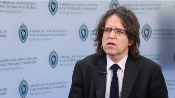
SEATTLE – The presentation of the late-breaking HISTORIC-AF Trial by Dr. Claudio Muneretto and his colleagues “is a very interesting one, which brings to the table a very different approach of hybrid procedures to treat stand-alone atrial fibrillation,” said Dr. Niv Ad of Inova Heart and Vascular Institute, Falls Church, Va.
Dr. Ad gave his comments in a video interview at the annual meeting of the American Association for Thoracic Surgery.
In his assessment, Dr. Ad noted that such studies are useful and can stimulate discussion, even if he would prefer a prospective, comparative study of all procedures. “I hope someday we can create an algorithm where everything has a place: catheter ablation, hybrid procedures where you do catheter ablation and surgical procedure together or in stage, and the stand-alone Maze procedure on pump,” Dr. Ad said.
The video associated with this article is no longer available on this site. Please view all of our videos on the MDedge YouTube channel
AT THE AATS ANNUAL MEETING
Mammographic breast density is a strong risk factor for breast cancer
Breast density is a strong, prevalent, and potentially modifiable risk factor for breast cancer, which makes it of special interest to clinicians whose jobs involve breast cancer risk prediction. That was the theme of a talk by Karla Kerlikowske, MD, of the UCSF Helen Diller Family Comprehensive Cancer Center in San Francisco. Dr. Kerlikowske delivered the John I. Brewer Memorial Lecture May 3 at the 2015 Annual Clinical Meeting of the American College of Obstetricians and Gynecologists in San Francisco.
Mammographic breast density is a radiologic term, Dr. Kerlikowske explained. “The only way to really know someone’s breast density is if they have a mammogram.” The whiter the mammogram, the denser the breast. The darker the mammogram, the fattier the breast.
According to the American College of Radiology, the following 4 categories of breast composition are defined by the “visually estimated” content of fibroglandular-density tissue within the breasts:
A. The breasts are almost entirely fatty.
B. There are scattered areas of fibroglandular density.
C. The breasts are heterogeneously dense, which may obscure small masses.
D. The breasts are extremely dense, which lowers the specificity of mammography.
Categories C and D signify dense breasts, which contain a high degree of collagen, epithelial cells, and stroma. In the United States, more than 25 million women are thought to have dense breasts.
Women who have a family history of breast cancer are more likely to have dense breasts. And women who have dense breasts have an elevated risk of breast cancer. They also have a higher risk of advanced disease, as well as a higher risk of large, high-grade, and lymph node-positive tumors, said Dr. Kerlikowske.
Breast-density legislation is increasing
Twenty-two states now have laws mandating that women found to have heterogeneously dense or extremely dense breasts be notified of their status, said Dr. Kerlikowske. That prompts the question: How should these patients be managed?
Breast density declines with age. Breast density also is influenced by body mass index (BMI). As BMI increases, density declines.
Breast density also can be affected by medications, such as hormone therapy and tamoxifen, Dr. Kerlikowske said.
For example, breast density declines about 1% to 2% per year in postmenopause. In postmenopausal women who take estrogen alone, breast density increases slightly. “But the real increase is for people who take estrogen plus progestin,” said Dr. Kerlikowske. “It’s thought that the progestin component is what increases breast density.” Estrogen-progestin therapy confers the same risk of breast cancer as that faced by a premenopausal woman with dense breasts.
As for tamoxifen, it reduces breast density by 2% to 3% per year in postmenopausal women, Dr. Kerlikowske said. “People who have a decrease of more than 10% in breast density are those who have a reduction in breast cancer.” If a woman doesn’t have that reduction with tamoxifen—about half of women don’t—there is no reduction in breast cancer mortality.
“There’s some thought that you should look at mammograms during the first year of tamoxifen use and, if you don’t see a change, consider switching to another medication,” said Dr. Kerlikowske.
More frequent mammograms and supplemental imaging are options for detecting cancers early. Among the modalities that have been studied in this regard are ultrasonography, tomosynthesis, and breast magnetic resonance imaging (MRI).
“If you do more tests, such as ultrasound, you will definitely find additional lesions,” said Dr. Kerlikowske. “There’s no question. But what are the harms?”
The biopsy rate almost doubles after ultrasonography, compared with mammography. And the number needed to screen to detect cancer is fairly high. For mammography, that number is about 250. For ultrasonography, tomosynthesis, and breast MRI, it is higher.
Tomosynthesis is more cost-effective than supplemental ultrasonography because it decreases the number of false positives, Dr. Kerlikowske said.
What’s the bottom line?
Not every woman with dense breasts is at high risk for breast cancer, said Dr. Kerlikowske. And although breast density is prevalent, it is potentially modifiable.
Nevertheless, breast density confers an elevated risk of breast cancer and can also mask tumors. Women with dense breasts likely should avoid the use of postmenopausal hormone therapy. They also may be candidates for more frequent mammography and/or supplemental imaging.
The Breast Cancer Surveillance Consortium (BCSC) Risk Calculator is the only tool that incorporates breast density. In the works is a new model that also incorporates benign breast disease.
Risk-prediction tool considers density and other factors
A risk-prediction tool from the Breast Cancer Surveillance Consortium (BCSC) is the only model to incorporate breast density. The BCSC Risk Calculator is available free of charge for the iPhone and iPad (an Android version is in the works). The tool takes 5 factors into consideration in estimating a woman’s 5-year risk of developing invasive breast cancer:
• age
• race/ethnicity
• breast density
• family history of breast cancer (first-degree relative)
• personal history of breast biopsy.
The tool is designed for use by health professionals. It is not appropriate for determining risk in women younger than 35 years or older than 79 years; women with a previous diagnosis of breast cancer, lobular carcinoma in situ, ductal carcinoma in situ, or atypical ductal hyperplasia; or women who have undergone breast augmentation. Other risk-prediction models are more appropriate for women with a BRCA mutation.
Share your thoughts! Send your Letter to the Editor to [email protected]. Please include your name and the city and state in which you practice.
Best interest
A dense broth of tedium, a sprinkle of annoyance, and a dash of trepidation – these are the ingredients of the mental soup that simmers in my head when I answer a middle-of-the-night parent phone call.
At least they were until the task of solving the community’s nighttime phone “crises” fell upon the able, eager shoulders of our new interns.

Every now and then, the universe throws up a surprise. One night, my intern was placating a very angry man who was upset with his inconsolable baby. He was the foster father of a baby with neonatal abstinence syndrome (NAS) who had been discharged earlier that day.
“I feel like this baby’s been thrown at us! He wasn’t ready to go home!” he cried. Having never met the baby (foster mom had taken the baby home), this man had returned from a long work shift to find a screaming baby at home. He was aggressive and rude as he ranted at my intern. She empathized and explained that other symptoms might include rapid breathing, irritability, sleep problems, etc.
Although she handled the call with consummate grace and professionalism, afterward an uneasy feeling crept over us. An angry, tired man pushed to his limits, and a very difficult, strange new baby at home was a setup for the perfect storm. Our duty in this situation was clear – we were the protectors of this baby’s best interest. That was all that mattered.
With a rising sense of dread, we juggled frantic phone calls between Child Protective Services, the attending physician, the foster family, and the nearest emergency department. All I could think of was how badly we had failed this baby. Something somewhere had gone horribly wrong for this innocent child to be in danger now.
An hour later, when I finally got through to the foster father, he sounded calmer. “We can’t do this,” he stated simply. He informed me he had called Child Protective Services himself. The baby would come back to us temporarily, while we found him a safe home. Relief swept over us.
Perhaps the baby should never have gone home with this family. Perhaps we had no way of knowing this would happen. Perhaps the family did not realize the responsibility they were assuming. We are all only human. We make mistakes. But we pick up the pieces and we try again, guided by the best interest of the children we swear to protect. This desire to keep trying is what makes all the difference in the world. By making the call himself, this man reaffirmed my faith in the process in which we all play a part. May the quest for the best interest of our children endure forever.
Dr. Behere is a third-year resident in pediatrics at Children’s Hospital at Dartmouth-Hitchcock, Lebanon, N.H. E-mail him at [email protected].
A dense broth of tedium, a sprinkle of annoyance, and a dash of trepidation – these are the ingredients of the mental soup that simmers in my head when I answer a middle-of-the-night parent phone call.
At least they were until the task of solving the community’s nighttime phone “crises” fell upon the able, eager shoulders of our new interns.
Every now and then, the universe throws up a surprise. One night, my intern was placating a very angry man who was upset with his inconsolable baby. He was the foster father of a baby with neonatal abstinence syndrome (NAS) who had been discharged earlier that day.
“I feel like this baby’s been thrown at us! He wasn’t ready to go home!” he cried. Having never met the baby (foster mom had taken the baby home), this man had returned from a long work shift to find a screaming baby at home. He was aggressive and rude as he ranted at my intern. She empathized and explained that other symptoms might include rapid breathing, irritability, sleep problems, etc.
Although she handled the call with consummate grace and professionalism, afterward an uneasy feeling crept over us. An angry, tired man pushed to his limits, and a very difficult, strange new baby at home was a setup for the perfect storm. Our duty in this situation was clear – we were the protectors of this baby’s best interest. That was all that mattered.
With a rising sense of dread, we juggled frantic phone calls between Child Protective Services, the attending physician, the foster family, and the nearest emergency department. All I could think of was how badly we had failed this baby. Something somewhere had gone horribly wrong for this innocent child to be in danger now.
An hour later, when I finally got through to the foster father, he sounded calmer. “We can’t do this,” he stated simply. He informed me he had called Child Protective Services himself. The baby would come back to us temporarily, while we found him a safe home. Relief swept over us.
Perhaps the baby should never have gone home with this family. Perhaps we had no way of knowing this would happen. Perhaps the family did not realize the responsibility they were assuming. We are all only human. We make mistakes. But we pick up the pieces and we try again, guided by the best interest of the children we swear to protect. This desire to keep trying is what makes all the difference in the world. By making the call himself, this man reaffirmed my faith in the process in which we all play a part. May the quest for the best interest of our children endure forever.
Dr. Behere is a third-year resident in pediatrics at Children’s Hospital at Dartmouth-Hitchcock, Lebanon, N.H. E-mail him at [email protected].

VIDEO: Esophagectomy outcomes better in hospitals that handle complex cases
SEATTLE – Hospitals that perform at least one nongastric conduit esophageal reconstruction per year have half the esophagectomy mortality of hospitals that do not, according to a review by the Mayo Clinic in Rochester, Minn., of 11,211 esophagectomies in the Nationwide Inpatient Sample database from 2000 to 2011.
“There is tremendous variation in outcome after esophagectomy, and some advocate for regionalization to high-volume hospitals,” the investigators said. The findings suggest that case complexity could be one of the things that help define which hospitals do it best, they added.
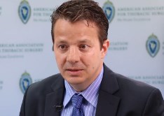
The study seems to confirm that hospital case volume makes a difference in surgical outcomes, said Dr. Nabil Rizk, a thoracic surgeon at Memorial Sloan-Kettering Cancer Center in New York.
Dr. Rizk, a discussant on the paper at the American Association for Thoracic Surgery annual meeting, explained how the study fits into regionalization trends, but also shared his concerns about the work in an interview at the meeting.
The video associated with this article is no longer available on this site. Please view all of our videos on the MDedge YouTube channel
SEATTLE – Hospitals that perform at least one nongastric conduit esophageal reconstruction per year have half the esophagectomy mortality of hospitals that do not, according to a review by the Mayo Clinic in Rochester, Minn., of 11,211 esophagectomies in the Nationwide Inpatient Sample database from 2000 to 2011.
“There is tremendous variation in outcome after esophagectomy, and some advocate for regionalization to high-volume hospitals,” the investigators said. The findings suggest that case complexity could be one of the things that help define which hospitals do it best, they added.
The study seems to confirm that hospital case volume makes a difference in surgical outcomes, said Dr. Nabil Rizk, a thoracic surgeon at Memorial Sloan-Kettering Cancer Center in New York.
Dr. Rizk, a discussant on the paper at the American Association for Thoracic Surgery annual meeting, explained how the study fits into regionalization trends, but also shared his concerns about the work in an interview at the meeting.
The video associated with this article is no longer available on this site. Please view all of our videos on the MDedge YouTube channel
AT THE AATS ANNUAL MEETING
VIDEO: Cardiosphere-derived cells improve outcomes in hypoplastic left heart syndrome
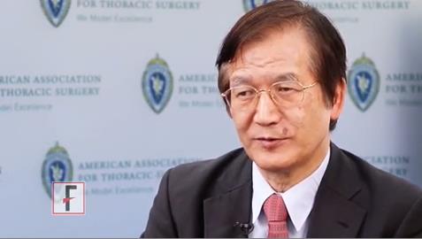
SEATTLE – Autologous stem cell therapy improves surgery outcomes in children with hypoplastic left heart syndrome, according to the results of a small prospective trial from Okayama University in Japan.
The investigators cultured cardiosphere-derived cells (CDCs) – cardiac progenitor cells – from right atrium samples taken during stage 2 or 3 surgical palliations in seven HLHS children. A month later, they injected 300,000 CDCs/kg into the children’s coronary arteries by catheter, with each child getting cells cultured from their own tissue.
The cells seemed to jump-start the intrinsic regenerative properties of very young hearts. At 30 months follow-up, right ventricular mass and ejection fractions were about 10% greater in CDC treated patients compared to seven controls. Treated children also had better growth.
It’s possible the technique could help older children, too, and even adults, said lead investigator Dr. Shunji Sano, professor and chairman of the department of cardiovascular surgery at Okayama. He explained the work, its implications, and the next phase of research in an interview at the annual meeting of the American Association for Thoracic Surgery.
The video associated with this article is no longer available on this site. Please view all of our videos on the MDedge YouTube channel
AT THE AATS ANNUAL MEETING
VIDEO: Less tricuspid regurgitation seen with Sano shunt in Norwood procedures
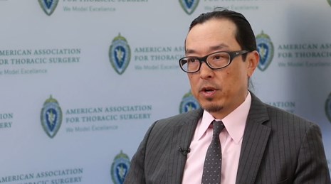
SEATTLE – Sano shunts outperform Blalock-Taussig shunts for Norwood procedures in neonates with hypoplastic left heart syndrome, according to a research registry study of 322 patients at the Cleveland Clinic and elsewhere.
The 166 newborns who had Sano shunts were matched to 166 who had Blalock-Taussig (BT) shunts.
“For comparable neonates with HLHS [hypoplastic left heart syndrome] undergoing Norwood operations, Sano offers better late survival [more than 3 years], less late tricuspid regurgitation, and perhaps less late right ventricular dysfunction than BT,” the investigators concluded.
Even so, Dr. Richard Ohye, professor of cardiac surgery at the University of Michigan, Ann Arbor, said the jury is still out on which shunt is best. He explained why in an interview at the American Association for Thoracic Surgery annual meeting. He also shared tips on shunt selection and explained a novel technique he has developed for doing a Sano shunt with a smaller hole in the right ventricle.
The video associated with this article is no longer available on this site. Please view all of our videos on the MDedge YouTube channel
AT THE AATS ANNUAL MEETING
Reversing glucocorticoid resistance in ALL

Photo courtesy of St. Jude
Researchers say they have identified a mechanism that helps leukemia cells resist glucocorticoids.
They believe the mechanism is responsible for about a third of steroid resistance in children and adolescents with acute lymphoblastic leukemia (ALL).
However, additional research is needed to determine if the process is at work in adults with ALL, where steroid resistance is more common and long-term survival lags.
The researchers described the mechanism in Nature Genetics.
“Based on these findings, research has already begun to identify small molecules with the potential to reverse glucocorticoid resistance, leading to more effective treatment and increased survival,” said study author William Evans, PharmD, of St. Jude Children’s Research Hospital in Memphis, Tennessee.
Dr Evans and his colleagues analyzed samples from 444 newly diagnosed ALL patients being treated at St. Jude or in clinical trials sponsored by the Dutch Childhood Oncology Group and the German Cooperative Study Group for Childhood ALL.
The team also analyzed samples collected at diagnosis and relapse from 49 pediatric ALL patients enrolled in clinical trials organized by the Children’s Oncology Group.
The researchers found differences in gene expression that correlated with sensitivity to steroids. CASP1 and NLRP3 were among the genes with increased activity in steroid-resistant leukemia cells.
The team also identified a reason for the increased gene activity. Leukemia cells overexpressing CASP1 and NLRP3 had lower levels of methylation compared to cells with normal expression.
Previous research showed that steroid resistance was more common in young ALL patients who relapsed than in newly diagnosed patients. Dr Evans and his colleagues found that expression of CASP1 and NLRP3 was significantly higher in ALL patients who relapsed.
The researchers also discovered that CASP1 blocks glucocorticoids by splitting the receptor where the drug binds and therefore blocks its access to the nucleus.
“Cells that overexpress CASP1 are chewing up their glucocorticoid receptor,” Dr Evans said. “That means when steroids enter the cell, there is no receptor for the drugs to bind to or fulfill its therapeutic function.”
To confirm that CASP1 cleavage of the steroid receptor is pivotal to ALL steroid resistance, the researchers engineered a receptor that lacked the CASP1 cleavage site. When they introduced the genetically engineered receptors into ALL cells that expressed high levels of CASP1, the cells remained sensitive to steroids.
Using a variety of techniques, the researchers showed that steroid resistance rose or fell in leukemia cells based on CASP1 levels. Overexpression of CASP1 rendered ALL cells 5 to 15 times more resistant to the glucocorticoids dexamethasone and prednisolone.
However, reducing CASP1 using genetic, pharmacologic, and other methods restored steroid sensitivity in leukemia cells. ![]()
Defining the role of TAMs in DLBCL

pseudopodia to engulf particles
PHILADELPHIA—New research suggests the prognostic value of tumor-associated macrophages (TAMs) is disease-specific as well as treatment-specific.
Investigators set out to determine if TAMs have a negative prognostic impact in diffuse large B-cell lymphoma (DLBCL), as previous studies produced conflicting results.
The team found that higher TAM levels are associated with worse survival in DLBCL, but only in patients who do not receive rituximab. The drug can overcome the poor prognosis TAMs confer in DLBCL.
Eri Matsuki, MD, PhD, of Memorial Sloan-Kettering Cancer Center in New York, New York, and her colleagues presented these findings in a poster at the AACR Annual Meeting 2015 (abstract 2371*).
To ascertain the role of TAMs in DLBCL, the investigators analyzed specimens from 103 DLBCL patients, 61 of whom received rituximab, 33 who did not, and 9 whose rituximab status was unknown. The team first looked at the expression of CD163 as a marker of TAMs.
“CD163 is more specific to M2-type macrophages, which have pro-tumor effects, compared to a more pan-macrophage marker which is widely used—CD68,” Dr Matsuki noted.
She and her colleagues found that a high level of CD163-positive cells (more than 150) was significantly associated with advanced-stage disease (P=0.016), non-GCB DLBCL (P=0.0071), and higher expression of c-Myc (P=0.0022).
“Our interpretation of that at this point is that, the more aggressive the tumor, the more likely that it would induce macrophages into the tissue,” Dr Matsuki said.
The investigators then assessed the impact of rituximab use. Among patients who didn’t receive rituximab, having a high level of CD163-positive cells (more than 150) was associated with inferior overall survival (OS, P=0.022). However, if patients did receive rituximab, there was no significant difference in OS.
Dr Matsuki said this supports previous studies showing that the prognostic effect of TAMs diminishes with rituximab use, as well as the in vitro finding that M2 macrophages exhibit increased phagocytosis of rituximab-opsonized tumor cells.
“So overall, what we’re seeing is that . . . the negative influence of TAMs can be overcome with rituximab use,” she summarized.
Dr Matsuki and her colleagues also looked at the patients’ lymphocyte-to-monocyte-ratio (LMR) because TAMs partly arise from peripheral blood monocytes.
The team found that having an LMR higher than 2.77 was significantly associated with superior OS (P=0.03) in patients who did not receive rituximab. And there was a trend toward improved OS with a higher LMR in patients who did receive the drug (P=0.07).
The investigators believe the differences they observed in the prognostic value of CD163 and LMR could be explained by the fact that TAMs are derived from both circulating monocytes and resident macrophages.
Dr Matsuki said this research has improved her group’s understanding of DLBCL, but they are still working to identify additional biomarkers associated with prognosis in this disease. ![]()
*Information in the abstract differs from that presented at the meeting.
New model simulates human MM

Photo by Ben Skála
By growing human tumors on chicken embryos, scientists have created a new model system for screening drugs to treat multiple myeloma (MM).
Several MM drug candidates have shown promising activity in this system, according to the researchers.
Gerold Untergasser, PhD, of Innsbruck Medical University in Austria, and his colleagues described how they developed the system in the Journal of Visualized Experiments.
First, the researchers prepared cultures of the MM cell lines OPM2 and RPMI 8226, as well as human mesenchymal stem cells. They then transfected MM cells with enhanced green fluorescent protein, making the cells easy to observe through a fluorescence microscope.
Next, the team cultured MM cells with mesenchymal cells and collagen to create 3-dimensional cell spheres, thereby simulating the natural microenvironment of the tumor.
The researchers then transferred their cell spheres to the outer membrane of chicken embryos. This choriallantoid membrane provides a suitable base for growing miniature human tumors in culture.
The team has introduced anti-myeloma drugs, such as bortezomib, into this system and assessed the drugs’ ability to target MM cells and prevent tumor growth and angiogenesis. They were also able to evaluate toxicity.
“The chicken egg is much easier to handle and cheaper than mice, and it reduces the number of animal experiments,” Dr Untergasser said. “In our new video publication, we give detailed information on how our system works.”
“We provide an easy-to-repeat protocol for broad use within the research community. Our vision for the future is that a separate test is performed for each patient to determine which drug is best suitable.” ![]()