User login
Inhibitor improves OS in poor-prognosis MDS

WASHINGTON, DC—A small-molecule inhibitor can improve overall survival (OS) in certain patients with previously treated myelodysplastic syndromes (MDS), results of a phase 3 trial suggest.
Overall, patients who received the dual PI3K/PLK pathway inhibitor rigosertib along with best supportive care (BSC) did not see a significant improvement in OS compared to patients who received BSC alone.
However, rigosertib did improve OS in patients with poor prognosis.
Guillermo Garcia-Manero, MD, of the MD Anderson Cancer Center in Houston, Texas, and his colleagues presented these results at the 13th International Symposium on Myelodysplastic Syndromes (abstract 112).
The trial, known as ONTIME, was sponsored by Onconova Therapeutics, Inc., the company developing rigosertib.
The trial included 299 higher-risk MDS patients with excess blasts (5% to 30% bone marrow blasts) who had failed to respond to (25%), progressed on (37%), or relapsed after (38%) treatment with hypomethylating agents (HMAs).
Patients were randomized 2:1 to receive rigosertib plus BSC or BSC alone. Patients treated with rigosertib received 1800 mg every 24 hours for 72 hours as a continuous, intravenous, ambulatory infusion, every 2 weeks for the first 16 weeks, then every 4 weeks.
The treatment arms were generally balanced in terms of baseline characteristics. The majority of patients were male (66%) and white (82%). The median age was 74 years. Most patients (85%) had an Eastern Cooperative Oncology Group score of 0 or 1.
The median duration of the last HMA therapy was 8.8 months for patients in the rigosertib arm and 10.3 months for patients in the BSC arm.
The researchers found no significant difference in OS between the treatment arms. The median OS was 8.4 months in the rigosertib arm and 5.9 months in the BSC arm, and the 12-month OS was 35% and 25%, respectively (hazard ratio[HR]=0.87, P=0.31).
On the other hand, certain patients did see a significant improvement in OS with rigosertib. Among patients with primary HMA failure (those who failed to respond to or progressed during HMA therapy), the median OS was 8.6 months in the rigosertib arm and 5.3 months in the BSC arm (HR=0.69, P=0.040).
For patients who received HMAs for less than 9 months, the median OS was 7.7 months in the rigosertib arm and 4.5 months in the BSC arm (HR=0.55, P=0.003). Among patients younger than 75 years of age, the median OS was 9.7 months in the rigosertib arm and 4.1 months in the BSC arm (HR=0.52, P=0.0004).
And for patients with very high-risk disease according to the Revised International Prognostic Scoring System, the median OS was 7.6 months in the rigosertib arm and 3.2 months in the BSC arm (HR=0.56, P=0.005).
The researchers said there were no obvious differences between the treatment arms with regard to overall adverse events (AEs) or grade 3 or higher AEs.
Overall, 99% of patients in the rigosertib arm and 85% in the BSC arm experienced treatment-emergent AEs. The incidence of grade 3 or higher AEs was 79% and 68%, respectively.
Treatment-emergent AEs of all grades—occurring in the rigosertib and BSC arms, respectively—included nausea (35% vs 18%), diarrhea (33% vs 20%), constipation (31% vs 11%), fatigue (30% vs 18%), pyrexia (27% vs 21%), anemia (23% vs 9%), peripheral edema (21% vs 16%), and thrombocytopenia (21% vs 8%).
Considering the study results together, Dr Garcia-Manero and his colleagues concluded that rigosertib is likely most effective in high-risk MDS patients with the worst prognosis, and these patients can safely receive the drug. ![]()
Society of Hospital Medicine (SHM)-American Academy of Family Physicians (AAFP) Joint Statement on Hospitalists Trained in Family Medicine
Hospitalists are physicians whose primary professional focus is the general medical care of hospitalized patients. Both the Society of Hospital Medicine (SHM) and the American Academy of Family Physicians (AAFP) hold that the opportunity to participate as a Hospitalist should be granted to all physicians commensurate with their documented training and/or experience, demonstrated abilities and current competencies.
During their training Family Physicians acquire the necessary attitudes, skills, and knowledge that enable them to provide continuing and comprehensive medical care across the spectrum of care settings, including the inpatient setting. Education in the primary management of hospitalized patients occurs during the required general inpatient ward and intensive care unit experiences. In addition, Family Physicians are required to train with general surgeons and surgical subspecialists, enhancing recognition and understanding of surgical disease states upon which Hospitalists are frequently asked to consult or co-manage. Family Medicine training also encompasses additional skills essential to the practice of Hospital Medicine, including participation in quality improvement, addressing the psychosocial needs of patients, coordinating across levels of care, and functioning as members of interdisciplinary teams.
Given this training, many Family Physicians effectively manage their patients in an inpatient setting after the completion of their residency.
Demand for Hospitalists continues to outweigh supply in the United States, including needs in underserved and rural areas. Hospitalists Trained in Family Medicine (HTFM) fulfill an important public health need by providing frontline inpatient services in a variety of geographic settings. In addition, while many HTFM focus exclusively on the care of adults, others are providing inpatient care across the spectrum of ages, as well as providing obstetric services. More than two-thirds of HTFM are also involved in the training of residents and medical students, enhancing the skills of our future physicians.
Recognition of achievement by HTFM from the SHM is available by meeting standards set for all Hospitalists, regardless of residency training, in the form of the designation of Fellow of Hospital Medicine. HTFM also have the opportunity to professionally qualify and sit for the Recognition of Focused Practice in Hospital Medicine board examination. This examination is administered and recognized jointly by the American Board of Family Medicine and the American Board of Internal Medicine.
In consideration of the above factors, both the Society of Hospital Medicine and the American Academy of Family Physicians endorse and encourage the growing contribution of Hospitalists Trained in Family Medicine.
Society of Hospital Medicine Welcomes New Fellows Class
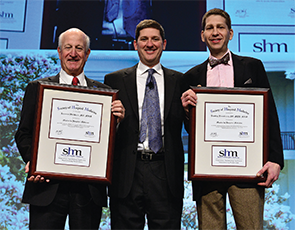
Last month, more than 230 hospitalists were inducted as Fellows in Hospital Medicine (FHM), Senior Fellows in Hospital Medicine (SFHM), and Masters in Hospital Medicine (MHM) by SHM at the 2015 annual meeting at the Gaylord National Resort and Convention Center in National Harbor, Md.
This year represents the largest fellows class in history, with 175 FHM and 61 SFHM honorees.
“Through their commitment to the specialty, through education and self-improvement, hospitalists earning the Fellow and Senior Fellow designations represent the very best of the hospital medicine movement and its goal to improve the care of hospitalized patients,” says SHM President Bob Harrington, MD, SFHM. “I hope you will join me in congratulating them in this professional milestone.”
Fellows and Senior Fellows have earned the right to use the “FHM” and “SFHM” designation.
SHM also inducted two new Masters in Hospital Medicine, the highest honor from SHM: Bradley Flansbaum, DO, MPH, MHM, and Larry Wellikson, MD, MHM.
Dr. Flansbaum was a founding member of SHM and served as a board member and officer; today, he is a hospitalist at Lenox Hill Hospital in New York City and physician editor for SHM’s blog, The Hospital Leader.
Dr. Wellikson joined SHM in January 2000 and serves as SHM’s chief executive officer.
Drs. Flansbaum and Wellikson join 16 other leaders in the specialty, including co-founders Win Whitcomb, MD, MHM, and John Nelson, MD, MHM, along with Bob Wachter, MD, MHM, who published the seminal article for the hospitalist movement in a 1996 New England Journal of Medicine article.
Brendon Shank is SHM’s associate vice president of communications.
Medicare Develops Next Generation Accountable Care Organization Model
The Centers for Medicare and Medicaid Services (CMS) Innovation Center recently announced the development of a new accountable care organization (ACO) model—the Next Generation ACO—that hopes to move closer to the goal of efficient, coordinated care for Medicare beneficiaries.
“This ACO model provides for greater engagement of beneficiaries, a more predictable, prospective financial model, and more tools to coordinate care for beneficiaries,” writes Patrick Conway, MD, MSc, chief medical officer and deputy administrator for innovation and quality at CMS, in a blog post announcing the Next Generation ACO.
ACOs align hospitals, physicians, nursing facilities, and other critical healthcare providers as a sort of one-stop shop for seamless patient care across settings and among providers. By bringing together the full range of services, ACOs aim to provide higher quality coordinated care while reducing costs for patients and Medicare.
Since the passage of the Affordable Care Act, CMS has overseen two distinct tracks for ACOs: the Medicare Shared Savings Program and the Pioneer ACO. The Shared Savings Program was a first step in moving toward streamlined healthcare delivery systems while incentivizing care coordination across settings. Pioneer ACOs, on the other hand, were designed as a test for more aggressive reforms that promised higher potential rewards in exchange for higher risk, while moving participants toward population-based payments.
The Next Generation ACO builds off of the Pioneer and Shared Savings Program ACO models to test whether the fundamental concepts behind an ACO—improving care and reducing costs—can be achieved using stronger financial incentives. Notably, the Next Generation ACO establishes stable, prospective targets for benchmarking expenditures and offers an array of payment mechanisms, including capitation.
ACO goals read like a laundry list of hospitalist goals and practice, such as reducing readmissions, maximizing efficiency, improving care transitions, and reducing length of stay.
Participants of the Next Generation ACO model will have new tools to help coordinate patient care, including expanded coverage for telehealth and home health services and increased access for skilled nursing facility coverage without prior hospitalizations. Because the Next Generation ACO model comes from the CMS Innovation Center, it’s specifically designed to help policymakers evaluate the impact of reimbursement and system changes with an eye toward scalability. The knowledge gained from this model could help structure the Medicare payment system of tomorrow.
Hospitalists have long been interested in the impact of ACOs on their practices, with good reason. Hospitals form an integral part of an ACO, and hospitalists serve critical roles within their hospitals. ACO goals read like a laundry list of hospitalist goals and practice, such as reducing readmissions, maximizing efficiency, improving care transitions, and reducing length of stay. The Next Generation ACO model offers the potential to further capitalize on the expertise of hospitalists as the healthcare system explores ways to move away from traditional fee-for-service payments.
The way in which Medicare pays providers is evolving rapidly as CMS seeks to reimburse for the quality rather than the quantity of services provided to beneficiaries. Over the next five years, CMS has set aggressive targets for transitioning fee-for-service payments into value-based payment systems; the Next Generation ACO is one tool for helping to push that goal onward.
Joshua Lapps is SHM’s manager of government relations.
Society of Hospital Medicine's HM15 Meeting Draws Thousands
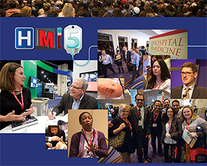
NATIONAL HARBOR, Md.—Cherry trees weren’t the only things that blossomed around Washington last month. SHM’s annual meeting, with roughly 2,500 attendees, featured 100 educational sessions, a day of Congressional lobbying, and plenaries from the “Checklist Doctor” and the Dean of Hospital Medicine. Pre-courses, the popular poster competition, and updates on everything from anticoagulants to VTE helped round out HM15, the specialty’s biggest annual event.
“I come to the meeting,” says new SHM President Robert Harrington, Jr., MD, SFHM, “and then for the next 362 days, this is enough to get me through the rest of the year ‘til I come back.”
Hospitalists Have Stake in Improving Quality, Patient Safety
NATIONAL HARBOR, Md.—Don Lee, MD, MPH, is building what one might call an analog quality improvement (QI) project focused on reducing readmissions. What the medical director for clinical integration at Columbia St. Mary’s in Milwaukee does is work with patient navigators to make follow-up phone calls after discharge to get ahead of potential issues.
What he wants to do is design a system that ensures that happens.
So, he came to HM15 for help.
“I’m very interested in continuous quality improvement. I wanted to work on how to not only get the project off the ground, but also to make sure what we are doing is good, and it’s doing what it’s supposed to be doing,” Dr. Lee says.
Well, he came to the right place. Quality and patient safety are hallmarks of the annual meeting; this year’s gathering was no exception. Plenaries provided advice from national thought leaders on improving safety by improving the patient experience; breakout sessions focused on how to build, maintain, and sustain QI projects; and SHM unveiled a new educational track dubbed the “Doctor-Patient Relationship.”
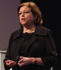
Hospital medicine, and healthcare in a broader sense, needs to be able to define safety better to attack it proactively, says Maureen Bisognano, president and CEO of the Institute for Healthcare Improvement (IHI). She compared medicine to NASA, which tracks its missions in a continuum of both successes and failures to understand what processes and protocols lay behind each.
Medicine has no such pathway laid out to date, though Bisognano said her task for the next year is to try to define one.
“We don’t know what the system of safety looks like,” she said. “We don’t know how many times we duplicate tests on admission because we haven’t connected with primary care. We don’t know how many times we send somebody home with inadequate social support, no food, and no way to pick up their prescription.
“We don’t have a sense of where our near-misses are, so we don’t have a vision of safety.”
Hospitalist Kedar Mate, MD, senior vice president for innovation at IHI, says that QI projects can seem daunting in the midst of daily censuses, hospital committee meetings, and a myriad of other responsibilities physicians face. But much of that fear is perception. A project can be simple or system-wide. The trick is just getting started in the face of perceived hurdles, he adds.
“Language around quality improvement tends to confuse and create mystery, and the jargon and so on creates interference,” says Dr. Mate, an assistant professor of medicine at Weill Cornell Medical College in New York City and a research fellow at Harvard Medical School’s Division of Global Health Equity. “It’s not that mysterious. It’s kind of a straightforward thing, actually, if you work through it logically and stepwise.”
And, as front-line providers, hospitalists are primed to lead healthcare systems in how to deliver care, he said.
“Formerly, physicians were iterant, right?” Dr. Mate adds. “They would come in and out of institutions and didn’t really have a stake in the game, on some level, of institutional quality. That’s totally different now.”
But, while the individual hospitalist has a responsibility to embrace safety initiatives, employers and industry groups have a duty to provide the proper resources to make that connection easier.
“The individual’s responsibility is to try to access that information to carry on in the face of busy schedules and busy lives,” Dr. Mate says. “SHM, IHI, and others have a responsibility to try to make those that are inclined able to continue and able to build and move their efforts forward in an even more productive way.”
Inclined docs like Dr. Lee, who know that their hospitals collect reams of data that can be useful for patient safety projects, many times have no idea how to extract said data. He has learned that partnering with “gatekeepers” is a way to help others help him.
“We are collecting data every second, every minute,” Dr. Lee says. “It’s amazing how much data we have, but to actually sift through it and make it meaningful is very difficult. You have to know what questions to ask and you have to get buy-in from the [gatekeepers], because they get thousands of requests for data extraction.”
Richard Quinn is a freelance writer in New Jersey.
NATIONAL HARBOR, Md.—Don Lee, MD, MPH, is building what one might call an analog quality improvement (QI) project focused on reducing readmissions. What the medical director for clinical integration at Columbia St. Mary’s in Milwaukee does is work with patient navigators to make follow-up phone calls after discharge to get ahead of potential issues.
What he wants to do is design a system that ensures that happens.
So, he came to HM15 for help.
“I’m very interested in continuous quality improvement. I wanted to work on how to not only get the project off the ground, but also to make sure what we are doing is good, and it’s doing what it’s supposed to be doing,” Dr. Lee says.
Well, he came to the right place. Quality and patient safety are hallmarks of the annual meeting; this year’s gathering was no exception. Plenaries provided advice from national thought leaders on improving safety by improving the patient experience; breakout sessions focused on how to build, maintain, and sustain QI projects; and SHM unveiled a new educational track dubbed the “Doctor-Patient Relationship.”
Hospital medicine, and healthcare in a broader sense, needs to be able to define safety better to attack it proactively, says Maureen Bisognano, president and CEO of the Institute for Healthcare Improvement (IHI). She compared medicine to NASA, which tracks its missions in a continuum of both successes and failures to understand what processes and protocols lay behind each.
Medicine has no such pathway laid out to date, though Bisognano said her task for the next year is to try to define one.
“We don’t know what the system of safety looks like,” she said. “We don’t know how many times we duplicate tests on admission because we haven’t connected with primary care. We don’t know how many times we send somebody home with inadequate social support, no food, and no way to pick up their prescription.
“We don’t have a sense of where our near-misses are, so we don’t have a vision of safety.”
Hospitalist Kedar Mate, MD, senior vice president for innovation at IHI, says that QI projects can seem daunting in the midst of daily censuses, hospital committee meetings, and a myriad of other responsibilities physicians face. But much of that fear is perception. A project can be simple or system-wide. The trick is just getting started in the face of perceived hurdles, he adds.
“Language around quality improvement tends to confuse and create mystery, and the jargon and so on creates interference,” says Dr. Mate, an assistant professor of medicine at Weill Cornell Medical College in New York City and a research fellow at Harvard Medical School’s Division of Global Health Equity. “It’s not that mysterious. It’s kind of a straightforward thing, actually, if you work through it logically and stepwise.”
And, as front-line providers, hospitalists are primed to lead healthcare systems in how to deliver care, he said.
“Formerly, physicians were iterant, right?” Dr. Mate adds. “They would come in and out of institutions and didn’t really have a stake in the game, on some level, of institutional quality. That’s totally different now.”
But, while the individual hospitalist has a responsibility to embrace safety initiatives, employers and industry groups have a duty to provide the proper resources to make that connection easier.
“The individual’s responsibility is to try to access that information to carry on in the face of busy schedules and busy lives,” Dr. Mate says. “SHM, IHI, and others have a responsibility to try to make those that are inclined able to continue and able to build and move their efforts forward in an even more productive way.”
Inclined docs like Dr. Lee, who know that their hospitals collect reams of data that can be useful for patient safety projects, many times have no idea how to extract said data. He has learned that partnering with “gatekeepers” is a way to help others help him.
“We are collecting data every second, every minute,” Dr. Lee says. “It’s amazing how much data we have, but to actually sift through it and make it meaningful is very difficult. You have to know what questions to ask and you have to get buy-in from the [gatekeepers], because they get thousands of requests for data extraction.”
Richard Quinn is a freelance writer in New Jersey.
Hospitalists Raise Healthcare Issues on Capitol Hill
NATIONAL HARBOR, Md.—Armed with blue folders chockablock with agendas, talking points, and fact sheets, about 100 hospitalists boarded three charter buses and descended on Capitol Hill last month like a swarm of erudite high schoolers on a class trip.
Clad in state-themed ties, suits, and dresses, the group’s goal was singular: Introduce the concept of hospital medicine to every senator, representative, and Congressional staffer who would take the time to meet them, and let those folks know that SHM and its members stand at the ready to serve as a resource for politicians.
“We don’t go to Washington and say, ‘You need to pay hospitalists more money,’” says SHM CEO Larry Wellikson, MD, MHM. “We go and we say, ‘You have a problem. We have a solution. Why don’t we work together to create the future?’ This is what people need to hear. This is a breath of fresh air, and that’s why we get invited back and we’re part of the discussion.”
This year’s discussion was formally titled Hospitalists on the Hill, version 2015. The turnout always improves when the annual meeting is just across the Potomac River at the Gaylord National Resort & Convention Center, as it has been for three of the past six years. The society ferried hospitalists to the offices of Washington power players with three goals this year:
- Push for support for the Improving Access to Medicare Coverage Act of 2015 (H.R. 1571 and S. 843), as it would adjust Medicare rules to allow observation status to be counted toward the three-day inpatient rule for coverage of care in skilled nursing facilities.
- Ask for support for the Personalize Your Care Act, a soon-to-be-reintroduced bill from U.S. Rep. Earl Blumenauer (D-Ore.) authorizing Medicare to pay for end-of-life care discussions and building in opportunities for patients to participate in their long-term care planning.
- Push for Congress to repeal the sustainable growth rate (SGR) formula and create a “pathway towards payment models that reward quality and efficiency.” This legislative “ask,” to use lobbying parlance, is an evergreen that has been an SHM priority for years.
Jodi Strong, director of operations at Novant Health, a 12-hospital group based in Charlotte, N.C., says that she joined this year’s advocacy pilgrimage for the first time because, in a time of generational upheaval in the American healthcare system, every voice should be heard.
“One vote does make a difference, and I want to be a part of that process,” she says, adding, “Hospitalists are very instrumental in the patient, the care that they receive, where they go after they’ve had a hospital visit, how they connect with the patient’s primary care physician.”
The trick of lobbying is getting those in power to see the world as those in practice do. It helps when the two are friends. H.E. “Chip” Walpole Jr., MS, MD, regional medical director of Select Medical of Greenville, S.C., has known U.S. Rep. Trey Gowdy (R-S.C.) for years. When they talk about medical issues, it helps the congressman get a stethoscope-on-the-ground view.

—H.E. “Chip” Walpole, Jr., MS, MD
“He’ll say, ‘I know I can trust Chip and he’ll give me a straight answer for a problem,’” Dr. Walpole says. “Then it’s about inviting them, to say ‘Hey, come and see. You want to learn a little bit more about what we do in the hospital? Come and see our facility.’”
And, while many first-time Hill Day attendees get nervous about trying to impress the Beltway, Dr. Walpole views it from the flip side.
“Any time you get to have face-to-face time with one of your Congressional leaders, whether it be a representative or senator, you talk to the people that actually directly influence and impact not only the work that we do, but the work that we do for our patients,” he says. “In that regard, we represent a voice for them, to explain to them who we are and what we do and what our patients’ needs are.
“They depend on us.”
That’s the message that Stephanie Vance, who founded Washington-based Advocacy Associates LLC, pushed as she prepped the laymen lobbyists for more than an hour before sending them off to their meetings. Vance, a 25-year veteran of the political scene, reminded hospitalists during the breakfast prep session that those in Congress are elected to serve—and that means they’re elected to listen.
Hospitalist Gordon Johnson, MD, FACP, FHM, got the message. He’s president of the SHM’s Oregon Chapter, but he had never done a lobbying trip like this before. The appeal was simple and effective to him.
“The more of us that are involved, the more meaningful it is,” he says. “When [members of Congress and their staffs] have people coming from their constituency, that carries a message. It does carry a stronger message.”
But, as with patient discharge, the message is always strongest with good follow-up. Vance, known to many as “the advocacy guru,” urged hospitalists to follow up after their meetings—an occasional phone call or e-mail to let the person know that, should they have any questions, a hospitalist is standing by to provide answers. To Dr. Walpole, a connection like that can be worth more than hiring a white-shoed lobbying firm.
“When you put a face with someone—‘Oh, I know Chip, I know Richard from back home,’—they make a connection with someone that is real and personal to them,” he says. “And, ultimately, that can probably make a bigger difference in influencing how they represent us than anything else.”
Richard Quinn is a freelance writer in New Jersey.
NATIONAL HARBOR, Md.—Armed with blue folders chockablock with agendas, talking points, and fact sheets, about 100 hospitalists boarded three charter buses and descended on Capitol Hill last month like a swarm of erudite high schoolers on a class trip.
Clad in state-themed ties, suits, and dresses, the group’s goal was singular: Introduce the concept of hospital medicine to every senator, representative, and Congressional staffer who would take the time to meet them, and let those folks know that SHM and its members stand at the ready to serve as a resource for politicians.
“We don’t go to Washington and say, ‘You need to pay hospitalists more money,’” says SHM CEO Larry Wellikson, MD, MHM. “We go and we say, ‘You have a problem. We have a solution. Why don’t we work together to create the future?’ This is what people need to hear. This is a breath of fresh air, and that’s why we get invited back and we’re part of the discussion.”
This year’s discussion was formally titled Hospitalists on the Hill, version 2015. The turnout always improves when the annual meeting is just across the Potomac River at the Gaylord National Resort & Convention Center, as it has been for three of the past six years. The society ferried hospitalists to the offices of Washington power players with three goals this year:
- Push for support for the Improving Access to Medicare Coverage Act of 2015 (H.R. 1571 and S. 843), as it would adjust Medicare rules to allow observation status to be counted toward the three-day inpatient rule for coverage of care in skilled nursing facilities.
- Ask for support for the Personalize Your Care Act, a soon-to-be-reintroduced bill from U.S. Rep. Earl Blumenauer (D-Ore.) authorizing Medicare to pay for end-of-life care discussions and building in opportunities for patients to participate in their long-term care planning.
- Push for Congress to repeal the sustainable growth rate (SGR) formula and create a “pathway towards payment models that reward quality and efficiency.” This legislative “ask,” to use lobbying parlance, is an evergreen that has been an SHM priority for years.
Jodi Strong, director of operations at Novant Health, a 12-hospital group based in Charlotte, N.C., says that she joined this year’s advocacy pilgrimage for the first time because, in a time of generational upheaval in the American healthcare system, every voice should be heard.
“One vote does make a difference, and I want to be a part of that process,” she says, adding, “Hospitalists are very instrumental in the patient, the care that they receive, where they go after they’ve had a hospital visit, how they connect with the patient’s primary care physician.”
The trick of lobbying is getting those in power to see the world as those in practice do. It helps when the two are friends. H.E. “Chip” Walpole Jr., MS, MD, regional medical director of Select Medical of Greenville, S.C., has known U.S. Rep. Trey Gowdy (R-S.C.) for years. When they talk about medical issues, it helps the congressman get a stethoscope-on-the-ground view.
—H.E. “Chip” Walpole, Jr., MS, MD
“He’ll say, ‘I know I can trust Chip and he’ll give me a straight answer for a problem,’” Dr. Walpole says. “Then it’s about inviting them, to say ‘Hey, come and see. You want to learn a little bit more about what we do in the hospital? Come and see our facility.’”
And, while many first-time Hill Day attendees get nervous about trying to impress the Beltway, Dr. Walpole views it from the flip side.
“Any time you get to have face-to-face time with one of your Congressional leaders, whether it be a representative or senator, you talk to the people that actually directly influence and impact not only the work that we do, but the work that we do for our patients,” he says. “In that regard, we represent a voice for them, to explain to them who we are and what we do and what our patients’ needs are.
“They depend on us.”
That’s the message that Stephanie Vance, who founded Washington-based Advocacy Associates LLC, pushed as she prepped the laymen lobbyists for more than an hour before sending them off to their meetings. Vance, a 25-year veteran of the political scene, reminded hospitalists during the breakfast prep session that those in Congress are elected to serve—and that means they’re elected to listen.
Hospitalist Gordon Johnson, MD, FACP, FHM, got the message. He’s president of the SHM’s Oregon Chapter, but he had never done a lobbying trip like this before. The appeal was simple and effective to him.
“The more of us that are involved, the more meaningful it is,” he says. “When [members of Congress and their staffs] have people coming from their constituency, that carries a message. It does carry a stronger message.”
But, as with patient discharge, the message is always strongest with good follow-up. Vance, known to many as “the advocacy guru,” urged hospitalists to follow up after their meetings—an occasional phone call or e-mail to let the person know that, should they have any questions, a hospitalist is standing by to provide answers. To Dr. Walpole, a connection like that can be worth more than hiring a white-shoed lobbying firm.
“When you put a face with someone—‘Oh, I know Chip, I know Richard from back home,’—they make a connection with someone that is real and personal to them,” he says. “And, ultimately, that can probably make a bigger difference in influencing how they represent us than anything else.”
Richard Quinn is a freelance writer in New Jersey.
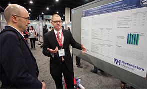
Society of Hospital Medicine’s RIV Poster Contest Draws Best, Brightest
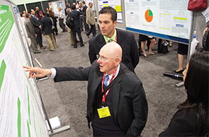
NATIONAL HARBOR, Md.—On one end of the cavernous exhibit hall space at HM15 stood Brendan Sullivan, OMS-II, a second-year medical student, practically grinning as he showcased his poster on the effects of bedside rounds with nurses. On the other side stood Donald Tashkin, MD, a pulmonologist who began his training in the 1960s and was talking like a younger man about his poster on drug therapies for exacerbated cases of COPD.
Both men were first-time presenters at SHM’s annual Research, Innovations, and Clinical Vignettes (RIV) poster competition. The contest has become one of the meeting’s most popular rites, growing so big it now spans two of the conference’s four days. This year’s competition drew a record 1,297 abstracts, topping the prior record of 1,132 and fully double the 634 abstracts submitted for HM10, according to SHM.
What makes the contest popular is that its posters are as varied as the presenters’ motives.
Take Sullivan, a student at Midwestern University Chicago College of Osteopathic Medicine in Downers Grove, Ill. His poster, “Examining the Future of Hospitalist Medicine: Impact of Bedside Rounding with Nurses on Patient Care,” served as his introduction to the specialty.
“You can see the tangible results [hospitalists] have,” he says. “Working with the nurses, the nurses recognize [hospitalists] as a continuous part of hospital life. It just seems like, as a field, there’s definitely a lot of opportunity for medical students like me, who want to go into internal medicine but [are] not really sure what aspect of internal medicine. Hospital medicine is definitely a very viable career option.”
Sullivan’s project came about because of work with his faculty mentor, a second-year hospitalist. At HM15, with the titans of the field walking around him, Sullivan showed his work off proudly but respectfully.
“It’s definitely a learning experience for me,” he says. “I’m just taking a backseat and soaking it all in. I realize that being one of the youngest and more inexperienced members here, I have a lot to learn .… I spent eight weeks in a field they’ve been doing for 20 years.”
But experience doesn’t mean a poster presenter has been here before. Dr. Tashkin, a veteran pulmonologist at UCLA’s David Geffen School of Medicine in Los Angeles, had never been to an SHM annual meeting. He presented two related posters on COPD drug therapies.
Where Sullivan was awed by the experience, Dr. Tashkin was in it for the academic stimulation that comes with bouncing medical ideas off of medical minds.
“It’s an intellectual enjoyment,” he says. “You can learn things when you talk to people, because they give you certain insights that you never thought of before. It’s not about ego; I’ll tell you that.”
Poster presenters say that a lot. The sharing of projects isn’t about adulation, they say. It’s about finding fellow hospitalists who are dealing with the kinds of issues that plague all hospital medicine groups. That’s why Greta Boynton, MD, SFHM, enjoys the RIV sessions.
“When you walk around and see all the great work that other people have done, most people are working on very similar things, like readmission rates or quality or [patient] satisfaction,” says Dr. Boynton, division chief of hospital medicine for Baystate Health in Springfield, Mass. “You get a lot of practical suggestions for things that you could implement in your own group.”
Dr. Boynton, regional medical director for the Northeast for Sound Physicians, has thought that for years, but this year she took the added step of presenting her first two posters. While showcasing one titled “Unit Medical Director as Career Development for Young Hospitalist,” she said years of seeing work similar to her own left her wondering why she didn’t present.
“I’ve done a lot of practice management and process improvement initiatives over the years, and I have not brought them forward here,” she says. “Then when you see other people working on similar things, you kind of kick yourself for not showing how you did it.”
So she did it. And now she’s glad she did.
“I feel proud of my hospitalist team,” Dr. Boynton says. “The fact that people are interested in it, the fact that they’re asking questions—practical questions on how it might look on a smaller team—very rewarding.”
Rehan Qayyum, MBBS, medical director of the academic hospitalist program at University of Tennessee College of Medicine in Chattanooga, Tenn., has found one reward particularly useful: peer review.
Over the course of roughly 10 posters presented over the years, he has used the comments of passersby to hone his writing skills. He is now transforming his poster, “Effect of HCAHPS Reporting Patient Satisfaction with Physicians,” into a paper he plans to publish.
The RIV session is free editing.
“They’re talking about what they think about how I should be looking at things that are not very clear, or things they may have interest in,” Dr. Qayyum says. “When I’m writing the discussion part or when I’m writing the methods and results part, I may focus on those [comments], add those parts, maybe. Or highlight those things in discussion where people show interest.
“I may be more focused in what I’m doing and may lose what may be important for other people. But being here and letting other people see my work and discuss it with me … that helps a lot.”
Richard Quinn is a freelance writer in New Jersey.
Just-In-Time CPR Training Studied in Pediatric Cardiopulmonary Arrest
Clinical question: Does the use of “just-in-time” (JIT) CPR training, with or without visual feedback (VisF), improve the quality of CPR in simulated pediatric cardiopulmonary arrest (CPA)?
Background: Rates of survival to discharge after in-hospital pediatric CPA range from 25%-50%, with three-quarters of survivors having good neurological outcomes.1 The quality of basic life support interventions has been found to be a critical factor influencing survival outcomes.1 Traditional basic life support (BLS) training has not been found to significantly increase compliance with 2010 AHA BLS Guidelines, however.2 Two recent advances have been found to improve the ability of CPR providers to estimate chest compression (CC) depth:
- JIT CPR training, where learners are given video-based training immediately before simulated CPA and
- real-time VisF, where learners are given feedback during CPR regarding rate and depth of CC by a small electronic device.
Visual CPR feedback devices used in recent studies are small (credit card-sized), are placed in the middle of the chest, and use accelerometer technology to provide real-time data regarding CC rate and depth. Prior studies utilizing VisF technology have found learners overestimate their compliance with target CC depth and rate.3
Study design: Prospective, randomized, 2 x 2 factorial-design trial.
Setting: Ten tertiary care teaching hospitals in the U.S. and Canada.
Synopsis: Researchers recruited participants from 10 tertiary care teaching hospitals that are part of the International Network for Simulation-Based Pediatric Innovation, Research, and Education (INSPIRE). Participants included medical students, resident/fellow physicians, nurses, and nurse practitioners. Participants were organized into teams of three, with one participant designated as team leader and two others assigned to perform CCs. Teams were then randomized into four arms as follows:
- Arm 1: No JIT / no VisF
- Arm 2: No JIT / + VisF
- Arm 3: + JIT / no VisF
- Arm 4: + JIT / + VisF
All participants watched a standard video orientation to the study, practiced CPR for two minutes, and participated in a pediatric septic shock simulation scenario (to minimize the Hawthorne effect of being videotaped). Depending on randomization, some teams received JIT CPR training prior to a simulated pediatric CPA scenario. Randomization also determined which teams would utilize a VisF device during CPR to give feedback regarding rate and depth of CCs. Actors were used to play roles of respiratory therapist and medication nurse, and all sites used standardized locations of defibrillator and medication cart.
Overall, quality of CPR was poor, but the JIT CPR training and VisF real-time feedback did result in improvement in CC depth and rate compliance:
- JIT CPR training resulted in a 20% absolute increase in CC depth compliance and a 12% increase in CC rate compliance;
- Real-time VisF resulted in a 15% absolute increase in CC depth compliance and a 40% absolute increase in CC rate compliance; and
- Use of both JIT CPR training and real-time VisF during CPA resulted in the highest rates of CC depth and rate compliance, but no significant interaction effect was observed.
Bottom line: Use of JIT CPR training prior to pediatric CPA and a real-time visual feedback device during CPR improves compliance with CC rate and depth guidelines during simulated pediatric CPA.
Citation: Cheng A, Brown LL, Duff JP. Improving cardiopulmonary resuscitation with a CPR feedback device and refresher simulations (CPR CARES study): a randomized clinical trial. JAMA Pediatr. 2015;169(2):137-144.

References
- Topjian AA, Nadkarni VM, Berg RA. Cardiopulmonary resuscitation in children. Curr Opin Crit Care. 2009;15(3):203-208.
- Sutton RM, Wolfe H, Nishisaki A. Pushing harder, pushing faster, minimizing interruptions but falling short of 2010 cardiopulmonary resuscitation targets during in-hospital pediatric and adolescent resuscitation. Resuscitation. 2013;84(12):1680-1684.
- Cheng A, Overly F, Kessler D, et al. Perception of CPR quality: influence of CPR feedback, Just-in-Time CPR training and provider role. Resuscitation. 2015;87: 44-50.
Clinical question: Does the use of “just-in-time” (JIT) CPR training, with or without visual feedback (VisF), improve the quality of CPR in simulated pediatric cardiopulmonary arrest (CPA)?
Background: Rates of survival to discharge after in-hospital pediatric CPA range from 25%-50%, with three-quarters of survivors having good neurological outcomes.1 The quality of basic life support interventions has been found to be a critical factor influencing survival outcomes.1 Traditional basic life support (BLS) training has not been found to significantly increase compliance with 2010 AHA BLS Guidelines, however.2 Two recent advances have been found to improve the ability of CPR providers to estimate chest compression (CC) depth:
- JIT CPR training, where learners are given video-based training immediately before simulated CPA and
- real-time VisF, where learners are given feedback during CPR regarding rate and depth of CC by a small electronic device.
Visual CPR feedback devices used in recent studies are small (credit card-sized), are placed in the middle of the chest, and use accelerometer technology to provide real-time data regarding CC rate and depth. Prior studies utilizing VisF technology have found learners overestimate their compliance with target CC depth and rate.3
Study design: Prospective, randomized, 2 x 2 factorial-design trial.
Setting: Ten tertiary care teaching hospitals in the U.S. and Canada.
Synopsis: Researchers recruited participants from 10 tertiary care teaching hospitals that are part of the International Network for Simulation-Based Pediatric Innovation, Research, and Education (INSPIRE). Participants included medical students, resident/fellow physicians, nurses, and nurse practitioners. Participants were organized into teams of three, with one participant designated as team leader and two others assigned to perform CCs. Teams were then randomized into four arms as follows:
- Arm 1: No JIT / no VisF
- Arm 2: No JIT / + VisF
- Arm 3: + JIT / no VisF
- Arm 4: + JIT / + VisF
All participants watched a standard video orientation to the study, practiced CPR for two minutes, and participated in a pediatric septic shock simulation scenario (to minimize the Hawthorne effect of being videotaped). Depending on randomization, some teams received JIT CPR training prior to a simulated pediatric CPA scenario. Randomization also determined which teams would utilize a VisF device during CPR to give feedback regarding rate and depth of CCs. Actors were used to play roles of respiratory therapist and medication nurse, and all sites used standardized locations of defibrillator and medication cart.
Overall, quality of CPR was poor, but the JIT CPR training and VisF real-time feedback did result in improvement in CC depth and rate compliance:
- JIT CPR training resulted in a 20% absolute increase in CC depth compliance and a 12% increase in CC rate compliance;
- Real-time VisF resulted in a 15% absolute increase in CC depth compliance and a 40% absolute increase in CC rate compliance; and
- Use of both JIT CPR training and real-time VisF during CPA resulted in the highest rates of CC depth and rate compliance, but no significant interaction effect was observed.
Bottom line: Use of JIT CPR training prior to pediatric CPA and a real-time visual feedback device during CPR improves compliance with CC rate and depth guidelines during simulated pediatric CPA.
Citation: Cheng A, Brown LL, Duff JP. Improving cardiopulmonary resuscitation with a CPR feedback device and refresher simulations (CPR CARES study): a randomized clinical trial. JAMA Pediatr. 2015;169(2):137-144.
References
- Topjian AA, Nadkarni VM, Berg RA. Cardiopulmonary resuscitation in children. Curr Opin Crit Care. 2009;15(3):203-208.
- Sutton RM, Wolfe H, Nishisaki A. Pushing harder, pushing faster, minimizing interruptions but falling short of 2010 cardiopulmonary resuscitation targets during in-hospital pediatric and adolescent resuscitation. Resuscitation. 2013;84(12):1680-1684.
- Cheng A, Overly F, Kessler D, et al. Perception of CPR quality: influence of CPR feedback, Just-in-Time CPR training and provider role. Resuscitation. 2015;87: 44-50.
Tips for Hospitalists to Understand, Promote Patient Satisfaction

Patient satisfaction—“the patient experience”—is given great weight by hospitals and the public alike. Physicians have always aspired to take excellent care of patients. What has changed is that assessments of the patient experience are now being used to measure and report the quality of our care. Although there are many venues for patients to share their opinions, including reviews and online ratings, only the HCAHPS (Hospital Consumer Assessment of Healthcare Providers and Systems) survey is standardized and allows for comparisons nationwide.
Given that HCAHPS is the standard by which hospitals, health systems, and individual hospitalists are judged, it is vital for us to understand the core drivers of measured patient experience—especially the factors within our control. Armed with this knowledge, we can more effectively promote a positive experience within our daily patient care.
Understanding HCAHPS
HCAHPS (H-caps) is a national, standardized, and publicly reported survey of patients’ experiences in the hospital. The Centers for Medicare and Medicaid Services (CMS) and the Agency for Healthcare Research and Quality (AHRQ) developed and piloted the survey in 2002 and launched it in October 2006, with results first published in March 2008 on the Hospital Compare website.
The survey must be administered to a random sample of hospital inpatients 48 hours to six weeks after discharge, and it is offered in multiple languages, either by phone or mail. Twenty-one core questions cover seven composites (communication with doctors, communication with nurses, responsiveness of hospital staff, pain control, communication about new medications, discharge information and planning, and cleanliness/quietness) and two global items (patients’ overall rating of the hospital and likelihood to recommend it to family and friends). There are several additional questions adjusting for patient mix between hospitals, as well as any supplementary questions desired by individual hospitals.1
With the exception of the two global items, all core HCAHPS questions ask “how often” a patient experienced a particular aspect of hospital care. Possible answers are “Always,” “Usually,” “Sometimes,” or “Never”; credit is given to the hospital only for a “top box” score of “Always.” The three questions that are most applicable to hospitalists make up the “communication with doctors” composite; they focus on the quality of physician-patient communication:
- During this hospital stay, how often did doctors treat you with courtesy and respect?
- During this hospital stay, how often did doctors listen carefully to you?
- During this hospital stay, how often did doctors explain things in a way you could understand?
Importance to Hospitalists
A tremendous amount is tied to HCAHPS scores: hospital reimbursement from CMS (through value-based purchasing), hospital rating and “brand,” patients’ choice of hospital, and, in some cases, hospitalist performance bonuses. Hospitals and health systems therefore emphasize HCAHPS heavily. This emphasis has sparked some controversy, particularly surrounding the risk that inappropriate medical decisions (e.g. prescribing antimicrobials or pain medications when not indicated) will be made in order to generate higher patient satisfaction scores. Given the evidence that physicians’ biomedical skill and interpersonal qualities are equally important in determining patient satisfaction, however, we can remain optimistic that time spent explaining the rationale for appropriate medical care is highly valued by patients.2,3
The more satisfied patients are with the care they receive, the more likely they are to continue to seek care from the same provider, hospital, and/or clinic. Improved continuity can help increase adherence, improve patient safety, and decrease healthcare costs. Positive healthcare experiences are also correlated with improved patient compliance with treatment regimens and medical advice, which will lead to better outcomes.
We must also recognize that both the patient experience and physician-patient communication impact clinical care. First, a positive patient experience is linked to higher continuity of care.4 The more satisfied patients are with the care they receive, the more likely they are to continue to seek care from the same provider, hospital, and/or clinic. Improved continuity can help increase adherence, improve patient safety, and decrease healthcare costs. Positive healthcare experiences are also correlated with improved patient compliance with treatment regimens and medical advice, which will lead to better outcomes.5,6
Additionally, higher patient satisfaction is associated with decreased readmission rates. An analysis of more than 2,500 hospitals demonstrated a statistically significant correlation between lower 30-day risk-standardized readmission rates, higher patient satisfaction with discharge planning, and higher overall patient satisfaction with care.7
Furthermore, high quality physician-patient communication has been linked to improved health outcomes. A meta-analysis of 21 separate studies evaluating the effect of communication on health outcomes demonstrated a direct positive correlation with five outcome measures—emotional health, symptom resolution, functional status, physiologic measures (blood pressure and glycemic control), and pain control.8
Finally, higher patient satisfaction and improved physician-patient communication are inversely correlated with medical malpractice risk.9,10
Current data regarding the effect of patient satisfaction on mortality and healthcare utilization/expenditures are conflicting. Jaipaul and Rosenthal found that higher patient satisfaction was associated with decreased mortality.11 Conversely, Fenton and colleagues found an association between high patient satisfaction and both increased mortality and higher healthcare utilization/costs.12 More long-term data will be helpful in clarifying this question.
For hospitalists, the importance of patient satisfaction might reach beyond its clinical impact. Both new residency graduates and more seasoned hospitalists will find that their personal HCAHPS scores can either be highlighted as a strength or work to their detriment when they apply for new positions. Many physicians find that they are asked about their patient satisfaction scores during job interviews. Being knowledgeable about both the patient experience and whether your patients perceive you positively can be an asset.
What Influences Patient Satisfaction, and How Do We Promote It?
Studies show that excellent medical care and strong interpersonal qualities are equally important influences on patients’ satisfaction with physicians.2,3 Having a high quality interaction with their doctor—during which patients feel that they are valued and listened to, that their opinions are taken into consideration, and that they have received a clear explanation—is more important to patients than having a lengthy visit with their provider.13
Consequently, interventions that focus on improving the humanistic aspects of our care and enhancing the quality of our communication will be the most effective strategies for improving patient satisfaction. Remembering to practice empathy for our patients in the midst of our very busy and stressful workdays is an excellent start. We can also utilize the following proven practices for enhancing physician-patient communication:
- Sit down at the bedside;
- Use patient-centered communication techniques, such as asking open-ended questions, using the teach-back method and shared decision-making, and avoiding jargon;
- Clearly outline the plan for the day and explain how it fits into the overall goal of the hospitalization;
- Invite questions; and
- Utilize patient whiteboards.
In addition, demonstrating to patients that we collaborate and effectively communicate with the rest of the healthcare team can also enhance their experience.
Final Thoughts
Ultimately, patient satisfaction should not be regarded as an extraneous amenity for our patients or as a necessary evil to placate hospital administrators. Instead, improving our patients’ hospital experience can help improve their overall care and health.
Strong physician-patient partnerships and high patient satisfaction increase continuity of care and adherence to treatment, while also resulting in better health outcomes and decreased hospital readmission rates. Furthermore, if hospitalists emphasize a positive patient experience by fostering effective communication and positive relationships, they can also decrease their malpractice risk.
We must therefore find ways to foster patient satisfaction while maintaining safe, effective, quality-driven patient care. Emphasizing humanism and communication, while providing safe and high quality care, is the optimal way to promote patient satisfaction. In this way, we can improve not only the patient experience but also health outcomes.
Dr. Bergin is an academic hospitalist for the internal medicine residency program at Banner-University Medical Center Phoenix in Arizona and a clinical assistant professor at the University of Arizona College of Medicine. Dr. O’Malley is the internal medicine residency program director at Banner and an assistant professor of medicine at the University of Arizona College of Medicine. She currently serves as SHM’s representative on the Alliance for Academic Internal Medicine’s Internal Medicine Education Redesign Advisory Board. Dr. Donahue is assistant professor of medicine at the University of Massachusetts Medical School in Worcester.
References
- Agency for Healthcare Research and Quality. HCAHPS Fact Sheet (CAHPS Hospital Survey) – August 2013. Available at: http://www.hcahpsonline.org/files/August_2013_HCAHPS_Fact_Sheet3.pdf. Accessed April 9, 2015.
- Matthews DA, Sledge WH, Lieberman PB. Evaluation of intern performance by medical inpatients. Am J Med. 1987;83(5):938-944.
- Matthews DA, Feinstein AR. A new instrument for patients’ ratings of physician performance in the hospital setting. J Gen Intern Med. 1989;4(1):14-22.
- Safran DG, Montgomery JE, Change H, Murphy J, Rogers WH. Switching doctors: Predictors of voluntary disenrollment from a primary physician’s practice. J Fam Pract. 2001;50(2):130-136.
- DeMatteo MR. Enhancing patient adherence to medical recommendations. JAMA. 1994:271(1):79, 83.
- Safran DG, Taira DA, Rogers WH, Kosinski M, Ware JE, Tarlov AR. Linking primary care performance to outcomes of care. J Fam Pract. 1998;47(3):213-220.
- Boulding W, Glickman SW, Manary MP, Schulman KA, Staelin R. Relationship between patient satisfaction with inpatient care and hospital readmission within 30 days. Am J Manag Care. 2011;17(1):41-48.
- Stewart MA. Effective physician-patient communication and health outcomes: a review. CMAJ. 1995;152(9):1423-1433.
- Tan SY. Issues in medical malpractice IX. Doctors most prone to lawsuits. Hawaii Med J. 2007;66(3):78-79.
- Beckman HB, Markakis KM, Suchman AL, Frankel RM. The doctor-patient relationship and malpractice: Lessons from plaintiff depositions. Arch Intern Med. 1994;154(12):1365-1370.
- Jaipaul CK, Rosenthal GE. Do hospitals with lower mortality have higher patient satisfaction? A regional analysis of patients with medical diagnoses. Am J Med Qual. 2003;18(2):59-65.
- Fenton JJ, Jerant AF, Bertakis KD, Franks P. The cost of satisfaction: a national study of patient satisfaction, health care utilization, expenditures, and mortality. Arch Intern Med. 2012;172(5):405-411.
- Blanden AR, Rohr RE. Cognitive interview techniques reveal specific behaviors and issues that could affect patient satisfaction relative to hospitalists. J Hosp Med. 2009;4(9):E1-6.