User login
Patient Satisfaction Surveys Not Accurate Measure of Hospitalists’ Performance

Feeling frustrated with your group’s patient-satisfaction performance? Wondering why your chief (fill in the blank) officer glazes over when you try to explain why your hospitalist group’s Hospital Consumer Assessment of Healthcare Providers and System (HCAHPS) scores for doctor communication are in a percentile rivaling the numeric age of your children?
It is likely that the C-suite administrator overseeing your hospitalist group has a portion of their pay based on HCAHPS or other patient-satisfaction (also called patient experience) scores. And for good reason: The Centers for Medicare & Medicaid Services (CMS) Hospital Value-Based Purchasing (HVBP) program that started Oct. 1, 2012, has placed your hospital’s Medicare reimbursement at risk based on its HCAHPS scores.
HVBP and Patient Satisfaction
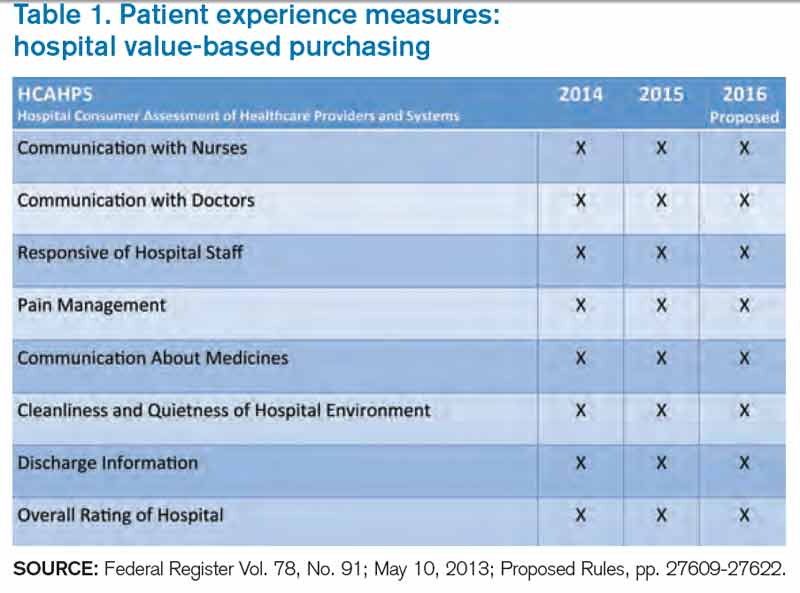
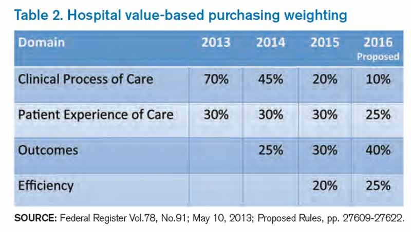
Patient satisfaction will remain an important part of HVBP in the coming years. Table 1 (below) shows the domains that will be included in fiscal years 2014 (which starts Oct. 1, 2013), 2015, and 2016. Table 2 (below) depicts the percent weighting the patient-satisfaction domain will receive through 2016. You may recall that HVBP is a program in which all hospitals place 1% to 2% (2013 through 2017, starting at 1% and increasing each year by 0.25% so that by 2017%, it is 2%) of their CMS inpatient payments in a withhold pool and, based on performance, can make back some, all, or an amount in excess of the amount placed in the withhold pool.
Source: Federal Register Vol. 78, No. 91; May 10, 2013; Proposed Rules, pp. 27609-27622.
Source: Federal Register Vol.78, No.91; May 10, 2013; Proposed Rules, pp. 27609-27622.
End In Itself
A colleague of mine recently asked, “Is an increase in patient satisfaction associated with higher quality of care and better patient safety?” The point here: It doesn’t matter. Patient satisfaction is an end in itself, and we should strive to maximize it, or at least put ourselves in the place of the patient and design care accordingly.
For Hospitalists: A Starting Point
There is a conundrum for hospitalists vis-à-vis patient satisfaction. Follow this chain of logic: The hospitals at which we work are incented to perform well on the HCAHPS domains. Hospitals pay a lot for hospitalists. Hospitalists can impact many of the HCAHPS domains. So shouldn’t hospitalists be judged according to HCAHPS scores?
Yes and no.
HCAHPS as a survey is intended to measure a patient’s overall experience of receiving care in the hospital. For example, from the “Doctor Communication” domain, we have questions like “how often did doctors treat you with courtesy and respect?” And “how often did doctors explain things in a way you could understand?”
These questions, like all in HCAHPS, are not designed to get at individual doctor performance, or even performance of a group of doctors, such as hospitalists. Instead, they are designed to measure a patient’s overall experience with the hospitalization, and “Doctor Communication” questions are designed to assess satisfaction with “doctors” collectively.
The Need for Hospitalist-Specific Satisfaction Surveys
So while HCAHPS is not designed to measure hospitalist performance with regard to patient satisfaction, it is a reasonable interim step for hospitals to judge hospitalists according to HCAHPS. However, this should be a bridge to a strategy that adopts hospitalist-specific patient-satisfaction questionnaires in the future and not an end in itself.
Why? Perhaps the biggest reason is that HCAHPS scores are neither specific nor timely enough to form the basis of improvement efforts for hospitalists. If a hospitalist receives a low score on the “Doctor Communication” domain, the scores are likely to be three to nine months old. How can we legitimately assign (and then modify) behaviors based on those scores?
Further, because the survey is not built to measure patient satisfaction specifically with hospitalists, the results are unlikely to engender meaningful and sustained behavior change. Hospitalists I talk to are generally bewildered and confused by HCAHPS scores attributed to them or their groups. Even if they understand the scores, I almost never see true quality improvement (plan-do-study-act) based on specific HCAHPS results. Instead, I see hospitalists trying to adhere to “best practices,” with no adjustments made along the way based on performance.
Nearly all the prominent patient satisfaction vendors have developed a survey instrument specifically designed for hospitalists. Each has an approach to appropriately attribute performance to the hospitalist in question, and each has a battery of questions that is designed to capture patient satisfaction with the hospitalist. Although use of these surveys involves an added financial commitment, I submit that because hospitalists have an unparalleled proximity to hospitalized patients, such an investment is worthy of consideration and has an accompanying business case, thanks to HVBP. The results of these surveys may form the basis of legitimate, targeted feedback to hospitalists, who may then adjust their approach to patient interactions. Such performance improvement should result in improved HCAHPS scores.
In sum, hospitalists should pay close attention to patient satisfaction and embrace HCAHPS. However, we should be looking beyond HCAHPS to survey instruments that fairly and accurately measure our performance. Such surveys will be more widely accepted by the hospitalists they are measuring, and will allow hospitalists to perform meaningful quality improvement based on the results. Although hospitalist-specific surveys will require an investment, the increased patient satisfaction that results should be the basis of a favorable return on that investment.
Dr. Whitcomb is medical director of healthcare quality at Baystate Medical Center in Springfield, Mass. He is co-founder and past president of SHM. Email him at [email protected].
Feeling frustrated with your group’s patient-satisfaction performance? Wondering why your chief (fill in the blank) officer glazes over when you try to explain why your hospitalist group’s Hospital Consumer Assessment of Healthcare Providers and System (HCAHPS) scores for doctor communication are in a percentile rivaling the numeric age of your children?
It is likely that the C-suite administrator overseeing your hospitalist group has a portion of their pay based on HCAHPS or other patient-satisfaction (also called patient experience) scores. And for good reason: The Centers for Medicare & Medicaid Services (CMS) Hospital Value-Based Purchasing (HVBP) program that started Oct. 1, 2012, has placed your hospital’s Medicare reimbursement at risk based on its HCAHPS scores.
HVBP and Patient Satisfaction
Patient satisfaction will remain an important part of HVBP in the coming years. Table 1 (below) shows the domains that will be included in fiscal years 2014 (which starts Oct. 1, 2013), 2015, and 2016. Table 2 (below) depicts the percent weighting the patient-satisfaction domain will receive through 2016. You may recall that HVBP is a program in which all hospitals place 1% to 2% (2013 through 2017, starting at 1% and increasing each year by 0.25% so that by 2017%, it is 2%) of their CMS inpatient payments in a withhold pool and, based on performance, can make back some, all, or an amount in excess of the amount placed in the withhold pool.
Source: Federal Register Vol. 78, No. 91; May 10, 2013; Proposed Rules, pp. 27609-27622.
Source: Federal Register Vol.78, No.91; May 10, 2013; Proposed Rules, pp. 27609-27622.
End In Itself
A colleague of mine recently asked, “Is an increase in patient satisfaction associated with higher quality of care and better patient safety?” The point here: It doesn’t matter. Patient satisfaction is an end in itself, and we should strive to maximize it, or at least put ourselves in the place of the patient and design care accordingly.
For Hospitalists: A Starting Point
There is a conundrum for hospitalists vis-à-vis patient satisfaction. Follow this chain of logic: The hospitals at which we work are incented to perform well on the HCAHPS domains. Hospitals pay a lot for hospitalists. Hospitalists can impact many of the HCAHPS domains. So shouldn’t hospitalists be judged according to HCAHPS scores?
Yes and no.
HCAHPS as a survey is intended to measure a patient’s overall experience of receiving care in the hospital. For example, from the “Doctor Communication” domain, we have questions like “how often did doctors treat you with courtesy and respect?” And “how often did doctors explain things in a way you could understand?”
These questions, like all in HCAHPS, are not designed to get at individual doctor performance, or even performance of a group of doctors, such as hospitalists. Instead, they are designed to measure a patient’s overall experience with the hospitalization, and “Doctor Communication” questions are designed to assess satisfaction with “doctors” collectively.
The Need for Hospitalist-Specific Satisfaction Surveys
So while HCAHPS is not designed to measure hospitalist performance with regard to patient satisfaction, it is a reasonable interim step for hospitals to judge hospitalists according to HCAHPS. However, this should be a bridge to a strategy that adopts hospitalist-specific patient-satisfaction questionnaires in the future and not an end in itself.
Why? Perhaps the biggest reason is that HCAHPS scores are neither specific nor timely enough to form the basis of improvement efforts for hospitalists. If a hospitalist receives a low score on the “Doctor Communication” domain, the scores are likely to be three to nine months old. How can we legitimately assign (and then modify) behaviors based on those scores?
Further, because the survey is not built to measure patient satisfaction specifically with hospitalists, the results are unlikely to engender meaningful and sustained behavior change. Hospitalists I talk to are generally bewildered and confused by HCAHPS scores attributed to them or their groups. Even if they understand the scores, I almost never see true quality improvement (plan-do-study-act) based on specific HCAHPS results. Instead, I see hospitalists trying to adhere to “best practices,” with no adjustments made along the way based on performance.
Nearly all the prominent patient satisfaction vendors have developed a survey instrument specifically designed for hospitalists. Each has an approach to appropriately attribute performance to the hospitalist in question, and each has a battery of questions that is designed to capture patient satisfaction with the hospitalist. Although use of these surveys involves an added financial commitment, I submit that because hospitalists have an unparalleled proximity to hospitalized patients, such an investment is worthy of consideration and has an accompanying business case, thanks to HVBP. The results of these surveys may form the basis of legitimate, targeted feedback to hospitalists, who may then adjust their approach to patient interactions. Such performance improvement should result in improved HCAHPS scores.
In sum, hospitalists should pay close attention to patient satisfaction and embrace HCAHPS. However, we should be looking beyond HCAHPS to survey instruments that fairly and accurately measure our performance. Such surveys will be more widely accepted by the hospitalists they are measuring, and will allow hospitalists to perform meaningful quality improvement based on the results. Although hospitalist-specific surveys will require an investment, the increased patient satisfaction that results should be the basis of a favorable return on that investment.
Dr. Whitcomb is medical director of healthcare quality at Baystate Medical Center in Springfield, Mass. He is co-founder and past president of SHM. Email him at [email protected].
Hospitalists’ Capitol Hill Advocacy Effort Produces Results

On May 12, 113 hospitalists descended on Capitol Hill for “Hospitalists on the Hill 2013,” the public-advocacy highlight of SHM’s annual meeting. Hospitalists from all parts of the country engaged with congressional representatives in a daylong series of meet-and-greets that may seem to some people useless in the face of political obstinacy in Washington. But the trip worked.
Josh Boswell, SHM’s senior manager of government relations, reports many Hill Day objectives were achieved:
- The number of legislators co-sponsoring a bill regarding the “three-day observation rule” more than tripled in the House of Representatives and doubled in the Senate. SHM officials note that the added support has come from both political parties.
- A Congressional Budget Office (CBO) review of the bill has been formally requested by those legislators.
- A congressman from Washington state asked for—and received—a letter of support for a proposed measure, the Improved Health Care at a Lower Cost Act of 2013 (H.R. 1487).
- Multiple reports of continued dialogue between congressional staffers and SHM members nationwide. When planning the advocacy day, SHM officials noted that one of the most valuable results is creating relationships at the local level.
Observation Legislation
One of the three talking points hospitalists took into their legislative meetings was solving the dilemmas surrounding observation status. Currently, time spent on observation status does not count toward the required three consecutive overnights an inpatient needs to qualify for Medicare benefits at a skilled nursing facility (SNF).
Hospitalists have been pushing to change that rule, in large part by supporting the Improving Access to Medicare Coverage Act of 2013 (H.R. 1179, S. 569), sponsored by Rep. Joe Courtney (D-Conn.), Rep. Tom Latham (R-Iowa), and Sen. Sherrod Brown (D-Ohio).1 In addition to the status reclassification, the proposal would establish a 90-day appeal period for those who have been denied the benefit.
The issue is important to hospitalists because of the penalties hospitals face for readmissions—and also in part because hospitalists increasingly are providing care at SNFs and other post-acute-care facilities. SHM says that after the Hill visits—and the ensuing follow-up communications—the number of co-sponsors in the House jumped to 70 from 22. The Senate version doubled its list of co-sponsors.
And, perhaps more important, a CBO analysis has been requested for the observation bills. That review, known as a CBO score, weighs the financial impacts of proposed laws and is considered a necessary precursor to successfully passing any legislation.
All in all, SHM was pleased with the progress on the observation-status bill and will continue to push for its passage, whether it is in this congressional session or the next.
“Rep. Courtney’s bill is now getting significant traction,” Boswell says. “Hospitalists should be proud to know this is in no small part due to their advocacy efforts.”
Political Networking
Hospitalist David Ramenofsky, MD, who works at Northwest Hospital and Medical Center in Seattle, wasn’t sure how much traction he was going to be able to generate at his first Hill Day. SHM had arranged meetings with the offices of three local politicians: Rep. Jim McDermott (D-Wash.), Sen. Patty Murray (D-Wash.), and Sen. Maria Cantwell (D-Wash.).
Dr. Ramenofsky sat with two of McDermott’s staffers, one of whom sounded knowledgeable and enthused about health-care-policy issues. Although the congressman couldn’t sit in on the meeting, he knew Dr. Ramenofsky’s name and took the time to say hello.
“It was really interesting to me that these staffers wanted to hear what I had to say and learn about my experience,” Dr. Ramenofsky adds. “My views may affect how they work with their bosses to make policy changes. It was surprising to me how much my opinions mattered to them.”
After the meeting and another briefing SHM arranged with another local hospitalist, McDermott reached out to SHM. He asked for support for his proposed bill to expand protections from anti-kickback laws and regulations, to provide safe harbor protection for gainsharing, and other incentive-payment systems.
SHM responded in July with a letter of support that thanked the congressman for his efforts.2
“We look forward to working with you,” the letter ended.
Dr. Ramenofsky says he’s proud his efforts led to a working relationship between his professional society and his local legislator. He says he’s looking forward to participating in future Hill Day activities and acting as a local liaison for SHM.
He laments that he has not received much post-meeting feedback from his discussions with the senators’ offices, but says he understands how busy politicians are. And a 1-for-3 showing is pretty good, given his status as a political novice.
“Given overall public perception of Congress, I’m amazed that my visits caused one of three offices to engage in further policy discussions with SHM,” he says. “I’m encouraged to remain engaged in political activities through SHM.”
Richard Quinn is a freelance writer in New Jersey.
References
- Society of Hospital Medicine. Letter to Congress members. Society of Hospital Medicine website. Available at: http://www.hospitalmedicine.org/AM/Template.cfm?Section=Letters_to_Congress_and_ Regulatory_Agencies&Template=/CM/ContentDisplay.cfm&ContentID=33169. Accessed July 15, 2013.
- Society of Hospital Medicine. Letter to Congressman Jim McDermott. Society of Hospital Medicine website. Available at: http://www.hospitalmedicine.org/AM/Template.cfm?Section=Letters_to_Congress_and_Regulatory_Agencies&Template=/CM/ContentDisplay.cfm&ContentID=34169. Accessed July 15, 2013.
Goals, Patient-Centered Decisions Drive Hospitalist Julianna Lindsey


Growing up on a farm in rural Kentucky could have led to a career in the family business for Julianna Lindsey, MD, MBA, FHM. Except she knew at an early age that she wanted to be a doctor.
“My family physician was very influential on my decision to become a physician,” she says. “[He] mentored and encouraged me from a young age; it was very powerful for me.”
Dr. Lindsey earned bachelor’s degrees in biomedical science from the University of South Alabama and biochemistry from Western Kentucky University. She graduated from the University of Kentucky College of Medicine and completed her internal-medicine residency at the University of Kentucky. In 2011, she earned her master’s in business administration from the University of Tennessee.
Immediately following residency, she worked for the Veterans Affairs Medical Center in Lexington, Ky., as an ED physician. In 2002, she latched on to a career in HM when she and her husband, a gastroenterologist, relocated to Knoxville, Tenn. She recently launched a startup company, Synergy Surgicalists, with two orthopedic surgeons, and also provides process-improvement and leadership-development consulting.
She says she was told early in her career to know your goals and stay focused.
“That has been the guiding light for me throughout my career,” says Dr. Lindsey, one of nine new Team Hospitalist members, The Hospitalist’s volunteer editorial advisory group. “My goal is to make medical care better and safer for hospitalized patients. We increasingly need to figure out how to do that with fewer and fewer resources. Regardless, we can never move backward on delivering better and safer care to patients.”
Question: How did you decide to become a hospitalist?
Answer: I have always been drawn to the practice of acute-care medicine. I enjoy taking care of patients and their families in their times of need. From the purely diagnostic standpoint, I very much enjoy the critical decision-making required in the diagnosis and treatment of the acutely ill patient.
Q: What do you like most about working as a hospitalist?
A: I enjoy the opportunity to “dig in” and positively affect processes and patient outcomes throughout hospitals.
Q: What do you dislike most?
A: Fighting the “scope creep” that is continually pushing on us as hospitalists. Hospitalists are constantly being asked to admit patients whose problems are outside the scope of our practice as medically trained physicians. A few examples of this include acute surgical abdomens, intracranial hemorrhages, and blunt-trauma cases.
Q: What’s the biggest change you’ve seen in HM in your career?
A: The explosion of hospitalist programs throughout the country. Hospitalists programs are now even being built by payors and long-term-care facilities.
Q: For group leaders, why is it important for you to continue seeing patients?
A: In order to improve upon a process, you must know the process; to truly know the process, you must live the process. If you are not at the bedside delivering care to patients, there will be a disconnect between you, as a leader, and your physicians, who are at the bedside delivering care.
Q: What are your interests outside of patient care?
A: I believe the success—or failure—of a hospital, physician group, corporation, etc. is directly related to leadership. I enjoy leadership development because I see that as “mission critical” to the success of delivering better and safer patient care in any health-care system. As physicians, most of us never receive meaningful leadership training, yet are expected to come out of residency ready to lead. I enjoy providing physicians the tools to lead effectively. It makes the careers of physician leaders more fulfilling, as well as the careers of those physicians who are “following.”
Q: What is your biggest professional challenge?
A: Continuing to provide better and safer patient care with diminishing resources.
Q: What is your biggest professional reward?
A: Making a difference in the lives of patients. It is very rewarding to me to be able to come into a hospital and put processes in place, then actually see the risk-adjusted mortality rates improve. One of my teams’ biggest wins was taking over an HM program in a hospital with a mortality rate of 4, and seeing that mortality rate cut literally in half within six months.
Q: When you aren’t working, what is important to you?
A: My husband and children are the most important aspect of my life. My husband is a gastroenterologist; we have been married for 13 years. We have two healthy, happy kiddos ages 8 and 10.
Q: What’s next professionally? Where do you see yourself in 10 years?
A: I am partnering with two orthopedic surgeons in a startup company, Synergy Surgicalists. Our company mirrors the hospitalist model utilizing general and orthopedic surgeons. It’s very exciting to have the opportunity to bring value to hospitals and patients on a larger scale. Also, for the immediate future, I have accepted the role of interim executive medical director for hospital medicine for University of Texas Southwestern and Parkland hospitals. We are completely restructuring those programs in preparation for moving into two beautiful new (and very large) hospitals. I’m very excited about working with a truly excellent group of physicians and leaders while we are recruiting a permanent executive director and expanding our ranks.
Q: If you weren’t a doctor, what would you be doing right now?
A: I cannot imagine not being a physician. I suppose if pressed, I imagine I would have landed somewhere in the financial industry. I am also a musician, but have a hard time seeing myself employed in that industry.
Q: What’s the best book you’ve read recently?
A: “Widow Walk” by Gerard LaSalle. He is a physician author who pens a beautiful story. It’s just an enjoyable read of American historical fiction set in the Pacific Northwest.
Q: How many Apple products do you interface with in a given week?
A: Sadly, I interface with 11 (11!) different Apple products in any given week. (Even sadder: I just came into an iPod Shuffle, so I’m up to 12 … )
Q: What’s next in your Netflix queue?
A: “Fringe,” Season 2, Episode 19.
Richard Quinn is a freelance writer in New Jersey.
Intravenous Immunoglobulin Most Common Retreatment Approach for Refractory Kawasaki Disease

Clinical question: How is refractory Kawasaki disease (rKD) treated in the United States?
Background: Kawasaki disease (KD) is an immunologically mediated disease of primarily small to medium-sized arteries. It is the most common cause of acquired heart disease in children in the United States.
The current standard of care for KD treatment is a single 2 g/kg dose of intravenous immunoglobulin (IVIG), infused over 10 to 12 hours, accompanied by aspirin (80 to 100 mg/kg/day by mouth in four divided doses). Fevers persistent more than 36 hours after initial treatment represent refractory Kawasaki disease (rKD). There are no current national guidelines or standards for rKD treatment, although a 2004 joint statement from the American Academy of Pediatrics and the American Heart Association suggested a second dose of IVIG for rKD.
Study design: Multicenter, retrospective, cross-sectional study.
Setting: Forty freestanding children’s hospitals.
Synopsis: Researchers examined data obtained from the Pediatric Health Information System (PHIS), a clinical and financial database of care provided at 43 nonprofit, freestanding children’s hospitals in the United States. Data from 40 of these hospitals were deemed complete enough for analysis and were collected from Jan. 1, 2005, to June 30, 2009. Subjects were included if they received at least one dose of IVIG and had a principal diagnosis of KD. To be considered rKD, the subject must have received additional treatment after the initial diagnosis of rKD.
The most commonly used treatment after initial IVIG treatment was retreatment with IVIG (65%), followed by intravenous methylprednisolone (27%), then infliximab (8%). Significant regional variation was observed, with hospitals in the Northeast using methylprednisolone most frequently for rKD (55%). Infliximab was used at a much higher frequency in the West (29%) compared with other regions.
Bottom line: Retreatment with IVIG is the most common treatment for rKD, but significant regional variation exists, possibly due to the influence of regional experts.
Citation: Ghelani SJ, Pastor W, Parikh K. Demographic and treatment variability of refractory Kawasaki Disease: a multicenter analysis from 2005 to 2009. Hospital Pediatrics. 2012;2:71-76.
Reviewed by Pediatric Editor Weijen Chang, MD, SFHM, FAAP, associate clinical professor of medicine and pediatrics at the University of California at San Diego School of Medicine, and a hospitalist at both UCSD Medical Center and Rady Children’s Hospital.
Proposed Bill Would Open Door to Gainsharing Arrangements for Hospitals, Physicians
There is little dispute in the potential for cost savings when gainsharing arrangements incentivize things like product standardization, substitution of lower-cost products, and, most notably for hospitalists, medically appropriate decreases in length of stay. However, well-meaning but overly inclusive federal law makes the legal risk of establishing these arrangements so great that providers recoil at the prospect.
This doesn’t mean that gainsharing isn’t occurring. Currently, Medicare accountable-care organizations (ACOs) have been granted official waivers to establish such arrangements; smaller-scale pilot projects implemented by Medicare also have been granted similar waivers in the past. As availability is limited to participants within officially sanctioned programs, most providers are not able to tap into these cost-saving efforts, though this has not been for lack of trying.
Hospitals and physicians are engaging in a number of clinical joint ventures that have spurred them to seek their own gainsharing waivers by approaching the Office of the Inspector General (OIG). The OIG is the arm of the U.S. Department of Health and Human Services charged with enforcing the applicable laws affecting gainsharing. The OIG responded by cautioning that gainsharing arrangements violate the Social Security Act’s “Civil Monetary Penalty” prohibition against limitation of services to publicly insured patients, in addition to violating the federal Anti-Kickback Law and possibly the “Stark” law. Nonetheless, the OIG concluded it would not impose sanctions for the violations. In short, the OIG declared the proposals illegal but gave the go-ahead. The caveat, of course, is that these opinions are nonbinding, so providers remain understandably timid.
As a result, gainsharing currently remains more or less out of reach for those not participating in a Medicare ACO. This makes little sense at a time when Medicare and the entire health-care system are focusing on how to deliver high-quality, cost-conscious care. For example, if hospitalists are capable of reducing length of stay without detriment to the patient, they should not be legally prohibited from sharing any of the resulting cost savings. Fortunately, U.S. Rep. Jim McDermott (D-Wash.) agrees with this sentiment and has introduced legislation to address the problem.
McDermott introduced the Improved Health Care at Lower Cost Act of 2013 (H.R. 1487) in April. It seeks to exempt monetary incentive payments made by hospitals to physicians from federal anti-kickback and other sanctions. Such exemptions, or safe harbors, would be automatically granted to gainsharing arrangements that meet a pre-determined set of requirements. This means no formal application process or participation in a specific federal program would be required.
Passage of the bill would be a major step in the right direction for providers lacking the resources to navigate legal minefields or establish a full-scale ACO. If well-implemented, it could also generate significant cost savings for Medicare.
It is for these reasons that SHM supports H.R. 1487 and looks forward to working with McDermott in securing its passage.
In the coming months, members of SHM’s Grassroots Network will be encouraging Congress to make this important change to facilitate practice arrangements that provide high-value coordinated care for patients. Stay informed and take action when SHM issues Legislative Action Alerts by signing up for the Grassroots Network at www.hospitalmedicine.org/grassroots.
Josh Boswell is SHM’s senior manager of government relations.
Academic Hospitalist Academy Provides Resources for Success
SHM asked Academic Hospitalist Academy course co-directors Jeffrey Glasheen, MD, SFHM, and Bradley Sharpe, MD, SFHM, why academic hospitalists should attend this year’s academy.
Question: What has you personally excited about this year’s Academic Hospitalist Academy?
Dr. Sharpe: I’ll be honest—it is one of my favorite weeks of the year. It is a tremendous opportunity to engage with academic hospitalists and help them develop the core skills they need to be successful.
Dr. Glasheen: For me, it’s about the energy, the talent, and the excitement that the attendees bring to the course. It ends up being a tremendously energizing week for me personally. To see the talent in the room begin to find outlets for success is truly invigorating.
Q: If you were talking one on one with an academic hospitalist, what would you say to encourage them to attend?
Dr. Sharpe: Based on previous attendees, these four days could truly change your life. We are confident you will leave with newfound energy and enthusiasm and key building blocks to help you be successful when you go back home. Don’t miss it.
Dr. Glasheen: There is a magical transformation that happens every year. Very talented individuals enter the program. Nearly uniformly, they are struggling with the same issues around mentorship, sense of purpose, direction, and resources for success. They all want to be successful but sense there is something that is missing, and within the course of four days, they find it.
Q: What have you heard from previous AHA attendees?
Dr. Sharpe: Here are a couple of emails I received: “I can’t say enough about the AHA. It was possibly the most important 3 days of my young career. Thank you both for the knowledge and guidance.” “I have fond memories of the whole AHA conference. The great Jeff Wiese!!! But most of all, the small group sessions were extremely helpful.”
Dr. Glasheen: To a person, they all say the meeting is “transformational.” They gain skills in teaching, confidence in evaluating learners, methods for scholarly success, and a roadmap for navigating the tricky world on academic medicine. But beyond that, they gain a peer network. They leave tied in with 80 other national colleagues that are struggling with the same issues. This network becomes their home away from home—people they can turn to with a question, catch up with at a national meeting, and look to as exemplars in the field.
Movers and Shakers in Hospital Medicine

Hospitalist Robert McNab, DO, has been named the new medical education director of Freeman Health System in Joplin, Mo. He will continue as a hospitalist and director of the internal-medicine residency program. Dr. McNab brings more than 10 years of teaching experience to the Freeman Graduate Medical Education Program.
Brian Harte, MD, SFHM, Cleveland Clinic hospitalist and president of South Pointe Hospital in Warrensville Heights, Ohio, is now president of Hillcrest Hospital in Mayfield Heights, Ohio. Dr. Harte is an SHM board member and serves as the board treasurer. In addition to his new role, Dr. Harte will continue to practice as a hospitalist at Hillcrest Hospital.
Anita Dhople, MD, is the new hospitalist medical director for the Rockledge, Fla.-based Health First health system. Dr. Dhople will oversee all hospitalist services at four affiliated hospitals. Dr. Dhople comes to Health First from the Piedmont Physicians Group in Atlanta.
Matthew Heinz, MD, has been appointed director of provider outreach in the U.S. Department of Health and Human Services’ Office of Intergovernmental and External Affairs (IEA). Dr. Heinz is a practicing hospitalist at Tucson (Ariz.) Medical Center and a former representative in the Arizona state legislature.
Business Moves


IPC: The Hospitalist Company, based in North Hollywood, Calif., has announced the acquisition of two private hospitalist practices: Sound Senior Geriatrics LLC (SSG) in Mystic, Conn., and Cape Coral Hospitalists Inc. (CCH) based in Fort Meyers, Fla. IPC contracts hospitalist services in 28 states.
Bayhealth hospital network in Dover, Del., has partnered with Apogee Physicians to provide 24-hour hospitalist services at Kent General Hospital in Dover and Milford Memorial Hospital in Milford. Bayhealth has been serving communities in central and southern Delaware for more than 75 years. Phoenix-based Apogee Physicians has been providing contracted hospitalist services since 2002.
Cogent Healthcare is partnering with South Georgia Medical Center (SGMC) to provide hospitalist services at the 285-bed acute-care nonprofit main campus and the 55-bed Smith Northview Campus in Valdosta, Ga. SGMC currently serves 15 counties in south-central Georgia and north Florida. Cogent provides contracted hospitalist services at more than 100 hospitals throughout the United States.
Cogent Healthcare Inc., based in Brentwood, Tenn., recently announced Dean Weiland as the private hospitalist staffing company’s new president and CEO. Weiland served as an executive during a merger between Renal Advantage and Liberty Dialysis in 2010. Before that, he served as CEO of The Work Institute.
Hill Country Memorial Hospital (HCM) in Fredericksburg, Texas, has announced plans to expand its 24-hour hospitalist service from only weekends to seven days a week. The decision comes as a reaction to the positive benefits HCM has experienced since it began its weekend hospitalist service in 2011.
Hospitalist Robert McNab, DO, has been named the new medical education director of Freeman Health System in Joplin, Mo. He will continue as a hospitalist and director of the internal-medicine residency program. Dr. McNab brings more than 10 years of teaching experience to the Freeman Graduate Medical Education Program.
Brian Harte, MD, SFHM, Cleveland Clinic hospitalist and president of South Pointe Hospital in Warrensville Heights, Ohio, is now president of Hillcrest Hospital in Mayfield Heights, Ohio. Dr. Harte is an SHM board member and serves as the board treasurer. In addition to his new role, Dr. Harte will continue to practice as a hospitalist at Hillcrest Hospital.
Anita Dhople, MD, is the new hospitalist medical director for the Rockledge, Fla.-based Health First health system. Dr. Dhople will oversee all hospitalist services at four affiliated hospitals. Dr. Dhople comes to Health First from the Piedmont Physicians Group in Atlanta.
Matthew Heinz, MD, has been appointed director of provider outreach in the U.S. Department of Health and Human Services’ Office of Intergovernmental and External Affairs (IEA). Dr. Heinz is a practicing hospitalist at Tucson (Ariz.) Medical Center and a former representative in the Arizona state legislature.
Business Moves
IPC: The Hospitalist Company, based in North Hollywood, Calif., has announced the acquisition of two private hospitalist practices: Sound Senior Geriatrics LLC (SSG) in Mystic, Conn., and Cape Coral Hospitalists Inc. (CCH) based in Fort Meyers, Fla. IPC contracts hospitalist services in 28 states.
Bayhealth hospital network in Dover, Del., has partnered with Apogee Physicians to provide 24-hour hospitalist services at Kent General Hospital in Dover and Milford Memorial Hospital in Milford. Bayhealth has been serving communities in central and southern Delaware for more than 75 years. Phoenix-based Apogee Physicians has been providing contracted hospitalist services since 2002.
Cogent Healthcare is partnering with South Georgia Medical Center (SGMC) to provide hospitalist services at the 285-bed acute-care nonprofit main campus and the 55-bed Smith Northview Campus in Valdosta, Ga. SGMC currently serves 15 counties in south-central Georgia and north Florida. Cogent provides contracted hospitalist services at more than 100 hospitals throughout the United States.
Cogent Healthcare Inc., based in Brentwood, Tenn., recently announced Dean Weiland as the private hospitalist staffing company’s new president and CEO. Weiland served as an executive during a merger between Renal Advantage and Liberty Dialysis in 2010. Before that, he served as CEO of The Work Institute.
Hill Country Memorial Hospital (HCM) in Fredericksburg, Texas, has announced plans to expand its 24-hour hospitalist service from only weekends to seven days a week. The decision comes as a reaction to the positive benefits HCM has experienced since it began its weekend hospitalist service in 2011.
Pediatric Hospital Medicine Marks 10th Anniversary
With a record number of attendees, Pediatric Hospital Medicine 2013 (PHM) swept into New Orleans last month, carrying with it unbridled enthusiasm about the past, present, and future.
Virginia Moyer, MD, MPH, vice president for maintenance of certification and quality for the American Board of Pediatrics (ABP) and professor of pediatrics and chief of academic general pediatrics at Texas Children’s Hospital, delivered a keynote address to 700 attendees that focused on the challenges and opportunities of providing evidence-based, high-quality care in the hospital, as well as ABP’s role in meeting these challenges.
“If evidence-based medicine is an individual sport,” Dr. Moyer said, “then quality improvement is a team sport.”
Barriers to quality improvement (QI)— such as lack of will, lack of data, and lack of training—can be surmounted in a team environment, she said. ABP is continuing in its efforts to support QI education through its Maintenance of Certification (MOC) Part 4 modules, as well as other educational activities.
Other highlights of the 10th annual Pediatric Hospital Medicine meeting:
- The addition of a new “Community Hospitalists” track was given high marks by those in attendance. It covered such topics as perioperative management of medically complex pediatric patients, community-acquired pneumonia, and osteomyelitis.
- A 10-year retrospective of pediatric hospital medicine was given by a panel of notable pediatric hospitalists, including Erin Stucky Fisher, MD, FAAP, MHM, chief of hospital medicine at Rady Children’s Hospital in San Diego; Mary Ottolini, MD, MPH, chief of hospital medicine at Children’s National Medical Center in Washington; Jack Percelay, MD, MPH, FAAP, associate clinical professor at Pace University; and Daniel Rauch, MD, FAAP, pediatric hospitalist program director at the NYU School of Medicine in New York City. A host of new programs has been established by the PHM community, including the Quality Improvement Innovation Networks (QuIIN); the Value in Pediatrics (VIP) network; the International Network for Simulation-Based Pediatric Innovation, Research, and Education (INSPIRE); patient- and family-centered rounds; and the I-PASS Handoff Program. The panel also discussed future challenges, including reduction of unnecessary treatments, interfacing, and perhaps incorporating “hyphen hospitalists,” and learning from advances made by the adult HM community.
- The ever-popular “Top Articles in Pediatric Hospital Medicine” session was presented by H. Barrett Fromme, MD, associate professor of pediatrics at the University of Chicago, and Ben Bauer, MD, director of pediatric hospital medicine at Riley Hospital for Children at Indiana University Health in Indianapolis, which was met with raucous approval by the audience. The presentation not only educated those in attendance about the most cutting-edge pediatric literature, but it also included dance moves most likely to attract the opposite sex and clothing appropriate for the Australian pediatric hospitalist.
- The three presidents of the sponsoring societies—Thomas McInerney, MD, FAAP, of the American Academy of Pediatrics, David Keller, MD, of the Academic Pediatric Association, and Eric Howell, MD, SFHM, of SHM—presented each society’s contributions to the growth of PHM, as well as future areas for cooperative sponsorship. These include the development of the AAP Section of Hospital Medicine Library website (sohmlibrary.org), the APA Quality Scholars program, and SHM’s efforts to increase interest in hospital medicine in medical students and trainees. “Ask not what hospital medicine can do for you,” Dr. Howell implored, “ask what you can do for hospital medicine!”
- Members of the Joint Council of Pediatric Hospital Medicine (JCPHM) presented the recent recommendations of the council arising from an April 2013 meeting with the ABP in Chapel Hill, N.C. Despite acknowledgements that no decision will be met with uniform satisfaction by all the stakeholders, the JCPHM concluded that the path that would best advance the field of PHM, provide for high-quality care of hospitalized children, and ensure the public trust would be a two-year fellowship sponsored by ABP. This would ultimately lead to approved certification eligibility for fellowship graduates by the American Board of Medical Specialties (ABMS); it would also make provisions for “grandfathering” in current pediatric hospitalists. Concerns from med-peds, community hospitalists, and recent residency graduate communities were addressed by the panel.
- A recurrent theme of reducing unnecessary treatments, interventions, and, perhaps, hospitalizations was summarized eloquently by Alan Schroeder, MD, director of the pediatric ICU and chief of pediatric inpatient care at Santa Clara (Calif.) Valley Health. Barriers to reducing unnecessary care can be substantial, including pressure from families, pressure from colleagues, profit motive, and the “n’s of 1,” according to Dr. Schroeder. Ultimately, however, avoiding testing and treatments that have no benefit to children will improve care. “Ask, ‘How will this test benefit my patient?’ not ‘How will this test change management?’” Dr. Schroeder advised.
Dr. Chang is The Hospitalist’s pediatric editor and a med-peds-trained hospitalist working at the University of California San Diego and Rady Children’s Hospital.
Career Boost a Benefit of Winning SHM’s Research, Innovations, and Clinical Vignettes Poster Competition
Back to the Furture Past RIV winners talk about what the recognition meant for their careers By Larry Beresford

After winning SHM’s annual Research, Innovations, and Clinical Vignettes (RIV) scientific abstract and poster competition for an abstract illustrating a program that promoted flu vaccinations for families of neonatal patients, Shetal Shah, MD, FAAP, became a leading advocate for two laws mandating that New York hospitals offer vaccinations to families.
A poster that described a VTE prevention program led Gregory Maynard, MD, MSc, SFHM, to join SHM’s VTE Prevention Collaborative and, eventually, to become senior vice president of the society’s Center for Hospital Innovation and Improvement.
A prize-winning innovations poster for improving team communication by Vineet Chopra, MD, MS, FACP, FHM, and colleagues later took off as a new technology company.
Leonard Feldman, MD, FAAP, SFHM, won for a poster that explained online CME curriculum for hospitalists as consultants; the curriculum now resides on SHM’s website.
The evidence is clear: RIV abstracts are a vital part of hospital medicine.
Nearly 800 abstracts were submitted for HM13.
Awards are given in three categories:
- Research posters report clinical or basic science data, systematically review a clinical problem, or address efficiency, cost, or method of health-care delivery or medical decision-making;
- Innovations posters describe an existing innovative program in hospital medicine, often with preliminary data; and
- Clinical vignettes, either adult or pediatric, report on one or more cases illustrating a new disease entity, a prominent or unusual feature of an established disease, or an area of clinical controversy.
The Hospitalist asked 11 past RIV winners what the poster contest meant to their careers. Some added more data and analysis and went on to be published in such medical journals as the Journal of Hospital Medicine. Some used the recognition to launch or boost research-oriented careers; others saw their careers go in different directions.
“Winning a national poster competition gives you the confidence to continue to pursue your interest and take it to a higher level, like successfully competing for funding and publishing your line of inquiry,” says hospitalist and researcher Vineet Arora, MD, MPP, FHM, of the University of Chicago, who won the 2006 RIV research competition. “Sometimes, presenting posters can be lonely, but at SHM, you get a lot of traffic. You get a chance to practice your spiel, communicating science and research in a very concise way, which is an important skill to have.”
David Metzger, MD, PhD, also from the University of Chicago, who won the RIV research award in 2005, says recognition is a big deal, but “one of the biggest values of the RIV competition is just getting information out to colleagues, with the opportunity to talk with your peers. That’s the real prize.
“I’ve been involved in presenting posters at SHM every year that the society has been in existence,” he says. “I’ve met so many people and talked about what they’re doing. That’s what a medical society should do—bring people together like this.”

Title: Administrator, academic consult service; teaching staff physician
Institution: Saint Joseph Mercy Hospital, Ypsilanti, Mich.
Year: 2008
RIV: “A Case of Salty Voluminous Urine” (clinical vignette)
Dr. Tassava was honored two years in a row for topics drawn from her experience as a hospitalist working in the surgical ICU. Her HM08 entry won top poster, and her HM09 poster, “Permissive Hypernatremia: Co-Management of Intracranial Pressure in a Patient with Diabetes Insipidus,” was selected for an oral presentation.
The HM09 vignette described how the hypernatremia that occurs with diabetes insipidus could be used in a novel way to control intracranial pressure in a 17-year-old patient who had a traumatic brain injury from an auto accident.
“She had a beautiful outcome,” Dr. Tassava says. “She started college and she came back to our unit for a visit after her recovery.”
Dr. Tassava enjoyed the opportunity to explain to her peers how diabetes insipidus presented and how she managed the case. “I was a little surprised at how much discussion was generated by my case,” she says, “even though I knew this was an important and novel approach.”
When her hospital added intensivists, her work and research in the ICU ended and her career moved more toward hospitalist administration. She now runs the academic consult service at St. Joseph, serves as lead physician for the orthopedic surgery floor, instructs and mentors medical residents, and chairs the hospital’s Coagulation Collaborative Practice Team (Coagulation CPT). She credits the RIV honors with helping her to gain recognition as an academic hospitalist who was nominated for leadership roles. She has moved out of research for now but plans to pursue anticoagulation research in the future.
“I really appreciated the recognition for my curiosity and scientific approach, which was acknowledged by my surgical colleagues,” Dr. Tassava says. “I absolutely love the CPT. I am the hospital’s principal educator with regard to anticoagulation. Over the past year, I have given medicine and cardiology grand rounds, and have presented on the newest anticoagulants.”
Dr. Tassava still collaborates with her residents on abstracts, several of which have been submitted to SHM, the American College of Physicians, and other medical societies.
“I still love research,” she says. “I have a million ideas.”

Title: Chief of the division of hospital medicine; senior vice president, SHM’s Center for Innovation and Improvement
Institution: University of California at San Diego (UCSD)
Year: 2008
RIV: “Prevention of Hospital-Acquired Venous Thromboembolism: Prospective Validation of a VTE Risk Assessment Model and Protocol” (research)
Citations: Maynard G, Stein J. Designing and implementing effective VTE prevention protocols: lessons from collaboratives. J Thromb Thrombolysis. 2010;29(2):159-166. Maynard G, Morris T, Jenkins I, et al. Optimizing prevention of hospital acquired venous thromboembolism: prospective validation of a VTE risk assessment model. J Hosp Med. 2010;5(1):10-18.
Dr. Maynard’s abstract described a project funded by the federal Agency for Healthcare Research and Quality to design and implement an organized, comprehensive protocol for VTE prevention within the hospital setting. The project also included a toolkit to help other hospitals do the same thing. The same group received SHM’s Award of Excellence for Teamwork.
This work, combined with similar efforts by Jason Stein, MD, and colleagues at Emory University in Atlanta and others, provided the foundation for SHM’s VTE resource room and the mentored implementation program of SHM’s VTE Prevention Collaborative, which had been launched in 2007 as one of the society’s first large-scale quality-improvement (QI) initiatives.
“SHM wanted to do something about VTE prevention, and when we got our AHRQ grant, I was interested in doing the same,” Dr. Maynard says. “We published our implementation guides on the AHRQ and SHM websites, along with a lot of valuable supporting materials.”
Dr. Maynard later took on leadership roles with SHM’s quality initiatives on glycemic control and care transitions, which made him the logical choice to become senior vice president of SHM’s Center for Hospital Innovation and Improvement.
He says the RIV honor lifted his profile not only within SHM, but also throughout the field, and it was instrumental in obtaining continued funding to advance the VTE initiative. “We did this tremendous work—with great results,” he says. “But I don’t think our local administrators appreciated it quite as much until we started to get external, national recognition.”
Dr. Maynard earned his master’s degree in biostatistics and clinical research design from the University of Michigan—skills he later brought to the academic setting at UCSD.
“It was a nice way for a hospitalist, who’s really a medical generalist, to become an expert in something,” he says. “I could never be more of an expert in cardiology than a cardiologist, or more of an expert in DVT than a hematologist or critical-care specialist. But I could help both of them do what they couldn’t do as effectively, which was to implement protocols reliably using a QI framework.”

Title: Assistant professor of general internal medicine, hospital medicine, and public health
Institution: Vanderbilt University, Nashville, Tenn.
Year: 2009
RIV: “Predictors of Early Post-Discharge Mortality in Critically Ill Patients: Lessons for Quality Performance and Quality Assessment” (research)
Citation: Vasilevskis EE, Kuzniewicz MW, Cason BA, et al. Predictors of early post-discharge mortality in critically ill patients: a retrospective cohort study from the California Intensive Care Outcomes project. J Crit Care. 2011;26(1):65-75.
Dr. Vasilevskis has submitted abstracts to the RIV competition almost every year since 2007, when he was completing a fellowship at the University of California at San Francisco’s Institute for Health Policy Studies. He was honored in 2009 for a project based on the California Intensive Care Outcomes Project, which drew data from 35 hospitals to demonstrate that shortening ICU length of stay was predictive of early post-discharge mortality in the most severely ill patients.
He has continued to research quality and safety in the ICU, and he has published dozens of journal articles.
“My initial focus was on traditional mortality and length-of-stay outcomes,” he says. “I am now pursuing additional outcomes, most notably delirium in the ICU patient. I work with an amazing group of researchers that are trying to better measure, define, and treat delirium in the ICU—an outcome associated with a number of poor patient outcomes.”
Dr. Vasilevskis also is researching the causes of hospital readmissions and the development of novel ways to improve care transitions for elderly patients. He is pursuing a master’s of public health at Vanderbilt, and is co-principal investigator of an investigation of the Hospital Medicine Reengineering Network to improve transitions of care, supported by the Association of American Medical Colleges.
In addition to his 2009 win, he captured the HM10 and HM12 research categories. His HM12 poster, “Veterans Administration Acute Care 30-Day Mortality Model: Development, Validation and Performance Variation,” was singled out by the judging committee for its impressive sample size (1,114,327 patients in a retrospective cohort study of 131 VA hospitals), as well as for how it combined administrative and clinical risk models.
Dr. Vasilevskis says the opportunity to present his research at SHM and the recognition he received encouraged him to continue as a hospitalist engaged in medical research. He has been a member of SHM’s Research Committee since 2009, an RIV judge at HM11, and chaired the HM13 RIV competition subcommittee.

Title: Assistant professor of medicine
Institution: University of Michigan Health System, Ann Arbor
Year: 2009
RIV: “MComm: Redefining Medical Communications in the 21st Century” (innovations)
Early in his career, Dr. Chopra was curious about how to improve the way patient care is delivered in the hospital setting. He was particularly interested in the inordinate amount of time hospitalists spend every day on communication.
“I saw one-way paging systems as a problem for communication between members of the medical team,” he says. “Doctors get paged and break off from what they’re doing to return the page—to someone who often isn’t there to take the call back. Sometimes the system gives us the wrong number or a cryptic message that makes no sense.”
A technological solution to this problem, which he and hospitalist Prasanth Gogineni, MD, conceived, designed, and created, then tested at the University of Michigan, is called MComm. Dr. Chopra describes it as a novel, uniform way of messaging for the entire medical team using wireless servers, PUSH technology, and iPhones. MComm was built around existing hospital workflow and patient-specific task lists, assigning priority to each message and documenting that it was delivered. The junior faculty members submitted an abstract about their innovative application, not really expecting it to get accepted. But when it won the poster competition and was selected for a plenary presentation, things got busy in a hurry. Specifically, the university hospital’s Office of Technology Transfer took a keen interest.
“We met with a number of people who had business experience in the health-care-technology space and found a CEO for the company we formed to develop MComm,” Dr. Chopra says. “I found myself getting pulled into it very quickly, with a lot of conversations about commercialization, revenue-sharing models, intellectual property, and the like.”
But running a company was not something Dr. Chopra wanted to do. Two years ago, that company, Synaptin, went one way and he went another—he stayed at Michigan as a medical researcher. He remains deeply interested in how care is delivered to hospitalized patients, but with a focus on such patient-safety questions as how to prevent negative outcomes from indwelling venous catheters.
“Winning the poster competition opened doors for me—there’s no doubt in my mind,” he says. “We demonstrated the ability to deliver a project of significance, from concept to prototype, without formal training in this area. If we didn’t have that recognition, I’m not sure I would have been ready to step into a research career as quickly. It helped me realize that medical research was what I wanted to do.”
Title: Associate program director, internal-medicine residency; assistant dean of scholarship and discovery
Institution: Pritzker School of Medicine, University of Chicago
Year: 2006
RIV: “Measuring Quality of Hospital Care for Vulnerable Elders: Use of ACOVE Quality Indicators” (research)
Citation: Arora VM, Fish M, Basu A, et al. Relationship between quality of care of hospitalized vulnerable elders and postdischarge mortality. J Am Geriatrics. 2010;58:1642-1648.

Title: Associate professor, department of medicine; associate faculty member, Harris School and the Department of Economics
Institution: University of Chicago
Year: 2005
RIV: “Effects of Hospitalists on Outcomes and Costs in a Multicenter Trial of Academic Hospitalists” (research)
Dr. Meltzer was the lead author, with 11 other prominent hospitalists, of an abstract based on a multisite study of the cost and outcome implications of the hospitalist model—still a relatively new concept in 2001, when the research began. Although the study did not uncover large cost savings realized from the hospitalist model of care, as some advocates had hoped, important findings and implications for the emerging field were teased out of the data.
At the time, only a few randomly controlled, multisite studies of costs and outcomes for the hospitalist model had been performed. The study, Dr. Meltzer says, required a complicated analysis to discover that hospitalists, in fact, saved their facilities money, with their most important impact realized post-hospitalization, such as on nursing-home costs. It was important to control for spillover effect and the fact that hospitalists do a better job of teaching house staff, while a physician’s years of experience was another important variable, he says.
Dr. Meltzer was a medical researcher interested in medical specialization when the term “hospitalist” was first coined in 1996. “I thought, here was a chance to study a medical specialty in its formative stages,” he says.
He still works as a hospitalist, although with limited clinical time. In addition to his administrative work as division chief, he directs the Center for Health and the Social Sciences at the University of Chicago. His research interests include cost-effectiveness, technology assessment, and information research.
In 2010, his poster “Effects of Hospitalists on 1-Year Post-Discharge Resource Utilization by Medicare Beneficiaries” took the top prize in the HM10 research competition. In 2011, he was appointed to the methodology committee of the federal Patient-Centered Outcomes Research Institute (PCORI), which was created by the Affordable Care Act to advise the government on clinical-effectiveness research. He also sits on the Advisory Council to the National Institute of General Medical Sciences at the Institute of Medicine, and on the Congressional Budget Office’s panel of health advisors.
In a career full of recognition, Dr. Meltzer says it’s hard to pinpoint the impact of winning the poster contest. But he has continued to submit abstracts to SHM every year and appreciates the opportunities for interaction with peers at the poster exhibits.

Title: Director of perioperative and consultative medicine
Institution: University of Michigan, Ann Arbor
Year: 2006
RIV: “Disseminated Histoplasmosis Presenting As Painful Oral Ulcers” (clinical vignettes)
Dr. Grant’s winning vignette presented a patient with a complex medical history, including heart disease and four months of painful oral ulcers, for which prior evaluations had been inconclusive, despite conducting biopsies. Following administration of high-dose corticosteroids, the patient’s condition worsened on multiple fronts. The vignette showed how the medical team was able to diagnose an unusual presentation of a fungal infection called histoplasmosis, which is prevalent in parts of the Midwest surrounding the Ohio and Mississippi river valleys.
“We see a lot of cases in the hospital where there are different angles you could take to turn it into a clinical vignette or a nice poster with good teaching points,” Dr. Grant says. “In this case, just digging deeper into the actual diagnosis was important because the empiric use of steroids can be fatal for some patients. Steroids are given for a lot of good reasons, but in this patient they caused immune suppression, allowing a smoldering infection to become very active.”
Dr. Grant did not submit the vignette for publication. “That was probably a mistake on my part,” he says, acknowledging the common complaint of too little time and too many competing priorities. But his interest in research has continued.
“I became involved at a national level with issues of perioperative medicine and last August published a textbook on the subject,” he reports.1 “VTE is another area of interest I have developed since my hospital medicine fellowship.”
He serves as the VTE resource expert on the Michigan Hospital Medicine Safety Consortium, a quality collaborative of more than 40 hospitals with Blue Cross/Blue Shield of Michigan. “It’s exciting to be able to look at the risk factors, what kinds of patients get VTEs, and whether they were appropriately prophylaxed in the hospital,” he says.
VTE is a national quality priority, and Dr. Grant expects abstracts to emerge from the consortium’s work.
He says he appreciates the opportunities that arise from participating in poster sessions at SHM, where medical students, residents, and working hospitalists talk to the presenters of interesting cases.
“It gives you a real back-and-forth, which is good for the person asking the question and for the presenter,” he says, noting hospitalists from other parts of the country were not as familiar with histoplasmosis.
He says winning the HM06 poster contest helped him “get his feet wet” and feel more prepared for a career in academic hospital medicine. “I’m sure the award solidified my employers’ satisfaction in hiring me—and in giving me more desirable academic roles and responsibilities,” he adds.

Title: Assistant professor of medicine pediatrics; director of the general internal-medicine comprehensive consultation service
Institution: Johns Hopkins Hospital, Baltimore
Year: 2009
RIV: “An Internet-Based Consult Curriculum for Hospitalists” (innovations)
Dr. Feldman’s poster described an online CME curriculum for hospitalists acting as medical consultants. The concept grew out of a perceived deficiency in his own medical education when, in 2004, he was asked to lead the consultation service at Johns Hopkins—just six months after finishing his residency.
“I had no idea what I was doing as a general-internal-medicine consultant,” he says. “I maybe received two weeks of experience as a consultant during my residency. I was willing to take it on, learning on the job and asking for help. But it occurred to me that I’m probably not alone in feeling unprepared.”
In his quest for self-education, Dr. Feldman wondered whether he should write a textbook on the subject. “But the information changes so quickly, I thought I’d have a better chance to reach people online,” he notes.
After talking to publishers and CME companies, he came up with the concept of learning modules on perioperative and consultative medicine topics, which could be taken online while earning CME credits. Johns Hopkins served as the CME certifier, and medical-education company Advanced Studies in Medicine joined as a partner. Once the project got off the ground, a medical advisory committee was convened.
“Winning the SHM poster competition is a great honor to have on a CV. It really helps to legitimize your name in the world of hospital medicine,” Dr. Feldman says. “It also provided confirmation that we were on the right track with the curriculum project. People valued what we were doing.”
Dr. Feldman and SHM have since become affiliated, and the “Consultative and Perioperative Medicine Essentials for Hospitalists” modules are available on SHM’s website (www.shmconsults.com). The site has 12,000 registered members completing 500 CME modules every month.
“I do a lot of the editing still,” Dr. Feldman says. “We update the modules every two years and are still creating new ones.”
Dr. Feldman also pursues a number of clinical-research interests, including resident education and costs of care.

Title: Assistant professor of medicine
Institution: Medical University of South Carolina, Charleston
Year: 2009
RIV: “Intensivists versus Hospitalists in the ICU: A Prospective Cohort Study Comparing Mortality and Length of Stay Between Two Staffing Models” (research) Citation: Wise KR, Akopov VA, Williams BR, Ido MS, Leeper KV, Dressler DD. Hospitalists and intensivists in the medical ICU: a prospective and observational study comparing mortality and length of stay between two staffing models. J Hosp Med. 2012;7(3):183-189.
Dr. Wise was recognized for research that began while she worked at Emory University in Atlanta, comparing hospitalists and intensivists in such outcomes as length of stay and mortality rates for patients in the ICU. The study was one of the first statistically rigorous examinations of this critical quality question. With an eye toward improving patient safety, national quality advocates such as the Leapfrog Group have called for hospitals to employ intensivists (critical-care specialists) to manage the care of ICU patients. In reality, Dr. Wise says, there aren’t enough intensivists to meet the need.
“Hospitalists are in the ICU anyway,” she says. “We just don’t have enough data to answer how well they do [in comparison to intensivists].”
Through a prospective cohort study of more than 1,000 patients, Dr. Wise’s group found that there was essentially no statistical difference in mortality rates between patients treated by intensivist teams or hospitalist ICU teams.
“We were also able to look at some of the intermediate-acuity patients—fairly complicated but not requiring ventilators,” she explains. “Our study wasn’t sufficiently powered for this subgroup, but it was an interesting piece of data to raise the question: Where should we deploy this scarce resource of intensivists? Which pockets of patients?”
Presenting her abstract at SHM’s annual meeting was a “good experience.”
“I’d done public speaking before, but never with an audience of about 500 people,” she says. “To go out there and field their questions was a real professional growing experience. Several people interested in the topic sought me out at the conference, introduced themselves, and we have subsequently stayed in touch.”
The manuscript published in JHM has been cited four times, including in a position paper from SHM and the Society of Critical Care Medicine.3 Another outgrowth of the research was being asked to contribute a chapter on hospitalists’ role in the ICU to a textbook on hospital medicine. Based on her still-fresh HM presentation, Dr. Wise was one of the few publicly identified experts on the subject. The chapter, co-authored by fellow Emory hospitalist Michael Heisler, MD, MPH, “The Role of the Hospitalist in Critical Care” was included in Principles and Practices of Hospital Medicine.4

Title: Neonatal intensivist
Institution: Stony Brook University Hospital, Great Neck, N.Y.
Year: 2006
RIV: “Administration of Inactivated Trivalent Influenza Vaccine (TIV) to Parents of Infants in the Neonatal Intensive Care Unit (NICU): A Novel Strategy to Increase Vaccination Rates” (innovations)
Citation: Shah SI, Caprio M, Hendricks-Munoz K. Administration of inactivated trivalent influenza vaccine to parents of high-risk infants in the neonatal intensive care unit. Pediatrics. 2007;120;e617-e621.
Dr. Shah was in his final year of a fellowship in neonatology at New York University when he took on the challenge of improving immunization access to protect premature, highly vulnerable patients in the NICU from influenza infections. Because these children are too young to be vaccinated directly, the concept of cocooning them from infection involves extending protection to everyone around them.
“We came up with the idea of offering flu vaccinations 24/7 in the NICU to the children’s parents,” he says. “It worked well for us as a way to define an indicated therapy for a defined population, even if it was a little outside the box. By the end of the flu season, 95% of the parents were vaccinated.”
SHM recognized the project as the top RIV innovations poster at HM06, but that was just the beginning.
“When I moved to SUNY Stony Brook, I continued to study and advocate for these vaccinations,” Dr. Shah says. “We were giving 500 to 700 vaccinations a year. Then I wrote a national resolution for the American Academy of Pediatrics, which was significant because it meant AAP was behind the project.”
Dr. Shah later became chair of AAP’s Long Island Chapter Legislative Committee and joined a statewide pediatric advocacy group. In 2009, the New York legislature enacted the Neonatal Influenza Protection Act, which required hospitals in the state to offer parents the vaccine, with Dr. Shah’s research and advocacy providing an essential basis for its passage. He’s even been recognized for his research in congressional citations.
Based on that success with influenza vaccinations, Dr. Shah and his colleagues looked at other diseases, starting with pertussis, and then tetanus, diphtheria, and whooping cough.5 All the while, they continued tracking immunization rates. A second state law, passed in 2011, added pertussis to the vaccinations. Next on his advocacy agenda is a project to promote smoking-cessation interventions in the NICU.6
“These parents come to see us every day,” he says. “What can we do, through the parents, to promote the health and well-being of their high-risk newborns?”

Title: Assistant professor of medicine; medical director of inpatient palliative-care consultation
Institution: University of Texas Health Sciences Center, San Antonio
Year: 2009
RIV: “When to Depend on the Kinins of Strangers: An Unusual Case of Abdominal Pain” (clinical vignettes)
Publication: An article on the ethics of determining code status for patients with advanced cancer and a book chapter on the “last hours of life” for a forthcoming book on palliative care and hospital medicine.
As a medical resident, Dr. Morrow met a 27-year-old woman who had chronic abdominal pain and had made multiple visits to the ED for this complaint. The patient had a history of substance abuse and requested dilaudid for her pain—making it easy for staff to consign her to the stereotype of the difficult patient.
“I met her after an interesting finding,” he says. “It turns out that on the previous emergency room visit, she received a CAT scan, which showed duodenal and small-bowel thickening consistent with hereditary angioedema, although with an unusual presentation. As it happened, we had onsite a world expert in angioedema.”
The expert was able to confirm the diagnosis, Dr. Morrow says.
“By giving her this ‘legitimate,’ organic diagnosis, it just changed the whole dynamic of her relationship with her doctors,” he says. “She knew that they knew something was really wrong. The residents were empowered to have something to hang their hats on. And we were able to get better control of her pain.”
Dr. Morrow says he came on the scene late in the discovery process, but he helped to solve the puzzle, and then put together the abstract and poster that told the story of making the diagnosis.
“In my previous job, I was hired as a hospitalist but helped to build the palliative-care program within the hospital-medicine service,” he says. “In my current job, I was brought in to build the inpatient palliative-care-consultation service, although I still moonlight as a hospitalist to stay sharp.”
Dr. Morrow says he enjoys sharing stories of difficult cases and submitting case studies about them to medical conferences, often with clever titles incorporating puns (e.g. the 2009 SHM poster citing kinins, polypeptides in the blood that cause inflammation). Another example is “The Angina Monologues,” a story of an 82-year-old patient with chronic angina pectoris and complex pain syndromes that were difficult to bring under control. Palliative care also emphasizes patients’ stories, he says, in order to understand the person behind the diagnosis.
Larry Beresford is a freelance writer in San Francisco. References available at www.the-hospitalist.org.
References
2. Yoder J. Association between hospital noise levels and inpatient sleep among middle-aged and older adults: Far from a quiet night. Abstract, Society of Hospital Medicine, 2011.
3. McKean SC, Ross JJ, Dressler DD, Brotman DJ, Ginsberg JS. Principles and Practice of Hospital Medicine. McGraw-Hill Medical; New York City: 2012.
4. Siegal EM, Dressler DD, Dichter JR, Gorman MJ, Lipsett PA. Training a hospitalist workforce to address the intensivist shortage in American hospitals: a position paper from the Society of Hospital Medicine and the Society of Critical Care Medicine. J Hosp Med. 2012;7:359-364.
5. Dylag A, Shah SI. Administration of tetanus, diphtheria, and acellular pertussis vaccine to parents of high-risk infants in the neonatal intensive care unit. Pediatrics. 2008;122:e550-e555.
6. Shah S. Smoking cessation counseling and PPSV 23-valent pneumococcal polysaccharide vaccine administration parents of neonatal intensive care unit (NICU)-admitted infants: A life-changing opportunity. J Neonatal-Perinatal Med. 2011;4:263-267.
Back to the Furture Past RIV winners talk about what the recognition meant for their careers By Larry Beresford
After winning SHM’s annual Research, Innovations, and Clinical Vignettes (RIV) scientific abstract and poster competition for an abstract illustrating a program that promoted flu vaccinations for families of neonatal patients, Shetal Shah, MD, FAAP, became a leading advocate for two laws mandating that New York hospitals offer vaccinations to families.
A poster that described a VTE prevention program led Gregory Maynard, MD, MSc, SFHM, to join SHM’s VTE Prevention Collaborative and, eventually, to become senior vice president of the society’s Center for Hospital Innovation and Improvement.
A prize-winning innovations poster for improving team communication by Vineet Chopra, MD, MS, FACP, FHM, and colleagues later took off as a new technology company.
Leonard Feldman, MD, FAAP, SFHM, won for a poster that explained online CME curriculum for hospitalists as consultants; the curriculum now resides on SHM’s website.
The evidence is clear: RIV abstracts are a vital part of hospital medicine.
Nearly 800 abstracts were submitted for HM13.
Awards are given in three categories:
- Research posters report clinical or basic science data, systematically review a clinical problem, or address efficiency, cost, or method of health-care delivery or medical decision-making;
- Innovations posters describe an existing innovative program in hospital medicine, often with preliminary data; and
- Clinical vignettes, either adult or pediatric, report on one or more cases illustrating a new disease entity, a prominent or unusual feature of an established disease, or an area of clinical controversy.
The Hospitalist asked 11 past RIV winners what the poster contest meant to their careers. Some added more data and analysis and went on to be published in such medical journals as the Journal of Hospital Medicine. Some used the recognition to launch or boost research-oriented careers; others saw their careers go in different directions.
“Winning a national poster competition gives you the confidence to continue to pursue your interest and take it to a higher level, like successfully competing for funding and publishing your line of inquiry,” says hospitalist and researcher Vineet Arora, MD, MPP, FHM, of the University of Chicago, who won the 2006 RIV research competition. “Sometimes, presenting posters can be lonely, but at SHM, you get a lot of traffic. You get a chance to practice your spiel, communicating science and research in a very concise way, which is an important skill to have.”
David Metzger, MD, PhD, also from the University of Chicago, who won the RIV research award in 2005, says recognition is a big deal, but “one of the biggest values of the RIV competition is just getting information out to colleagues, with the opportunity to talk with your peers. That’s the real prize.
“I’ve been involved in presenting posters at SHM every year that the society has been in existence,” he says. “I’ve met so many people and talked about what they’re doing. That’s what a medical society should do—bring people together like this.”
Title: Administrator, academic consult service; teaching staff physician
Institution: Saint Joseph Mercy Hospital, Ypsilanti, Mich.
Year: 2008
RIV: “A Case of Salty Voluminous Urine” (clinical vignette)
Dr. Tassava was honored two years in a row for topics drawn from her experience as a hospitalist working in the surgical ICU. Her HM08 entry won top poster, and her HM09 poster, “Permissive Hypernatremia: Co-Management of Intracranial Pressure in a Patient with Diabetes Insipidus,” was selected for an oral presentation.
The HM09 vignette described how the hypernatremia that occurs with diabetes insipidus could be used in a novel way to control intracranial pressure in a 17-year-old patient who had a traumatic brain injury from an auto accident.
“She had a beautiful outcome,” Dr. Tassava says. “She started college and she came back to our unit for a visit after her recovery.”
Dr. Tassava enjoyed the opportunity to explain to her peers how diabetes insipidus presented and how she managed the case. “I was a little surprised at how much discussion was generated by my case,” she says, “even though I knew this was an important and novel approach.”
When her hospital added intensivists, her work and research in the ICU ended and her career moved more toward hospitalist administration. She now runs the academic consult service at St. Joseph, serves as lead physician for the orthopedic surgery floor, instructs and mentors medical residents, and chairs the hospital’s Coagulation Collaborative Practice Team (Coagulation CPT). She credits the RIV honors with helping her to gain recognition as an academic hospitalist who was nominated for leadership roles. She has moved out of research for now but plans to pursue anticoagulation research in the future.
“I really appreciated the recognition for my curiosity and scientific approach, which was acknowledged by my surgical colleagues,” Dr. Tassava says. “I absolutely love the CPT. I am the hospital’s principal educator with regard to anticoagulation. Over the past year, I have given medicine and cardiology grand rounds, and have presented on the newest anticoagulants.”
Dr. Tassava still collaborates with her residents on abstracts, several of which have been submitted to SHM, the American College of Physicians, and other medical societies.
“I still love research,” she says. “I have a million ideas.”
Title: Chief of the division of hospital medicine; senior vice president, SHM’s Center for Innovation and Improvement
Institution: University of California at San Diego (UCSD)
Year: 2008
RIV: “Prevention of Hospital-Acquired Venous Thromboembolism: Prospective Validation of a VTE Risk Assessment Model and Protocol” (research)
Citations: Maynard G, Stein J. Designing and implementing effective VTE prevention protocols: lessons from collaboratives. J Thromb Thrombolysis. 2010;29(2):159-166. Maynard G, Morris T, Jenkins I, et al. Optimizing prevention of hospital acquired venous thromboembolism: prospective validation of a VTE risk assessment model. J Hosp Med. 2010;5(1):10-18.
Dr. Maynard’s abstract described a project funded by the federal Agency for Healthcare Research and Quality to design and implement an organized, comprehensive protocol for VTE prevention within the hospital setting. The project also included a toolkit to help other hospitals do the same thing. The same group received SHM’s Award of Excellence for Teamwork.
This work, combined with similar efforts by Jason Stein, MD, and colleagues at Emory University in Atlanta and others, provided the foundation for SHM’s VTE resource room and the mentored implementation program of SHM’s VTE Prevention Collaborative, which had been launched in 2007 as one of the society’s first large-scale quality-improvement (QI) initiatives.
“SHM wanted to do something about VTE prevention, and when we got our AHRQ grant, I was interested in doing the same,” Dr. Maynard says. “We published our implementation guides on the AHRQ and SHM websites, along with a lot of valuable supporting materials.”
Dr. Maynard later took on leadership roles with SHM’s quality initiatives on glycemic control and care transitions, which made him the logical choice to become senior vice president of SHM’s Center for Hospital Innovation and Improvement.
He says the RIV honor lifted his profile not only within SHM, but also throughout the field, and it was instrumental in obtaining continued funding to advance the VTE initiative. “We did this tremendous work—with great results,” he says. “But I don’t think our local administrators appreciated it quite as much until we started to get external, national recognition.”
Dr. Maynard earned his master’s degree in biostatistics and clinical research design from the University of Michigan—skills he later brought to the academic setting at UCSD.
“It was a nice way for a hospitalist, who’s really a medical generalist, to become an expert in something,” he says. “I could never be more of an expert in cardiology than a cardiologist, or more of an expert in DVT than a hematologist or critical-care specialist. But I could help both of them do what they couldn’t do as effectively, which was to implement protocols reliably using a QI framework.”
Title: Assistant professor of general internal medicine, hospital medicine, and public health
Institution: Vanderbilt University, Nashville, Tenn.
Year: 2009
RIV: “Predictors of Early Post-Discharge Mortality in Critically Ill Patients: Lessons for Quality Performance and Quality Assessment” (research)
Citation: Vasilevskis EE, Kuzniewicz MW, Cason BA, et al. Predictors of early post-discharge mortality in critically ill patients: a retrospective cohort study from the California Intensive Care Outcomes project. J Crit Care. 2011;26(1):65-75.
Dr. Vasilevskis has submitted abstracts to the RIV competition almost every year since 2007, when he was completing a fellowship at the University of California at San Francisco’s Institute for Health Policy Studies. He was honored in 2009 for a project based on the California Intensive Care Outcomes Project, which drew data from 35 hospitals to demonstrate that shortening ICU length of stay was predictive of early post-discharge mortality in the most severely ill patients.
He has continued to research quality and safety in the ICU, and he has published dozens of journal articles.
“My initial focus was on traditional mortality and length-of-stay outcomes,” he says. “I am now pursuing additional outcomes, most notably delirium in the ICU patient. I work with an amazing group of researchers that are trying to better measure, define, and treat delirium in the ICU—an outcome associated with a number of poor patient outcomes.”
Dr. Vasilevskis also is researching the causes of hospital readmissions and the development of novel ways to improve care transitions for elderly patients. He is pursuing a master’s of public health at Vanderbilt, and is co-principal investigator of an investigation of the Hospital Medicine Reengineering Network to improve transitions of care, supported by the Association of American Medical Colleges.
In addition to his 2009 win, he captured the HM10 and HM12 research categories. His HM12 poster, “Veterans Administration Acute Care 30-Day Mortality Model: Development, Validation and Performance Variation,” was singled out by the judging committee for its impressive sample size (1,114,327 patients in a retrospective cohort study of 131 VA hospitals), as well as for how it combined administrative and clinical risk models.
Dr. Vasilevskis says the opportunity to present his research at SHM and the recognition he received encouraged him to continue as a hospitalist engaged in medical research. He has been a member of SHM’s Research Committee since 2009, an RIV judge at HM11, and chaired the HM13 RIV competition subcommittee.
Title: Assistant professor of medicine
Institution: University of Michigan Health System, Ann Arbor
Year: 2009
RIV: “MComm: Redefining Medical Communications in the 21st Century” (innovations)
Early in his career, Dr. Chopra was curious about how to improve the way patient care is delivered in the hospital setting. He was particularly interested in the inordinate amount of time hospitalists spend every day on communication.
“I saw one-way paging systems as a problem for communication between members of the medical team,” he says. “Doctors get paged and break off from what they’re doing to return the page—to someone who often isn’t there to take the call back. Sometimes the system gives us the wrong number or a cryptic message that makes no sense.”
A technological solution to this problem, which he and hospitalist Prasanth Gogineni, MD, conceived, designed, and created, then tested at the University of Michigan, is called MComm. Dr. Chopra describes it as a novel, uniform way of messaging for the entire medical team using wireless servers, PUSH technology, and iPhones. MComm was built around existing hospital workflow and patient-specific task lists, assigning priority to each message and documenting that it was delivered. The junior faculty members submitted an abstract about their innovative application, not really expecting it to get accepted. But when it won the poster competition and was selected for a plenary presentation, things got busy in a hurry. Specifically, the university hospital’s Office of Technology Transfer took a keen interest.
“We met with a number of people who had business experience in the health-care-technology space and found a CEO for the company we formed to develop MComm,” Dr. Chopra says. “I found myself getting pulled into it very quickly, with a lot of conversations about commercialization, revenue-sharing models, intellectual property, and the like.”
But running a company was not something Dr. Chopra wanted to do. Two years ago, that company, Synaptin, went one way and he went another—he stayed at Michigan as a medical researcher. He remains deeply interested in how care is delivered to hospitalized patients, but with a focus on such patient-safety questions as how to prevent negative outcomes from indwelling venous catheters.
“Winning the poster competition opened doors for me—there’s no doubt in my mind,” he says. “We demonstrated the ability to deliver a project of significance, from concept to prototype, without formal training in this area. If we didn’t have that recognition, I’m not sure I would have been ready to step into a research career as quickly. It helped me realize that medical research was what I wanted to do.”
Title: Associate program director, internal-medicine residency; assistant dean of scholarship and discovery
Institution: Pritzker School of Medicine, University of Chicago
Year: 2006
RIV: “Measuring Quality of Hospital Care for Vulnerable Elders: Use of ACOVE Quality Indicators” (research)
Citation: Arora VM, Fish M, Basu A, et al. Relationship between quality of care of hospitalized vulnerable elders and postdischarge mortality. J Am Geriatrics. 2010;58:1642-1648.
Title: Associate professor, department of medicine; associate faculty member, Harris School and the Department of Economics
Institution: University of Chicago
Year: 2005
RIV: “Effects of Hospitalists on Outcomes and Costs in a Multicenter Trial of Academic Hospitalists” (research)
Dr. Meltzer was the lead author, with 11 other prominent hospitalists, of an abstract based on a multisite study of the cost and outcome implications of the hospitalist model—still a relatively new concept in 2001, when the research began. Although the study did not uncover large cost savings realized from the hospitalist model of care, as some advocates had hoped, important findings and implications for the emerging field were teased out of the data.
At the time, only a few randomly controlled, multisite studies of costs and outcomes for the hospitalist model had been performed. The study, Dr. Meltzer says, required a complicated analysis to discover that hospitalists, in fact, saved their facilities money, with their most important impact realized post-hospitalization, such as on nursing-home costs. It was important to control for spillover effect and the fact that hospitalists do a better job of teaching house staff, while a physician’s years of experience was another important variable, he says.
Dr. Meltzer was a medical researcher interested in medical specialization when the term “hospitalist” was first coined in 1996. “I thought, here was a chance to study a medical specialty in its formative stages,” he says.
He still works as a hospitalist, although with limited clinical time. In addition to his administrative work as division chief, he directs the Center for Health and the Social Sciences at the University of Chicago. His research interests include cost-effectiveness, technology assessment, and information research.
In 2010, his poster “Effects of Hospitalists on 1-Year Post-Discharge Resource Utilization by Medicare Beneficiaries” took the top prize in the HM10 research competition. In 2011, he was appointed to the methodology committee of the federal Patient-Centered Outcomes Research Institute (PCORI), which was created by the Affordable Care Act to advise the government on clinical-effectiveness research. He also sits on the Advisory Council to the National Institute of General Medical Sciences at the Institute of Medicine, and on the Congressional Budget Office’s panel of health advisors.
In a career full of recognition, Dr. Meltzer says it’s hard to pinpoint the impact of winning the poster contest. But he has continued to submit abstracts to SHM every year and appreciates the opportunities for interaction with peers at the poster exhibits.
Title: Director of perioperative and consultative medicine
Institution: University of Michigan, Ann Arbor
Year: 2006
RIV: “Disseminated Histoplasmosis Presenting As Painful Oral Ulcers” (clinical vignettes)
Dr. Grant’s winning vignette presented a patient with a complex medical history, including heart disease and four months of painful oral ulcers, for which prior evaluations had been inconclusive, despite conducting biopsies. Following administration of high-dose corticosteroids, the patient’s condition worsened on multiple fronts. The vignette showed how the medical team was able to diagnose an unusual presentation of a fungal infection called histoplasmosis, which is prevalent in parts of the Midwest surrounding the Ohio and Mississippi river valleys.
“We see a lot of cases in the hospital where there are different angles you could take to turn it into a clinical vignette or a nice poster with good teaching points,” Dr. Grant says. “In this case, just digging deeper into the actual diagnosis was important because the empiric use of steroids can be fatal for some patients. Steroids are given for a lot of good reasons, but in this patient they caused immune suppression, allowing a smoldering infection to become very active.”
Dr. Grant did not submit the vignette for publication. “That was probably a mistake on my part,” he says, acknowledging the common complaint of too little time and too many competing priorities. But his interest in research has continued.
“I became involved at a national level with issues of perioperative medicine and last August published a textbook on the subject,” he reports.1 “VTE is another area of interest I have developed since my hospital medicine fellowship.”
He serves as the VTE resource expert on the Michigan Hospital Medicine Safety Consortium, a quality collaborative of more than 40 hospitals with Blue Cross/Blue Shield of Michigan. “It’s exciting to be able to look at the risk factors, what kinds of patients get VTEs, and whether they were appropriately prophylaxed in the hospital,” he says.
VTE is a national quality priority, and Dr. Grant expects abstracts to emerge from the consortium’s work.
He says he appreciates the opportunities that arise from participating in poster sessions at SHM, where medical students, residents, and working hospitalists talk to the presenters of interesting cases.
“It gives you a real back-and-forth, which is good for the person asking the question and for the presenter,” he says, noting hospitalists from other parts of the country were not as familiar with histoplasmosis.
He says winning the HM06 poster contest helped him “get his feet wet” and feel more prepared for a career in academic hospital medicine. “I’m sure the award solidified my employers’ satisfaction in hiring me—and in giving me more desirable academic roles and responsibilities,” he adds.
Title: Assistant professor of medicine pediatrics; director of the general internal-medicine comprehensive consultation service
Institution: Johns Hopkins Hospital, Baltimore
Year: 2009
RIV: “An Internet-Based Consult Curriculum for Hospitalists” (innovations)
Dr. Feldman’s poster described an online CME curriculum for hospitalists acting as medical consultants. The concept grew out of a perceived deficiency in his own medical education when, in 2004, he was asked to lead the consultation service at Johns Hopkins—just six months after finishing his residency.
“I had no idea what I was doing as a general-internal-medicine consultant,” he says. “I maybe received two weeks of experience as a consultant during my residency. I was willing to take it on, learning on the job and asking for help. But it occurred to me that I’m probably not alone in feeling unprepared.”
In his quest for self-education, Dr. Feldman wondered whether he should write a textbook on the subject. “But the information changes so quickly, I thought I’d have a better chance to reach people online,” he notes.
After talking to publishers and CME companies, he came up with the concept of learning modules on perioperative and consultative medicine topics, which could be taken online while earning CME credits. Johns Hopkins served as the CME certifier, and medical-education company Advanced Studies in Medicine joined as a partner. Once the project got off the ground, a medical advisory committee was convened.
“Winning the SHM poster competition is a great honor to have on a CV. It really helps to legitimize your name in the world of hospital medicine,” Dr. Feldman says. “It also provided confirmation that we were on the right track with the curriculum project. People valued what we were doing.”
Dr. Feldman and SHM have since become affiliated, and the “Consultative and Perioperative Medicine Essentials for Hospitalists” modules are available on SHM’s website (www.shmconsults.com). The site has 12,000 registered members completing 500 CME modules every month.
“I do a lot of the editing still,” Dr. Feldman says. “We update the modules every two years and are still creating new ones.”
Dr. Feldman also pursues a number of clinical-research interests, including resident education and costs of care.
Title: Assistant professor of medicine
Institution: Medical University of South Carolina, Charleston
Year: 2009
RIV: “Intensivists versus Hospitalists in the ICU: A Prospective Cohort Study Comparing Mortality and Length of Stay Between Two Staffing Models” (research) Citation: Wise KR, Akopov VA, Williams BR, Ido MS, Leeper KV, Dressler DD. Hospitalists and intensivists in the medical ICU: a prospective and observational study comparing mortality and length of stay between two staffing models. J Hosp Med. 2012;7(3):183-189.
Dr. Wise was recognized for research that began while she worked at Emory University in Atlanta, comparing hospitalists and intensivists in such outcomes as length of stay and mortality rates for patients in the ICU. The study was one of the first statistically rigorous examinations of this critical quality question. With an eye toward improving patient safety, national quality advocates such as the Leapfrog Group have called for hospitals to employ intensivists (critical-care specialists) to manage the care of ICU patients. In reality, Dr. Wise says, there aren’t enough intensivists to meet the need.
“Hospitalists are in the ICU anyway,” she says. “We just don’t have enough data to answer how well they do [in comparison to intensivists].”
Through a prospective cohort study of more than 1,000 patients, Dr. Wise’s group found that there was essentially no statistical difference in mortality rates between patients treated by intensivist teams or hospitalist ICU teams.
“We were also able to look at some of the intermediate-acuity patients—fairly complicated but not requiring ventilators,” she explains. “Our study wasn’t sufficiently powered for this subgroup, but it was an interesting piece of data to raise the question: Where should we deploy this scarce resource of intensivists? Which pockets of patients?”
Presenting her abstract at SHM’s annual meeting was a “good experience.”
“I’d done public speaking before, but never with an audience of about 500 people,” she says. “To go out there and field their questions was a real professional growing experience. Several people interested in the topic sought me out at the conference, introduced themselves, and we have subsequently stayed in touch.”
The manuscript published in JHM has been cited four times, including in a position paper from SHM and the Society of Critical Care Medicine.3 Another outgrowth of the research was being asked to contribute a chapter on hospitalists’ role in the ICU to a textbook on hospital medicine. Based on her still-fresh HM presentation, Dr. Wise was one of the few publicly identified experts on the subject. The chapter, co-authored by fellow Emory hospitalist Michael Heisler, MD, MPH, “The Role of the Hospitalist in Critical Care” was included in Principles and Practices of Hospital Medicine.4
Title: Neonatal intensivist
Institution: Stony Brook University Hospital, Great Neck, N.Y.
Year: 2006
RIV: “Administration of Inactivated Trivalent Influenza Vaccine (TIV) to Parents of Infants in the Neonatal Intensive Care Unit (NICU): A Novel Strategy to Increase Vaccination Rates” (innovations)
Citation: Shah SI, Caprio M, Hendricks-Munoz K. Administration of inactivated trivalent influenza vaccine to parents of high-risk infants in the neonatal intensive care unit. Pediatrics. 2007;120;e617-e621.
Dr. Shah was in his final year of a fellowship in neonatology at New York University when he took on the challenge of improving immunization access to protect premature, highly vulnerable patients in the NICU from influenza infections. Because these children are too young to be vaccinated directly, the concept of cocooning them from infection involves extending protection to everyone around them.
“We came up with the idea of offering flu vaccinations 24/7 in the NICU to the children’s parents,” he says. “It worked well for us as a way to define an indicated therapy for a defined population, even if it was a little outside the box. By the end of the flu season, 95% of the parents were vaccinated.”
SHM recognized the project as the top RIV innovations poster at HM06, but that was just the beginning.
“When I moved to SUNY Stony Brook, I continued to study and advocate for these vaccinations,” Dr. Shah says. “We were giving 500 to 700 vaccinations a year. Then I wrote a national resolution for the American Academy of Pediatrics, which was significant because it meant AAP was behind the project.”
Dr. Shah later became chair of AAP’s Long Island Chapter Legislative Committee and joined a statewide pediatric advocacy group. In 2009, the New York legislature enacted the Neonatal Influenza Protection Act, which required hospitals in the state to offer parents the vaccine, with Dr. Shah’s research and advocacy providing an essential basis for its passage. He’s even been recognized for his research in congressional citations.
Based on that success with influenza vaccinations, Dr. Shah and his colleagues looked at other diseases, starting with pertussis, and then tetanus, diphtheria, and whooping cough.5 All the while, they continued tracking immunization rates. A second state law, passed in 2011, added pertussis to the vaccinations. Next on his advocacy agenda is a project to promote smoking-cessation interventions in the NICU.6
“These parents come to see us every day,” he says. “What can we do, through the parents, to promote the health and well-being of their high-risk newborns?”
Title: Assistant professor of medicine; medical director of inpatient palliative-care consultation
Institution: University of Texas Health Sciences Center, San Antonio
Year: 2009
RIV: “When to Depend on the Kinins of Strangers: An Unusual Case of Abdominal Pain” (clinical vignettes)
Publication: An article on the ethics of determining code status for patients with advanced cancer and a book chapter on the “last hours of life” for a forthcoming book on palliative care and hospital medicine.
As a medical resident, Dr. Morrow met a 27-year-old woman who had chronic abdominal pain and had made multiple visits to the ED for this complaint. The patient had a history of substance abuse and requested dilaudid for her pain—making it easy for staff to consign her to the stereotype of the difficult patient.
“I met her after an interesting finding,” he says. “It turns out that on the previous emergency room visit, she received a CAT scan, which showed duodenal and small-bowel thickening consistent with hereditary angioedema, although with an unusual presentation. As it happened, we had onsite a world expert in angioedema.”
The expert was able to confirm the diagnosis, Dr. Morrow says.
“By giving her this ‘legitimate,’ organic diagnosis, it just changed the whole dynamic of her relationship with her doctors,” he says. “She knew that they knew something was really wrong. The residents were empowered to have something to hang their hats on. And we were able to get better control of her pain.”
Dr. Morrow says he came on the scene late in the discovery process, but he helped to solve the puzzle, and then put together the abstract and poster that told the story of making the diagnosis.
“In my previous job, I was hired as a hospitalist but helped to build the palliative-care program within the hospital-medicine service,” he says. “In my current job, I was brought in to build the inpatient palliative-care-consultation service, although I still moonlight as a hospitalist to stay sharp.”
Dr. Morrow says he enjoys sharing stories of difficult cases and submitting case studies about them to medical conferences, often with clever titles incorporating puns (e.g. the 2009 SHM poster citing kinins, polypeptides in the blood that cause inflammation). Another example is “The Angina Monologues,” a story of an 82-year-old patient with chronic angina pectoris and complex pain syndromes that were difficult to bring under control. Palliative care also emphasizes patients’ stories, he says, in order to understand the person behind the diagnosis.
Larry Beresford is a freelance writer in San Francisco. References available at www.the-hospitalist.org.
References
2. Yoder J. Association between hospital noise levels and inpatient sleep among middle-aged and older adults: Far from a quiet night. Abstract, Society of Hospital Medicine, 2011.
3. McKean SC, Ross JJ, Dressler DD, Brotman DJ, Ginsberg JS. Principles and Practice of Hospital Medicine. McGraw-Hill Medical; New York City: 2012.
4. Siegal EM, Dressler DD, Dichter JR, Gorman MJ, Lipsett PA. Training a hospitalist workforce to address the intensivist shortage in American hospitals: a position paper from the Society of Hospital Medicine and the Society of Critical Care Medicine. J Hosp Med. 2012;7:359-364.
5. Dylag A, Shah SI. Administration of tetanus, diphtheria, and acellular pertussis vaccine to parents of high-risk infants in the neonatal intensive care unit. Pediatrics. 2008;122:e550-e555.
6. Shah S. Smoking cessation counseling and PPSV 23-valent pneumococcal polysaccharide vaccine administration parents of neonatal intensive care unit (NICU)-admitted infants: A life-changing opportunity. J Neonatal-Perinatal Med. 2011;4:263-267.
Medicare Penalties Make Hospital-Acquired-Infection Solutions a Priority
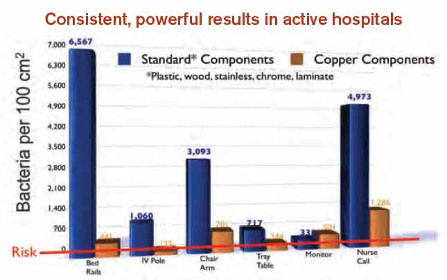
A shift in governmental regulations regarding reimbursement for hospital-acquired infections (HAIs) is forcing hospitals to take a closer look at how to reduce them. A recent study in Infection Control and Hospital Epidemiology shows that copper-alloy surfaces may be one such solution.3 According to the study, although only 9% of the touch surfaces in each ICU were replaced with copper components, there were 58% fewer HAI cases.1
“Before these regulations, hospitals didn’t necessarily want technology to decrease HAI rates, because the more infections and complications, the longer the length of patient stay, the greater the reimbursement, and the better the bottom line,” says Archelle Georgiou, MD, president of Georgiou Consulting LLC in Minneapolis and an advisor to the Copper Development Association.
Three regulations that have resulted in reimbursements to hospitals getting cut include:
- The Deficit Reduction Act of 2005, which was implemented on Oct. 1, 2008, which states that Medicare will not reimburse for certain types of HAIs;
- Section 3025 of the Affordable Care Act (signed into law in 2010), which incentivizes hospitals to decrease their readmission rates, which frequently are caused by HAIs. Beginning this fall, hospitals are getting reduced reimbursement when their readmission rates exceed a certain threshold. The maximum penalty in 2013 is 1% and will increase to 3% by 2015; and
- Section 1886 of the Affordable Care Act, which describes value-based purchasing and makes hospitals eligible to receive incentive payments for achieving better care on certain quality metrics. Funding for the program comes from withholding payment from poor-performing hospitals. The financial impact to hospitals started this year. In 2014, urinary tract infections and vascular-catheter-associated infections will be among the targeted conditions measured by CMS to calculate incentives and penalties.
“Hospitals are now feeling a direct impact from all of this,” Dr. Georgiou says. “Back in 2008, hospitals were noticing, but it was hard to get their attention since only one program was impacting their bottom line. But, pretty soon, hospitals risk losing upwards of 5% of their Medicare reimbursement for decreased quality.
“Reducing HAIs is clearly on the priority list of chief operating officers. They are very aware of the impact to their bottom line. They are looking to their vendors and suppliers to develop strategies to work with their hospitals to improve performance around these metrics.”
Karen Appold is a freelance writer in Pennsylvania.