User login
This month in the journal CHEST®
Editor’s picks
Fertility and pregnancy in cystic fibrosis By Dr. M. Shteinberg, et al.Changes in purchases for intensive care medicines during the COVID-19 pandemic: A global time series study By Dr. K. Kim, et al.The impact of obesity in critical illness By Drs. M.R. Anderson and M. Shashaty
Strategies to improve bedside clinical skills teaching By Drs. B. Garibaldi and S.W. RussellAntithrombotic therapy for VTE Disease: Second update of the CHEST guideline and expert panel report – executive summary By Dr. S.M. Stevens, et al.
Editor’s picks
Editor’s picks
Fertility and pregnancy in cystic fibrosis By Dr. M. Shteinberg, et al.Changes in purchases for intensive care medicines during the COVID-19 pandemic: A global time series study By Dr. K. Kim, et al.The impact of obesity in critical illness By Drs. M.R. Anderson and M. Shashaty
Strategies to improve bedside clinical skills teaching By Drs. B. Garibaldi and S.W. RussellAntithrombotic therapy for VTE Disease: Second update of the CHEST guideline and expert panel report – executive summary By Dr. S.M. Stevens, et al.
Transitioning from fellow to attending
It’s day 1 of “attendingship,” and I’m back to wearing my white coat after years of being confident enough in myself to think I didn’t need it to look like “the doctor.” Is it okay to park here? Does my clinic have a staff bathroom? Will my log in work? Oh my gosh, how do I place orders?! I remember this feeling – it’s intern year all over again, except there’s no senior resident to rescue me now – here we go!

Starting off
As a new attending, the amount of responsibility can be intimidating and overwhelming. It is important to remember that you are not alone, you have a whole team supporting you whether you are in clinic or the ICU. Be sure to introduce yourself to those who you will be working with, get to know them, their roles, and figure out the best way that you can help each other with the ultimate goal of helping patients. In addition to meeting your own team, it is important to introduce yourself to your new colleagues – especially if you are new to the institution. Drs. Fielder and Sihag suggest putting together an introductory email to those who may be referring to you that includes an overview of what you do and how you can help, as well as your contact information. They also suggest maintaining an open line of communication and keeping the referring provider updated on your mutual patient (Fiedler AG, Sihag S. J Thorac Cardiovasc Surg. 2020 Mar;159[3]:1156-60). While this may sound antiquated, in my experience thus far, my colleagues have greatly appreciated this gesture.
Finding support
Even though you will be surrounded by a plethora of new colleagues, the transition to attending can be lonely – especially if you are moving to a new institution. Be sure to keep in touch with your co-residents, co-fellows, mentors, and, of course, your friends and family. Studies have shown that support mitigates stress and reduces job strain, which can lead to better health outcomes in the long term (Fiedler AG, Sihag S. [above]). Another great source of support for me is my CHEST colleagues. If you have not already, I highly suggest joining the CHEST Network(s) that aligns with your career interests. This is a great way to not only network with those who share the same niche as you but also to explore academic opportunities outside of your institution. Through the CHEST Home Mechanical Ventilation and Sleep NetWorks, I have gained mentors, made friends, and have become more involved in CHEST’s annual meeting, chairing my first session this year.
Staying organized
Adjusting to your new schedule can be just as hard as adjusting to a new role or new institution. After years of moving through the well-oiled, regimented machine that is medical training, there are suddenly no more rotations, no more research blocks, and no more protected time for learning. Dr. Okereke suggests creating a weekly calendar, which blocks time for not only your clinical duties but for studying (as you will be taking boards during your first year), academic endeavors (teaching and/or research), and, most importantly – for fun (Okereke I. J Thorac Cardiovasc Surg. 2020 Mar;159[3]:1161-2). Being cognizant about maintaining work-life balance is key once you become an attending. It is finally time to learn how to take time off, away from all things work, and to not feel guilty about it.
Saying no
This brings me to saying “no.” We are taught to say “yes” to every opportunity throughout our careers and, while that can certainly help us get far, it can also lead to burnout. Once you’re an attending, you’re in it for the long haul, so best to say yes to the things you are most interested in and “spark joy,” as Marie Kondo says, and say no to the things that do not make you happy and are not congruent with your overall goals. Fielder and Sihag (above) note that your division director or chief typically has a vision in mind for you within the department. It is important to communicate with leadership so that everyone is on the same page and the administrative and academic opportunities afforded to you are in alignment with your career goals going forward.
Teaching trainees
To prepare for teaching as an attending, Dr. Greco recommends starting during your own training. She suggests cataloging your study materials and notes for later reference, curating talks throughout your training, and exploring different rounding styles prior to graduation (Greco, A. CHEST Thought Leader Blog. 2021 June). To get more experience in formal speaking, Dr. Shen and colleagues encourage getting involved in resident noon conferences (Shen JZ, Memon AA, Lin C. Stroke. 2019 Sep;50[9]:e250-e252). A benefit of being a critical care attending is that you can gain experience teaching not only with the internal medicine residents but with emergency medicine, anesthesiology, and critical care advanced practice provider residents, as well.
While lecturing is one thing, teaching on service is a whole different ball game. No matter how young, fun, and relatable you think you are, you’re the boss now. You’re the giver of grades and the writer of evaluations. It is important to be self-aware of your influence and be deliberate with the environment you create on rounds and in clinic. Set expectations on day 1 so that everyone understands. Be open with what you are working on. For example, I make daily goals for myself that I share with the team before rounds. Drs. Fielder and Sihag (above) suggest sharing anecdotes from your own time in training that can help both you and your trainees remember that you were just in their shoes. Allowing yourself to be vulnerable creates a safe space in which your learners feel more comfortable doing the same.
Lastly, delegation is key. While many of us have done this since residency, Dr. Shen et al (above) suggest deliberately practicing this during fellowship. If you were the fellow who was able to handle a lot on your own, trust that your own fellows will be able to do that. Delegating to your trainees helps you improve personal and team efficiency, provides fellows with needed autonomy, and allows you to further grow into the role of attending physician.
Conclusion
While you may be nervous starting out, trust that you have been well trained and have the clinical knowledge and skills you need to do your job – you are ready. Get to know the staff you will be working with, your colleagues, and keep in touch with your co-trainees and mentors who have helped you along the way. Make daily goals for yourself, and make time to read and reflect so that you can continue to learn and grow. Most of all, make time for yourself, your friends, and your family, because after years of supporting you through all of your hard work, you’ve finally made it – congrats!
Dr. Greer is Assistant Professor of Medicine, Emory University Division of Pulmonary, Allergy, Critical Care, and Sleep Medicine, Atlanta, GA.
It’s day 1 of “attendingship,” and I’m back to wearing my white coat after years of being confident enough in myself to think I didn’t need it to look like “the doctor.” Is it okay to park here? Does my clinic have a staff bathroom? Will my log in work? Oh my gosh, how do I place orders?! I remember this feeling – it’s intern year all over again, except there’s no senior resident to rescue me now – here we go!
Starting off
As a new attending, the amount of responsibility can be intimidating and overwhelming. It is important to remember that you are not alone, you have a whole team supporting you whether you are in clinic or the ICU. Be sure to introduce yourself to those who you will be working with, get to know them, their roles, and figure out the best way that you can help each other with the ultimate goal of helping patients. In addition to meeting your own team, it is important to introduce yourself to your new colleagues – especially if you are new to the institution. Drs. Fielder and Sihag suggest putting together an introductory email to those who may be referring to you that includes an overview of what you do and how you can help, as well as your contact information. They also suggest maintaining an open line of communication and keeping the referring provider updated on your mutual patient (Fiedler AG, Sihag S. J Thorac Cardiovasc Surg. 2020 Mar;159[3]:1156-60). While this may sound antiquated, in my experience thus far, my colleagues have greatly appreciated this gesture.
Finding support
Even though you will be surrounded by a plethora of new colleagues, the transition to attending can be lonely – especially if you are moving to a new institution. Be sure to keep in touch with your co-residents, co-fellows, mentors, and, of course, your friends and family. Studies have shown that support mitigates stress and reduces job strain, which can lead to better health outcomes in the long term (Fiedler AG, Sihag S. [above]). Another great source of support for me is my CHEST colleagues. If you have not already, I highly suggest joining the CHEST Network(s) that aligns with your career interests. This is a great way to not only network with those who share the same niche as you but also to explore academic opportunities outside of your institution. Through the CHEST Home Mechanical Ventilation and Sleep NetWorks, I have gained mentors, made friends, and have become more involved in CHEST’s annual meeting, chairing my first session this year.
Staying organized
Adjusting to your new schedule can be just as hard as adjusting to a new role or new institution. After years of moving through the well-oiled, regimented machine that is medical training, there are suddenly no more rotations, no more research blocks, and no more protected time for learning. Dr. Okereke suggests creating a weekly calendar, which blocks time for not only your clinical duties but for studying (as you will be taking boards during your first year), academic endeavors (teaching and/or research), and, most importantly – for fun (Okereke I. J Thorac Cardiovasc Surg. 2020 Mar;159[3]:1161-2). Being cognizant about maintaining work-life balance is key once you become an attending. It is finally time to learn how to take time off, away from all things work, and to not feel guilty about it.
Saying no
This brings me to saying “no.” We are taught to say “yes” to every opportunity throughout our careers and, while that can certainly help us get far, it can also lead to burnout. Once you’re an attending, you’re in it for the long haul, so best to say yes to the things you are most interested in and “spark joy,” as Marie Kondo says, and say no to the things that do not make you happy and are not congruent with your overall goals. Fielder and Sihag (above) note that your division director or chief typically has a vision in mind for you within the department. It is important to communicate with leadership so that everyone is on the same page and the administrative and academic opportunities afforded to you are in alignment with your career goals going forward.
Teaching trainees
To prepare for teaching as an attending, Dr. Greco recommends starting during your own training. She suggests cataloging your study materials and notes for later reference, curating talks throughout your training, and exploring different rounding styles prior to graduation (Greco, A. CHEST Thought Leader Blog. 2021 June). To get more experience in formal speaking, Dr. Shen and colleagues encourage getting involved in resident noon conferences (Shen JZ, Memon AA, Lin C. Stroke. 2019 Sep;50[9]:e250-e252). A benefit of being a critical care attending is that you can gain experience teaching not only with the internal medicine residents but with emergency medicine, anesthesiology, and critical care advanced practice provider residents, as well.
While lecturing is one thing, teaching on service is a whole different ball game. No matter how young, fun, and relatable you think you are, you’re the boss now. You’re the giver of grades and the writer of evaluations. It is important to be self-aware of your influence and be deliberate with the environment you create on rounds and in clinic. Set expectations on day 1 so that everyone understands. Be open with what you are working on. For example, I make daily goals for myself that I share with the team before rounds. Drs. Fielder and Sihag (above) suggest sharing anecdotes from your own time in training that can help both you and your trainees remember that you were just in their shoes. Allowing yourself to be vulnerable creates a safe space in which your learners feel more comfortable doing the same.
Lastly, delegation is key. While many of us have done this since residency, Dr. Shen et al (above) suggest deliberately practicing this during fellowship. If you were the fellow who was able to handle a lot on your own, trust that your own fellows will be able to do that. Delegating to your trainees helps you improve personal and team efficiency, provides fellows with needed autonomy, and allows you to further grow into the role of attending physician.
Conclusion
While you may be nervous starting out, trust that you have been well trained and have the clinical knowledge and skills you need to do your job – you are ready. Get to know the staff you will be working with, your colleagues, and keep in touch with your co-trainees and mentors who have helped you along the way. Make daily goals for yourself, and make time to read and reflect so that you can continue to learn and grow. Most of all, make time for yourself, your friends, and your family, because after years of supporting you through all of your hard work, you’ve finally made it – congrats!
Dr. Greer is Assistant Professor of Medicine, Emory University Division of Pulmonary, Allergy, Critical Care, and Sleep Medicine, Atlanta, GA.
Chronically interrupted: The importance of communication with patient and family during the COVID-19 pandemic
Case narrative
A 35-year-old woman has worsening alcoholic cirrhosis and repeated admissions for ascites, hepato-renal syndrome, and alcoholic hepatitis. Upon recognition of her grave prognosis, we proceeded with a shared-management approach involving medicine, gastroenterology, social work, chaplaincy, and palliative care. When the team spoke with the patient’s health care proxy (HCP), family, and friends for collateral information and involvement in goals of care conversation, we realized that none were aware of her months-long decline and poor prognosis for recovery to hospital discharge.

Although several factors contributed to the disconnect between the patient and her support system, the obstacles were greatly exacerbated by profound changes in hospital protocol because of the COVID-19 pandemic. Physicians feel underprepared and challenged by prognostication and discussion of end of life during normal times; we believe COVID-19 has limited this essential physician role and led to tragic delays in effective communication and end of life planning.
Closing the loop
For patients with complex medical issues or those reaching end of life, effective communication within the health care system is critical. While inpatient teams often drive the plan, they care for their patients during a snapshot in time; contrarily, primary care providers and specialists often have established longitudinal relationships with their patients. Ergo, clinicians should communicate directly, and ideally with both patients and families, to achieve patient-centered and goal-concordant care.

For medically complex patients, PCPs tend to prefer verbal hand-offs. Timely and reliable communication between inpatient and outpatient providers has also been shown to prevent medical adverse events.1 Despite this, direct communication occurs infrequently.2 Given that hospitalists serve as primary inpatient providers for most general admissions, it is their responsibility to communicate with outpatient providers.
A multidisciplinary team redesigned the process by which PCPs were contacted following patient discharge. The transmission of information should ideally occur prior to discharge.3 Deficits in communication are extremely common and may negatively impact patient care, patient satisfaction, and patient safety.
Changes during the COVID-19 era
During the pandemic, patients have only one visitor per day, restricted visiting hours, and limited interactions with clinicians per implemented policies. Along with the increased burdens from personal protective equipment, remote hospital providers (social workers, case managers), and increased bureaucratic duties, COVID-19 has elucidated limitations in medical capacity and revealed the difficulties that clinicians face in communicating with patients and families, especially about serious illness.

Tasks include facilitating virtual goodbyes between dying patients and families, conducting family meetings via teleconference, and discussing patient care with specialists through virtual technologies.4 While these tasks are arguably more important during a global disaster, COVID-19 paradoxically restricts physical presence and severely hinders communication.5 Clinicians should continue to utilize core skills like building rapport, assessing patient/family perspectives and agenda, and using empathy.6 Patients tend to more frequently value functional outcomes while clinicians tend to default to treatment modalities.7 Additionally, goals of care and end of life discussions are associated with improved quality of life, fewer aggressive medical interventions near death, and even increased survival.
Given the limited resources and difficulties in communication during the pandemic, clinicians should place greater emphasis on values-based shared decision-making. Internet-based solutions are essential and widely used, and videoconferencing has been initiated at the institutional scale at many hospitals. Many clinicians with little experience are broadly implementing these technologies.7 Despite these technological innovations, issues still arise in how to communicate effectively in the hospital setting, and we must acknowledge that strategies require devices, Internet access, and technological literacy, highlighting disparities in access to quality health care.6 Conversations during the pandemic will require listening, empathy, responsive action, and the acknowledgment of the social determinants of health.7
Improving communication and transition of care
Multiple steps will be warranted to implement the safe transition process and improve communication. High-quality patient care encompasses careful review of medications, communication between inpatient and outpatient providers, and close follow-up at discharge. These steps serve to increase our reliance on patient compliance and the exchange of information about global progression of disease.
The quantitative and qualitative steps of transition of care should overcome disconnect between teams, specifically deficit areas regarding postdischarge communication, monitoring, and understanding of prognosis around the relevance to this era of COVID-19.
Dr. Haddad is a resident physician in the psychiatry residency program at Brigham and Women’s Hospital, Boston. Dr. Halporn is clinic director, Division of Adult Palliative Care, in the department of psychosocial oncology and palliative care, Dana-Farber Cancer Institute and Brigham and Women’s Hospital. Dr. Barkoudah is associate director of the Hospital Medicine Unit at Brigham and Women’s Hospital.
References
1. Goldman L et al. Passing the clinical baton: 6 principles to guide the hospitalist. Am J Med. 2001;111(9B):36S-39S. doi: 10.1016/s0002-9343(01)00968-8.
2. Kripalani S et al. Deficits in communication and information transfer between hospital-based and primary care physicians. JAMA. 2007 Feb 28;297(8):831-41. doi: 10.1001/jama.297.8.831.
3. Scotten M et al. Minding the gap: Interprofessional communication during inpatient and post discharge chasm care. Patient Educ Couns. 2015 Jul;98(7):895-900. doi: 10.1016/j.pec.2015.03.009.
4. Back A et al. Communication skills in the age of COVID-19. Ann Intern Med. 2020 Jun 2;172(11):759-60. doi: 10.7326/M20-1376.
5. Hart JL et al. Family-centered care during the COVID-19 era. J Pain Symptom Manage. 2020 Aug;60(2):e93-7. doi: 10.1016/j.jpainsymman.2020.04.017.
6. Rubinelli S et al. Implications of the current COVID-19 pandemic for communication in healthcare. Patient Educ Couns. 2020 Jun;103(6):1067-9. doi: 10.1016/j.pec.2020.04.021.
7. Simpson N et al. Don’t forget shared decision-making in the COVID-19 crisis. Intern Med J. 2020 Jun;50(6):761-3. doi: 10.1111/imj.14862.
Case narrative
A 35-year-old woman has worsening alcoholic cirrhosis and repeated admissions for ascites, hepato-renal syndrome, and alcoholic hepatitis. Upon recognition of her grave prognosis, we proceeded with a shared-management approach involving medicine, gastroenterology, social work, chaplaincy, and palliative care. When the team spoke with the patient’s health care proxy (HCP), family, and friends for collateral information and involvement in goals of care conversation, we realized that none were aware of her months-long decline and poor prognosis for recovery to hospital discharge.
Although several factors contributed to the disconnect between the patient and her support system, the obstacles were greatly exacerbated by profound changes in hospital protocol because of the COVID-19 pandemic. Physicians feel underprepared and challenged by prognostication and discussion of end of life during normal times; we believe COVID-19 has limited this essential physician role and led to tragic delays in effective communication and end of life planning.
Closing the loop
For patients with complex medical issues or those reaching end of life, effective communication within the health care system is critical. While inpatient teams often drive the plan, they care for their patients during a snapshot in time; contrarily, primary care providers and specialists often have established longitudinal relationships with their patients. Ergo, clinicians should communicate directly, and ideally with both patients and families, to achieve patient-centered and goal-concordant care.
For medically complex patients, PCPs tend to prefer verbal hand-offs. Timely and reliable communication between inpatient and outpatient providers has also been shown to prevent medical adverse events.1 Despite this, direct communication occurs infrequently.2 Given that hospitalists serve as primary inpatient providers for most general admissions, it is their responsibility to communicate with outpatient providers.
A multidisciplinary team redesigned the process by which PCPs were contacted following patient discharge. The transmission of information should ideally occur prior to discharge.3 Deficits in communication are extremely common and may negatively impact patient care, patient satisfaction, and patient safety.
Changes during the COVID-19 era
During the pandemic, patients have only one visitor per day, restricted visiting hours, and limited interactions with clinicians per implemented policies. Along with the increased burdens from personal protective equipment, remote hospital providers (social workers, case managers), and increased bureaucratic duties, COVID-19 has elucidated limitations in medical capacity and revealed the difficulties that clinicians face in communicating with patients and families, especially about serious illness.
Tasks include facilitating virtual goodbyes between dying patients and families, conducting family meetings via teleconference, and discussing patient care with specialists through virtual technologies.4 While these tasks are arguably more important during a global disaster, COVID-19 paradoxically restricts physical presence and severely hinders communication.5 Clinicians should continue to utilize core skills like building rapport, assessing patient/family perspectives and agenda, and using empathy.6 Patients tend to more frequently value functional outcomes while clinicians tend to default to treatment modalities.7 Additionally, goals of care and end of life discussions are associated with improved quality of life, fewer aggressive medical interventions near death, and even increased survival.
Given the limited resources and difficulties in communication during the pandemic, clinicians should place greater emphasis on values-based shared decision-making. Internet-based solutions are essential and widely used, and videoconferencing has been initiated at the institutional scale at many hospitals. Many clinicians with little experience are broadly implementing these technologies.7 Despite these technological innovations, issues still arise in how to communicate effectively in the hospital setting, and we must acknowledge that strategies require devices, Internet access, and technological literacy, highlighting disparities in access to quality health care.6 Conversations during the pandemic will require listening, empathy, responsive action, and the acknowledgment of the social determinants of health.7
Improving communication and transition of care
Multiple steps will be warranted to implement the safe transition process and improve communication. High-quality patient care encompasses careful review of medications, communication between inpatient and outpatient providers, and close follow-up at discharge. These steps serve to increase our reliance on patient compliance and the exchange of information about global progression of disease.
The quantitative and qualitative steps of transition of care should overcome disconnect between teams, specifically deficit areas regarding postdischarge communication, monitoring, and understanding of prognosis around the relevance to this era of COVID-19.
Dr. Haddad is a resident physician in the psychiatry residency program at Brigham and Women’s Hospital, Boston. Dr. Halporn is clinic director, Division of Adult Palliative Care, in the department of psychosocial oncology and palliative care, Dana-Farber Cancer Institute and Brigham and Women’s Hospital. Dr. Barkoudah is associate director of the Hospital Medicine Unit at Brigham and Women’s Hospital.
References
1. Goldman L et al. Passing the clinical baton: 6 principles to guide the hospitalist. Am J Med. 2001;111(9B):36S-39S. doi: 10.1016/s0002-9343(01)00968-8.
2. Kripalani S et al. Deficits in communication and information transfer between hospital-based and primary care physicians. JAMA. 2007 Feb 28;297(8):831-41. doi: 10.1001/jama.297.8.831.
3. Scotten M et al. Minding the gap: Interprofessional communication during inpatient and post discharge chasm care. Patient Educ Couns. 2015 Jul;98(7):895-900. doi: 10.1016/j.pec.2015.03.009.
4. Back A et al. Communication skills in the age of COVID-19. Ann Intern Med. 2020 Jun 2;172(11):759-60. doi: 10.7326/M20-1376.
5. Hart JL et al. Family-centered care during the COVID-19 era. J Pain Symptom Manage. 2020 Aug;60(2):e93-7. doi: 10.1016/j.jpainsymman.2020.04.017.
6. Rubinelli S et al. Implications of the current COVID-19 pandemic for communication in healthcare. Patient Educ Couns. 2020 Jun;103(6):1067-9. doi: 10.1016/j.pec.2020.04.021.
7. Simpson N et al. Don’t forget shared decision-making in the COVID-19 crisis. Intern Med J. 2020 Jun;50(6):761-3. doi: 10.1111/imj.14862.
Customizing pre-CAR T chemotherapy could improve ALL patient outcomes
The findings, if validated in a prospective study, could help cut the rate of relapses after initial response to CAR T-cell therapy, which currently approaches 50%, the investigators noted.
In 152 pediatric and young adult patients with relapsed or refractory B-cell acute lymphoblastic leukemia (B-ALL) who underwent CD-19-directed CAR T-cell therapy after cyclophosphamide/fludarabine lymphodepleting chemotherapy, estimated fludarabine exposure was associated with cumulative incidence of relapse (CIR) and a composite endpoint that included loss of B-cell aplasia (BCA) or relapse, Vanessa Fabrizio, MD, and colleagues found.
Dr. Fabrizio, a pediatric hematologist and oncologist at Children’s Hospital Colorado and the University of Colorado Cancer Center in Aurora, was a fellow at Memorial Sloan Kettering (MSK) Cancer Center during the study.
Optimal fludarabine exposure was identified by the investigators as an area under the curve (AUC) of at least 13.8 mg*hr/L. The fludarabine exposure AUC was calculated for each patient by using a validated pharmacokinetics population model.
Multivariable analyses controlling for baseline patient factors and fludarabine exposure showed that patients without optimal exposure had a 2.5-fold higher CIR (hazard ratio, 2.45), and a twofold higher risk of relapse or loss of BCA (HR, 1.96), compared with those who had optimal fludarabine exposure, they reported.
High pre-infusion disease burden was associated with an increased risk of relapse and death (HRs, 2.66 and 4.77, respectively), they said.
The study was published online Nov. 17 in Blood Advances.
“We know that [with] fludarabine ... everyone’s body clears it differently,” principal investigator Kevin J. Curran, MD, said in an interview.
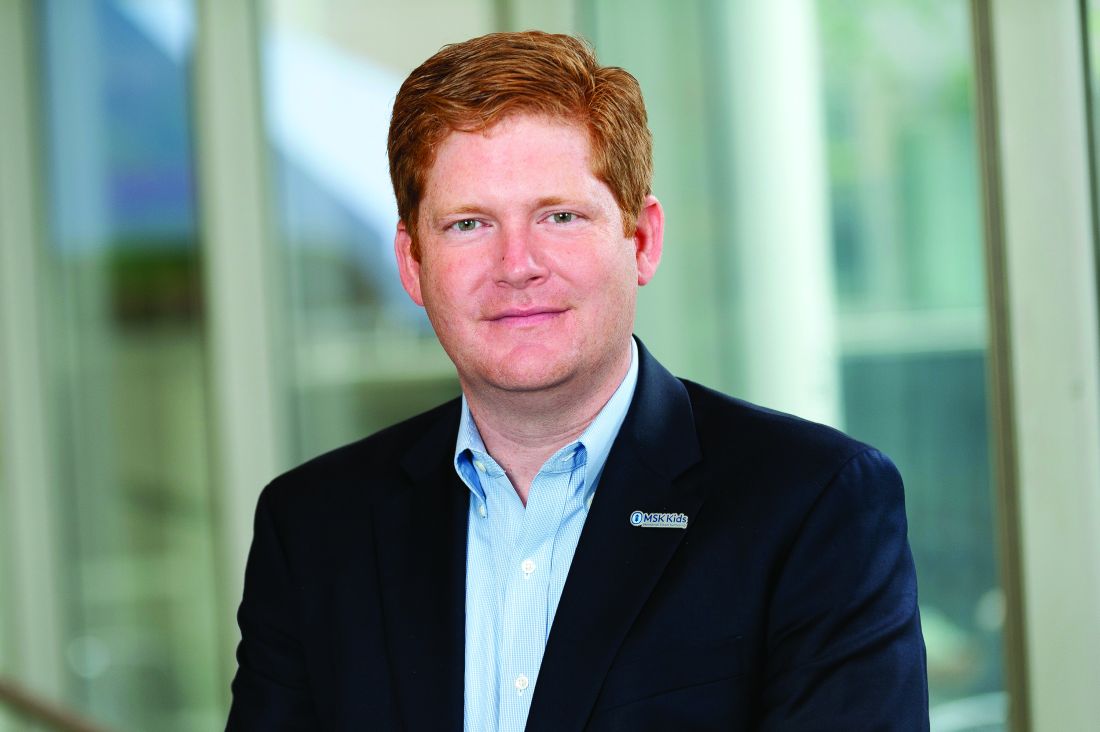
Factors affecting clearance include kidney function and weight, and it is simple to determine the optimal dose based on these factors and apply that in practice, said Dr. Curran, a pediatric oncologist and assistant attending physician specializing in cellular therapy at MSK Kids.
In fact, in prior studies, optimal fludarabine exposure in patients undergoing allogeneic hematopoietic cell transplantation has been shown to “decrease nonrelapse mortality due to improved immune reconstitution and subsequently improve survival,” he and his colleagues wrote, explaining the rationale for the study.
The participants, who were part of the Pediatric Real-World CAR Consortium (PRWCC), had a median age of 12.5 years, and 131 of 152 (86%) responded to CAR T-cell therapy. The 12-month OS was 75.1%, the 12-month CIR was 36.4%, and 67% of patients had optimal fludarabine exposure, the authors said.
The findings indeed suggest that one way to improve outcomes without changing the actual cell therapy is to tailor the lymphodepleting therapy prior to CAR T-cell therapy, said Dr. Curran.
“That’s what this does. It’s exciting because cell therapy is very effective [in terms of] initial response, but what we don’t like is the durability of the response,” he said “The next step is to prove it in a prospective study.”
A phase 2 study looking at personalized dosing, as opposed to the standard 30 mg/m2 that most patients receive, is planned for 2022, he noted.
The study was supported by a St Baldrick’s/Stand Up 2 Cancer Pediatric Dream Team Translational Cancer Research Grant, the Virginia and D.K. Ludwig Fund for Cancer Research, and a National Cancer Institute Cancer Center Support Grant. Dr. Curran has served as a consultant for Novartis and Mesoblast, and received research funding from Novartis and Celgene. Dr. Fabrizio reported having no disclosures.
The findings, if validated in a prospective study, could help cut the rate of relapses after initial response to CAR T-cell therapy, which currently approaches 50%, the investigators noted.
In 152 pediatric and young adult patients with relapsed or refractory B-cell acute lymphoblastic leukemia (B-ALL) who underwent CD-19-directed CAR T-cell therapy after cyclophosphamide/fludarabine lymphodepleting chemotherapy, estimated fludarabine exposure was associated with cumulative incidence of relapse (CIR) and a composite endpoint that included loss of B-cell aplasia (BCA) or relapse, Vanessa Fabrizio, MD, and colleagues found.
Dr. Fabrizio, a pediatric hematologist and oncologist at Children’s Hospital Colorado and the University of Colorado Cancer Center in Aurora, was a fellow at Memorial Sloan Kettering (MSK) Cancer Center during the study.
Optimal fludarabine exposure was identified by the investigators as an area under the curve (AUC) of at least 13.8 mg*hr/L. The fludarabine exposure AUC was calculated for each patient by using a validated pharmacokinetics population model.
Multivariable analyses controlling for baseline patient factors and fludarabine exposure showed that patients without optimal exposure had a 2.5-fold higher CIR (hazard ratio, 2.45), and a twofold higher risk of relapse or loss of BCA (HR, 1.96), compared with those who had optimal fludarabine exposure, they reported.
High pre-infusion disease burden was associated with an increased risk of relapse and death (HRs, 2.66 and 4.77, respectively), they said.
The study was published online Nov. 17 in Blood Advances.
“We know that [with] fludarabine ... everyone’s body clears it differently,” principal investigator Kevin J. Curran, MD, said in an interview.
Factors affecting clearance include kidney function and weight, and it is simple to determine the optimal dose based on these factors and apply that in practice, said Dr. Curran, a pediatric oncologist and assistant attending physician specializing in cellular therapy at MSK Kids.
In fact, in prior studies, optimal fludarabine exposure in patients undergoing allogeneic hematopoietic cell transplantation has been shown to “decrease nonrelapse mortality due to improved immune reconstitution and subsequently improve survival,” he and his colleagues wrote, explaining the rationale for the study.
The participants, who were part of the Pediatric Real-World CAR Consortium (PRWCC), had a median age of 12.5 years, and 131 of 152 (86%) responded to CAR T-cell therapy. The 12-month OS was 75.1%, the 12-month CIR was 36.4%, and 67% of patients had optimal fludarabine exposure, the authors said.
The findings indeed suggest that one way to improve outcomes without changing the actual cell therapy is to tailor the lymphodepleting therapy prior to CAR T-cell therapy, said Dr. Curran.
“That’s what this does. It’s exciting because cell therapy is very effective [in terms of] initial response, but what we don’t like is the durability of the response,” he said “The next step is to prove it in a prospective study.”
A phase 2 study looking at personalized dosing, as opposed to the standard 30 mg/m2 that most patients receive, is planned for 2022, he noted.
The study was supported by a St Baldrick’s/Stand Up 2 Cancer Pediatric Dream Team Translational Cancer Research Grant, the Virginia and D.K. Ludwig Fund for Cancer Research, and a National Cancer Institute Cancer Center Support Grant. Dr. Curran has served as a consultant for Novartis and Mesoblast, and received research funding from Novartis and Celgene. Dr. Fabrizio reported having no disclosures.
FROM BLOOD ADVANCES
FDA authorizes Pfizer boosters for 16- and 17-year-olds
, clearing the way for millions of teenagers to get a third dose of vaccine starting 6 months after their second dose.

The FDA said it was basing its emergency authorization of boosters for 16- and 17-year-olds on data from 200 individuals who were 18-55 years of age when they received a booster dose. They are requiring Pfizer to collect data on safety in postauthorization studies.
“The FDA has determined that the benefits of a single booster dose of the Pfizer-BioNTech COVID-19 Vaccine or Comirnaty outweigh the risks of myocarditis and pericarditis in individuals 16 and 17 years of age to provide continued protection against COVID-19 and the associated serious consequences that can occur including hospitalization and death,” the agency said in a news release.
Israel has been giving booster doses of Pfizer’s vaccine to everyone 12 and up since late August. Data from that country show that myocarditis cases continue to be very rare, even in younger age groups, and are mild and temporary.
The authorization comes as the effectiveness of the current vaccines against the new Omicron variant has become a point of intense scientific inquiry.
Early studies suggest that booster doses may be necessary to keep Omicron at bay, at least until new variant-specific vaccines are ready next spring.
Current evidence suggests that the protection of the vaccines is holding up well against severe disease and death, at least with Delta and early iterations of the virus.
How well they will do against Omicron, and how severe Omicron infections may be for different age groups, remain open questions.
On Dec. 8, the World Health Organization urged countries not to wait for all the science to come in, but to act now to contain any potential threat.
The first pieces of evidence on Omicron suggest that it is highly contagious, perhaps even more than Delta, though early reports suggest symptoms caused by this version of the new coronavirus may be less severe than in previous waves. Experts have cautioned that the true severity of Omicron infections isn’t yet known, since the first cases have been detected in younger people, who tend to have milder COVID-19 symptoms than those of adults and seniors.
“Vaccination and getting a booster when eligible, along with other preventive measures like masking and avoiding large crowds and poorly ventilated spaces, remain our most effective methods for fighting COVID-19,” Acting FDA Commissioner Janet Woodcock, MD, said in a news release. “As people gather indoors with family and friends for the holidays, we can’t let up on all the preventive public health measures that we have been taking during the pandemic. With both the Delta and Omicron variants continuing to spread, vaccination remains the best protection against COVID-19.”
In mid-November, the FDA authorized boosters of the Pfizer vaccine for all individuals 18 and older, but the agency held off on expanding the use of boosters for younger age groups, partly because they have the highest risk of myocarditis, a very rare side effect.
Myocarditis cases seem to be temporary, with patients making a full recovery, though they need to be monitored in the hospital. The risk of myocarditis with a COVID-19 infection is many times higher than it is from a vaccine.
There have been little data to support the need for boosters in this age group, because children and teens tend to experience milder COVID-19 disease, though they are still at risk for post–COVID-19 complications such as long COVID and a delayed reaction to the virus called Post Acute Sequelae of SARS-CoV2 Infection among Children, or PAS-C.
All that changed with the arrival of Omicron.
A version of this article first appeared on WebMD.com.
, clearing the way for millions of teenagers to get a third dose of vaccine starting 6 months after their second dose.
The FDA said it was basing its emergency authorization of boosters for 16- and 17-year-olds on data from 200 individuals who were 18-55 years of age when they received a booster dose. They are requiring Pfizer to collect data on safety in postauthorization studies.
“The FDA has determined that the benefits of a single booster dose of the Pfizer-BioNTech COVID-19 Vaccine or Comirnaty outweigh the risks of myocarditis and pericarditis in individuals 16 and 17 years of age to provide continued protection against COVID-19 and the associated serious consequences that can occur including hospitalization and death,” the agency said in a news release.
Israel has been giving booster doses of Pfizer’s vaccine to everyone 12 and up since late August. Data from that country show that myocarditis cases continue to be very rare, even in younger age groups, and are mild and temporary.
The authorization comes as the effectiveness of the current vaccines against the new Omicron variant has become a point of intense scientific inquiry.
Early studies suggest that booster doses may be necessary to keep Omicron at bay, at least until new variant-specific vaccines are ready next spring.
Current evidence suggests that the protection of the vaccines is holding up well against severe disease and death, at least with Delta and early iterations of the virus.
How well they will do against Omicron, and how severe Omicron infections may be for different age groups, remain open questions.
On Dec. 8, the World Health Organization urged countries not to wait for all the science to come in, but to act now to contain any potential threat.
The first pieces of evidence on Omicron suggest that it is highly contagious, perhaps even more than Delta, though early reports suggest symptoms caused by this version of the new coronavirus may be less severe than in previous waves. Experts have cautioned that the true severity of Omicron infections isn’t yet known, since the first cases have been detected in younger people, who tend to have milder COVID-19 symptoms than those of adults and seniors.
“Vaccination and getting a booster when eligible, along with other preventive measures like masking and avoiding large crowds and poorly ventilated spaces, remain our most effective methods for fighting COVID-19,” Acting FDA Commissioner Janet Woodcock, MD, said in a news release. “As people gather indoors with family and friends for the holidays, we can’t let up on all the preventive public health measures that we have been taking during the pandemic. With both the Delta and Omicron variants continuing to spread, vaccination remains the best protection against COVID-19.”
In mid-November, the FDA authorized boosters of the Pfizer vaccine for all individuals 18 and older, but the agency held off on expanding the use of boosters for younger age groups, partly because they have the highest risk of myocarditis, a very rare side effect.
Myocarditis cases seem to be temporary, with patients making a full recovery, though they need to be monitored in the hospital. The risk of myocarditis with a COVID-19 infection is many times higher than it is from a vaccine.
There have been little data to support the need for boosters in this age group, because children and teens tend to experience milder COVID-19 disease, though they are still at risk for post–COVID-19 complications such as long COVID and a delayed reaction to the virus called Post Acute Sequelae of SARS-CoV2 Infection among Children, or PAS-C.
All that changed with the arrival of Omicron.
A version of this article first appeared on WebMD.com.
Medical board stops warning docs against giving false COVID information
Under pressure from Republican state lawmakers, t
The board’s 7-3 vote on December 7 to delete the statement followed repeated threats by a powerful state House Republican to dissolve the board and appoint all new members if it did not immediately take it down.
The Tennessee board’s statement was a verbatim restatement of a warning to physicians issued by the Federation of State Medical Boards in July. The federation cited a “dramatic increase” in dissemination of misinformation and disinformation about the COVID-19 vaccine by physicians. It said that’s dangerous because physicians enjoy a high degree of public credibility.
Across the country, state medical licensing boards and state and national medical associations and specialty boards are struggling with how to respond to scientifically baseless public statements about COVID-19 by some physicians, which they say are increasing public confusion, political conflict, and preventable illnesses and deaths.
There have been only a small number of disciplinary actions by medical boards against physicians for spreading false COVID-19 information. Critics say the boards have been weak in responding to these dangerous violations of medical standards. As an example, they cite the State Medical Board of Ohio’s September renewal of the medical license of Sherri Tenpenny, DO, who had previously testified before Ohio lawmakers that COVID-19 vaccines magnetize their recipients and “interface” with cell phone towers.
“I’m not satisfied with what medical boards have done, and we are ramping up our efforts to press the boards to hold these physicians accountable,” said Nick Sawyer, MD, an emergency physician in Sacramento, Calif., who heads a group of healthcare professionals called No License for Disinformation.
Still, Tennessee board members insisted that the board’s policy of disciplining physicians who disseminate false information about COVID-19 vaccinations remains in effect, because state law empowers the board to take action against doctors whose unprofessional behavior endangers the public.
“COVID misinformation and disinformation has caused undue loss of life and jobs and other incalculable loss in our society,” said Melanie Blake, MD, MBA, a Chattanooga internist who’s president of the board. “Physicians have a responsibility to uphold their oath and put forward consensus-driven medical principles.”
But state Rep. John Ragan, the Republican co-chairman of the Joint Government Operations Committee, told the Tennessean newspaper that deleting the statement from the board’s website was equivalent to rescinding the policy. Ragan, who identifies himself as a business consultant and retired Air Force pilot, did not respond to a request for comment for this article.
Blake acknowledged that removing the statement from the board’s website has the potential to confuse Tennessee physicians. And the pressure from GOP lawmakers, who overwhelmingly control the Tennessee legislature, could discourage investigations and disciplinary actions against physicians who allegedly spread COVID-19 misinformation, she added. “It’s hard for me to answer whether this puts a chill on us,” she said.
In September, the Tennessee board, besides approving the general statement that physicians who spread COVID-19 disinformation could face licensure action, also directed the State Department of Health to prioritize investigations of physicians who spread outrageous claims. The board cited statements such as the vaccines are poisonous, cause infertility, contain microchips, or magnetize the body.
In response, the Tennessee General Assembly passed a bill in late October prohibiting the board from implementing any disciplinary process regarding the prescribing of “medication for COVID-19” without review and approval by Ragan’s committee. It’s not clear whether that language covers vaccines.
Last summer, in a similar move, Ragan threatened to dissolve the State Department of Health because its top vaccination official wrote a letter to medical providers explaining that state law allowed them to give COVID-19 vaccinations to minors older than 14 without parental consent. That official, Michelle Fiscus, MD, was fired in July.
Republican Sen. Richard Briggs, MD, a cardiothoracic surgeon who voted against the October legislation affecting COVID-related disciplinary actions, criticized his GOP colleagues’ interference in the medical board’s licensure decisions. “The mission of the board is to protect the health and safety of Tennessee citizens, and this was in complete conflict with that mission,” he said.
The Federation of State Medical Boards similarly condemned the Tennessee lawmakers’ moves. “The FSMB strongly opposes restricting a board’s authority to evaluate the standard of care and assess potential risk for patient harm,” a spokesman said. “Any interference, politically motivated or otherwise, is unhelpful and dangerous.”
But Arthur Caplan, PhD, a professor of bioethics at NYU School of Medicine, doubts that state medical boards are up to the task of policing disinformation spread by physicians. That’s because they ultimately are under the control of elected state officials, who may force the boards to base policy on ideology rather than science.
He said medical board members in Florida and another GOP-controlled state have told him they do not want to pursue disciplinary actions against physicians for COVID-19 misinformation for fear of political backlash.
Michele Heisler, MD, medical director of Physicians for Human Rights, agreed that the Tennessee situation highlights the looming political threat to the independence of state medical boards. She urged other medical organizations, particularly medical specialty boards, to step in.
“As a profession, we need to take a stance against this,” said Heisler, who’s a professor of internal medicine and public health at the University of Michigan. “Our credibility as physicians is at stake.”
A version of this article first appeared on Medscape.com.
AMA, hospital group sue federal government over surprise billing law
which tilts toward using prevailing rates paid for services.
The American Hospital Association and American Medical Association said they will ask the U.S. District Court for the District of Columbia to try to prevent implementation of certain provisions of new federal rules on surprise bills. This court is often a venue for fights over federal rules. Also joining the suit are Nevada-based Renown Health, UMass Memorial Health, and two physicians based in North Carolina, AHA and AMA said.
Federal agencies, including the Department of Health & Human Services, in September had unveiled the rule on surprise medical bills that will take effect Jan. 1.
Under this rule, a key benchmark for payment disputes would be the qualifying payment amount (QPA), which is pegged to median contracted rates. In the dispute-resolution process outlined in the rule, there is a presumption that the QPA is the appropriate out-of-network rate.
The rule allows for exceptions in which the independent mediating organization handling the payment dispute resolution has “credible information” as to why the QPA is materially different from the appropriate out-of-network rate.
In the view of the federal agencies that issued the rule, this approach “encourages predictable outcomes,” which likely would reduce the number of disputes that go through the resolution process while also “providing equitable and clear standards” for cases to appropriately deviate from QPA. HHS was joined in issuing the rule by the Treasury and Labor Departments and the Office of Personnel Management.
AMA and AHA disagree with their view, seeing this approach as a boon for insurers at the expense of physicians and hospitals.
In a press release, they said the rule’s approach to surprise billing would “all but ensure that hospitals, physicians, and other providers will routinely be undercompensated by commercial insurers, and patients will have fewer choices for access to in-network services.”
The rule is part of the implementation of a federal law passed in December 2020, known as the No Surprises Act. In their statement, AHA and AMA said their legal challenge would not prevent “core patient protections’’ of that law from moving forward.
“No patient should fear receiving a surprise medical bill,” Rick Pollack, AHA president and chief executive, said in the statement. “That is why hospitals and health systems supported the No Surprises Act to protect patients and keep them out of the middle of disputes between providers and insurers. Congress carefully crafted the law with a balanced, patient-friendly approach and it should be implemented as intended.”
AMA President Gerald E. Harmon, MD, added the approach used in the rule on surprise billing could create “an unsustainable situation for physicians.”
“Our legal challenge urges regulators to ensure there is a fair and meaningful process to resolve disputes between health care providers and insurance companies,” Dr. Harmon said.
AHA and AMA included with their statement a link to a November letter from more than 150 members of Congress, who also objected to the approach taken in designing the independent dispute-resolution (IDR) process.
“This directive establishes a de facto benchmark rate, making the median in-network rate the default factor considered in the IDR process. This approach is contrary to statute and could incentivize insurance companies to set artificially low payment rates, which would narrow provider networks and jeopardize patient access to care – the exact opposite of the goal of the law,” wrote the members of Congress, including Rep. Raul Ruiz, MD, a California Democrat, and Rep. Larry Bucshon, MD, an Indiana Republican.
A version of this article first appeared on Medscape.com.
Risk for severe COVID-19 and death plummets with Pfizer booster
Both studies were completed before the advent of the Omicron variant.
In one study that included data on more than 4 million patients, led by Yinon M. Bar-On, MSc, of the Weizmann Institute of Science in Rehovot, Israel, the rate of confirmed SARS-CoV-2 infection was lower in the booster group than in the nonbooster group by a factor of about 10.
This was true across all five age groups studied (range among the groups [starting with age 16], 9.0-17.2).
The risk for severe COVID-19 in the primary analysis decreased in the booster group by a factor of 17.9 (95% confidence interval, 15.1-21.2), among those aged 60 years or older. Risk for severe illness in those ages 40-59 was lower by a factor of 21.7 (95% CI, 10.6-44.2).
Among the 60 and older age group, risk for death was also reduced by a factor of 14.7 (95% CI, 10.0-21.4).
Researchers analyzed data for the period from July 30 to Oct. 10, 2021, from the Israel Ministry of Health database on 4.69 million people at least 16 years old who had received two Pfizer doses at least 5 months earlier.
In the main analysis, the researchers compared the rates of confirmed COVID-19, severe disease, and death among those who had gotten a booster at least 12 days earlier with the rates in a nonbooster group.
The authors wrote: “Booster vaccination programs may provide a way to control transmission without costly social-distancing measures and quarantines. Our findings provide evidence for the short-term effectiveness of the booster dose against the currently dominant Delta variant in persons 16 years of age or older.”
Death risk down by 90%
A second study, led by Ronen Arbel, PhD, with the community medical services division, Clalit Health Services (CHS), Tel Aviv, which included more than 800,000 participants, also found mortality risk was greatly reduced among those who received the booster compared with those who didn’t get the booster.
Participants aged 50 years or older who received a booster at least 5 months after a second Pfizer dose had 90% lower mortality risk because of COVID-19 than participants who did not get the booster.
The adjusted hazard ratio for death as a result of COVID-19 in the booster group, as compared with the nonbooster group, was 0.10 (95% CI, 0.07-0.14; P < .001). Of the 843,208 eligible participants, 758,118 (90%) received the booster during the 54-day study period.
The study included all CHS members who were aged 50 years or older on the study start date and had received two Pfizer doses at least 5 months earlier. CHS covers about 52% of the Israeli population and is the largest of four health care organizations in Israel that provide mandatory health care.
The authors noted that, although the study period was only 54 days (Aug. 6–Sept. 29), during that time “the incidence of COVID-19 in Israel was one of the highest in the world.”
The authors of both original articles pointed out that the studies are limited by short time periods and that longer-term studies are needed to see how the booster shots stand up to known and future variants, such as Omicron.
None of the authors involved in both studies reported relevant financial relationships.
A version of this article first appeared on Medscape.com.
FROM THE NEW ENGLAND JOURNAL OF MEDICINE
Underinsurance rises among U.S. children
The proportion of U.S. children who are underinsured for health care increased by 3.4% from 2016 to 2019, reflecting approximately 2.4 million underinsured children, based on data from the National Survey of Children’s Health.
Children with inconsistent or inadequate medical coverage are more likely to forgo medical care, including preventive well-child visits, and to have unmet medical needs such as prescription medications, Justin Yu, MD, of the Children’s Hospital of Pittsburgh, and colleagues wrote. Although the American Academy of Pediatrics and the Healthy People 2030 guidelines have endorsed increasing the proportion of children with adequate coverage, recent studies suggest that advances in insuring children in the wake of the Affordable Care Act have stalled, and trends in child insurance have not been well described, the researchers said.
In a study published in Pediatrics, the researchers reviewed data from the combined 2016-2019 datasets of the National Survey of Children’s Health, a survey funded by the Maternal and Child Health Bureau of the Health Resources and Services Administration.
Adequate insurance was defined as a composite with three questions; whether the benefits “usually” or “always” meet the child’s needs; the benefits “usually” or “always” allow the child to see needed providers; and whether out-of-pocket expenses are either absent or “usually” or “always” reasonable.
Overall, the proportion of children with underinsurance increased from 30.6% in 2016 to 34.0% in 2019.
Underinsurance was significantly associated with increased health complexity and private insurance, with adjusted odds ratios of 1.9 and 3.5, respectively. In addition, underinsurance was significantly associated with child age of 6 years or older, non-Black racial identity, U.S. nonnative status, and a family income of at least 100% above the Federal Poverty Level. Notably, underinsurance grew significantly among White children living in “middle-income” families, the researchers said.
The increase in underinsurance was driven primarily by increased insurance inadequacy, which rose from 24.8% to 27.9% over the study period. The increase in insurance inadequacy was described primarily as unreasonable out-of-pocket medical expenses, according to the survey respondents.
The study findings were limited by several factors including the inability to show causality or to describe changes in outcomes for individual children, the researchers noted. Other limitations include the reliance on parent reports and the lack of a definitive definition of underinsurance.
However, the results highlight the ongoing problem of underinsurance in children, and the need to address the factors that contribute to inadequate insurance for children, the researchers said.
“Our data, demonstrating a shift from public to private insurance that is more likely to be inadequate, in conjunction with existing literature linking Medicaid/CHIP [Children’s Health Insurance Program] coverage with improved access to medical care as well as improved long-term outcomes in adulthood, should give policy makers and payers pause as they contemplate strategies to improve child health,” they concluded.
Nationwide action needed to fight underinsurance
The authors should be commended for highlighting the disturbing trend in underinsurance among children in the United States, Tim Joos, MD, a Seattle-based clinician with a combination internal medicine/pediatrics practice, said in an interview.
“With the passage of the Affordable Care Act, the population of uninsured and underinsured had shrunk quite a bit, but in the past few years, the numbers are growing again. This population has often been called the working poor; the vast majority are legal residents who make too much to qualify for Medicaid/CHIP programs, and whose employers don’t offer affordable robust health care coverage,” Dr. Joos said.
“These families have to make the risky decisions of how much of the family budget to spend on insurance plans, often to the detriment of their own and their children’s health,” he explained. “If you believe the old adage about ‘an ounce of prevention,’ then the money we spend on preserving the health of our children will more than pay for itself in benefits of increased productivity and health care savings in the 1-2 decades later when they reach adulthood. It is time for us as a nation to come up with a more comprehensive baseline coverage for all pediatric patients and take away any barriers for families to access basic health care for children.”
The study received no outside funding. The researchers had no financial conflicts to disclose. Dr. Joos had no financial conflicts to disclose and serves on the editorial advisory board of Pediatric News.
FROM PEDIATRICS
Mumps: Sometimes forgotten but not gone
The 7-year-old boy sat at the edge of a stretcher in the emergency department, looking miserable, as his mother recounted his symptoms to a senior resident physician on duty. Low-grade fever, fatigue, and myalgias prompted rapid SARS-CoV-2 testing at his school. That test, as well as a repeat test at the pediatrician’s office, were negative. A triage protocol in the emergency department prompted a third test, which was also negative.

“Everyone has told me that it’s likely just a different virus,” the mother said. “But then his cheek started to swell. Have you ever seen anything like this?”
The boy turned his head, revealing a diffuse swelling that extended down his right cheek to the angle of his jaw.
“Only in textbooks,” the resident physician responded.
It is a credit to our national immunization program that most practicing clinicians have never actually seen a case of mumps. Before vaccination was introduced in 1967, infection in childhood was nearly universal. Unilateral or bilateral tender swelling of the parotid gland is the typical clinical finding. Low-grade fever, myalgias, decreased appetite, malaise, and headache may precede parotid swelling in some patients. Other patients infected with mumps may have only respiratory symptoms, and some may have no symptoms at all.
Two doses of measles-mumps-rubella vaccine have been recommended for children in the United States since 1989, with the first dose administered at 12-15 months of age. According to data collected through the National Immunization Survey, more than 92% of children in the United States receive at least one dose of measles-mumps-rubella vaccine by 24 months of age. The vaccine is immunogenic, with 94% of recipients developing measurable mumps antibody (range, 89%-97%). The vaccine has been a public health success: Overall, mumps cases declined more than 99% between 1967 and 2005.
But in the mid-2000s, mumps cases started to rise again, with more than 28,000 reported between 2007 and 2019. Annual cases ranged from 229 to 6,369 and while large, localized outbreaks have contributed to peak years, mumps has been reported from all 50 states and the District of Columbia. According to a recently published paper in Pediatrics, nearly a third of these cases occurred in children <18 years of age and most had been appropriately immunized for age.
Of the 9,172 cases reported in children, 5,461 or 60% occurred between 2015 and 2019. Of these, 55% were in boys. While cases occurred in children of all ages, 54% were in children 11-17 years of age, and 33% were in children 5-10 years of age. Non-Hispanic Asian and/or Pacific Islander children accounted for 38% of cases. Only 2% of cases were associated with international travel and were presumed to have been acquired outside the United States
The reason for the increase in mumps cases in recent years is not well understood. Outbreaks in fully immunized college students have prompted concern about poor B-cell memory after vaccination resulting in waning immunity over time. In the past, antibodies against mumps were boosted by exposure to wild-type mumps virus but such exposures have become fortunately rare for most of us. Cases in recently immunized children suggest there is more to the story. Notably, there is a mismatch between the genotype A mumps virus contained in the current MMR and MMRV vaccines and the genotype G virus currently circulating in the United States.
With the onset of the pandemic and implementation of mitigation measures to prevent the spread of COVID-19, circulation of some common respiratory viruses, including respiratory syncytial virus and influenza, was sharply curtailed. Mumps continued to circulate, albeit at reduced levels, with 616 cases reported in 2020. In 2021, 30 states and jurisdictions reported 139 cases through Dec. 1.
Clinicians should suspect mumps in all cases of parotitis, regardless of an individual’s age, vaccination status, or travel history. Laboratory testing is required to distinguish mumps from other infectious and noninfectious causes of parotitis. Infectious causes include gram-positive and gram-negative bacterial infection, as well as other viral infections, including Epstein-Barr virus, coxsackie viruses, parainfluenza, and rarely, influenza. Case reports also describe parotitis coincident with SARS-CoV-2 infection.
When parotitis has been present for 3 days or less, a buccal swab for RT-PCR should be obtained, massaging the parotid gland for 30 seconds before specimen collection. When parotitis has been present for >3 days, a mumps Immunoglobulin M serum antibody should be collected in addition to the buccal swab PCR. A negative IgM does not exclude the possibility of infection, especially in immunized individuals. Mumps is a nationally notifiable disease, and all confirmed and suspect cases should be reported to the state or local health department.
Back in the emergency department, the mother was counseled about the potential diagnosis of mumps and the need for her son to isolate at home for 5 days after the onset of the parotid swelling. She was also educated about potential complications of mumps, including orchitis, aseptic meningitis and encephalitis, and hearing loss. Fortunately, complications are less common in individuals who have been immunized, and orchitis rarely occurs in prepubertal boys.
The resident physician also confirmed that other members of the household had been appropriately immunized for age. While the MMR vaccine does not prevent illness in those already infected with mumps and is not indicated as postexposure prophylaxis, providing vaccine to those not already immunized can protect against future exposures. A third dose of MMR vaccine is only indicated in the setting of an outbreak and when specifically recommended by public health authorities for those deemed to be in a high-risk group. Additional information about mumps is available at www.cdc.gov/mumps/hcp.html#report.
Dr. Bryant is a pediatrician specializing in infectious diseases at the University of Louisville (Ky.) and Norton Children’s Hospital, also in Louisville. She said she had no relevant financial disclosures. Email her at [email protected].
The 7-year-old boy sat at the edge of a stretcher in the emergency department, looking miserable, as his mother recounted his symptoms to a senior resident physician on duty. Low-grade fever, fatigue, and myalgias prompted rapid SARS-CoV-2 testing at his school. That test, as well as a repeat test at the pediatrician’s office, were negative. A triage protocol in the emergency department prompted a third test, which was also negative.
“Everyone has told me that it’s likely just a different virus,” the mother said. “But then his cheek started to swell. Have you ever seen anything like this?”
The boy turned his head, revealing a diffuse swelling that extended down his right cheek to the angle of his jaw.
“Only in textbooks,” the resident physician responded.
It is a credit to our national immunization program that most practicing clinicians have never actually seen a case of mumps. Before vaccination was introduced in 1967, infection in childhood was nearly universal. Unilateral or bilateral tender swelling of the parotid gland is the typical clinical finding. Low-grade fever, myalgias, decreased appetite, malaise, and headache may precede parotid swelling in some patients. Other patients infected with mumps may have only respiratory symptoms, and some may have no symptoms at all.
Two doses of measles-mumps-rubella vaccine have been recommended for children in the United States since 1989, with the first dose administered at 12-15 months of age. According to data collected through the National Immunization Survey, more than 92% of children in the United States receive at least one dose of measles-mumps-rubella vaccine by 24 months of age. The vaccine is immunogenic, with 94% of recipients developing measurable mumps antibody (range, 89%-97%). The vaccine has been a public health success: Overall, mumps cases declined more than 99% between 1967 and 2005.
But in the mid-2000s, mumps cases started to rise again, with more than 28,000 reported between 2007 and 2019. Annual cases ranged from 229 to 6,369 and while large, localized outbreaks have contributed to peak years, mumps has been reported from all 50 states and the District of Columbia. According to a recently published paper in Pediatrics, nearly a third of these cases occurred in children <18 years of age and most had been appropriately immunized for age.
Of the 9,172 cases reported in children, 5,461 or 60% occurred between 2015 and 2019. Of these, 55% were in boys. While cases occurred in children of all ages, 54% were in children 11-17 years of age, and 33% were in children 5-10 years of age. Non-Hispanic Asian and/or Pacific Islander children accounted for 38% of cases. Only 2% of cases were associated with international travel and were presumed to have been acquired outside the United States
The reason for the increase in mumps cases in recent years is not well understood. Outbreaks in fully immunized college students have prompted concern about poor B-cell memory after vaccination resulting in waning immunity over time. In the past, antibodies against mumps were boosted by exposure to wild-type mumps virus but such exposures have become fortunately rare for most of us. Cases in recently immunized children suggest there is more to the story. Notably, there is a mismatch between the genotype A mumps virus contained in the current MMR and MMRV vaccines and the genotype G virus currently circulating in the United States.
With the onset of the pandemic and implementation of mitigation measures to prevent the spread of COVID-19, circulation of some common respiratory viruses, including respiratory syncytial virus and influenza, was sharply curtailed. Mumps continued to circulate, albeit at reduced levels, with 616 cases reported in 2020. In 2021, 30 states and jurisdictions reported 139 cases through Dec. 1.
Clinicians should suspect mumps in all cases of parotitis, regardless of an individual’s age, vaccination status, or travel history. Laboratory testing is required to distinguish mumps from other infectious and noninfectious causes of parotitis. Infectious causes include gram-positive and gram-negative bacterial infection, as well as other viral infections, including Epstein-Barr virus, coxsackie viruses, parainfluenza, and rarely, influenza. Case reports also describe parotitis coincident with SARS-CoV-2 infection.
When parotitis has been present for 3 days or less, a buccal swab for RT-PCR should be obtained, massaging the parotid gland for 30 seconds before specimen collection. When parotitis has been present for >3 days, a mumps Immunoglobulin M serum antibody should be collected in addition to the buccal swab PCR. A negative IgM does not exclude the possibility of infection, especially in immunized individuals. Mumps is a nationally notifiable disease, and all confirmed and suspect cases should be reported to the state or local health department.
Back in the emergency department, the mother was counseled about the potential diagnosis of mumps and the need for her son to isolate at home for 5 days after the onset of the parotid swelling. She was also educated about potential complications of mumps, including orchitis, aseptic meningitis and encephalitis, and hearing loss. Fortunately, complications are less common in individuals who have been immunized, and orchitis rarely occurs in prepubertal boys.
The resident physician also confirmed that other members of the household had been appropriately immunized for age. While the MMR vaccine does not prevent illness in those already infected with mumps and is not indicated as postexposure prophylaxis, providing vaccine to those not already immunized can protect against future exposures. A third dose of MMR vaccine is only indicated in the setting of an outbreak and when specifically recommended by public health authorities for those deemed to be in a high-risk group. Additional information about mumps is available at www.cdc.gov/mumps/hcp.html#report.
Dr. Bryant is a pediatrician specializing in infectious diseases at the University of Louisville (Ky.) and Norton Children’s Hospital, also in Louisville. She said she had no relevant financial disclosures. Email her at [email protected].